Faculté de Médecine
Unité de Néphrologie
Chloride Transporters and Vacuolar H
+
-ATPase
in Nephrogenesis and Congenital Tubulopathies
François JOURET
Docteur en Médecine
Promoteur : Prof. Olivier DEVUYST
Thèse présentée en vue de l’obtention du grade de Docteur en Sciences Biomédicales
Faculté de Médecine
Unité de Néphrologie
Chloride Transporters and Vacuolar H
+
-ATPase
in Nephrogenesis and Congenital Tubulopathies
François JOURET
Docteur en Médecine
Promoteur : Prof. Olivier DEVUYST
Thèse présentée en vue de l’obtention du grade de Docteur en Sciences Biomédicales
à Céline, à Chloé et Baptiste, à mes parents,
Recognizing that we have the kind of blood we have because we have the kind of kidneys we have, we must acknowledge that our kidneys constitute the major foundation of our philosophical freedom.
Only because they work the way they do has it become possible for us to have bones, muscles, glands and brains. Superficially, it might be said that the function of the kidney is to make urine;
but in a more considered view one can say that the kidneys make the stuff of philosophy itself.
Homer W. Smith Boston, 1953
Au Professeur B. Coulie, Recteur, à son prédécesseur, le Professeur M. Crochet, au Professeur J-F. Denef, Prorecteur aux Affaires Médicales, aux Professeurs J-J. Rombouts et D. Moulin, respectivement actuel et ancien Doyens de la Faculté de Médecine, ainsi qu’au Professeur J. Melin, Coordonnateur Général des Cliniques Universitaires St-Luc, j’adresse toute ma reconnaissance pour la formation médicale et scientifique que j’ai reçue au sein de l’Université catholique de Louvain.
Dès ma deuxième candidature en Médecine (1996), le Professeur J-F. Denef m’accueille au sein du laboratoire d’Histologie, et m’initie à la démarche expérimentale avec beaucoup de disponibilité et de rigueur. Je tiens à lui exprimer ici ma profonde gratitude d’avoir suscité en moi l’attrait pour la recherche fondamentale.
Par la qualité de son enseignement et son enthousiasme scientifique, le Professeur O. Devuyst me convainc rapidement de la richesse clinique et fondamentale de la Néphrologie, et m’ouvre les portes de son laboratoire en 1999. Promoteur de cette thèse, puisse-t-il recevoir ici l’expression de mes sincères remerciements pour la qualité de son encadrement, son esprit d’analyse et sa motivation à toute épreuve. Merci d’avoir alimenté jour après jour ma curiosité scientifique, et d’avoir permis tant de collaborations nationales et internationales !
Qu’il me soit permis de remercier ici les Professeurs R. Beauwens (Université Libre de Bruxelles), E.I. Christensen (University of Aarhus, Aarhus, Denmark), J-P. Cosyns, P.J. Courtoy, Ph. Gailly, A. Goffinet et J-N. Octave, Président du Jury, d’avoir accepté de faire partie du jury de cette thèse. Leur « passion intellectuelle » ouverte à l’interprétation originale des données primaires et inscrite dans une approche critique et intégrative de la physiologie humaine a développé mon esprit de questionnement et d’investigation (inquiry, lat. quaere verum, chercher le vrai) tout au long de ce doctorat. Leurs conseils judicieux et commentaires constructifs lors de la rédaction m’ont en outre permis d’améliorer tant le fond que la forme de cette thèse.
Ce travail n’aurait pas été possible sans l’aide bienveillante et la qualité technique de V. Beaujean, Y. Cnops, H. Debaix, S. Druart et N. Van Oost au sein du laboratoire de Néphrologie. Je tiens à les remercier vivement pour leur temps consacré à mon apprentissage scientifique, leur engouement et leur complicité durant mon séjour au laboratoire. Merci à S. Ruttens, Ph. Camby et L. Wenderickx du laboratoire
Hermans, T. Leal et P. Lebecque des Cliniques Universitaires St-Luc, ainsi qu’à F. Jamar, S. Pauwels, et S. Walrand du laboratoire d’Imagerie Moléculaire et de Radiothérapie Expérimentale pour leur précieuse aide logistique, leur efficacité, et leurs conseils avisés lors de nos collaborations passés, présentes,… et, j’espère, à venir !
Nos travaux de recherche ont en outre bénéficié des échanges fructueux avec les équipes scientifiques de J-J. Cassiman (Katholieke Universiteit Leuven, Leuven), H.R. De Jonge (Erasmus Universiteit, Rotterdam), W.B. Guggino (Johns Hopkins University, Baltimore), F.E. Karet (University of Cambridge, Cambridge), S.J. Scheinman (State University of New-York, Syracuse), P. Steels (Universiteit Hasselt, Diepenbeek), R.V. Thakker (University of Oxford, Oxford), T. Willnow (Max Delbrück Center, Berlin).
During my stay in the lab, I got the unique opportunity to meet and work every day with people from (almost) all around the World. Cultural and scientific exchanges, as well as the daily practice of English, allowed me (… us) to learn more about our various traditions and lifestyle, thereby improving our own personality. Again, my thanks to you all, A. Ahrabi, H. Belge, P. Nguyen, J. Ni, T. Nishino, K. Parreira, E. Riveira-Munoz, A. Sacré, F. Turan, and J. Zhang, for being who you are. Merci également à tout le staff clinique du service de Néphrologie pour leur intérêt porté à nos travaux et leurs encouragements durant toute la durée de ce doctorat.
In fine, mes remerciements vont à mon épouse et mes enfants pour leur soutien
et patience, ainsi qu’à mes parents, frère et sœur, pour l’exemple de vie qu’ils me donnent chaque jour et la confiance qu’ils m’ont toujours accordée. Qu’ils trouvent ici l’expression de toute mon affection.
Ces travaux ont bénéficié du soutien du Fonds National de la Recherche Scientifique au travers d’un mandat d’Aspirant, et se sont déroulés au sein de l’école doctorale de Physiologie, Pharmacologie et Morphologie Cellulaires de la Faculté de Médecine de l’Université catholique de Louvain.
I. Introduction... 1
1. The proximal tubule ... 2
1.1. Anatomy and ultrastructure ... 2
1.2. Main functions... 5
1.3. Receptor-mediated endocytosis... 6
1.3.1. Megalin... 9
1.3.2. Cubilin... 13
1.3.3. The V-ATPase... 16
1.3.4. Lignac-de Toni-Debré-Fanconi syndrome ... 20
2. The intercalated cells of the collecting duct... 22
2.1. Distribution and ultrastructure ... 22
2.2. Role in acid-base homeostasis... 22
2.3. Kidney-specific isoforms of V-ATPase subunits... 25
2.4. Hereditary distal renal tubular acidosis... 26
3. Chloride transporters in the kidney... 29
3.1. Dent’s disease and ClC-5 ... 30
3.1.1. Dent’s disease... 30
3.1.2. ClC-5 ... 32
3.1.3. Mouse models of Dent’s disease ... 38
3.1.4. Evidence for genetic heterogeneity in Dent’s disease ... 39
3.2. Cystic fibrosis and CFTR ... 41
3.2.1. Cystic fibrosis... 41
3.2.2. CFTR... 44
3.2.3. Mouse models of cystic fibrosis ... 47
4. Nephrogenesis... 51
4.1. Major steps in kidney organogenesis ... 52
4.2. The handling of low-molecular-weight proteins in the developing kidney ... 56
4.2.1. Acquisition of cell polarity... 56
4.2.2. Ontogeny of megalin and cubilin ... 58
4.2.3. Ontogeny of V-ATPase subunits... 59
4.2.4. Ontogeny of CFTR... 60
4.3. Acid-base homeostasis during nephrogenesis... 61
4.3.1. Maturation of acid-base transport in the proximal tubule ... 62
4.3.2. Differentiation of the intercalated cells ... 62
III. Ubiquitous and kidney-specific subunits of the V-ATPase are
differentially expressed during nephrogenesis ... 81
IV. Vacuolar H
+-ATPase d2 subunit: molecular characterization,
developmental regulation, and localization to specialized proton
pumps in kidney and bone... 95
V. Cystic fibrosis is associated with a defect in apical
receptor-mediated endocytosis in mouse and human kidney... 109
VI. Type III carbonic anhydrase: a novel renal isoform that plays a
role in Dent’s disease and proximal tubule dysfunction... 133
VII. In vivo investigation of proximal tubule dysfunction in conscious
mice by single photon emission computed tomography... 155
VIII. Discussion and Perspectives ... 173
Personal contributions ... 183
References ... 185
ABC ATP-binding cassette AE1/SLC4A1 Type I anion exchanger
AF amniotic fluid
AFLP amplified fragment length polymorphism AMN amnionless
AQP aquaporin
ARF ADP-ribosylation factor
CA carbonic anhydrase
CaCC Ca++-activated Cl- channel
CBAVD congenital bilateral absence of the vas deferens CC16 Clara cell protein 16kD
CD collecting duct
CF cystic fibrosis
CFTR cystic fibrosis transmembrane conductance regulator
COP coat protein
ΔF508 deletion of phenylalanine at position 508
E11 embryonic day 11
EBCR epithelial basolateral Cl- conductance regulator EGF epidermal growth factor
EM electron microscopy
ENaC epithelium Na+ channel
ER endoplasmic reticulum
GAPDH glyceraldehyde 3-phosphate dehydrogenase GFR glomerular filtration rate
GW gestational week
HE hydroethidine HK-2 human kidney (2) cells
HPRT hypoxanthine guanine phosphoribosyltransferase
HRP horseradish peroxidase IC intercalated cell IF intrinsic factor KO knock-out LDL low-density lipoprotein LMW low-molecular-weight MDR multidrug resistance
MRP multidrug resistance protein
MM metanephric mesenchyme
NaC Na+-dependent dicarboxylate transporter NBC Na+/HCO3- co-transporter
NBD nucleotide-binding domain
OCRL oculo-cerebro-renal syndrome of Lowe
OK opossum kidney
OMIM Online Mendelian Inheritance in ManTM ORCC outwardly rectifying chloride channel PCNA proliferative cell nuclear antigen PCT proximal convoluted tubule PET positron emission tomography PST proximal straight tubule
PIP2 phosphoinositide (4,5) bisphosphate
PKA protein kinase A
PT proximal tubule
PTH parathyroid hormone
RAP receptor-associated protein
ROS reactive oxygen species RTA renal tubular acidosis
RT-PCR reverse-transcription polymerase chain reaction
SOD superoxide dismutase
SPECT single-photon emission computed tomography TAL thick ascending limb
99m
Tc-DMSA 99mTechnetium dimercaptosuccinic acid
99m
Tc-MAG3 99mTechnetium mercaptoacetyltriglycine
TGN trans-Golgi network
tnAP tissue-nonspecific alkaline phosphatase
TMD transmembrane domain
UB ureteric bud
V-ATPase vacuolar H+-ATPase
Websites http://mordor.cgb.ki.se/cgi-bin/CONSITE/consite http://organogenesis.ucsd.edu/ http://us.expasy.org/ http://www.genet.sickkids.on.ca/cftr/ http://www.gene.ucl.ac.uk/nomenclature/ http://www.hgmd.cf.ac.uk/hgmd0.html
C
HAPTERI. I
NTRODUCTIONThe kidney serves essential functions in water, electrolyte, acid-base, and organic solute homeostasis by ultrafiltrating blood and producing urine. Likewise, the kidney removes metabolic products and exogenous toxins from the body, and excretes them into the urine. In addition, specialized renal cells are involved in hormone production, thereby participating in calcium metabolism, erythrogenesis, and blood pressure regulation. The functional unit of the kidney is the nephron consisting of a glomerulus responsible for blood ultrafiltration, and a tubule organized into structurally and functionally distinct epithelial segments. In this thesis, we address the role of chloride transporters and vacuolar proton-ATPase (V-ATPase) in the developing and mature tubule, and more specifically in the proximal tubule (PT) cells and the intercalated cells (IC) of the collecting duct (CD).
The general structure and main functions of the PT are summarized in Section 1, with a particular emphasis on the reabsorption of filtered low-molecular-weight (LMW) proteins. This function is ensured by the receptor-mediated endocytic pathway and plays a central role in hormone and vitamin homeostasis, as well as in the salvage of amino acids. The Section 2 outlines the distribution and functional properties of the IC, as well as their crucial role in acid-base homeostasis.
The importance of Cl- and H+ transporters in the physiology of both PT cells and IC is highlighted by the severe phenotype observed in inherited and acquired human diseases affecting these proteins. The Section 3 details the pathophysiology of two
“chloride channelopathies” further investigated in this project, i.e. Dent’s disease (defect of the Cl- transporter ClC-5) and cystic fibrosis (defect of the Cl- channel CFTR). The attention is especially drawn on the mouse models available to further investigate these disorders.
The last part of the introduction addresses the embryonic development of the kidney. Consistent observations of early PT dysfunction and acid-base disorders in infants with mutations of genes encoding Cl- and H+ transporters support that the tubular maturation is essentially achieved around birth and during early infancy. Additional clinical and experimental evidences for such functional differentiation of kidney tubules during organogenesis are presented and discussed in Section 4.
The co-distribution of Cl- and H+ transporters in specialized cells along the nephron supports mutual interactions between these proteins, as well as their implication in various physiological processes disrupted in human diseases and animal models.
1. The proximal tubule
1.1. Anatomy and ultrastructure
Each kidney consists of 700,000 to 1,200,000 nephrons in man, and about 10,000 to 20,000 nephrons in mouse. Nephrons in the mammalian kidney can be classified according to the position of their glomerulus in the cortex (superficial, midcortical, or juxta-medullary) or according to the length (short or long) of their loop of Henle. The loop of Henle consists of the straight part of the PT, the descending thin limb, the ascending and thick ascending limb (Figure 1.1). Most superficial nephrons have short loops that bend within the outer medulla, whereas the loops of juxta-medullary nephrons extend to the inner medulla. However, all three types of glomeruli may be associated with short as well as long loops. The ratio of short to long loops is about 85:15 in man, 82:18 in mouse, 70:30 in rat, and 34:66 in rabbit, which reflects species differences in urine-concentrating mechanisms (Zhai, 2006). Such structural organization of the distinct segments of the nephron in the cortex and the medulla has important implications in understanding the mechanisms of renal function. Two architectural zones can particularly be distinguished in the mammalian cortex: the
the outer stripe of the outer medulla and are regularly located between the larger tracts of the cortical labyrinth. The glomeruli, proximal and distal convoluted tubules, connecting tubules, initial CD, and most of the vascular network, are located within the cortical labyrinth, whereas the medullary rays carry the straight segments of proximal and distal tubules, the cortical CD, and associated capillaries. Within medullary rays, the straight tubules from the most superficial nephrons are arranged as central bundles which are surrounded by tubules originating in the deeper cortex. This illustrates the morphologic and likely also functional heterogeneity of the different zones of the renal cortex.
Figure 1.1. Anatomy of the nephron
The nephron represents the structural and functional unit of the kidney, and consists of a glomerulus and a tubule organized into distinct segments. The first segments of the tubule are the proximal convoluted tubule, further subdivided into S1 and S2, and the proximal straight tubule, S3. The loop of Henle includes the descending thin limb, the ascending thin limb, and the thick ascending limb (alias the distal straight tubule). The distal convoluted tubule continues into the connecting tubule (CNT). The CNT leads further into the cortical, outer medullary, and inner medullary collecting duct that joins the duct of Bellini.
The PT begins at the urinary pole of the glomerulus as a continuation of the parietal epithelium of the Bowman space (Figure 1.1). The PT is approximately 14 mm long in man, and between 8 to 10 mm in rodents. At low magnification, the PT can be divided into a convoluted part (proximal convoluted tubule, PCT) and a straight part (proximal straight part, PST). Furthermore, ultrastructural examinations distinguish three morphologically distinct consecutive segments: S1, S2, and S3. The S1 and S2 segments cover respectively the first and the second portions of the PCT located in the cortical labyrinth, whereas the S3 segment corresponds to the PST and runs in the medullary rays and the outer stripe of the outer medulla. At the junction of the outer and inner stripes of the outer medulla, the straight segments of PT end abruptly, and give rise to the descending thin limbs of the loops of Henle. Note that this segmental variation in the ultrastructure of the PT is not as clear in mouse as in rat and human kidney (Zhai, 2003).
The epithelial cells lining the PT are characterized by highly specialized apical and basolateral membrane domains (Figure 1.2). The luminal membrane shows a typical sensory primary cilium and long densely packed microvilli forming a “brush border” system. This enlargement of the apical surface area correlates with the main role of the PT, i.e. the reabsorption of the bulk of the ultrafiltrate. On the other side, the basolateral membrane shows numerous invaginations and interdigitations between adjacent cells. This rearrangement generates an extensive intercellular space that is separated from the tubular lumen by the tight junctions or zonula occludens and delimited from the interstitium by the basement membrane. In strong contrast with those found in the distal nephron, the structure of PT tight junctions is typical of “leaky epithelia”, which favours a number of paracellular transport mechanisms. In addition, PT cells are characterized by numerous mitochondria, a well-developed endocytic/lysosomal system, as well as a prominent Golgi apparatus responsible for protein synthesis, sorting and targeting. Cell complexity progressively declines from S1 to S3 segments, correlating with a gradual decrease of reabsorptive rates along the PT. Of note, differences in the volume density of lysosomes and large endosomes have
dependent variations in the glomerular filtration of macromolecules and the reabsorptive and degradative capacity of the endocytic apparatus in the PT (Zhai, 2003).
Figure 1.2. Electron microscopy of mouse PT cell
The ultrastructural morphology of PT epithelial cells includes a well-developed brush border and endosomal apparatus, numerous mitochondria, and a thin basement membrane (A). Higher magnification illustrates a tall and well-differentiated brush border (B), and numerous apical endosomes demonstrated here by peroxidase cytochemistry upon injection of horseradish peroxidase (C). PT cells are connected to each other by apical intercellular junctions (D, arrowheads).
Bars in (A), 5 μm; (B), 1 μm; (C), 0.5 μm; (D), 0.8 μm. Courtesy of P.J. Courtoy and P. Van der Smissen
1.2. Main functions
The epithelial cells lining the PT are highly specialized to reabsorb the ultrafiltrate, including approximately two-thirds of the filtered salt and water and all filtered organic solutes (primarily glucose and amino acids). The solutes are absorbed isotonically, in that the osmotic potential of the fluid leaving the proximal tubule is the same as that of the initial glomerular filtrate. This is mainly ensured by the transport of Na+ from the lumen into the blood driven by the Na+/K+-ATPase located at the basolateral side of PT cells, which in turn allows the reabsorption of glucose, amino acids, and inorganic phosphate via secondary active transport through apical
Na+/solute co-transporters (Féraille, 2001). The uptake of albumin and LMW proteins is achieved by receptor-mediated endocytosis in the first PT segments (Birn, 2006). Such PT endocytic activity also prevents the urinary loss of essential hormones and vitamins like parathyroid hormone (PTH), vitamin D, retinol, vitamin B12, and folates, and participates in their processing, i.e. degradation, storage or activation and release into the bloodstream. In addition, PT cells are involved in the regulation of acid-base balance by reabsorbing the bulk of filtered HCO3- and secreting NH4+. This nephron
segment also reabsorbs divalent ions, such as Ca2+, HPO42-, and SO42-. Finally, several
organic anion and cation transporters, mostly distributed along the S3 PT, actively secrete a variety of substances, including drugs and metabolites, from the blood into the urine (Wright, 2004).
1.3. Receptor-mediated endocytosis
A significant amount of albumin and plasma LMW proteins are filtered daily through the glomerulus and avidly reabsorbed in S1 and S2 segments of the PCT, as well as in the straight part S3 (Birn, 2006). As an example, albumin concentration in the renal ultrafiltrate has been estimated in the range of 22-32 mg/liter, which corresponds to a daily filtered load of albumin of 3300-5760 mg, but less than 1% of filtered albumin is excreted in the final urine (Gekle, 2005). By definition, LMW proteins are characterized by a molecular weight below that of albumin (∼69kDa). They include hormones (PTH, insulin, growth hormone), carrier or storage proteins (retinol-, vitamin D- and folate-binding proteins), enzymes (cytochrome C, lysozyme), cell surface antigen components (β2-microglobulin), immunoglobulin light chains, and
other proteins (cystatin C, Clara cell CC16 protein, and α1-microglobulin). Most of
their filtered load is reabsorbed and catabolized by PT cells, and the human urine is virtually devoid of plasma proteins under physiological conditions. Such massive uptake of proteins accounts for as much as 80% of the total metabolic clearance of small proteins and peptides, and plays a key role in hormone and vitamin homeostasis. Of note, the fraction of reabsorbed albumin compared to the filtered load is smaller in rodents with relatively short PT than in larger species with longer PT.
the PT. During receptor-mediated endocytosis, substances are concentrated at the cell membrane, and the concentration in the endocytic invagination exceeds the concentration in the extracellular space several fold. The mechanisms underlying receptor-mediated endocytosis can be roughly subdivided into three types with respect to vesicle formation: (i) endocytosis via clathrin-coated pits; (ii) caveolae-mediated endocytosis; and (iii) clathrin- and caveolae-independent endocytosis (for a detailed review, see Conner, 2003). Clathrin-mediated endocytosis represents the predominant pathway for protein uptake across the apical membrane of PT cells. The apical endocytic pathway of PT cells consists of five main interrelated compartments: (i) microvilli and clathrin-coated pits, (ii) early endosomes, (iii) dense apical tubules responsible for apical recycling, (iv) late endosomes, and (v) lysosomes (Figure 1.3).
Figure 1.3. Organization of the endocytic apparatus in PT cell
The complex ligand-receptor forms at the apical brush border of PT cells, is internalized into coated vesicle, and transferred to the endosomal network. Progression along the endocytic pathway depends on the acidification of the intravesicular lumen, as well as on the actin cytoskeleton and the microtubules. Ligand-receptor complexes dissociate in early endosomes, with further recycling of the receptor through the DAT network and transfer of the ligand to lysosomes for degradation or storage. In each organelle lumen, the upper and lower figures represent pH and [Cl-] concentration (mM) values, respectively.
Two multiligand receptors, megalin and cubilin, are abundantly expressed at the brush border of PT cells (Christensen, 2002a). Ligand binding and interactions between both receptors induce their internalization into coated vesicles and their subsequent delivery to endosomes and lysosomes for ligand processing and receptor degradation or recycling. Pharmacological studies have shown that receptor-mediated endocytosis of albumin depends on the integrity of the actin cytoskeleton (stabilization of the microvilli), as well as on the microtubules (acceleration of vesicle movement from the plasma membrane to the endocytic network) (Gekle, 1997). In addition, the endocytic process is dependent on a progressive acidification from early to late endosomes and finally to lysosomes (Shi, 1991; Faundez, 2004). Indeed, the drop in pH in the successive endocytic compartments triggers receptor-ligand dissociation and modulates vesicle trafficking, endosomal fusion events, and coat formation. In PT cells, the endosomal acidification is driven by the electrogenic V-ATPase (Figure 1.4), whose inhibition by pharmacological agents like bafilomycin A-1 or toxic agents like the heavy metal cadmium, has been demonstrated to severely impair the uptake of albumin and LMW proteins in vitro and in vivo (Wang, 2005; Herak-Kramberger, 1998). The translocation of H+ from the cytoplasm into the endosomes generates a transmembrane electrical potential (ΔΨ), resulting in a rapid inhibition of V-ATPase activity. Thus, in order to limit the formation of an endosomal-positive membrane potential, either negative charge carriers have to concurrently enter vesicles or positive charge carriers have to leave. In most cases, the acidification of intracellular vesicles is dependent on a parallel Cl- conductance that provides the electrical shunt necessary to neutralize the H+ electrical gradient (Jentsch, 2002). Furthermore, the intravesicular Cl -concentration itself could directly affect the activity of the V-ATPase (Moriyama, 1987), as well as the vesicle recycling, independently of its effect on pH (Faundez, 2004). Of note, the involvement of Na+/H+-exchanger isoform 3 (NHE-3) in endosomal acidification has been recently demonstrated in PT cells (Wang, 2005). Most likely, NHE-3 participates in the acidification of early endosomes, in which there is a sufficient Na+ outward gradient (vesicle-to-cytosol) to drive NHE-3 in the appropriate direction. Indeed, pharmacological inhibition of NHE-3 in vitro disturbs the early vesicular acidification and retards albumin endocytosis. Moreover, genetic
Figure 1.4. Partners of the endosomal acidification
The endosomal acidification is achieved by ATP-driven transport of cytosolic H+ through the V-ATPase. The positive electrical gradient (ΔΨ) is dissipated by cation (H+, K+) leakage, as well as by parallel Cl- permeability through ClC-5 and most likely other anion transporters. Two recent studies have demonstrated that ClC-5 acts as voltage-dependent Cl-/H+ exchanger (Picollo, 2005; Scheel, 2005). The electro-neutral Na+/H+ exchanger NHE3 also participates in endosomal pH regulation (Wang, 2005).
1.3.1. Megalin
Megalin is a multiligand endocytic receptor belonging to the low-density lipoprotein (LDL) receptor family, which was originally identified in rat glomerular podocytes as the antigen in Heymann membranous glomerulonephritis (Kerjaschki, 1982-83).
Megalin co-distributes with cubilin in many absorptive epithelia, like the small intestine, the renal PT, the visceral yolk sac and the placenta (Christensen, 2002a). In addition, megalin has been identified in the choroid plexus, endometrium, epididymis, lung, inner ear, parathyroid and thyroid glands. The subcellular distribution of megalin
in rodent and human kidney includes the brush border and the apical endocytic apparatus, as well as lysosomal structures, of PT cells (Chatelet, 1986; Christensen, 1995). Megalin has been shown to rapidly recycle between apical clathrin-coated pits and early and late endosomes, with delivery of luminal ligands to lysosomes for hormone and vitamin homeostasis and amino acid recovery (Christensen, 1998; Nagai, 2003). The normal expression of megalin is finely regulated by its interaction with the chaperone RAP (receptor-associated protein, 45 kDa) in the endoplasmic reticulum (ER), which protects freshly synthesized megalin from the premature binding of ligands and its subsequent degradation (Birn, 2000). In addition, RAP may be involved in the correct folding of megalin (Bu, 1996).
The structure of megalin is characterized by a large extracellular domain, a single transmembrane domain, and a short cytoplasmic tail harbouring two NPxY motifs necessary for the clustering into clathrin-coated pits (Takeda, 2003) (Figure 1.5). In addition to NPxY motifs, the C-terminal tail of megalin contains distinct targeting sequences, such as a related VENQNY motif involved in apical sorting and several Src homology 3 and 2 recognition sites likely implicated in signal transduction (Hjalm, 1996). The extracellular domain is made of four clusters of cysteine-rich complement-type/LDL-receptor type A repeats forming the ligand binding regions. These domains are separated by a total of 17 epidermal growth factor (EGF)-type repeats and eight spacer regions containing YWTD sequences. Ligand binding to megalin is considered as Ca++-dependent and favored by cationic sites on the ligands (Christensen, 1992; Moestrup, 1995).
The role of megalin in kidney PT cells has been suggested by the investigations of megalin-deficient mouse models (Willnow, 1996; Leheste, 2003). The absence of megalin does not affect the overall architecture of the epithelial PT cells and no changes in glucose, electrolyte and amino acid transports have been reported in
Figure 1.5. Structure of megalin, cubilin and amnionless
Megalin is a 4600-amino-acid transmembrane protein. The extracellular domain contains four cysteine-rich clusters of LDL receptor-type A repeats constituting the ligand binding regions separated and followed by a total of 17 EGF-type repeats and eight spacer regions containing YWTD repeats. The cytoplasmic C-terminal tail contains two NPXY sequences and one VENQNY sequence responsible for apical sorting.
Cubilin is a 3600-amino-acid protein with no transmembrane domain. The extracellular domain contains 27 CUB domains where interactions with multiple ligands take place. The CUB domains are preceded by a stretch of 106 amino acids followed by eight EGF-type repeats. The amino-terminal region contains a potential palmitoylation site and an amphipathic helix structure with some similarity to the lipid binding regions of apolipoproteins.
Amnionless (AMN) is a 434-amino-acid single-transmembrane protein, with no known, closely related proteins. The extracellular domain includes a cysteine-rich stretch of 70 amino acids that shares similarities with modules present in bone morphogenic protein (BMP) inhibitors. The cytoplasmic domain exhibits 2 highly conserved FXNPXF sequences implicated in ligand-independent internalization via clathrin-coated pits.
Table 1.1. Ligands for megalin and cubilin
Megalin Cubilin Vitamin carrier proteins
transcobalamin II-vitamin B12 intrinsic factor-vitamin B12
vitamin D-binding protein, vitamin D vitamin D-binding protein, vitamin D retinol-binding protein
folate-binding protein
sex hormones-binding globulin
Other carrier proteins
albumin albumin hemoglobin, myoglobin hemoglobin, myoglobin
lactoferrin transferrin odorant-binding protein
transthyretin Lipid binding proteins
apolipoproteins B, E, J, H, M apolipoprotein A1
high-density lipoprotein
Hormones, hormone precursors, and signaling proteins parathyroid hormone
insulin epidermal growth factor
prolactin thyroglobin angiotensin II
leptin Enzymes and enzyme-inhibitors
plasminogen plasminogen activator inhibitor-type 1
plasminogen activator inhibitor-type 1-urokinase plasminogen activator inhibitor-type 1-tissue plasminogen activator pro-urokinase lipoprotein lipase α-amylase α1-microglobulin cystatin lysozyme
Immune- and stress-response related proteins immunoglobulin light chains immunoglobulin light chains pancreatitis-associated protein 1 Clara cell secretory protein advanced glycation end products
β2-microglobulin
Receptors and transmembrane proteins
cubilin megalin
heavy metallothionein Amnionless
Drugs and toxics aminoglycosides, polymyxin B
aprotinin, trichosantin somatostatin analogues
Others
receptor-associated protein receptor-associated protein Ca++
cells, resulting in impaired uptake of filtered LMW proteins (Leheste, 1999). Similar defects are observed in patients with inherited or acquired PT dysfunction. Moreover, numerous diseases with tubular proteinuria, including Dent’s disease (see section 3.1.1.), have been associated with decreased urinary excretion of megalin itself (Norden, 2002). These clinical observations reflect the defective recycling of megalin to PT brush border, with subsequent impaired receptor-mediated reabsorption of urinary ligands (Christensen, 2003). Significant progress has been made in the identification of ligands that can interact with megalin (Christensen, 2002a). These include LMW plasma proteins, peptide hormones, vitamin-binding carriers, apolipoproteins, enzymes, Ca++, and polybasic drugs such as aminoglycosides (Table 1.1). Although the affinity of megalin ligands varies from high to rather low, the abundant expression of megalin (and cubilin) receptors at PT brush border is regarded as a high-capacity system in constant and fast recycling that ensures the constitutive reabsorption of most filtered LMW proteins. Thus, megalin appears to play a key role in the homeostasis of distinct classes of molecules, ranging from ions to lipid carriers, hormones and vitamins.
1.3.2. Cubilin
Cubilin, also known as the intestinal intrinsic factor (IF)-B12 receptor, is a multiligand endocytic receptor that was originally identified as the target of teratogenic antibodies in rats (Brent, 1961).
Cubilin co-distributes with megalin in numerous absorptive epithelia, as mentioned above. In PT cells, cubilin has been located at the brush border and along the endocytic apparatus, including the coated pits, the endosomes and the dense apical tubule network that provides for the recycling of apical membrane and receptors (Christensen, 1998). Smaller amounts of cubilin have been also detected in lysosomal structures (Seetharam, 1997). The normal subcellular distribution of cubilin depends on its reciprocal interaction with the transmembrane protein amnionless (AMN, 45 kDa) identified as a key factor for mouse gastrulation (Figure 1.5) (Kalantry, 2001).
First AMN binds the EGF-type domains of cubilin, ensuring membrane attachment and export of the compound from the ER. The apical sorting of the cubilin-AMN (cubam) complex is then dependent on the correct glycosylation of cubilin extracellular domains. Cubam complexes are finally directed from the ER to the plasma membrane, where they participate in receptor-mediated endocytosis (Coudroy, 2005). Cubilin contributes ligand-binding regions of cubam complexes, whereas AMN ensures the membrane anchorage, biosynthetic processing, and recycling of the complexes at the plasma membrane (Fyfe, 2004). Indeed, defective apical insertion of cubilin has been reported in dogs with a mutation in AMN gene and in AMN-deficient mouse epithelial cells (He, 2005; Strope, 2004). Of note, although RAP has been demonstrated to bind cubilin, its role in cubilin processing and trafficking remains debated (Birn, 1997).
Cubilin is a highly conserved membrane glycoprotein of 460 kDa that contains 13-14% carbohydrates. Its structure consists of a N-terminal membrane anchoring domain followed by eight EGF-like repeats and 27 CUB domains, which encompass complement sub-components C1r/C1s, Uegf (EGF-related sea urchin protein), and bone morphogenic protein-1 domains (Figure 1.5) (Bork, 1993). CUB domains are frequently found in developmental proteins and are known for their ability to bind a variety of ligands. Interestingly, cubilin contains a cleavable signal sequence that allows the polypeptide chain to enter the ER, but it lacks a transmembrane domain or a glycosylphosphatidylinositol anchor. Its membrane association may involve a putative amphipathic helix as well as palmitoylation and myristoylation (Kristiansen, 1999). Recent biochemical and immunomorphological data support that AMN is essential not only for the trafficking and membrane anchoring of cubilin, but also for its internalization (Fyfe, 2004). In addition, high-affinity binding of purified megalin to cubilin N-terminal region (CUB domains 1 and 2) has been reported in vitro, suggesting that megalin also participates in the endocytosis and intracellular trafficking of cubam complexes (Moestrup, 1998).
The role of cubilin in PT function has been suggested by the investigations of an inbred strain of dogs with inherited intestinal cobalamin malabsorption due to a defective intracellular processing of cubilin, likely caused by the functional loss of AMN (Fyfe, 1991; He, 2003). These dogs have LMW proteinuria in addition to megaloblastic anaemia. Likewise, loss-of-function mutations in CUBN or AMN genes have been found in patients with Imerslund–Gräsbeck disease (also known as juvenile megaloblastic anemia, OMIM #261100), a rare autosomal recessive disorder characterized by selective intestinal malabsorption of vitamin B12 (Aminoff, 1999; Tanner, 2003). Most patients show an increased urinary excretion of LMW proteins, indicating a role of cubilin and AMN in protein reabsorption by PT cells. The variable severity of tubular proteinuria may reflect the functional consequences of CUBN mutations on its multiligand properties or only on its IF-B12 binding site. These observations indicating a role of cubilin in the renal handling of LMW proteins are further supported by the investigation of the Clcn5 KO mouse model of Dent’s disease. Indeed the absence of ClC-5 leads to impaired trafficking and enhanced degradation of cubilin (and megalin) in PT cells, resulting in the urinary loss of their ligands (Christensen, 2003). Of note, no mouse model deficient in cubilin has been reported so far. Genetic inactivation of cubilin in mice would probably lead to early embryonic lethality, as suggested by the role of cubilin in yolk-sac function and the severe fœtal malformations observed in rodents after the injection of anti-cubilin antibodies (Sahali, 1988). However, this limitation might be practically overcome in the near future by strategies based on conditional inactivation (e.g. Cre recombinase) of the Cubn gene in the kidney, similarly to what has been successfully achieved for the megalin gene (Leheste, 2003).
The comparison between megalin- and cubilin-ligands reveals that both receptors participate in the uptake of common peptides, such as albumin, hemoglobin, DBP, apolipoproteins, and immunoglobulin light chains (Table 1.1). In addition, cubilin-specific ligands have been identified and include the IF-vitamin B12, transferrin, apolipoprotein A-I, and Clara cell protein CC16. However, the in vitro uptake of these cubilin-specific ligands is inhibited by anti-megalin antibodies, as well as by megalin
antisense oligonucleotides (Birn, 2006). Conversely, the urinary excretion of megalin-specific ligands, such as α1- and β2-microglobulin, is increased in genetically
well-defined patients with mutations in CUBN (Wahlstedt-Fröberg, 2003). These observations further support the functional interaction between cubilin and megalin necessary to its internalization and intracellular trafficking.
1.3.3. The V-ATPase
The V-ATPase is a ubiquitous multisubunit complex responsible for ATP-driven transport of H+ across membranes (Nishi, 2002; Wagner, 2004). The V-ATPase belongs to the super-family of ATPases that is subdivided into three subgroups: (1) P-type ATPases, such as Na+/K+-ATPase, Ca+ +-ATPases and H+/K+-ATPase; (2) mitochondrial F1F0-ATPase; and (3) V-type ATPase
(http://www.gene.ucl.ac.uk/nomenclature/). The latter two sub-classes share many structural homologies, such as subunit composition and organization, although they function in an opposite way (Gruber, 2001). The mitochondrial F1F0-ATPase
consumes H+ gradients for ATP synthesis, whereas the V-ATPase uses ATP hydrolysis to generate pH gradient. Accordingly, structure and function of the V-ATPase have been mostly established from data obtained from either the F1F0-ATPase
or the yeast vacuolar H+-ATPase (Nelson, 1999).
The distribution of the V-ATPase includes a variety of intracellular compartments, such as endosomes, the Golgi/TGN apparatus, and lysosomes (Nelson, 1999). The function of these organelles depends on acidic intravesicular pH to maintain optimal enzyme activity. In the kidney, the V-ATPase is particularly found in the submicrovillar area of PT cells (Breton, 2000), where it ensures vesicular acidification along the endocytic pathway (Marshansky, 2002). Besides its intracellular distribution, the V-ATPase has also been located at the plasma membrane of specialized cells, where it mediates H+ extrusion from the cell (Wagner, 2004). For example, osteoclasts and macrophages acidify their immediate environment to dissolve
cells lining the inner ear and the epididymis regulate finely the extracellular pH of closed compartments. In the kidney, the V-ATPase is present at the surface of two cell types involved in urine acidification, i.e. PT cells and the IC of the CD (Giebisch, 2003b). In PT cells, the V-ATPase has been located at the base and on the microvilli of the brush border, where it participates in the reabsorption of ~70-80% of filtered HCO3- (Brown, 1988). The features of the V-ATPase present in the IC are detailed in
section 2.3.
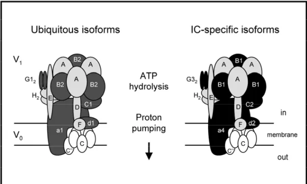
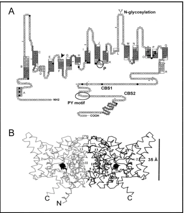
Figure 1.6. Structure of the vacuolar H+-ATPase
The transmembrane V0 domain of the mammalian V-ATPase is organized into a complex of [a(c)4-5c’’d], whereas the cytosolic V1 domain has a stoichiometry of [A3BB3CDEFG2H2].
The ubiquitous and the IC-specific alternate isoforms of V0 and V1 subunits are shaded. In the plasma membrane of murine IC, a4, d2, B1, C2, G3 substitute ubiquitous a1, d1, B2, C1 and G1, respectively. This model was modified from the yeast model to represent the mammalian V-ATPase complex. Note that the existence of the c’ subunit in mammals is uncertain (Smith, 2002).
Adapted from Borthwick, 2002
The structure of the V-ATPase includes at least 13 different subunits forming two functional domains, V0 and V1 (Figure 1.6) (Wagner, 2004; Wilkens, 2004). According to quantitative amino acid analysis and single molecule electron microscopy imaging, the transmembrane V0 domain contains five different subunits
organized into a [a(c)4-5c’’d] channel responsible for H+ translocation. The cytosolic
V1 domain involves eight subunits with a stoichiometry of [A3BB3CDEFG2H2], even
though the exact number of copies of each E and G per V-ATPase remains debated. The A and B subunits are organized in an alternating manner to form a pseudo-hexagonal head-piece, which ensures ATP hydrolysis necessary for active H transport. The other V1 subunits form the “stalk” that connects A and B subunits to the V0 domain. Interestingly, it is thought that ATP binding to B and subsequent hydrolysis by A lead to a rotation of the “stalk” structure relative to A
+
3B3 domain,
which may then induce the motion and the opening of the V0 channel (Nelson, 1999 ; Gruber, 2001). When located in the plasma membrane, the overall structure of V-ATPase is very similar but specific isoforms of V0 and V1 subunits are present (Figure 1.6) (Borthwick, 2002). These isoforms are encoded by distinct genes, with tissue-specific expression patterns (see section 2.3.).
The acidification of intracellular organelles along the endocytic pathway is a prerequisite for important processes in PT cells, such as ligand-receptor dissociation, receptor recycling and ligand degradation, storage, or intracellular targeting (Nelson, 1999). The mechanism by which the endosomal pH regulates trafficking processes remains unclear and may involve the vesicular recruitment of specialized coat proteins (COP), such as β-COP and small GTPases (Arf1, Arf6) of the ADP-ribosylation factor (ARF) family (Zeusem, 1992; Gu, 2000). Recent studies have demonstrated that functional interactions between coat proteins and particular V-ATPase V0 subunits regulate protein trafficking between early and late endosomes, indicating a pivotal role of the V-ATPase along the endocytic and degradative pathway (Hurtado-Lorenzo, 2006). Moreover, an endosomal pH-sensing machinery, not yet fully identified, has been suggested to initiate COP proteins recruitment in response to a V-ATPase-driven drop in luminal pH (Maranda, 2001).
Na+/H+ exchanger NHE3 (Giebisch, 2003b). The secreted H+ combine with luminal HCO3- under the influence of the membrane-bound carbonic anhydrase (CA) IV to
form H2O and CO2. After diffusion into PT cell, CO2 is reversibly hydrated by the
cytosolic CAII isozyme, thereby generating H+ for apical H+ extrusion and HCO3- for
basolateral exit via the co-transporter NBC1 (Figure 1.7). This process of HCO3
-reabsorption in the PT is intricately linked to Na+ and water homeostasis and thus finely modulated by hormones and metabolic status (Wagner, 1998). Thus, the V-ATPase plays pivotal roles in PT functions at both intracellular and plasma membrane locations. The severe PT cellular dysfunction observed in case of in vitro or in vivo disruption of the V-ATPase further highlights the importance of this pump in this segment.
Figure 1.7. Schematic model of HCO3- reabsorption in PT cells
Protons are secreted via the apical Na+/H+ exchanger NHE3 and the vacuolar H+-ATPase. The secreted H+ combine with filtered HCO3- under the influence of a membrane-bound carbonic anhydrase (CAIV) to form H2O and CO2. After diffusion into PT cells, CO2 is re-hydrated by the cytosolic CAII into H+ that are secreted back to PT lumen, and HCO3 -released into the interstitium via the basolateral Na+/HCO3- co-transporter NBC1.
1.3.4. Lignac-de Toni-Debré-Fanconi syndrome
The Lignac - de Toni - Debré - Fanconi syndrome (renal Fanconi syndrome) is characterized by a generalized defect in PT reabsorption of filtered solutes, such as glucose, phosphate, calcium, uric acid, amino acids, as well as LMW proteins. Clinical features can also include a metabolic acidosis (Type 2 renal tubular acidosis due to HCO3- losses and impaired NH4+ generation), rickets and growth retardation in
children and osteomalacia in adults (reduced vitamin D synthesis), nephrolithiasis related to increased urinary Ca++ excretion, hypocitraturia and impaired urine acidification, and progressive renal failure. The renal Fanconi syndrome can be inherited or acquired.
The list of acquired causes of renal Fanconi syndrome is largely heterogeneous and includes multiple myeloma, light chain deposition disease, and renal transplantation. In addition, various toxic compounds and drugs have been associated with PT defects, especially heavy metals (cadmium, uranium, lead and mercury), aminoglycoside antibiotics, as well as some anti-retroviral drugs (e.g. azidothymidine) and cytotoxics (e.g. ifosfamide, cisplatin). Most of these compounds affect the endocytic/lysosomal system and the mitochondrial function, which might explain their particular toxicity for the PT (Izzedine, 2003).
Inherited forms of the renal Fanconi syndrome are rare (approximately 1/40,000 births), transmitted as a recessive trait and mostly diagnosed during childhood (Table 1.2). Of note, the prevalence of cystinosis (OMIM #219800), the most frequent congenital PT disorder, has wide ranging estimates (from 0.03-0.4 per 10,000 live births) depending on the population studied. The genetic deficiencies affect cellular energy metabolism, membrane trafficking, or ion and solute transports (Bergeron, 1995). They include autosomal recessive disorders like cystinosis, tyrosinaemia, fructosaemia, galactosaemia, Type I glycogen storage disease, and cytochrome c oxidase deficiency; and X-linked recessive diseases like Dent’s disease and Lowe
Table 1.2. Inherited causes of renal Fanconi syndrome
Disorder OMIM Gene Protein Inheritance
COX deficiency #220110 MTC01-03 MTTS1 COX10 SC01-02
Cytochrome c oxidase AR
Cystinosis #219800 CTNS (17p13) Lysosomal cystine
transporter
AR
Dent’s disease (1) #300009 CLCN5 (Xp11.22) H+/Cl- exchanger XR
Dent’s disease (2) #300555 OCRL (Xq26.1) PIP2 5-phosphatase XR
Fanconi-Bickel syndrome
#227810 GLUT2 (3q26.1-3) Glucose transporter
GLUT2 AR
Fructosaemia +229600 ALDOB (9q22.3) Fructose-bisphosphate
aldolase B AR
Galactosaemia #230400 GALT (9p13) Galactose 1-phosphate
uridylyltransferase AR Imerslund-Gräsbeck disease #261100 CUBN (10p12.1) AMN (14q32) Cubilin Amnionless AR
Lowe syndrome #309000 OCRL (Xq26.1) PIP2 5-phosphatase XR
Tyrosinaemia +276700 FAH (15q23-25) Fumarylacetoacetase AR
von Gierke disease +232200 G6PC (17q21) Glucose
6-phosphatase AR Wilson disease #277900 ATP7B (13q14.3-21.1) Copper-transporting
ATPase 2 AR
A “number” symbol (#) indicates that the phenotype is not linked to a unique locus, whereas a “plus” sign (+) means that the entry associates a gene with a phenotype (AR: autosomal recessive; XR: X-linked recessive).
2. The intercalated cells of the collecting duct
2.1. Distribution and ultrastructure
The CD system has three segments designated after their distribution in kidney cortex, outer medulla or inner medulla, and continues the distal tubule of the nephron (Figure 1.1). The adult CD is composed of principal and intercalated cells that exhibit striking morphological and functional differences (Giebisch, 2003a). Principal cells represent about two thirds of CD cell population and mediate salt and water reabsorption under hormonal influence. They have a light cytoplasm with relatively few organelles and sparse mitochondria scattered randomly in the cytoplasm. Typically, principal cells show prominent infoldings of the basal membrane, no lateral interdigitations, and a single central cilium on the apical surface. In contrast, the IC are involved in acid-base transport and are characterized by a more electron dense cytoplasm and numerous dark staining mitochondria (hence, their previous name “dark cells”). The IC are the only cells along the urinary system that do not exhibit a primary cilium.
The IC can be further subdivided into two major groups, namely α- and β-type IC (Bastani, 1997). The existence of a non-α-non-β-type IC (also called γ-IC), which may represent an interconvertible state between α- and β-phenotypes, remains debated (Schwartz, 1985; Al-Awqati, 1996; Wagner, 2004). The α-type IC are present in all CD segments and have prominent apical micro-projections and typical tubulo-vesicular structures beneath the apical surface, both coated with studs on the cytoplasmic face. In contrast, the β-type IC are most frequently found in the cortical CD and show a fairly smooth apical surface, a zone free of organelles beneath the apical membrane, and abundant cytoplasmic vesicles. This distinction between α- and β-type IC is not only structural, but also (and mostly) functional as discussed hereafter.
2.2. Role in acid-base homeostasis
Together with the lungs, the kidneys play an essential role in acid-base homeostasis by reabsorbing virtually all filtered HCO - and secreting H+ into the urine
ascending limb, TAL), whereas the fine regulation of acid-base excretion and absorption occurs in the CD. The cortical CD either reabsorbs or secretes H+ and HCO3-, while the outer medullary CD secretes only H+. The average diet in the
“Western” world is rich in proteins and generates daily 1-1.5 mmol hydrogen/kg body weight. The net urinary acid excretion is therefore essential and urine pH can drop as low as 4.5.
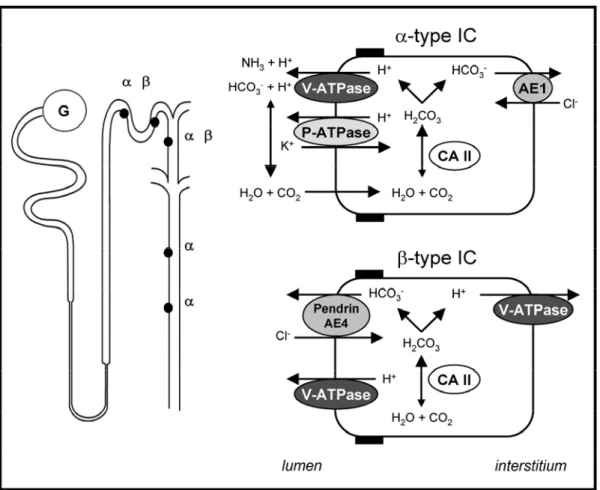
The IC are the main cells involved in acid-base transport along the CD. Both α- and β-type IC are characterized by high activity of cytosolic CAII (Figure 1.8). However, they differ by the selective polarity of the V-ATPase and the exclusive expression of a basolateral (α-IC) or apical anion-exchanger (β-IC) (Giebisch, 2003b). The α-type IC secrete H+
into the urine via the V-ATPase that is present at high density on the luminal membrane. Studies in animal models have shown that the V-ATPase is also present within specialized intracellular tubulovesicles close to the membrane, allowing fast recruitment of additional pumps to the membrane in response to stimuli, like systemic acidosis (Penney, 1999; Brown, 2000a). Animal studies have also identified an additional P-type ATPase at the apical surface of α-IC that exchanges H+ for K+ (Wingo, 1990). However, the overall contribution of such pump in human acid-base physiology remains unclear. On the basolateral side, the α-IC are characterized by the expression of the kidney-specific truncated version of the band 3/AE1 (SLC4A1) Cl-/HCO3- exchanger (Alper, 1989). In contrast, the β-type IC
show a basolateral or bipolar expression of the V-ATPase and an apical anion exchanger different from AE1, whose molecular identity remains debated. Two Cl -/HCO3- exchangers have been identified in β-IC: pendrin and AE4. Pendrin resides in
the apical membrane of all non-α-type IC and is regulated by acid-base status (Wagner, 2002). However, the genetic loss of pendrin causes Pendred syndrome (OMIM #274600) of deafness and goiter in man and mouse, with no significant metabolic alkalosis at baseline (Royaux, 2001). AE4 is also expressed in non-α-type IC, but its subcellular localization seems to be species-specific (apical in rabbit, basolateral in mouse and rat) and its role remains, therefore, uncertain (Tsuganezawa, 2001).
Figure 1.8. Schematic model of H+ and HCO3
secretion in α- and β-type IC
The α-type IC are found in the connecting tubule (CNT), cortical collecting (CCD), and both outer and inner medullary CD, whereas the β-type IC are present only in the CNT and CCD.
The α-type IC are characterized by the expression of the basolateral Cl
-/HCO3
exchanger AE-1 and the apical V-ATPase. The cytosolic CAII generates H+ and HCO3- that are secreted into the lumen and the interstitium, respectively. An apical H+/K+-ATPase has been identified in α-type IC of certain species.
The β-type IC are defined by the absence of the basolateral AE-1 exchanger, and the presence of the V-ATPase on both sides of the cell. The molecular identity of the apical Cl-/HCO3- exchanger responsible for HCO3- secretion remains debated. Two putative anion exchangers, pendrin and AE4, have been proposed.
Apparent plasticity of molecular targeting according to ambient pH in vitro and/or acid and alkali load in vivo has suggested that α- and β-type IC may represent molecular mirror images of each other (Schwartz, 2005). Indeed, the various patterns of V-ATPase distribution observed in IC support the hypothesis that all IC are phenotypic variants of the same cell type, and that the precise cellular location of the V-ATPase is determined by the acid-base status. Moreover, the functional phenotype
(Schwartz, 1985). Hensin was proposed to induce terminal differentiation in IC, which was reflected by the α-cell phenotype. This change in functional activity of IC involves the concerted action of microtubules and microfilaments and requires de novo protein synthesis (Schwartz, 2002). It must be emphasized that the change in phenotype from α- to β-type involves more than a simple inversion of polarity of membrane transporters, as α-IC and β-IC have been distinguished by a differential expression of proteins, including the anion exchangers and NHE-RF (Na+/H+ exchanger regulatory factor) (Wagner, 2004). In any event, the daily acid load provided by an omnivorous human diet dictates that the majority of IC will be acid-secretory (α-type). The following sections address the specificity of the V-ATPase present in the IC and review the causes of inherited dysfunction of the IC resulting in distal renal tubular acidosis.
2.3. Kidney-specific isoforms of V-ATPase subunits
Tissue-specific isoforms of some V0 and V1 subunits, encoded by distinct genes, are present in the V-ATPase located at the plasma membrane of specialized cells, such as the IC of the CD (Figure 1.6) (Borthwick, 2002). The V1 B subunit was the first to be identified as such (Nelson, 1992). Although B2 is regarded as the ubiquitous isoform, B1 expression seems restricted to kidney, inner ear and male genital tract (Nelson, 1992; Breton, 2000). Three V1 G and two V1 C subunit isoforms have been identified in man, with G3 and C2 showing kidney-specific distribution (Smith, 2002). The V0 a subunit is even more complex, since four isoforms have been described in both man and mouse (Smith, 2001; Oka, 2001). All four a subunits are expressed in the kidney as detected by Northern blotting and RT-PCR analyses, with distribution patterns associated with various regions of the nephron (Wagner, 2004). The a4 isoform is particularly found in all subtypes of IC, as well as in the inner ear and along the epididymis (Smith, 2001). Finally the V0 d2 subunit isoform has been recently identified and located in kidney and bone (Smith, 2002).
Recent studies have addressed the roles of individual V-ATPase subunits, with a particular interest for those with tissue-specific expression. In contrast to the ubiquitous B2, the B1 subunit contains a C-terminal DTAL motif that may interact with the PDZ protein NHE-RF to generate, maintain or modulate the V-ATPase
polarity in β-IC, which are characterized by a highly variable pattern of intracellular localization of the V-ATPase (Breton, 2000). Whether PDZ proteins are involved in either the targeting, the trafficking, or the anchoring of the V-ATPase in specialized membrane domains remains to be determined. Of note, the α-IC that insert the V-ATPase uniquely into their apical domain contain little or no detectable NHE-RF. The distinct distribution of the V0 a isoforms also suggests that this subunit may be involved in the assembly and/or targeting of the V-ATPase. Indeed, the N-terminal domain of this subunit appears to play a major role in V-ATPase targeting to organelles and in vivo dissociation in yeast, whereas its C-terminal region controls the coupling of ATP hydrolysis and H+ translocation (Kawasaki-Nishi, 2001). Altogether, these data support that particular isoforms of V-ATPase subunits interact with different proteins and/or confer specific sorting signals, resulting to a differential distribution of the pump in the cell.
2.4. Hereditary distal renal tubular acidosis
Renal tubular acidosis (RTA) refers to a group of tubular dysfunctions characterized by a hyperchloremic metabolic acidosis due to a failure of renal HCO3
-reabsorption or H+ excretion that is not related to a reduction in the glomerular filtration rate. These disorders may be inherited, with autosomal recessive or dominant modes of transmission, or acquired (Igarashi, 2002; Karet, 2002). The classification of RTAs is based on the perceived roles of the different nephron segments in the acid-base regulation. Proximal RTA (type 2 RTA) is due to an impaired reabsorption of HCO3- by the PT, often associated with other signs of PT dysfunction (“renal Fanconi
syndrome”). The distal RTAs are due to a defective H+ excretion by the distal tubule and CD. They are frequently associated with hypercalciuria and low urinary citrate excretion, leading to nephrocalcinosis and nephrolithiasis. They are further divided into the hypokalemic distal (type 1 RTA) and hyperkalemic distal (type 4 RTA). Finally, mutations in CA2 gene cause Guibaud-Vainsel disease (type 3 RTA), an inherited syndrome characterized by renal tubular acidosis, osteopetrosis, and cerebral calcifications (Sly, 1985). In this case, the functional loss of CAII associates signs of both proximal and distal tubular dysfunction (Laing, 2005).
Type 1 distal RTAs are relatively rare in Western populations, but occur more commonly in regions where rates of parental consanguinity are high (Karet, 2002). The inheritance of type 1 RTA shows both autosomal recessive and dominant patterns, the most severe cases being inherited recessively (Table 1.3). Dominant Type 1a distal RTA is caused by mutations in SLC4A1 gene encoding the Cl-/HCO3- exchanger AE1 (Bruce,
1997; Karet, 1998). Both forms of recessive distal RTA are associated with defects in the IC-specific B1 and a4 subunits of the V-ATPase. The functional loss of B1, which is encoded by ATP6V1B1 gene and present in both kidney and inner ear, causes Type 1b RTA with deafness (Karet, 1999). Type 1c RTA with preserved hearing in childhood is caused by inactivating mutations of ATP6V0A4 (Smith, 2000). To note, some patients with distal RTA do not show linkage to either SLC4A1, ATP6V1B1 or ATP6V0A4 genes, suggesting that other distal RTA genes remain to be identified. These include (i) genes involved in IC differentiation; (ii) genes for V-ATPase subunit isoforms; (iii) genes with products that are required for trafficking of the V-ATPase in α-IC; (iv) and genes encoding molecules necessary for the generation of H+, absorption of HCO3-, recycling of
Cl-, or maintenance of the electrochemical gradients across the epithelial barrier.
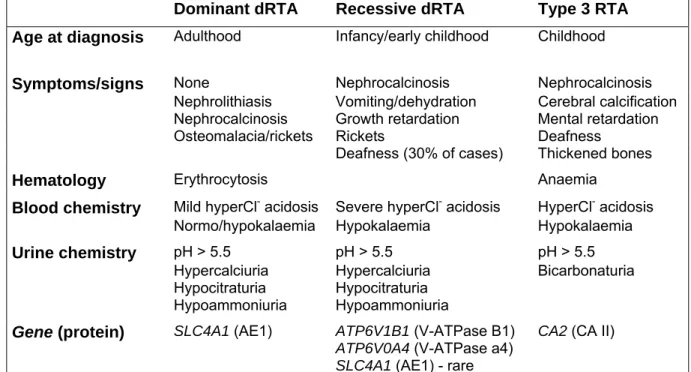
Table 1.3. Primary distal RTA: clinical and biochemical features
Dominant dRTA Recessive dRTA Type 3 RTA
Age at diagnosis Adulthood Infancy/early childhood Childhood
Symptoms/signs None Nephrocalcinosis Nephrocalcinosis Nephrolithiasis Vomiting/dehydration Cerebral calcification Nephrocalcinosis Growth retardation Mental retardation
Osteomalacia/rickets Rickets Deafness Deafness (30% of cases) Thickened bones
Hematology Erythrocytosis Anaemia
Blood chemistry Mild hyperCl- acidosis Severe hyperCl- acidosis HyperCl- acidosis Normo/hypokalaemia Hypokalaemia Hypokalaemia Urine chemistry pH > 5.5 pH > 5.5 pH > 5.5
Hypercalciuria Hypercalciuria Bicarbonaturia Hypocitraturia Hypocitraturia
Hypoammoniuria Hypoammoniuria Gene (protein) SLC4A1 (AE1) ATP6V1B1 (V-ATPase B1)
ATP6V0A4 (V-ATPase a4)
SLC4A1 (AE1) - rare
CA2 (CA II)
A mouse model deficient in B1 V-ATPase subunit has been very recently engineered by homologous recombination-mediated targeting of exons 7-11 in
Atp6v1b1 gene (Finberg, 2005). In contrast to human distal RTA caused by ATP6V1B1
mutations that presents in early infancy, Atp6v1b1 KO mice appear healthy and grow normally. This phenotypic discrepancy is likely related to dietary differences. However, Atp6v1b1 KO mice produce urine that is significantly more alkaline than that of wild-type littermates under standard diet and fail to acidify urine after oral acid challenge. Moreover, clearance studies performed after furosemide treatment have demonstrated that this defect in urine acidification results from IC dysfunction. Note that genetic inactivation of Atp6v1b1 in mice was not associated with hypercalciuria and skeletal abnormalities, male infertility, or hearing loss as reported in patients. The compensatory role of B2 isoform in V-ATPase complexes in case of Atp6v1b1 inactivation remains uncertain, but could provide a partial functional compensation in some organs in mice. Thus, these data demonstrate that plasma membrane V-ATPase represents the main pathway of urinary acidification in IC and propose a useful animal model to investigate in vivo its role in pH homeostasis.
Another mouse model of distal RTA has been recently reported (Blomqvist, 2004). These mice lack the forkhead transcription factor Foxi1 that plays a central role in IC differentiation during nephrogenesis. Accordingly, the epithelium lining the distal nephron in Foxi1 KO mice show no typical IC, but a single population of cells positive for markers of both principal and intercalated cells. The importance of these observations on our knowledge about cell differentiation along the distal nephron is further detailed in section 4.3.2.
3. Chloride transporters in the kidney
Chloride is the most abundant anion in plants and animal tissues and Cl- transport across cellular membranes is involved in the transepithelial transport of salt and water, membrane excitability, and regulation of cell volume and pH. In addition, Cl -transporters participate in the acidification and ionic homeostasis of intracellular organelles (Jentsch, 2002). Over the last few years, numerous Cl- transporters, including channels and exchangers, have been characterized in all segments of the nephron (Devuyst, 2002). Most of the Cl- ions filtered by the glomeruli are reabsorbed through different mechanisms operating in the apical and basolateral membranes of tubular epithelial cells. Several Cl- transporters use the energy generated by transmembrane gradients of other ions to move Cl- against its electrochemical gradient, whereas passive diffusion of Cl- through Cl- channels is involved in cell volume regulation. Transport of Cl- through Cl- / HCO3- exchangers in IC, or through
the Na+-linked Cl- / HCO3- exchanger operating in PT cells, participate in acid-base
homesostasis. In addition, Cl- ions are also important for acidification of intracellular vesicles by neutralizing the transmembrane potential generated by the electrogenic V-ATPase. The association of a large spectrum of human diseases affecting kidney function with mutations in distinct genes encoding Cl- transporters, has provided new insights into the diverse roles ensured by Cl- movement in cell physiology (Jentsch, 2005; Romero, 2005).
On one hand, two main families of Cl- exchangers have been implicated in kidney function, namely the SLC4 and SLC26 transporters. The SLC4 HCO3
-transporter family contains 10 members that move base equivalents (OH- or HCO3-)
across cell membranes to alter intracellular pH (Romero, 2005). There are at least 4 Cl -/ HCO3- exchangers in the SLC4 family: AE1-4 (also called SCL4A1-3 and SLC4A9).
The kidney-specific truncated version of the band 3/AE1 is found in the basolateral membrane of the α-type IC of the CD and mutations in SLC4A1 gene have been associated with distal RTA (see section 2.4). In addition, three isoforms of AE2 (SLC4A2a-c) have been located in the TAL and AE4 (SLC4A9) is present in the
apical membrane of rabbit β-type IC, where its role in acid-base homeostasis remains debated (see section 2.2). The SLC26 anion exchangers transport an expanding number of monovalent and divalent anions and play critical roles in skeletal development, synthesis of thyroid hormones, transepithelial Na+, Cl-, and HCO3
-transport (Mount, 2004). The distribution of SLC26 members includes different segments of the nephron. SLC26A6 has been detected in the apical membrane of PT cells and may represent the long-elusive apical Cl- entry site involved in PT Na+-Cl -reabsorption (Chernova, 2005). In addition, SLC26A4 (also called Pendrin) has been located in the luminal membrane of the β-type IC, which are known to secrete HCO3
-via a Cl- / HCO3- exchanger (see section 2.2).
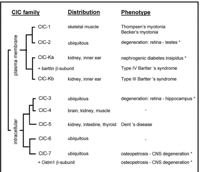
On the other hand, several Cl- channels have been identified along the nephron, based on single-channel properties and sensitivity to inhibitors. However, the molecular counterpart of many renal Cl- channels remains debated (Devuyst, 2002). This section will focus on two structural classes of Cl- transporters that have been extensively characterized: the CLC family and the CFTR (cystic fibrosis transmembrane conductance regulator). The emphasis will be particularly set on the role of the Cl- transporter ClC-5 and the Cl- channel CFTR in the pathophysiology of Dent’s disease and cystic fibrosis, respectively.
3.1. Dent’s disease and ClC-5
3.1.1. Dent’s disease
In 1964, C.E. Dent and M. Friedman described two unrelated English boys with hypercalciuric rickets associated with renal tubular damage including tubular proteinuria, aminoaciduria, phosphaturia and hypercalciuria (Dent, 1964). The term
Dent’s disease, first introduced in early nineties, identifies a group of X-linked renal
tubular disorders characterized by LMW proteinuria associated with hypercalciuria and nephrocalcinosis and/or nephrolithiasis (Wrong, 1994). This triad of manifestations has been variably named in the past as X-linked recessive