C
LINICAL BENEFIT TO PROGRAMMED DEATH
-1
INHIBITION FOR NON
-SMALL
-
CELL LUNG CANCER IS ASSOCIATED WITH HIGHER BLOOD
EOSINOPHIL LEVELS
A. Sibille, M. Henket, J. L. Corhay, R. Louis and B. Duysinx
CHU de Liege-Hopital du Sart Tilman, Liege, Belgium
Introduction
The use of immune checkpoint inhibitors (ICI) for non-smallcell lung cancer (NSCLC) is increasing. Currently validated indications include advanced and locally advanced disease [1]. Classically, response evaluation relies on radiological assessment with the REsponse Criteria In Solid Tumours (RECIST) version 1.1 [2]. However, in the setting of ICI these criteria seem imperfect. Indeed, atypical response patterns have been observed that make radiological evaluation less clear than it is with chemotherapy [3]. Pseudoprogression, one of these atypical radiological responses, is defined as radiological progression in the absence of clinical deterioration. It correlates with immune cell infiltration and/or transient tumour growth before response to ICI [3]. Discontinuing ICI in this case would mean stopping an efficient therapy. Alternative or additional tools for the evaluation of response could help to effectively and more accurately evaluate the efficacy of ICI. Blood eosinophil counts are routinely available at a negligible cost. Most of the objective responses to PD-1 inhibitors occur within the first two months of PD-1 inhibitor use [4, 5]. Here, we report on blood eosinophil evolution during the first months of treatment with anti-Programmed Death (PD)-1 antibodies and on their value as early indicators of response in patients treated for advanced stage NSCLC.
Materials and methods
Medical records from patients consecutively treated at our institution with any anti-PD-1/anti-PD-L1 in monotherapy for advanced stage NSCLC between 1/8/2015 and 30/4/2018 were investigated. In this time frame only two agents were used: pembrolizumab, given at the dose of 2 mg/kg every 3 weeks during the early access programm then at the fixed dose of 200 mg every 3 weeks, and nivolumab, given at 3 mg/kg every 2 weeks. We collected the following data: (i) patients characteristics (age at the start of immunotherapy, gender, smoking status, concurrent airway disease), (ii) lung cancer characteristics (histological subtype, stage, line of treatment of the anti-PD-1, PD-L1 status), (iii) treatment characteristics (agent, response at t1 (time of first evaluation, i.e. after three (for pembrolizumab) to four (for nivolumab) cycles of immunotherapy) and t2 (time of second evaluation, i.e. after four (for pembrolizumab) to six
(for nivolumab) additional cycles of immunotherapy) using REsponse Criteria for Solid Tumours (RECIST) v1.1), (iv) eosinophil counts (absolute and relative) at t0 (before treatment), t1 and t2 . Of the 191 patients identified the following patients were excluded: loss of follow-up (n = 8), treatment discontinuation before t2 due to toxicity (n = 2), progressive disease (n = 4), patient’s will (n =3) or death (n = 57). Response was assessed according to the RECIST criteria version 1.1 [2]. We further describe patients as responders (complete (CR) or partial (PR) response), stable or progressive. We focussed on the first two radiological evaluations as the majority of objective responses (i.e. CR and PR) occur in the first two months of treatment with anti-PD-1/-PD-L1 in monotherapy for NSCLC, corresponding to the first time point (t1) in our study [4,5]. We extended the evaluation period to the second radiological evaluation (t2) in order to include the patients showing a non-significant response at t1 further evolving towards PD or PR. Blood eosinophils were expressed as median number of cells/mL for the absolute eosinophil count (AEC) and in percentage of the total white blood cell count for the relative eosinophil count (REC) with interquartile range (IQR).
Regarding the statistical analyses paired comparisons of eosinophil values between the three visits of patients were performed with a non-parametric test: Wilcoxon’s signed rank test. The comparison of eosinophil levels of the 3 groups of patients ranked according to the response to the treatments were performed by an unpaired test for nonparametric continuous variables: Kruskal-Wallis test followed by the Dunn’s post-hoc testing if Kruskal-Wallis tests were positive. A p value <.05 was considered statistically significant. Analyses were conducted by the statisticians of the Pneumology laboratory Unit of the CHU de Liège using GraphPad Prism V.7.03 (GraphPad Software, La Jolla, California, USA) for the statistical analyses and for the figures.
Figure 1. Blood eosinophil levels of the entire study cohort. Results expressed as median ± IQR, confidence interval 10-90%.
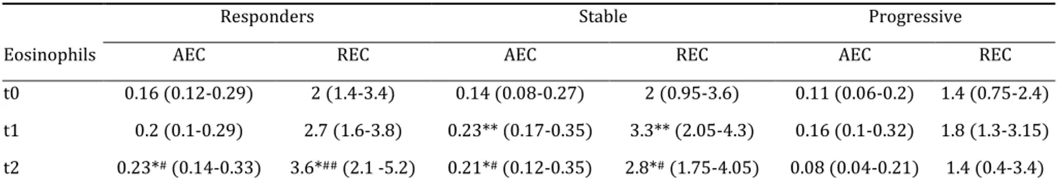
Table 1. Blood eosinophil levels according to the type of response.
Responders Stable Progressive
Eosinophils AEC REC AEC REC AEC REC
t0 0.16 (0.12-0.29) 2 (1.4-3.4) 0.14 (0.08-0.27) 2 (0.95-3.6) 0.11 (0.06-0.2) 1.4 (0.75-2.4) t1 0.2 (0.1-0.29) 2.7 (1.6-3.8) 0.23** (0.17-0.35) 3.3** (2.05-4.3) 0.16 (0.1-0.32) 1.8 (1.3-3.15) t2 0.23*# (0.14-0.33) 3.6*## (2.1 -5.2) 0.21*# (0.12-0.35) 2.8*# (1.75-4.05) 0.08 (0.04-0.21) 1.4 (0.4-3.4)
AEC: absolute eosinophil count; expressed as number of cells/mL; REC: relative eosinophil count; expressed as percentage of the total white blood cell count. Responders (n = 27), stable (n = 61) and progressive (n = 29) patients: according to the RECIST criteria (see materials and methods). Inter-group analysis: Kruskal-Wallis test followed, if positive, by Dunn’s test; p-value vs. progressive: ##p < .01; #p < .05. Intra-group analysis: Wilcoxon’s
paired test; p-value: **p < .01, *p < .05.
Results
In the 117 patients analysed baseline blood eosinophils were not statistically different in responders, stable or progressive patients. For the whole study population the AEC and REC were significantly raised at t1 compared to t0 (p < .01 for both AEC and REC) (Figure 1). Responders and stable patients had significantly higher eosinophils than progressive patients at t2 (p < .05 for AEC and p < .01 for REC for responders and p < .05 for both AEC and REC for stable patients). Stable patients showed an early (t1) and persistent (t2) significant rise in eosinophils (p < .01 for AEC and REC at t1 and p < .05 for AEC and REC at t2) (Table 1). Performing univariate analysis (two-way factorial ANOVA) we did not find any impact of histology, type of anti-PD-1 agent, smoking status (current versus former smoker) or PD-L1 status on those results.
Discussion
Response evaluation of patients treated with PD-1 inhibitors remains a challenge. In routine clinical practice RECIST criteria remain the core element for the evaluation of response. However, atypical response patterns have been described following anti-PD-1 use showing the limitations of these criteria. We hypothesised that blood eosinophil kinetics might be an early indicator of response to ICI.
Considering the whole study population, we found a significant and early rise in blood eosinophils, i.e. after two to three months of PD-1 inhibition, compared to baseline values. In a large series (n = 909) of patients treated with anti- PD-1 antibodies for various types of cancers the rise in AEC was seen from 3 months after the start of the treatment and peaked at a median of 6.4 months [
6
]. The significant increase at t1 in our cohort is in keeping with this previousstudy although we found no further significant rise between t1 and t2, possibly due to the lower number of patients.
Baseline eosinophil counts did not differ between responders, stable or progressive patients. This contrasts with retrospective data from one melanoma series treated with anti-PD-1 agent pembrolizumab where baseline REC > 1.5% was associated with an improved overall survival and more objective responses according to the RECIST criteria, although this positive prognostic and predictive value of eosinophils was only noted for the REC, not for the AEC, and in combination with the relative lymphocyte count [
7
]. The predictive role of a composite bloodbiomarker was retrospectively investigated in one series of NSCLC patients and showed a longer progression-free survival, defined as the time between the start of a PD-1 inhibitor and radiological progression, in patients showing the following baseline characteristics: an absolute lymphocyte count >1 cells/mL, an absolute eosinophil count >0.15 cells/mL and an absolute neutrophil count <7.5 cells/mL) [
8
].Our main results indicate a clear association between blood eosinophils kinetics and the type of response. Indeed, eosinophils were significantly higher in responders and in stable patients than in patients with progressive disease at the time of second evaluation. Also, stable patients showed an early and persistent significant increase in eosinophils. This was not the case for responders, possibly due to a lower number of patients (27 responders vs. 61 stable patients). To the best of our knowledge no data exist to date regarding the evolution of blood eosinophil levels and the type of response to ICI in NSCLC.
The exact role of eosinophils in (lung) cancer remains uncertain at present [
9
]. Indeed, somepreclinical studies have shown a lower incidence of squamous cell carcinoma in eosinophil-deficient mice. Most studies, however, highlight their multiple anti-tumour effects: maturation of dendritic cells, polarisation of macrophages to an M1 phenotype, inhibition and normalisation of tumour vasculature, recruitment and activation of T lymphocytes and NK-cells, direct cytotoxic effects on tumour cells [
9
,10
]. Even though we cannot state whether raised blood eosinophilsare a consequence or a driver of enhanced activity of PD-1 antibodies our results indicate that they might be indicators of response to anti-PD-1 drugs for NSCLC. Although we acknowledge the need for a validation study with a greater and ideally prospective cohort, we believe that the highly significant differences between eosinophils of responders and stable patients versus non-responders in our study warrant reporting.
In conclusion, our retrospective cohort suggests a role of blood eosinophils in the early response to PD-1 inhibitors in NSCLC patients.
References
[1] Planchard D, Popat S, Kerr K, et al. Metastatic non-small cell lung cancer: ESMO Clinical Practice Guidelines for diagnosis, treatment and follow-up. Ann Oncol. 2019 ; 30(5) : 863-870.
[2] Eisenhauer EA, Therasse P, Bogaerts J, et al. New response evaluation criteria in solid tumours: revised RECIST guideline (version 1.1). Eur J Cancer. 2009 ; 45(2) : 228-247.
[3] Borcoman E, Kanjanapan Y, Champiat S, et al. Novel patterns of response under immunotherapy. Ann Oncol. 2019 ; 30(3) : 385-396.
[4] Brahmer J, Reckamp KL, Baas P, et al. Nivolumab versus docetaxel in advanced Squamous-Cell Non-Small-Cell lung cancer. N Engl J Med. 2015 ; 373(2) : 123-135.
[5] Borghaei H, Paz-Ares L, Horn L, et al. Nivolumab versus docetaxel in advanced nonsquamous Non-Small-Cell lung cancer. N Engl J Med. 2015 ; 373(17) : 1627-1639.
[6] Bernard-Tessier A, Jeanville P, Champiat S, et al. Immune-related eosinophilia induced by anti-programmed death 1 or death-ligand 1 antibodies. Eur J Cancer. 2017 ; 81 : 135-137.
[7] Weide B, Martens A, Hassel JC, et al. Baseline biomarkers for outcome of melanoma patients treated with pembrolizumab. Clin Cancer Res. 2016 ; 22(22) : 5487-5496.
[8] Tanizaki J, Haratani K, Hayashi H, et al. Peripheral blood biomarkers associated with clinical outcome in non-small cell lung cancer patients treated with nivolumab. J Thorac Oncol. 2018 ; 13(1) : 97-105.
[9] Simon SCS, Utikal J, Umansky V. Opposing roles of eosinophils in cancer. Cancer Immunol Immunother. 2019 ; 68(5) : 823-833.
[10] Carretero R, Sektioglu IM, Garbi N, et al. Eosinophils orchestrate cancer rejection by normalizing tumor vessels and enhancing infiltration of CD8(+) T cells. Nat Immunol. 2015 ; 16(6) : 609-617.