Hydrogen Peroxide Enhances the Expression and Function
of Giα Proteins in Aortic Vascular Smooth Muscle cells from
Sprague Dawley rats: Role of Growth Factor Receptors
Transactivation
Par Nathan Mbong
Département de physiologie Université de Montréal
Mémoire présenté à la Faculté de Médecine en vue de l’obtention du grade de Maîtrise ès Sciences
en Physiologie
Novembre, 2010
Faculté de Faculté de Médecine
Ce mémoire intitulé :
Hydrogen peroxide enhances the expression and function of Giα proteins in aortic vascular smooth muscle cells from Sprague Dawley rats: role of growth factor receptors transactivation
Présenté par : Nathan Mbong
a été évalué par un jury composé des personnes suivantes :
Dr. Rémy Sauvé, président-rapporteur
Dre Madhu B. Anand-Srivastava, directrice de recherche Dre Josette Noël, membre du jury
Résumé
Nous avons récemment démontré que les espèces réactives oxygénées induisent une augmentation de l’expression des protéines Giα dans les cellules du muscle lisse vasculaire (CMLV) provenant d’aortes de rats spontanément hypertendus (SHR, de l’anglais spontaneously hypertensive rats). La présente étude a pour but d’étudier les effets du peroxyde d’hydrogène (H2O2), un oxydant qui induit le stress oxydatif, sur l’expression de Giα et sur l’activité de
l’adénylate cyclase, et d’explorer les voies de signalisation sous-jacentes responsables de cette réponse.
Nos résultats montrent que H2O2 induit une augmentation de l’expression des protéines Giα-2 et
Giα-3 de manière dose- et temps-dépendante avec une augmentation maximale de 40-50% à 100 µM après 1 heure, sans affecter l’expression de Gsα. L’expression des protéines Giα a été maintenue au niveau normal en presence de AG 1478, AG1295, PD98059 et la wortmannine, des inhibiteurs d’EGF-R (de l’anglais epidermal growth factor receptor), PDGFR-β (de l’anglais platelet-derived growth factor receptor β), de la voie de signalisation ras-ERK1/2 (de l’anglais extracellular regulated kinase1/2), et de la voie de la PI3Kinase-AKT (de l’anglais phosphatidyl inositol-3 kinase), respectivement. En outre, le traitement des CMLV avec H2O2 a induit une
augmentation du degré de phosphorylation d’EGF-R, PDGF-R, ERK1/2 et AKT; et cette expression a été maintenue au niveau témoin par leurs inhibiteurs respectifs. Les inhibiteurs d’EGF-R et PDGF-R ont aussi induit une diminution du degré de phosphorylation de ERK1/2, et AKT/PKB. En outre, la transfection des cellules avec le siRNA (de l’anglais, small interfering ribonucleic acid) de EGF-R et PDGFR-β a atténué la surexpression des protéines Giα-2 et Giα-3 induite par le traitement au H2O2.
La surexpression des protéines Giα induite par H2O2 a été corrélée avec une augmentation de la
fonction de la protéine Giα. L’inhibition de l’activité de l’adénylate cyclase par de faibles concentrations de GTPγS après stimulation par la forskoline a augmenté de 20% dans les cellules traitées au H2O2. En outre, le traitement des CMLV au H2O2 a aussi accru l’inhibition de
l’activité de l’adénylate cyclase par les hormones inhibitrices telles que l’angiotensine II, oxotrémorine et C-ANP4-23. D’autre part, la stimulation de l’adénylate cyclase induite par
GTPγS, glucagon, isoprotérénol, forskoline, et le fluorure de sodium (NaF) a été atténuée de façon significative dans les cellules traitées au H2O2. Ces résultats suggèrent que H2O2 induit la
surexpression des protéines Giα-2 and Giα-3 via la transactivation des récepteurs des facteurs de croissance EGF-R, PDGFR-β et l’activation des voies de signalisation ras-ERK1/2 et PI3K-AKT
Mot-cles: Protéines Giα, peroxyde d’hydrogène, stress oxydant, récepteurs des facteurs de
croissance, MAP kinases, adénylate cyclase, hypertension
Abstract
We recently have shown that reactive oxygen species contribute to the enhanced expression of Giα proteins in vascular smooth muscle cells (VSMC) from spontaneously hypertensive rats
(SHR). The present study was undertaken to examine if H2O2, an oxidant that induces oxidative
stress could also enhance the expression of Giα proteins and associated adenylyl cyclase signalling in aortic VSMC and to further explore the underlying signaling pathways responsible for this response. Treatment of cells with H2O2 increased the expression of Giα-2 and Giα-3
proteins but not that of Gsα proteins in a concentration- and time-dependent manner. A maximal increase of 40-50% was observed at 100µM and 1h. The enhanced expression of Giα proteins was restored to control levels by AG 1478, AG1295, PD98059 and wortmannin, inhibitors of epidermal growth factor receptor (EGF-R), platelet-derived growth factor receptor (PDGFR-β), the mitogen-activated protein kinase (MEK1/2), and PI3 kinase respectively. In addition, treatment of VSMC with H2O2 also increased the phosphorylation of EGF-R, PDGF-R, ERK1/2
and AKT and this increased phosphorylation was attenuated to control levels by the respective inhibitors, whereas the inhibitors of EGF-R and PDGE-R also attenuated the enhanced phosphorylation of ERK1/2 and AKT to control levels. Transfection of cells with EGF-R and PDGFR-β siRNA followed by H2O2 treatment restored the H2O2-induced enhanced expression of
Giα-2 and Giα-3 proteins to control levels. The increased expression of Giα proteins by H2O2
was reflected in the increased Gi functions. The inhibition of forskolin (FSK)-stimulated AC activity by low concentration of GTPγS (receptor- independent Gi functions) was increased by about 20% by H2O2 treatment. Moreover, treatment of cells with H2O2 also resulted in an
increased Ang II-, C-ANP4-23, and oxotremorine-mediated inhibition of AC (receptor-dependent
functions of Gi). On the other hand, Gsα-mediated stimulation of AC by GTPγS, glucagon, isoproterenol, FSK, and NaF was significantly decreased in H2O2-treated cells. These results
suggest that H2O2 increases the expression of Giα-2 and Giα-3 proteins in VSMC through the
transactivation of EGF-R, PDGFR-β and associated ERK1/2 and PI3K signalling pathways.
Keywords: Giα proteins, hydrogen peroxide, adenylyl cyclase, oxidative stress, MAP kinase,
Table of contents
Résumé ... iii Abstract ... v Table of contents ... vi List of tables ... ix List of figures ... x List of abbreviations ... xi Acknowledgements ... xiv Chapter 1 – Introduction ... 11.1 The Adenylyl cyclase signal transduction system ... 2
1.1.1 Signalling via transmembrane receptors... 2
1.1.1.1 G protein-coupled receptors as mechanism of signal transduction ... 3
1.1.2 Structure of guanine nucleotide binding proteins ... 4
1.1.2.1 Classification of Heterotrimeric G proteins ... 5
1.1.2.2 Activation of guanine nucleotide proteins ... 7
1.1.3 The enzyme adenylyl cyclase and its regulation ... 7
1.1.3.1 Structural organisation of adenylyl cyclases ... 7
1.1.3.2 Regulation of adenylyl cyclase activity ... 9
1.1.3.2.1 Regulation by G protein subunits ... 9
1.1.3.2.2 Regulation by phosphorylation ... 10
1.1.3.2.3 Regulation by Calcium and Forskolin ... 10
1.2 The Adenylyl cyclase system in cardiovascular diseases ... 10
1.2.1 Blood Pressure ... 11
1.2.2. Mechanisms of blood pressure regulation ... 11
1.2.2.1. The Renin-Angiotensin system in the regulation of blood pressure ... 12
1.2.3 Hypertension ... 12
1.2.3.1 Physiology of hypertension on vascular smooth muscle cells ... 13
1.2.3.2 Alteration of G proteins expression in hypertension ... 14
1.2.3.3 Alteration of adenylyl cyclase/cAMP activity in hypertension ... 15
1.3.1 Sources and chemistry of reactive oxygen species ... 16
1.3.1.1 Enzymatic sources of reactive oxygen species ... 18
1.3.1.2 Non enzymatic sources of reactive oxygen species ... 18
1.3.2 The NADPH oxidases ... 19
1.3.2.1 Structure and expression profile of vascular NADPH oxidases ... 19
1.3.3 Oxidative stress in hypertension ... 20
1.3.3.1 Oxidative stress in genetic models of hypertension ... 20
1.3.3.2 Oxidative stress in human hypertension ... 21
1.3.4 Vasoactive peptides as inducers of oxidative stress ... 22
1.3.4.1 Oxidative stress and angiotensin II ... 22
1.3.4.2 Oxidative stress and endothelin-1 ... 23
1.4 Oxidative stress and signalling ... 23
1.4.1 Receptor tyrosine kinase as mechanism of signal transduction ... 23
1.4.2 Signalling pathways activated by Reactive oxygen species ... 24
1.4.3 Reactive oxygen species and Mitogen activated proteins kinases ... 25
1.4.3.1 The extracellular-regulated kinase (ERK) pathway ... 26
1.4.3.2 The p38MAP kinase pathway ... 28
1.4.3.3 The c-Jun N-terminal protein kinase (JNK) pathway ... 28
1.4.4 The phosphoinositide 3-kinase signalling pathways ... 29
1.4.4.1 Activation mechanisms of the phosphoinositide 3-kinase ... 29
1.4.5 MAPK and PI3K activation by reactive oxygen species ... 30
1.4.6 Mechanism of activation of growth factor receptors ... 31
1.4.6.1 Activation of epidermal growth factor receptors ... 32
1.4.6.2 Activation of platelet-derived growth factor receptor ... 33
1.4.7 Activation of growth factor receptors by reactive oxygen species ... 33
1.5 Transactivation of growth factor receptors ... 34
1.6 Objectives ... 35
3. Results ... 44 4. Discussion. ... 49 Chapter 3 – Conclusion ... 68 General discussion ... 69 General conclusion ... 76 Future work ... 77 Bibliography ... 79
List of tables
List of figures
Chapter 1: Literature review...1
Figure 1. The adenylyl cyclase signal transduction system...5
Figure 2. Diagram of a typical mammalian adenylyl cyclase...8
Figure 3. Generation of O2- and H2O2 from NADPH oxidase...17
Figure 4. Redox depending signalling pathways by ROS in VSMC...25
Figure 5. MAPK signal transduction pathway...27
Figure 6. Signal transduction of GPCR and receptor tyrosine kinase...31
Figure 7. Mechanism responsible for the enhanced expression of Giα proteins by H2O2 in aortic vascular smooth muscle cells...74
List of abbreviations
AC adenylyl cyclase
ACE angiotensin converting enzyme
Ang II Angiotensin II
cAMP cyclic adenosine monophosphate
ATP adenosine triphosphate
CO carbon monoxide
DAG diacyl glycerol
DNA deoxyribonucleic acid
DPI diphenyleneiodonium
ERK1/2 extracellular regulated kinase 1 and 2
ET-1 endothelin-1
EGF epidermal growth factor
FGF fibroblast growth factor
FSK forskolin
Gs stimulatory G protein
Gi inhibitory G protein
Gbr2-Sos growth factor receptor-bound protein 2 – son of sevenless
GDP guanosine diphosphate GPCR G protein-coupled receptor GSH-Px glutathione peroxydase GTP guanosine triphosphate HO- hydroxyl anion H4B tetrahydrobiopterin H2O2 hydrogen peroxide IP3 Inositol-(1,4,5)-triphosphate
JNK c-jun-N-terminal kinase
kDa kilodalton(s)
MAPK mitogen activated protein kinase MEK mitogen-activated extracellular-signal kinase
mm Hg millimetres mercury
MMP matrix metalloprotease
NAC N-acetyl cystein
NADPH nicotinamide adenine dinucleotide phosphate
NaF sodium fluoride
NO nitric oxide
.O
2- superoxide anion
ONOO- peroxynitrite
PKA protein kinase A
PKB protein kinase B
PKC protein kinase C
PI3K phosphoinositide-3 kinases
PDGF platelet-derived growth factor
PIP2 phosphatidylinositol-4,5-biphosphate PLCβ phospholipase Cβ Ptdlns-3,4,5-P3 phosphatidylinositol-3,4,5-triphosphate Ptdlns-3,4-P2 phosphatidylinositol-3,4-bisphosphate Ptdlns-3-P phosphatidylinositol-3-phosphate Ptdlns-4,5-P2 phosphatidylinositol-4,5-diphosphate
PTPs protein tyrosine phosphatases
Ras rat sarcoma
R-COOሶ peroxyl radical
RTK receptor tyrosine kinase
SHR spontaneously hypertensive rats
SOD superoxide dismutase
VPS34 vacuolar protein sorting human VSMC vascular smooth muscle cells
Acknowledgements
I would like to thank my supervisor, Dr. Madhu Anand-srivastava not only for accepting me as a Master’s student in her laboratory, but also for her constant academic guidance and encouragement during the course of my graduate studies.
I would also like to thank the members of my jury Dr. Rémy Sauvé and Dr. Josette Noël for accepting to evaluate my graduate research work.
Finally my gratitude to the lab crew including, Dr Yuan Li, Dr. Magda Descorbeth, Jasmine El Andalousi, Yessica Gomez, Svetlana Gusan, and Mohamed Ehmedi Atef. Your scientific spirit and hard working mindset will continue to influence my future career in research.
Chapter 1 – Introduction
1.1 The Adenylyl cyclase signal transduction system
The hormone-sensitive adenylyl cyclase system is comprised of three types of plasma membrane-associated components: proteins-coupled receptors for a variety of hormones and neurotransmitters; heterotrimeric G proteins; and a catalytic entity (Schramm & Selinger, 1984). The first component of the Adenylyl cyclase system interacts with ligands to convert extracellular signals into intracellular reactions. The nature of the extracellular message may include extracellular signal molecules, such as low-molecular weight messenger proteins, or sensory signals (Krauss, 2003). Adenylyl cyclase systems in mammalian cells are activated by prostaglandins, and a variety of peptides hormones (Rodbell, Lin, & Salomon, 1974). It has been shown that adrenocorticotropic hormones, glucagon (Pohl, Krans, Kozyreff, Birnbaumer, & Rodbell, 1971), and epinephrine (Schramm, Feinstein, Naim, Lang, & Lasser, 1972) bind reversibly to specific sites in target cell plasma membranes containing adenylyl cyclase systems responding to these hormones (Schramm & Selinger, 1984). Cells can only respond to an extracellular message if they express receptors that specifically recognize and bind that particular messenger molecule. A given cell contains receptors for many different ligands, so the response to one particular ligand may depends on the level of affinity of the other ligands with the cell’s signal transduction machinery (Voet, Voet, & Pratt, 2008).
1.1.1 Signalling via transmembrane receptors
Transmembrane receptors are proteins that span the phospholipid bilayer of the cell membrane. These receptors include G protein coupled receptors (GPCRs), receptor protein-tyrosine kinases (RTKs), and ligand-gated channels (Karp, 2008). The signalling molecule binds on the extracellular side of the receptor, which is thereby activated. Transmission of the signal implies specific communication with effectors proteins, and conformational change of receptors. In this process, enzymatic activities can be triggered and/or the activated receptor interacts with downstream signalling proteins. Each protein in the signalling cascade acts by altering the conformation of the downstream protein, an event that activates or inhibits that protein. An intracellular chain is set in motion, whose outcome is to trigger a defined biochemical response of the target cell (Krauss, 2003).
1.1.1.1 G protein-coupled receptors as mechanism of signal transduction
GPCRs form the largest single family of transmembrane receptors that comprise the adenylyl cyclase signal transduction system. GPCRs transduce extracellular stimuli into intracellular signal through interaction of their intracellular domains with heterotrimeric G protein (Wheatley, et al., 2007). The first GPCR to be structurally characterized at the atomic level was rhodopsin, a light-sensing protein in the retina. The mammalian genome is estimated to contain more than 1000 different GPCRs. The importance of these receptors is also evident in the fact that more than 50% of therapeutic drugs target specific GPCRs. The GPCRs include the glucagon receptor, the β-adrenergic receptor (to which epinephrine binds), and other proteins that bind peptide hormones, odorants, and biogenic amines (Voet, et al., 2008).
A characteristic structural feature of GPCRs is the presence of 7 transmembrane helices. The transmembrane helices of GPCRs are generally uniform in size: 20 to 27 residues which span the lipid bilayer. However, their N and C-terminal segments and the loops connecting their transmembrane helices vary in length (Krauss, 2003). There are three loops present on the outside of the cell, which together form a ligand biding site. There are also three loops present on the cytoplasmic side of the plasma membrane that provide biding sites for intracellular signalling proteins. GPCRs and other cell-surface receptors function much like allosteric proteins such as hemoglobin by alternating between the active and inactive conformation. The inactive conformation is stabilized by non-covalent interactions between the transmembrane α helices. Ligand biding disturbs these interactions, thus causing the receptor to assume an active conformation which is translated by a rotation or movement of the transmembrane α helices relative to each other and a change in the conformation of the cytoplasmic loops. This in turn leads to an increase in the affinity of the receptor for a G protein present on the cytoplasmic surface of the plasma membrane, and a conformational change in the α subunit of the G protein with subsequent release of guanosine diphosphate ( GDP) followed by the binding of guanosine-5’-triphosphate (GTP) (Karp, 2008). One characteristic of GPCRs is that they adapt to long-term stimuli by reducing their response to them, a process named desensitization. In the case of the β-adrenergic receptor, continuous exposure to epinephrine leads to the phosphorylation of one or more of the receptor’s Ser residues. The phosphorylation decreases the GPCRs affinity for epinephrine, which leads to reduced levels of epinephrine and dephosphorylation of the receptor, thereby restoring the cell’s epinephrine sensitivity (Voet, et al., 2008).
1.1.2 Structure of guanine nucleotide binding proteins
Heterotrimeric G proteins were discovered, purified and characterized by Martin Rodbell, and Alfred Gilman and colleagues. The discovery confirmed the hypothesis of Rodell and his colleagues that a regulatory element is interposed between hormone receptors that control adenylyl cyclase activity and the enzyme itself. These proteins are referred to as G proteins because they bind guanine nucleotides, either GDP or GTP (Fleming, Wisler, & Watanabe, 1992). Heterotrimeric G proteins as their name implies, consist of α, β, and γ subunits. At least 20 different genes for α-subunits, 5 for β-subunits, and 12 for γ-subunits are known in mammals. The diversity of the heterotrimeric G proteins is mainly a function of their α-subunits. The α subunit confers functional specificity upon a G protein it binds, and also facilitates discrimination between different receptor and effectors molecules (Zolk, Kouchi, Schnabel, & Bohm, 2000). Most animal cells express at least 10 of the 20 α-subunit gene products. The large α-subunit of 39-46 kDa designated as Gα contains the nucleotide biding site and it forms contact with one side of the β-subunit and carries the GTPase activity. The whole assembly is anchored to the membrane by hydrophobic interactions, one at the N-terminal of the α-subunit and the other at the C-terminal of the γ-subunit. The β-subunit is tightly associated with the γ-subunit with such high affinity that they only dissociate under denaturing conditions, and together they behave as a single entity, the βγ-subunit (Gomperts, Tatham, & Kramer, 2002).
RS sα GDP γ β β AC γ + GDPsα iα GDP γ β β γ + iα GDP Ri Stimulatory External Signal Inhibitory External Signal GTP GDP H2O Gsα· GDP + Pi Cholera toxin H2O Giα· GDP + Pi
4AMP 4cAMP + R2C2 R2·cAMP4+ 2C
ATP GDP GTP Pertussis toxin 4PPi 4H2O phosphodiesterase ATP ADP Protein inactive Protein-p active Pi H2O P-protein phosphatase Cellular Response
Figure 1. The adenylyl cycase signalling system.The binding of hormone to a stimulatory receptor Rs induces the
binding of heterotrimeric G protein Gs, which in turn stimulates the Gsα subunit to exchange its bound GDP for GTP. The Gsα·GTP complex then dissociates from Gβγ and, until it catalyzes the hydrolysis of its bound GTP to GDP, stimulates adenylyl cyclase (AC) to convert ATP to cAMP. The binding of hormone to the inhibitory receptor Ri triggers the inhibition of adenylyl cyclase by Giα·GTP. R2C2 represents protein kinase A (PKA) whose catalytic subunit C, when activated by the dissociation of the regulatory dimer R2·cAMP4, activates various cellular proteins by catalyzing their phosphorylation. Source: From Voet. D, Voet.j.G., and pratt, C.W.(2008). Fundamental of Biochemistry: Life at the Molecular Level. 2nd edition. Jonh wiley & Son, inc
1.1.2.1 Classification of Heterotrimeric G proteins
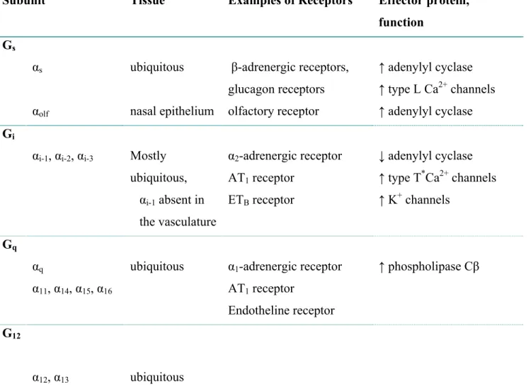
There are four classes of heterotrimeric G protein, including Gs, Gi, Gq and G12/13 (Table 1).
This classification is based on the comparison of amino acid sequences of the Gα subunits. Molecular cloning has revealed four different forms of Gsα (Gsα-1,Gsα-2, Gsα-3, Gsα-4) resulting from the differential splicing of one gene and three distinct forms of Giα, Giα-1 Giα-2, and Giα-3 encoded by three distinct genes (Bray, et al., 1986). A characteristic of the α-subunits of Gs is that they are inhibited by cholera toxin. Giα is distinguished by the ability of pertussis toxin to inactivate the protein by transfer of an ADP-ribose moiety from NAD to the α-subunit of the Giα protein. Giα also has been shown to inhibit adenylyl cyclase and directly couple cell membrane receptors to ion channels (Seamon & Daly, 1982).
Table 1 : classification of heterotrimeric G protein according to subunits
Subunit Tissue Examples of Receptors Effector protein,
function Gs
αs ubiquitous β-adrenergic receptors,
glucagon receptors
↑ adenylyl cyclase ↑ type L Ca2+ channels αolf nasal epithelium olfactory receptor ↑ adenylyl cyclase Gi
αi-1, αi-2, αi-3 Mostly
ubiquitous, αi-1 absent in the vasculature α2-adrenergic receptor AT1 receptor ETB receptor ↓ adenylyl cyclase ↑ type T*Ca2+ channels ↑ K+ channels Gq
αq ubiquitous α1-adrenergic receptor
AT1 receptor Endotheline receptor ↑ phospholipase Cβ α11, α14, α15, α16 G12 α12, α13 ubiquitous
Sources : Krauss, G. (2008). Biochemistry of signal transduction and regulation. (4e ed.). Weinheim; Chichester: Wiley-VCH .
*Lader, A. S., Xiao, Y. F., Ishikawa, Y., Cui, Y., Vatner, D. E., Vatner, S. F., et al. (1998). Cardiac Gsalpha
overexpression enhances L-type calcium channels through an adenylyl cyclase independent pathway. Proc Natl Acad Sci U S A, 95(16), 9669-9674.
Gαq is a 42 kDa protein that exhibit slow rate of GDP-GTP exchange and GTP hydrolysis in comparison to other Gα-subunits. Gαq activates PLCβ, but is refractory to ADP-ribosylation by cholera toxin or pertussis toxin. Go (G “other”) was first discovered as a 39 kDa pertussis toxin substrate in addition to Gi in the brain. Go is very similar to Gi, binds GTP, and has been shown to regulate muscarinic receptors for agonists in the brain (Fleming, et al., 1992).
1.1.2.2 Activation of guanine nucleotide proteins
In its unactivated state, a G protein maintains its heterotrimeric state and its Gα subunit binds GDP. The G protein is turned on by the interaction with an activated receptor (GPCRs) which induces the Gα subunit to exchange its bound GDP for GTP, followed by a dissociation of Gα-Gβγ complex (Fleming, et al., 1992). Subsequently, the Gα subunit dissociates from the Gα-Gβγ and binds to an effector such as adenylyl cyclase, activating the effector. The Gβγ dimer also binds to effectors such as ions channels, activate phospholipase Cβ, (PLCβ), and phospholipase A. Following hydrolysis of GTP, the Gα subunit will dissociate from the effector and reassociate with the Gβγ subunit to reform the inactive heterotrimeric G protein (Karp, 2008). The enzyme adenylyl cyclase is the catalytic unit of the hormone-sensitive adenylyl cyclase system (Voet, et al., 2008).
1.1.3 The enzyme adenylyl cyclase and its regulation
Many hormones and drugs interact with plasma membrane receptors to bring about the appropriate cellular response by generation of second messengers. The first such messenger to be identified was adenosine-3’,5’ cyclic monophosphate (cAMP), discovered by Sutherland and Rall in the late 1950s (Sutherland & Rall, 1958). Cellular functions are responsive to changes in concentrations of cAMP and thus to changes in the activities of adenylyl cyclases, the enzyme that catalyze the synthesis of cAMP from ATP (Voet, et al., 2008).
1.1.3.1 Structural organisation of adenylyl cyclases
Mammalian adenylyl cyclases are expressed in all tissues, but at very low levels, approximately 0.01-0.001% of membrane protein. Molecular cloning has permitted the identification of several novel isoforns of mammalian adenylyl cyclase. Ten different isoforms of adenylyl cylase have been identified, which differ in their primary sequence, tissue distribution and regulation (Taussig & Gilman, 1995). All of the isoforms of adenylyl cyclase are activated by the α-subunit of Gs, and by the diterpene forskolin. Certain isoforms (Type I, III) are also activated by Ca2+ -calmodulin, while some (Type I, V, VI) are inhibited by the α-subunit of the three Gi proteins, and Ca2+ (type V and VI). Type I and II adenylyl cyclase also appear to have independent sites for interaction with the βγ-subunits of G protein. The type I enzyme is strongly inhibited, while
type II, and IV adenylyl cyclase are activated provided Gsα is also present (Sunahara, Dessauer, & Gilman, 1996). N C1b C2b M1 M2 ATP cAMP + PPi 2Mg2+ Gsα, PKC, Gβγ Giα Ca2+, Ca2+ ·CaM, PKA C2a C1a
Figure 2: Diagram of a typical mammalian adenylyl cyclase.
The M1 and M2 domains each contain six transmembrane helices. C1a and C2a form the enzyme’s pseudosymetric catalytic core. The domains with which various regulatory proteins areknown to interact are indicated.
Abreviations: PKA, protein kinase A; PKC, protein kinase C; CaM, calmodulin. Source: From Voet. D, Voet.j.G., and pratt, C.W.(2006). Fundamental of Biochemistry: Life at the Molecular Level.2nd edition. Jonh wiley & Son, inc.
Adenylyl cyclases are large transmembrane proteins that consist of two bundles of six transmembrane segments. These 120 kD membrane-bound protein each consist of a small N-terminal domain, along with two repeats of a unit consisting of a transmembrane domain (M) followed by two consecutive cytoplasmic domains (C). The interaction between the C1 and C2
domains is essential for catalysis. The C1a and C2a domains associate to form the enzyme’s
catalytic core, whereas C1b as well as C1a and C2a bind regulatory molecules (Figure 2).The two
catalytic domains of the enzyme and their interactions provide a binding site for ATP that activates adenylyl cyclase to transform ATP to cAMP. Forskolin and Gsα bind to C2a to activate
adenylyl cyclase, and Giα binds to C1a to inhibit the enzyme. Other regulators of adenylyl
cyclase activity include Ca2+, calmodulin, protein kinase A (PKA), and protein kinase C (PKC) (Voet, et al., 2008).
1.1.3.2 Regulation of adenylyl cyclase activity
The identification of several isoforms of adenylyl cyclase, along with the discovery of distinct, type-specific modes of regulation allow for the classification of regulatory patterns.
1.1.3.2.1 Regulation by G protein subunits
Regulation of adenylyl cyclase by Gsα was the basis for the discovery of G protein. A ligand activated GPCR triggers the exchange of Gsα-GDP for GTP. GTP-bound Gsα subsequently undergoes conformational changes that facilitate dissociation from the βγ-subunit of G protein. Such dissociation allows the α-subunit to interact with adenylyl cyclase. Hydrolysis of Gsα-bound GTP to GDP terminates activation of adenylyl cyclase by Gsα-GTP after several seconds, then Gsα-GDP reassociates with βγ-subunits (Taussig, Tang, Hepler, & Gilman, 1994). The binding of G proteins to the receptors exposes the enzyme binding sites for ATP, which then catalyzes the transformation of ATP into cAMP. G protein βγ subunits inhibit type I adenylyl cyclase, and the effect of βγ is exerted directly on the enzyme. The concentrations of βγ subunits required to inhibit adenylyl cyclase are significantly higher than the concentration of Gsα required to activate the enzyme. The source of βγ subunits is presumed to be from Gi or Go since only low concentrations of βγ subunits can be achieved by activation of Gsα, while substantially higher concentrations can be obtained by activation of Gi or Go. Furthermore, inhibition of type I adenylyl cyclase activity by Giα is absent when the Gsα-stimulated activity is examined (Taussig & Gilman, 1995). The first evidences that Giα proteins could in fact inhibit adenylyl cyclases came from studies of a cell transfected with cDNAs encoding constitutively activated (GTPase deficient) mutants of various Gα subunits. Expression of activated Gsα-1,Gsα-2, Gsα-3 impaired accumulation of cAMP stimulated by either Gsα or forskolin (Sunahara, et al., 1996).
1.1.3.2.2 Regulation by phosphorylation
Adenylyl cyclase catalyzes the synthesis of cAMP from ATP. Cyclic AMP binds to regulatory subunits of the cAMP-dependent protein kinase A (PKA), thereby activating the kinase to phosphorylate specific Ser or Thr residues on target proteins in the cytosol, and nucleus (Voet, et al., 2008). Potential regulation of adenyly cyclase by protein kinase C has been explored. Studies have shown that phosphorylation of the enzyme by protein kinase C enhances the activity of Type II, VII, and V (type VI is inhibited) adenylyl cyclase. This is a consequence of the activation of GPCRs through Gq, followed by activation of PLCβ and generation of diacyl glycerol (DAG) (Gomperts, et al., 2002).
1.1.3.2.3 Regulation by Calcium and Forskolin
Calcium is an important regulator of adenylyl cyclases. Changes in intracellular Ca2+ concentration also affect the concentration of cAMP. In association with calmodulin, Ca2+ increases the activity of type I, III and VIII adenylyl cyclases. Intracellular concentrations of cAMP rise when transfected cells expressing isoforms of the enzyme are exposed to agonists that elevate intracellular Ca2+ (Taussig & Gilman, 1995). Another activator of adenylyl cyclase is forskolin, a diterpene isolated from the roots of Coleus forskohlii. Forskolin directly activates adenylyl cyclase, bypassing all upstream influences including receptors and GTP binding proteins. Forskolin also potentiates Gsα-mediated activation of adenylyl cyclase (Gomperts, et al., 2002).
1.2 The Adenylyl cyclase system in cardiovascular diseases
The Adenylyl cyclase/cAMP signal transduction system is one of the biochemical mechanisms that regulate arterial tone and reactivity. Hypertensive hormones such as epinephrine, isoproterenol increase cAMP formation in rat aorta through their stimulatory effect on adenylyl cyclase (Asano, Masuzawa, & Matsuda, 1988). Studies have shown that changes in cardiac contractility and myocardial metabolism that occur in heart diseases involved alteration of the membrane-bound adenylyl cyclase system (Baumann, et al., 1981). In heart failure and hypertension, several signal transduction defects leading to adenylyl cyclase desensitization have been demonstrated, such as β-adrenoceptor downregulation, increase of Giα proteins expression, and uncoupling of β-adrenergic receptors by an increase of receptor kinase activity (Castellano &
Bohm, 1997). Studies have provided evidence that the increase of Giα and desensitization of adenylyl cyclase are common features of cardiac hypertrophy in polygenic, monogenic, and acquired types of hypertension (Michel, Brodde, & Insel, 1993)
1.2.1 Blood Pressure
The hydrostatic force that blood exerts against the wall of a vessel and that propels blood is called blood pressure. Blood pressure is much greater in arteries than in veins and is highest in arteries when the heart contracts during ventricular systole (Campbell & Reece, 2005). The maximum pressure exerted in the arteries when blood is ejected into them during systole averages 120 mmHg in humans. The minimum pressure within the arteries when blood is drained off into the remainders of the vessels during diastole averages 80 mm Hg. In human, blood pressure is determined partly by cardiac output and partly by peripheral resistance. Contraction of smooth muscles in the walls of the arterioles constricts the tiny vessels, increases peripheral resistance, and therefore increases blood pressure upstream in the arteries. When the smooth muscles relax, the arterioles dilate, blood flow through the arterioles increases, and the pressure in the arteries falls (Guyton & Hall, 2006). Nerve impulses and hormones control arterioles wall muscles. Stress, both physical and emotional, can raise blood pressure by triggering nervous and hormonal responses that will constrict blood vessels. Cardiac output is adjusted in concert with changes in peripheral resistance. This coordination of regulatory mechanisms maintains adequate blood flow as the demand on the circulatory system changes (Sherwood, Klandorf, & Yancey, 2005).
1.2.2. Mechanisms of blood pressure regulation
Development and maintenance of a level of arterial blood pressure adequate to perfuse the tissues is required for the survival of all mammals. Rapid alterations of arterial blood pressure are stabilized by neural reflex and hormonal mechanisms. The nervous system detects changes in arterial pressure and provides both rapid stabilization and long term control of blood pressure by adjusting level of sympathetic tone (Cowley, 1992). The short and long-term regulation of arterial blood pressure is achieved by the interaction of the baroreceptors reflexes, the renin-angiotensin system and the sympathetic nervous system (Reid, 1992).
1.2.2.1. The Renin-Angiotensin system in the regulation of blood pressure
The renin-angiotensin system plays an important role in the regulation of renal sodium and water excretion, and thus in maintaining body sodium and fluid balance. All components of the renin-angiotensin system are present in the kidney. Renin is secreted by the juxtaglomerular cells in the form of prorenin after proteolytic removal of 43-amino acid residue at the N-terminus of prorenin. The active form of renin contains 339-343 amino acid residues, with a mass of 37 kDa. Renin provides a pathway for the secretion of Angiotensin I which is rapidly converted into Angiotensin II (Ang II) by angiotensin converting enzyme (ACE). Ang II, a peptide hormone, is the primary product of the renin-angiotensin system. The main effects of Ang II are control of cardiovascular, renal and adrenal functions (Carey, 2007). The kidneys secrete the hormone rennin in response to reduced sodium chloride, extracellular fluid volume, and arterial pressure. Renin then activates angiotensinogen, a plasma protein produced by the liver, into angiotensin I, which is converted into AngII. Ang II stimulates the adrenal cortex to secrete aldosterone, which stimulates sodium reabsorption by the kidneys. The increased sodium reabsorption by the distal portion of the tubule induces water retention, which helps restore the plasma volume, thus being important in the long-term control of blood pressure (Sherwood, et al., 2005).
1.2.3 Hypertension
Hypertension is a cardiovascular condition characterized by sustained high blood pressure. A mean arterial blood pressure greater than 110 mm Hg under resting condition is considered to be hypertensive; at that level, the diastolic blood pressure is greater than 90 mm Hg and the systolic greater than 135 to 140 mm (Guyton & Hall, 2000). Hypertension is generally classified as either primary (essential), or secondary hypertension. Essential hypertension or hypertension of unknown causes, accounts for more than 90% of cases of hypertension. Many factors have been implicated in the genesis of essential hypertension: overproduction of sodium-retaining hormones and vasoconstriction, long-term high sodium intake, sedentary lifestyle, and inappropriate renin secretion.
It is estimated that 43 million people in the United States have hypertension or are taking hypertensive medication, which represents 24% of the adult population (Carretero & Oparil, 2000). According to the Canadian hypertension society, hypertension is a substantial health concern in Canada, affecting over five millions people (Canadian Hypertension Society, 2009).
The incidence of hypertension increases with age. Its prevalence is approximately 50% in the population aged 60 to 69 years, and increases to 70% in those older than 70 years (Suri & Qureshi, 2006) Several studies have reported the impact of a variety of factors including age, race, gender, and body mass index on hypertension. Hypertension is a major risk factor for myocardial infarction, stroke, congestive heart failure, and renal diseases (Mulvany, 1991). Despite major research efforts, it remains uncertain what triggers hypertension in the general population. In most cases, genetic as well as environmental factors are responsible for the pathogenesis of hypertension (Lifton, Gharavi, & Geller, 2001). Studies have shown that mutations of a family of protein kinases genes WNK1, and WNK4 (With-no-Lysine Ks) cause Gordon’s syndrome, a rare Mendelian form of hypertension by increasing renal sodium retention (Huang, Kuo, & Toto, 2008). During hypertension, small arteries undergo functional and structural changes, resulting in reduced lumen size and increase peripheral resistance (Lehoux & Tedgui, 1998).
1.2.3.1 Physiology of hypertension on vascular smooth muscle cells
During hypertension, the vascular wall is constantly subjected to mechanical forces in the form of stretch or tensile stress due to blood pressure, and shear stress due to blood flow. Shear stress is principally sensed by endothelial cells located at the interface between the blood and the vessel wall. Functional changes in either shear stress or stretch result in vascular remodelling. The processes involved in vascular remodelling include cellular hypertrophy and hyperplasia, as well as enhanced protein synthesis or extracellular matrix protein reorganization (Lehoux & Tedgui, 1998). The primary hemodynamic characteristic of essential hypertension is increased peripheral vascular resistance which is associated with structural and functional alterations of the vasculature (Korner, Bobik, Angus, Adams, & Friberg, 1989). The structural changes include reduced vessel lumen diameter and media thickening. At the cellular level, there are hyperplasia, hypertrophy, and elongation of VSMC resulting in a smaller lumen and outer diameter (Korsgaard, Aalkjaer, Heagerty, Izzard, & Mulvany, 1993; Mulvany, Baandrup, & Gundersen, 1985). Many factors regulate VSMC function, including vasoactive peptides, such as Ang II and endothelin-1 (ET-1) that stimulate vasoconstriction and growth, and vasorelaxing factors, such as nitric oxide, prostacyclin, and C-type natriuretic peptide that induce vasodilation (Rubanyi, 1991). Vascular smooth muscle cells (VSMC) are central to these events. VSMCs detect
mechanicals stimuli resulting from pulsatile stretch and transduce them into intracellular signals leading to modulation of gene expression and cellular functions such as proliferation, apoptosis, migration, and remodelling (Haga, Li, & Chien, 2007).
Using neonatal rat aortic VSMCs, Wilson and colleagues have shown that cyclical mechanical strain induces the production of platelet-derived growth factor (PDGF) αα and ββ, and that the strain-induced growth of VSMC was dependent on the autocrine action of PDGF (Wilson, Mai, Sudhir, Weiss, & Ives, 1993). Stretching of VSMCs induces a rapid phosphorylation of PDGF receptor α independent of its ligand (PDGF) activation (Hu, Bock, Wick, & Xu, 1998). Stretch also induces the phosphorylation of epidermal growth factor receptor (EGFR) and its recruitment of adaptor proteins Shc and Grb2 (growth factor receptor-bound2), which in turn activate extracellular regulated kinases (ERK1/2) (Iwasaki, Eguchi, Ueno, Marumo, & Hirata, 2000). Several stretch-induced protein kinase molecules have been identified in VSMCs, including, Phosphoinositide-3 kinases (PI3Ks), protein kinase C, nuclear factor kappa-light-chain-enhancer of activated B cells (NFkB), Rho family GTPases, and mitogen activated protein kinase (MAPKs). Stretching of VSMCs induces PI3K and PKB/AKT activation which can be inhibited by pre-treatment of VSMCs with N-acetylcysteine, a scavenger of reactive oxygen species (ROS) suggesting the role of ROS in the mechanotransduction of VSMCs (Zhou, et al., 2003).
1.2.3.2 Alteration of G proteins expression in hypertension
Heterotrimeric guanine nucleotide proteins have been shown to be implicated in various pathological conditions including hypertension, diabetes, and heart failures. Alteration of Giα proteins levels and adenylyl cyclase activity have been reported in cardiovascular tissus from spontaneously hypertensive rats (SHR) and various model of hypertension including deoxycorticosterone acetate (DOCA)-salt hypertensive rats (Marcil, de Champlain, & Anand-Srivastava, 1998). An increased expression of Giα-1, and Giα-2 protein and mRNA in hearts and aortas from SHR, as well as in hearts from DOCA-salt hypertensive rats with established hypertension has been demonstrated (Marcil, et al., 1998). The enhanced expression of Giα protein was shown to occur before the onset of hypertension in SHR and DOCA-salt suggesting the implication of increased expression of Giα protein in hypertension (Marcil, Thibault, & Anand-Srivastava, 1997). Our laboratory has shown that overexpression of Giα-2 and Giα-3 proteins precedes the development of hypertension, and that inactivation of the enhanced Giα
proteins expression by pertussis toxin attenuates the development of high blood pressure in SHR (Li & Anand-Srivastava, 2002). Furthermore, we have reported that volume overload cardiac hypertrophy exhibits decreased expression of Gsα and not of Giα in hearts of Sprague-Dawley rats (Di Fusco, Hashim, & Anand-Srivastava, 2000). TsuTsui et al. have reported impaired levels of endothelial Giα proteins in atherosclerotic coronary arteries (Tsutsui, et al., 1994). Reduced function of Gsα in β adrenoreceptor-adenylyl cyclase system of femoral arteries isolated from SHR has also been reported (Asano, Masuzawa, Matsuda, & Asano, 1988).
1.2.3.3 Alteration of adenylyl cyclase/cAMP activity in hypertension
The elevation of blood pressure in essential hypertension is due to an increase in the peripheral resistance of vessels. The increase of the peripheral resistance is attributed to structural changes in the vessels, abnormalities in Ca2+ movement, and aberration in cyclic nucleotides metabolism (Anand-Srivastava, 1996). It has been shown that the adenylyl cyclase/cAMP system is one the biochemical mechanism participating in the regulation of arterial tone (Triner, Vulliemoz, Verosky, Habif, & Nahas, 1972). Decreased cAMP level in cardiovascular tissues have been implicated in the pathogenesis of hypertension (Ramanathan & Shibata, 1974). The adenylyl cyclase/cAMP system is involved in both the control of heart contractility, and vascular smooth muscle tone (Bar, 1974). Reduced adenylyl cyclase activity in response to β-adrenergic
stimulation has been demonstrated in mesenteric vasculature (Amer, Gomoll, Perhach, Ferguson, & McKinney, 1974), aorta and myocardium of spontaneously SHR (Anand-Srivastava, 1988). Bohm and colleagues have reported the positive inotropic effect of cAMP-phosphodiesterase inhibitor pimobendan in failing human myocardium, and the role played by other factors such as increased Ca2+ sensitivity of myofilaments in the increase in force of contraction (Bohm, et al., 1991). In addition to the effect of PKA on myosin light chain kinase (MLCK), G12/13 proteins
also play a key role in the regulation of vascular tone (Siehler, 2009). Activation of G12/13 by
ligands such as Ang II, and endothelin activates the guanine nucleotide exchange protein pp115-RhoGEF, which in turn catalyzes the exchange of bound GDP for GTP on the small GTPase RhoA. Downstream target of RhoA-GTP include ROCK, which mediates cell contraction through the inhibition of myosin light chain phosphatase (MLCP) (Sward, et al., 2003).
1.3 Oxidative stress, redox signalling and cardiovascular diseases
Oxidative stress is caused by the overproduction of reactive oxygen species (ROS). An imbalance between oxidants and antioxidants in favour of the oxidants leads to oxidative stress. Oxidants are produced as normal products of aerobic metabolism, but can also be formed at higher rates under pathological conditions. Antioxidants include enzymatic and non-enzymatic molecules capable of inhibiting the oxidation of other proteins or molecules thereby maintaining them in their reduced state (Baynes, 1991). Antioxidants include enzymes such as catalase, superoxide dismutase (SOD), glutathione peroxidase, and molecules such as tocopherols (Vitamine E), glutathione, ascorbic acid (Vitamine C), and flavonoids (Ames, Shigenaga, & Hagen, 1993). Reactive oxygen species (ROS) are highly reactive oxygen-containing molecules due to the presence of unpaired valence shell electrons. Reduction-oxidation (redox) reactions that generate ROS are important chemical processes that regulate signal transduction (Thannickal & Fanburg, 2000).
1.3.1 Sources and chemistry of reactive oxygen species
ROS include both free radicals, which have oxygen or nitrogen-based unpaired electrons, and other species such as hydrogen peroxide (H2O2) that act as oxidants. A primary form of reactive
oxygen species (ROS) is superoxide anion (O2-) but biological systems produce other species
including hydroxyl anion (HO-), peroxynitrite (ONOO-) and hydrogen peroxide (H2O2). In the
vasculature, ·O2- anion is produced by a one electron reduction of oxygen using Nicotinamide
adenine dinucleotide phosphate (NAD(P)H) as an electron donor (Taniyama & Griendling, 2003). Superoxide has an unpaired electron, which conferts high reactivity and renders the molecule unstable. Hydrogen peroxide is mainly produced from dismutation of ·O2-. This
reaction can be spontaneous or catalyzed by SOD, of which there are three isoforms, CuZnSOD, MnSOD, and extracellular SOD (EC-SOD) (Fridovich, 1997).
The SOD-catalyzed dismutation is favoured when the concentration of O2- is low and the
concentration of SOD high. Unlike ·O2-, H2O2 is not a free radical and is a much more stable
molecule. It is lipid soluble, crosses cell membrane, and has a longer half-life than ·O2- (Han,
Antunes, Canali, Rettori, & Cadenas, 2003). In biological system, H2O2 is scavenged by catalase
and glutathione peroxidase to produce H2O molecules (Schafer & Buettner, 2001). Hydrogen
containing-molecules such as Fe2+ (Fridovich, 1997). When ·O2- is produced in excess, a
significant amount of ·O2- reacts with oxide nitrate (NO) to produce ONOO-. In the vasculature, ·O2-, H2O2, ONOO-, NO, and HO- are produced to varying degrees. Their production is regulated
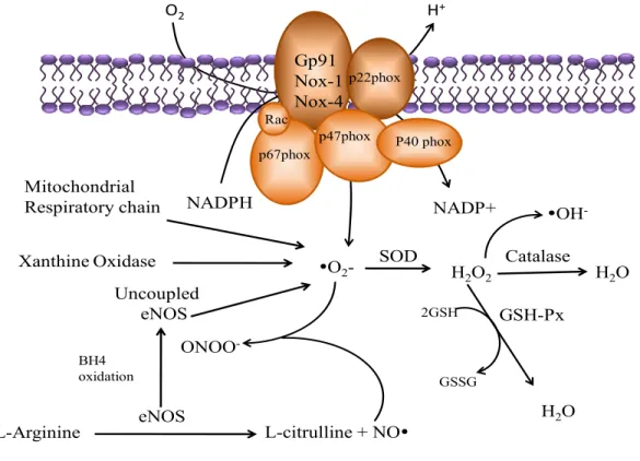
by anti-oxidant enzymes such as catalase, SOD, thioredoxin, glutathione (Thannickal & Fanburg, 2000). Catalase NADPH NADP+ yO2 -GSH-Px H2O2 GSSG 2GSH yOH -Gp91 Nox-1 Nox-4 p22phox p67phox p47phox Rac P40 phox O2 H2O ONOO -L-citrulline + NOy eNOS L-Arginine H2O BH4 oxidation Uncoupled eNOS Xanthine Oxidase Mitochondrial Respiratory chain H+ SOD
Figure 3. Generation of ·O2- and H2O2 from NAD(P)H oxidase. Many enzyme systems, including NAD(P)H
oxidase, xanthine oxidase, and uncoupled nitric oxide synthase (NOS) have the potential to generate reactive oxygen species. NAD(P)H oxidase is a multisubunit enzyme, comprising gp91phox( or its homologues, Nox1 and Nox4), p22phox, p47phox, p67phox, and p40phox. Abreviations: SOD, superoxide dismutase; GSH, glutathione ; GSH-PX, glutathione peroxydase ; GSSG, glutathione disulfide ; H2O2, hydrogen peroxide ; Mox-1, mitogen oxydase 1 ; NAD(P)+, nicotinamide-adenine-dinucleotide ; NAD(P)H, nicotinamide-adenine-dinucleotide-phosphate ; NAD(P)H oxidase, nicotinamide-adenine-dinucleotide-phosphate oxidase ; yNO-, nitric oxide ; yO2-, superoxide radical ; yOH-, hydroxyl radical ; ONOO-, peroxynitrite ; Rac, small G protein. Source. Tamara, M.P., Touyz, R.M. Redox signaling in hypertesion. (2006). Cardiovascular research.71 :247-258.
Under normal conditions, the rate of ROS production is balanced by the rate of elimination. Oxidative stress is the result of the imbalance between the production of ROS and the cellular
anti-oxidant capability of biological system. In biological system, ROS have enzymatic sources and non enzymatic sources multiple sources (Touyz & Schiffrin, 2004).
1.3.1.1 Enzymatic sources of reactive oxygen species
Cellular production of ROS occurs from both enzymatic and nonenzymatic sources. Any electron-transferring protein or enzymatic system can result in the formation of ROS as by-products of electron transfer reactions. The generation of ROS in the mitochondria accounts for 1-2% O2-, of total production under reducing conditions (Thannickal & Fanburg, 2000). In the
vasculature, several enzyme systems contribute to ROS formation, including the NAD(P)H oxidases, xanthine oxidase, endothelial NO synthases, enzymes of the mitochondrial respiratory chain, lipoxygenases, cytochrome P450 monoxygenases, and cyclooxygenases (Clempus & Griendling, 2006). Mitochondria generate ROS as byproducts during ATP production via electron transfer through cytochrome c oxidases. NAD(P)H oxidase is one of the major sources of ROS in the vasculature. Xanthine oxidoreductase catalyzes the oxidation of hypoxanthine into xanthine in the process of purine metabolism. Xanthine oxidoreductase exists in two interconvertible forms, either as xanthine dehydrogenase or xanthine oxidase. The former reduces NAD+, whereas the latter reduces molecular oxygen leading to the production of ·O2-
and H2O2 (Touyz, 2004).
Nitric oxide synthase (NOS) generates ·O2- in addition to NO. Members of NOS are encoded by
different genes. There are three isoforms including endothelia NOS (eNOSor NOS3), neuronal NOS (nNOS or NOS1) and inducible NOS (iNOS, or NOS1) (Gilkeson, et al., 1997). NOS uses L-arginine as a substrate to synthesize NO in a tetrahydrobiopterin (H4B)-dependent manner. If
the concentration of L-arginine or H4B is low, or if H4B is oxidized, eNOS becomes uncoupled
and generates significant amounts of ·O2- (Stuehr, Pou, & Rosen, 2001). eNOS uncoupling has
been demonstrated in atherosclerosis, diabetes, and hypertension, all of which are associated with activation of the renin-angiotensin system and production of O2- from eNOS (Taniyama &
Griendling, 2003).
1.3.1.2 Non enzymatic sources of reactive oxygen species
Autooxidation of small molecules such as dopamine, epinephrine, flavins and hydroquinones can be an important source of intracellular ROS production. In most cases, the direct product of such
autooxidation reactions is O2- (Freeman & Crapo, 1982). Peroxisomes are an important source of
total cellular H2O2 production. They contain a number of H2O2-generating enzymes including
glycolate oxidase, acid oxidase, urate oxidase, and fatty acyl-CoA oxidase (Thannickal & Fanburg, 2000). The cellular production of ROS may trigger the production of more ROS via a radical chain reaction. The reaction between ROS and polyunsaturated fatty acids within cell membrane may result in a fatty acid peroxyl radical (R-COO-) that will react with adjacent fatty acid side chains and initiate production of more lipid radicals (Zalba, et al., 2001). Arachidonic acid metabolism involving lipoxygenase and cyclooxygenase-dependent pathway leading to leukotriene synthesis has been reported to generate ROS. Lipoxygenase activity also has been implicated in redox-regulated signalling by Ang II, and EGF (Thannickal & Fanburg, 2000).
1.3.2 The NADPH oxidases
The NADPH oxidases are enzymes that catalyze the production of superoxide from oxygen and NADPH. They are present in vascular tissue and phagocytic cells such as neutrophils, macrophages, and eosinophils (Griendling, Sorescu, & Ushio-Fukai, 2000).
1.3.2.1 Structure and expression profile of vascular NADPH oxidases
The vascular NAD(P)H oxidase is a multimeric protein complex responsible for the formation of
·O2-. In vascular smooth muscle cells (VSMC), Oሶ2- and H2O2 production are mainly intracellular
(Griendling, Minieri, Ollerenshaw, & Alexander, 1994).The vascular NAD(P)H consists of four major subunits: a cytochrome b558, comprising of two cell membrane-associated gp91phox (or gp91phox (nox2) homologues, nox1 and nox4) and p22phox, and two cytosolic components, p47phox and p67phox. A low molecular weight G protein rac participates in subunits assembly and activation of the enzyme (Griendling, Sorescu, Lassegue, & Ushio-Fukai, 2000). The rac proteins are kept inactive by binding to a guanine nucleotide dissociation inhibitor, which prevents the exchange of guanine nucleotides from the rac proteins. An essential component of the NAD(P)H oxidase is gp91phox to which are bound the electrons carrying components of oxidase such as flavine adenine dinucleotide, and a pair of hemes molecules. P47phox is the protein that carries the cytosolic proteins to the membrane proteins to assemble the active oxidase. p67phox contains two Src homology 3(SH3) domains, one in the middle of the protein, and one near the carboxyl terminus. The SH3 domains interact with p22phox to activate the
enzyme. p22phox is located in the membrane, along with gp91phox and has a tail in the cytosol. When p22phox is phosphorylated, it binds to p47phox, an interaction that is critical in the activation of the enzyme (Brandes & Kreuzer, 2005). The distribution of catalytic subunits of NAD(P)H oxidase in VSMC is tissue and species-specific. Aortic smooth muscle cells express Nox1 and Nox4 in rodents, and also Nox5 in humans. In contrast, VSMC from human resistance arteries contain Nox2 and Nox4, but no Nox1 (Clempus & Griendling, 2006). Vascular NAD(P)H oxidase is a constitutive enzyme, but it is regulated by Ang II, platelet derived growth factors (PDGF), thrombin, and tumor growth factor-α (Zalba, et al., 2001).
1.3.3 Oxidative stress in hypertension
The impact of ROS in vascular functions and the development of hypertension has been studied extensively. It has been shown that·O2- inactivates endothelium-derived NO, one of the most
important vasodilator, thereby promoting vasoconstriction (Zicha, Dobesova, & Kunes, 2001). In order to counteract the hypertensive effect of ROS, several studies have used exogenous administration of antioxidants to reduce blood pressure in animal model (Boshtam, Rafiei, Sadeghi, & Sarraf-Zadegan, 2002), and in human hypertension (Boshtam, et al., 2002; Duffy, et al., 2001). Nevertheless, the results of such studies are not conclusive and the relationship between oxidative stress and hypertension is continuously being investigated.
1.3.3.1 Oxidative stress in genetic models of hypertension
Recent work by Suzuki and associates (1995) provided evidence regarding the role of ROS in the pathophysiology of hypertension. They demonstrated that O2- is increased in venules and
arterioles of SHR, and the administration of heparin-binding SOD, which is localized within the vessel wall normalised the blood pressure of SHR (Suzuki, Swei, Zweifach, & Schmid-Schonbein, 1995). Genetic models of hypertension, such as SHR and stroke-prone SHR exhibit enhanced NAD(P)H oxidase-mediated ·O2- generation in resistance arteries, aorta, and kidneys.
These processes are associated with increase expression of NAD(P)H oxidase subunits particularly p22phox, and p47phox (Touyz & Schiffrin, 2004). We have shown an increase expression of NAD(P)H oxidase subunits Nox4, and p47phox in VSMC from SHR (Saha, Li, & Anand-Srivastava, 2008). Furthermore, Fukui et al demonstrated that chronic infusion of Ang II in normotensive rats upregulates vascular p22phox mRNA and increases NADPH
oxidase-derived superoxide. Both the hypertension and the increase in p22phox mRNA were prevented by pretreatment with SOD (Fukui, Lassegue, Kai, Alexander, & Griendling, 1995). Diminished NO bioavailability as a consequence of enhanced vascular ·O2- generation also contribute to
oxidative stress in SHR and stroke-prone SHR. Treatment of rats with antioxidant vitamins, NADPH inhibitors, AT1 blockers, BH4, and SOD mimetics attenuate to varying degree the
development of hypertension in SHR and stroke-prone SHR (Sharma, Hodis, Mack, Sevanian, & Kramsch, 1996).Vascular oxidative stress has also been demonstrated in DOCA-salt rats. An enhanced ·O2- production present in the aorta of these rats is associated with an increased
NAD(P)H oxidase activity due to the increased vascular Ang II release as a consequence of nephrectomy (Zalba, et al., 2001). NO is an important endogenous antihypertensive factor that plays a role in sodium excretion and the regulation of blood pressure. A dysfunction of NOS in tissues has been observed in Dahl salt-sensitive rats. In addition, genetic deletion of eNOS has been proven to lead to hypertension in mice. NOS inhibitor L-nitro-arginine methyl ester (L-NAME) leads to a decrease in blood flow, the retention of sodium, and the development of hypertension in rats (Nakanishi, Mattson, & Cowley, 1995).
1.3.3.2 Oxidative stress in human hypertension
Clinical studies have shown the occurrence of increased ROS production in humans with essential hypertension. The level of ROS scavengers, such as vitamine E, glutathione, and SOD, have been reported to be decreased in hypertensive patients (Sagar, Kallo, Kaul, Ganguly, & Sharma, 1992). Berry et al have shown that NAD(P)H oxidase is the source of basal·O2-
production in human internal arteries and saphenous veins, and that Ang II increases ·O2- in
human arteries. This effect is mediated by NADPH oxidase and inhibited by the AT1 receptor antagonist losartan (Berry, et al., 2000). Activation of the renin-angiotensin system has been proposed as a major mediator of NAD(P)H oxidase activation and ROS production in human hypertension. Some of the therapeutic blood pressure-lowering effects of AT1 receptors blockers and ACE inhibitors have been attributed to inhibition of NADPH oxidase activity (Touyz & Schiffrin, 2004). Several reports have shown the role of Ang II in NADPH subunits expression and activation (Touyz, et al., 2003).
1.3.4 Vasoactive peptides as inducers of oxidative stress
Vasoactive peptides such as Ang II and endothelin-1 mediate part of their responses through ROS generation (Griendling & Ushio-Fukai, 2000; Li, Watts, et al., 2003). In conditions associated with vascular damage such as hypertension, these peptides increase ROS production by activating NADPH oxidase (Touyz, Yao, Viel, Amiri, & Schiffrin, 2004).
1.3.4.1 Oxidative stress and angiotensin II
Ang II, a key component of the renin-angiotensin system regulates blood pressure, plasma volume via aldosterone-regulated sodium excretion, sympathetic nervous activity, and also plays a role in vascular remodelling in hypertension (Touyz & Schiffrin, 2000). In mammalian cells, Ang II mediates its effects via at least two plasma membrane receptors, AT1 and AT2. These
receptors belong to the 7-transmembrane, GPCR family (Horiuchi, Akishita, & Dzau, 1999). Ang II, also regulates a variety of physiological functions including cell growth, and apoptosis (Paul & Ganten, 1992). Ang II stimulates many signalling pathways leading to cell contraction and cellular hypertrophy. In VSMCs, Ang II induces cellular hypertrophy by acting via G protein coupled AT1 receptors (Berk, Vekshtein, Gordon, & Tsuda, 1989; Geisterfer, Peach, & Owens, 1988). In endothelial and VSMc, Ang II increases the production of ·O2- via the activation of
membrane-associated NAD(P)H oxidase. The ·O2- production upon Ang II stimulation is rapidly
converted to H2O2 by SOD (Munzel, Hink, Heitzer, & Meinertz, 1999).
Griendling et al have demonstrated that Ang II-induced cellular hypertrophy is mediated by intracellularly produced H2O2. The reduction of NAD(P)H oxidase activity by transfection of
antisense p22phox inhibits both H2O2 production and hypertrophy. In addition, infusion of Ang
II in rats upregulated vascular p22phox mRNA and increased NAD(P)H oxidase-derived ·O2-
(Griendling, et al., 1994). It has been reported that Ang II treatment of A10 VSMC increases the production of ·O2- and the expression of Nox4 and p47phox proteins of NADPH oxidase (Saha, Li,
& Anand-Srivastava, 2008). Furthermore, the Ang II-induced phosphorylation of ERK1/2 is due to enhanced oxidative stress, since treatment of A10 VSMC with Diphenyleneiodonium (DPI) an inhibitor of NAD(P)H oxidase attenuates the Ang II-induced phosphorylation of ERK1/2 (Li, Lappas, & Anand-Srivastava, 2007).
1.3.4.2 Oxidative stress and endothelin-1
Endothelin-1 (ET-1) is a 21-amino-acid polypeptide produced by vascular endothelial cells. ET-1 has been implicated in the pathophysiology of many cardiovascular diseases including hypertension, atherosclerosis and hypercholesterolemia (Barton, et al., 1998). In addition to its vasoactive effects, ET-1 also induce cell proliferation, tissue remodelling and cell survival in VSMCs and endothelials cells (Salani, et al., 2000; Ziche, Morbidelli, Donnini, & Ledda, 1995). ET-1 has been shown to exert its biological effects through binding to specific G protein-coupled membrane receptors ETA, and ETB subtypes (Pollock, 2005). It has been shown that ET-1
activates NAD(P)H oxidase and induces ·O2- production in cultured endothelial and smooth
muscle cells (Callera, Tostes, Yogi, Montezano, & Touyz, 2006). Although Ang II is a major stimulus of vascular production of Oሶ2- in high-angiotensin hypertension, studies have
demonstrated that ET-1 plays a major role in increasing vascular ·O2- in low-renin hypertension,
such as DOCA-salt model, an effect that is partially mediated by the ETA receptor/NAD(P)H
oxidase pathway (Li, Fink, et al., 2003).
1.4 Oxidative stress and signalling
Reactive oxygen species can trigger the activation of several signalling pathways that influence cytotoxicity, cell proliferation, and apoptosis. ROS are responsible for the phosphorylation of a variety of proteins kinases and transcription factors (Wang, Martindale, Liu, & Holbrook, 1998).
1.4.1 Receptor tyrosine kinase as mechanism of signal transduction
Protein-tyrosine phosphorylation is a mechanism of signal transduction that appeared with the evolution of multicellular organisms. Over 90 differents protein-tyrosine kinases are encoded by the human genome. These kinases are involved in the regulation of growth, cell migration and differentiation, inflammation, and apoptosis. Protein-tyrosine kinases are divided in two groups: Receptor protein-tyrosine kinases (RTKs) which are integral membrane proteins with an extracellular ligand binding domain, and non-receptor or cytoplasmic protein tyrosine kinases (Karp, 2008). Insulin and many other growth factors such as EGF and PDGF do not act via GPCRs and cAMP dependent pathways. Instead, these hormones and growth factors bind to RTKs, whose C-terminal domains have tyrosine kinase activity. RTKs contain a single transmembrane segment and are monomers in the unliganded state. Ligand binding causes two
monomeric receptors to form a dimer, a process called dimerization. The insulin receptor is unusual in that it is a dimer in the unliganded state; therefore ligand biding does not induce conformational change in the receptor. When a RTK dimerizes, the cytoplasmic tyrosine kinase cross-phosphorylate each other on specific tyrosine residues. This autophosphorylation activates the tyrosine kinase so it can phosphorylate other substrates (Voet, et al., 2008).
1.4.2 Signalling pathways activated by Reactive oxygen species
ROS are involved in the regulation of many signal transduction pathways. They influence cellular processes associated with growth and inflammation. Exogenous H2O2 affects the
function of various proteins, including protein kinases, protein phosphatases, transcription factors, phospholipases, ion channels, and G proteins (Rhee, Bae, Lee, & Kwon, 2000). ROS activates mitogen-activated protein (MAP) kinases, ERK1/2, p38MAP kinase, c-jun N-terminal kinase (cJNK) (Touyz & Schiffrin, 2004). Non-receptor tyrosine kinases such as c-Src, Pyk2, janus kinase (JAK2) are regulated by ROS. Reactive oxygen species modulate intracellular free Ca2+ concentration, an important determinant of vascular contraction and dilation. H2O2
increases Ca2+ in VSMC and endothelial cells. These effects are attributed to redox-dependent inositol triphosphate-induced Ca2+ mobilization which increases Ca2+ influx and decreases Ca2+ -ATPase activation (Lounsbury, Hu, & Ziegelstein, 2000). Exogenous H2O2 induces tyrosine
phosphorylation and activation of PDGFR and EGFR, probably due to ROS-mediated inhibition of tyrosine phosphatases. Protein tyrosine phosphatases (PTP) are susceptible to oxidation and inactivation by ROS (figure 4) (Touyz & Schiffrin, 2004). Sue Goo Rhee et al, have shown that Growth factors-induced H2O2 production requires the activation of phosphatidyl inositide
3-kinase (PtdIns 3-3-kinase) which subsequently provide phosphatidylinositol-3,4,5-triphosphate that recruits and activates a guanine nucleotide exchange factor of Rac, which is required for the activation of NADPH oxidase (Rhee, Chang, Bae, Lee, & Kang, 2003).
Figure 4. Redox-dependent signaling pathways by ROS in VSMC. Intracellular ROS activates redox-sensitive
MAP kinases, tyrosine kinases, ions channels, and MMPs. Tyrosine phosphatases are negatively regulated by ROS, which further increases the activity of tyrosine kinase and MAP kinase. ROS also influence gene expression by activating transcription factors. Activation of these redox-sensitive pathways results in cellular responses including altered vascular tone, increased VSMC growth, inflammation, and increase deposition of extracellular matrix protein, leading to vascular remodelling, and hypertension. Abreviations: ROS, reactive oxygen species; AngII, angiotensin II; SOD, superoxide dismutase; MMP, matrix metalloproteinase; MAPK, mitogen activated protein kinase; +, stimulatory effect; -, inhibitory effect. Sources. Touyz M.R., (2005). Intracellular mechanisms involved in vascular remodelling of resistance arteries in hypertension: role of angiotensin II. Experimental Physiology.90:449-455. Touyz, R.M., Schiffrin, E.L. Reactive oxygen species in vascular biology: implications in hypertension.(2004).122:339-352.
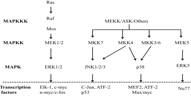
1.4.3 Reactive oxygen species and Mitogen activated proteins kinases
Mitogen-activated protein kinases (MAPK) are serine/threonine kinases whose phosphorylation mediates nuclear transduction of extracellular signals leading to activation of transcription factors, and enhance gene expression. The mammalian MAPKs are grouped into six major
subfamilies, among which three are best characterized: the extracellular-regulated kinase1/2 (ERK1/2) also known as p44-kDa MAPK and p42-kDa MAPK respectively, the c-jun N-terminal protein kinase (c-JNK), and the p38 MAPK. This classification is based on the presence of different amino acids in their phosphorylation motif, Glu for ERK1/2, Pro for c-JNK, and Gly for p38 MAPK (Touyz & Berry, 2002).
1.4.3.1 The extracellular-regulated kinase (ERK) pathway
The extracellular-regulated kinase (ERK) signaling pathway was the first MAP kinase cascade to be characterized. The ERK pathway is involved in cell growth, proliferation and survival. There are several ERKs, the best characterized are: the 44-kDa MAPK (ERK1), the 42-kDa MAPK (ERK2), and the 63k-Da MAPK (ERK3).The 42-kDa MAPK was the first mammalian MAPK to be identified as a 42-kDa protein that increases its phosphorylation upon stimulation by mitogens, hence the name. It was later found that other stimuli such as growth factors, cytokines, and ligands for G protein linked receptors also activate p42MAPK (Torres, 2003). In the heart, ERK1 is the most highly expressed ERK. Induction of MAPK activation involves phosphorylation by a MAPK also known as MEK. MEKs (MEK1/2) are regulated by other MEK kinases, including Raf-1, and Mos. MEK1 and MEK2 function as upstream MAPKK and the Raf proteins as MAPKKK (figure4). MEKs activate MAPKs by dual phosphorylation on a tyrosine and threonine residue lying within the phosphorylation motif (TyrXThr) in the activation loop (Ruwhof & van der Laarse, 2000).
The ERK pathway can be stimulated upon G protein coupled receptor activation by hormones such as ET-1, Ang II, and through receptors protein-tyrosine kinases activation by growth factors. The ERK pathway can also be stimulated by non receptor protein tyrosine kinase such as c-Src (Chang & Karin, 2001). The ERK phosphorylation cascade is initiated by the binding of a hormone such as Ang II to AT-1 receptors, which induces Shc-Grb2-Sos formation (tyrosine phosphoryaltion of Src homology domain), and activation of Raf. The activation of Raf may involve PLC and PKC, which is independent of tyrosine kinase and Ras (Kolch, et al., 1993).