Int. J. Biol. Chem. Sci. 3(1): 20-26, 2009
ISSN 1991-8631
© 2009 International Formulae Group. All rights reserved.
Original Paper http://indexmedicus.afro.who.int
Development of HY1 hybrid embryos between a cultivar of Phaseolus
vulgaris L. and a wild genotype of Phaseolus coccineus L.
Pamphile NGUEMA NDOUTOUMOU
1*, André TOUSSAINT
2and
Jean Pierre BAUDOIN
21 Université des Sciences et Techniques de Masuku, Institut National Supérieur d’Agronomie et de Biotechnologies (INSAB), B.P. 813 Franceville, Gabon.
2 Faculté Universitaire des Sciences Agronomiques de Gembloux, Unité de Phytotechnie Tropicale et Horticulture. Passage des Déportés, 2. B-5030, Gembloux, Belgium.
Tel.: +32 81 62 24 17, Fax: + 32 81 61 44 45, E-mail: toussaint.a@fsagx.ac.be.
* Corresponding author, Tel.: +241 07 71 10 01, Fax: +241 67 13 35, E-mail: pamphilen@hotmail.com
ABSTRACT
Reciprocal crosses between Phaseolus vulgaris and P. coccineus frequently lead to embryos abortion in all the developmental stages. However, the P. vulgaris NI637 cultivar and the wild form NI1108 of P. coccineus present abilities for combination. The abortion rate of pods is 93.93% when NI1108 is the maternal parent against 43.49% when it is the pollinator. The technique of histological resin sections of 2-Hydroxyethyl Methacrylate (HEMA) allows the analysis of the evolution of self-pollinated embryos and 3-14 days old hybrids after pollination. The growth rate does differ between the two types of embryos. On the basis of developmental stages, the form of embryo on one hand, and the ovary tissues on the other hand, present several specificities. The self-pollinated embryos regularly developed according to the initial polarity apex-base. In NI1108( )xNI637 hybrid embryos, the suspensor is hypertrophied over the embryo proper. These embryos of small size, present a slow growth compared to the self-pollinated ones and the NI637( )xNI1108 hybrids. These observations will allow the identification of the stages concerned by more abortions, explain their possible causes, and describe the expression of abortion symptoms in hybrid embryos during the embryogenesis within the Phaseolus genus, in view of improving the common bean production.
© 2009 International Formulae Group. All rights reserved.
Key words: Plant embryos, interspecific hybridization, abortion, suspensor INTRODUCTION
It can be useful to utilize interspecific hybridization for improving the genetic of common bean. However, crosses realized within the complex P. vulgaris Lam. – P.
polyanthus Greenman – P coccineus L. have
revealed incompatibilities barriers, especially post-zygotic (Baudoin et al., 2004), and limiting the rating success of these crosses (Sabja et al., 1990). Symptoms differ according to species and the biological status of crossed forms (Geerts et al., 2002). When
P. polyanthus is pollinated by P. vulgaris,
crosses often lead to embryo abortion at the early stage of embryogenesis (Geerts et al., 2002) following incoherent development between embryo and endosperm (Russell, 1993; Lecomte et al., 1998; Olsen et al., 1999). During crosses between P. coccineus ( ) and P. vulgaris, the abnormal development of endosperm and the important requirement of nutrients by embryos to the huge suspensor may be responsible to the abortion cases observed (Perata et al., 1990; Yeung and Meinke, 1993; Pullman and Buchanan, 2003; Nguema Ndoutoumou et al.,
2007). The abortion of embryo is initiated before the cotyledonary stage (Sallandrouze et al., 2002), and their first signs are seen within the mother tissues (Sage and Webster, 1990), whatever the developmental stage of the embryo.
It is usually observed that starch is altered (Chamberlin et al., 1994), tegumentary and nucellar cells are vacuolated and the cell wall of the middle layer of the teguments are deformed or degenerated (Lecomte et al., 1998). This often leads to abnormal embryo formation (Pullman and Buchanan, 2003). Interactions between the embryo, endosperm and mother tissues involved in embryo growth may be the main cause of the failures observed on the next generation embryos. The identification of abortion in relation to embryo growth contributes to a better determination of the adequate moment of immature embryo rescue. The early examination of the embryogenesis through the histological analysis is necessary to describe the self-pollinated hybrid embryos in order to understand the abortion of hybrid embryos.
MATERIAL AND METHODS
The wild form (NI1108) of P.
coccineus and the NI637 cultivar of P. vulgaris, respectively originating from
Mexico and Brasilia were chosen for their aptitude to flowering under our experimental conditions, their biological status and geographical origin, the genetic distance between species and their aptitude to hybridization (Hoover et al., 1985). They were grown in green house under the following parameters: a temperature day/night of 24 °C and 20 °C, a relative humidity of 75%, an irradition of 170 mol. m-2.s-1 and a photoperiod of 12h. Pollinations were realized, according to the method of Buishand (1956). Pods destinated to histological studies were daily collected from the 3rd to 14th day after pollination (DAP) and originated from self-pollination, or reciprocal crosses between
P. vulgaris and P. coccineus. Ovules were
fixed in a standard solution using 1.25% glutaraldehyde, 4% paraformaldehyde and cleaned thrice successively with a mixture of Na2HPO4.2H2O (0.3M) and NaH2PO4.2H2O (0.3M) and maintained at pH 6.6. Dehydration was conducted in six baths of increased ethanol gradient (30%, 50%, 70%, 90%, 95%
and 100%). The pre-infiltration was realized in a mixture of absolute ethanol and pure Technovit™ 7100 resin, followed by infiltration and embedding of objects. Longitudinal sections of 3 m were carried out on the axe of the embryo using a rotative microtome (Microm H360). They were then stained with toluidine (Gutmann, 1995) and examined under a Nikon Eclipse E800 microscope. Pictures were snapped using a Nikon Digital Sight DS-U1 camera and treated with the Lucia picture computer program. After crosses, pods were collected and ovules analyzed histologically and recorded. The abortion rate of pods was calculated by the following formula: number of aborted pods x 100 / number of pollinations. The length of suspensor and embryo were measured on self-pollinated samples, 64 hybrid ovules NI1108 ( ) x NI637 and 82 hybrid ovules NI637 ( ) x NI1108. Statistical analysis was performed using Minitab 1.4 computer program followed by the Tukey test to segregate between the means at 5% level of significance. The development of embryo structures of parents and hybrids was compared from 3 to 14 days after pollination.
RESULTS
Self and cross-pollinations
During the course of observation, the outer signs of embryos abortion are expressed initially by the slowness of pods length growth, a softening and yellowish, followed by a decline of tissues, and the abscission of pods. Table 1 indicates the number of self-pollinations and crosses effected, as well as the abortion rate of pods at 14 DAP. The abortion rate of pods was lower in the autogam genotype NI637 compared to
allogam genotype NI1108, and the
interspecific crosses. During hybridizations, the abortion rate of pods was greater (93.93%) in NI1108 ( ) x NI637 combination than the reciprocal cross (43.49%). Figure 1 presents the evolution of pods abortion rate between the 3 and 14 DAP, depending on the pollination type. Abortions of self-pollinated pods gradually increased from the 3rd to 14th DAP, but remained very limited. In contrast, for reciprocal crosses, the rate was more elevated and reached a maximum between 5 and 6 DAP. After 7 DAP, the abortion rate
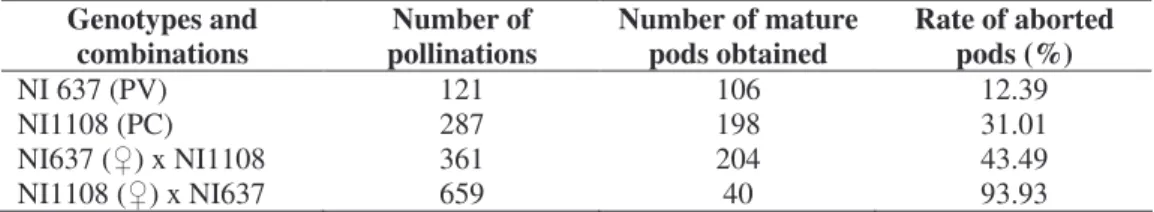
Table 1: Number of pollinations, mature pods and rate of pod abortions. Genotypes and
combinations pollinations Number of Number of mature pods obtained Rate of aborted pods (%)
NI 637 (PV) 121 106 12.39
NI1108 (PC) 287 198 31.01
NI637 ( ) x NI1108 361 204 43.49
NI1108 ( ) x NI637 659 40 93.93
NI = Identification Number ; PV = P. vulgaris ; PC = P. coccineus
0 10 20 30 40 50 60 70 80 90 100 3 4 5 6 7 8 9 10 11 12 13 14
Days after pollination (DAP)
P od s ab or tio n (% ) ( % )
NI1108xNI637 NI637xNI1108 NI637 NI1108
Figure 1: Rate of pods abortion from 3 to 14 DAP during self-pollination and reciprocal
crosses between P. coccineus and P. vulgaris. decreased in hybrids, while remaining constant in NI1108 ( ) x NI637.
Development of embryos
The evolution of the average length of the main embryo structures (suspensor and embryo), maternal parent (NI1108 and NI637) and reciprocal hybrids described by Nguema Ndoutoumou et al. (2007) revealed a progressive growth of the suspensor length in all the embryos. No significant difference was observed between the means. As far as the evolution of the average length of embryos is concerned, the genotype NI637 showed the mean values statistically higher (p < 0.05) than those of NI1108 and the reciprocal hybrids, as from 10 DAP.
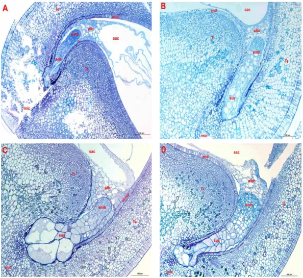
Figures 2 and 3 illustrate respectively the maternal genotypes embryos (NI637, NI1108) and reciprocal hybrids at the globular and heart stage of their development. The
globular stage was observed averagely at between 4 and 7 DAP in the maternal genotypes (NI637 and NI1108), compared to 8 DAP for hybrids NI1108 ( ) x NI637 and 7 DAP for hybrids NI637 ( ) x NI1108. The embryos of the maternal genotypes had a fusiform suspensor (Photo A & B), while hybrid embryos showed big cells at the basis of suspensor (Photo C). In the case of hybrid embryo NI1108 ( ) x NI637, the suspensor was hypertrophied at the cost of embryo proper. In all the self-pollinated ovules or hybrids, the endosperm cells persist in the neighbouring of the embryo proper, inside the embryo sac. The highly albuminated and nucleated cells border the endothelium, suggesting a nutrients transport from the endothelium to the embryo and the suspensor. In NI637, the heart stage was initiated toward the 5 DAP; while the embryo reached the heart stage later on at 7 DAP. The NI1108
Figure 2: Longitudinal median sections of globular embryos of P. vulgaris (NI637), P. coccineus
(NI1108) and reciprocal hybrids. Parental embryos NI637 (Photo A) and NI1108 (Photo B) have a fusiform suspensor (sus) in micropylar end (mic). Basal cells are filled out within reciprocal P. coccineus x P. vulgaris embryos (Photo C & D). The endosperm (alb) separates embryo sac (sac) from the embryo proper (emb). The transfer cells (*) develop between the endothelium (end) covered by inner and outer integuments (Te & Ti) and the endosperm in the one hand, and between the embryo proper and the suspensor in the other hand.
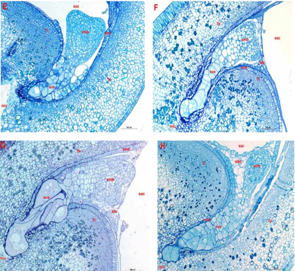
embryo and NI637 ( ) x NI1108 were at the heart stage at 9 DAP, while the hybrid embryo NI1108 ( ) x NI637 was at the heart stage at 10 DAP. A restricted zone links the suspensor and hypertrophied cells of the hybrid embryo to the embryo proper (Photo G). This may explain the nutrient deficiency of the embryo
via the suspensor. The endothelium was intact
around the embryo. The cellular endosperm was totally reabsorbed in the ovule of NI637 contrary to other cases. The embryo may get its nutrients preferentially through the links established between the endothelium and the endosperm. Later, the hybrid embryos reached the cotyledonary stage at 12 DAP, but this
stage was attained at 9 DAP in NI637 and 11 DAP in NI1108. In hybrid embryos, the suspensor drove in teguments, where it de-velops important invaginations useful for embryo nutrition. The contact between the endosperm and cotyledons was established for the same reason. The endosperm disappeared in the neighbouring of the embryo proper, but was developed quite in part of the embryo sac. The endothelium was crushed under the pres-sure of basal cells of the suspensor. The transferred cells were seen in the contact points between the endothelium and the embryo, the suspensor and the endosperm.
Figure 3: Longitudinal median sections of heart stage of embryos from P. vulgaris (NI637),
P. coccineus (NI1108) and reciprocal hybrids. Within NI637 (Photo E) and NI1108 (Photo F) embryos, the suspensor (sus) is slender. Within reciprocal crosses P. coccineus x P. vulgaris (Photo G & H), the suspensor is enormous compared to the embryo proper (emb). It is anchored into teguments (Ti & Te) near to the micropyle (mic). A thin layer of albuminated cells (alb) delimits the embryo from the embryo sac (sac). The transfer cells (*) are in the extension of the endothelium (end) within self-pollinated ovules.
The intracellular thicknesses were also observed in the endothelium and the tegumentary layers, with numerous grains of starch.
DISCUSSION
Embryo abortion arises from self-pollination and during hybridization between
P. vulgaris and P. coccineus. In the first case,
this is often explained by intrinsic phenomena in plants, in accordance with phonologic stages. In the second case, barriers raise at all
the developmental stages of the hybrid embryo, despite the aptitude to combination suggested between the cultivated species of P.
vulgaris and the wild form P. coccineus.
Several turgescent ovules hybrid are deprived from living embryos. In addition to the biological form of crossed genotypes, the direction of cross also determines the success of interspecific hybridization between P.
vulgaris and P. coccineus. Crosses are easier
when P. vulgaris is the maternal parent. During these crosses, a great number of
abortions occur 5 to 6 DAP. It is also a critical period in the development of hybrid embryos between P. vulgaris ( ) and P. coccineus. Similar observations were reported during crosses between P. vulgaris and P. polyanthus (Yeung and Meinke, 1993; Lecomte et al., 1998; Geerts et al., 2002). Like other authors (Sage and Webster, 1990; Lecomte et al., 1998; Nguema Ndoutoumou et al., 2007) observations were made that incompatibility barriers in Phaseolus were post-zygotic and were interpreted by embryo abortion for nutritional reasons essentially. During the early embryogenesis, the interactions between the suspensor, the embryo proper, the endosperm and the maternal parent arise during the determination of embryos growth. The embryo genotype NI637 of P. vulgaris is developed faster than that of P. coccineus (NI1108) and those of the reciprocal hybrids. The influence of the genotype NI1108 may be
preponderant in the dynamic of
embryogenesis of reciprocal hybrids.
Whatever the direction of crossing, the divisions of zygote hybrid is initiated later compared to the maternal embryos and lead to delay development of hybrid embryos. Similar results were obtained during crosses between
P. vulgaris and P. polyanthus (Lecomte et al.,
1998; Geerts, et al., 2002). In the maternal parent genotypes, the nutrition of embryo is organized early, as from the globular stage. The nuclear endosperm, in contact with the proembryo and the endothelium, is partitioned off.
The transferred cells were seen between the endothelium and the endosperm on one hand, and between the embryo and the suspensor on the other hand (Figure 2). On serial sections, the links are well seen at advanced stages. In hybrid ovules, no direct contact exists between the embryo proper and the endothelium. A thin layer of the transferred cells is seen between structures. The contact is narrow between the endothelium and the basis of suspensor. The endosperm borders the apical part of embryo and touches the upper part of the suspensor. The transferred cells link the base of the suspensor to endosperm and the body of suspensor to endothelium. This reveals the difficulties of providing embryo with nutrients. The suspensors of maternal embryos evolve more slowly after the late globular
stage. The embryo is provided with nutrients
via the contacts between cellular endosperm
and the body of the suspensor, and between the embryo proper and the endosperm. The suspensors and hybrid embryos develop
important invaginations towards the
micropyle end (Figure 3), suggesting an important nutritive demand (Maheshwari, 1950; Yeung and Sussex, 1979; Lecomte et al., 1998; Nguema Ndoutoumou et al., 2007). The embryo proper was supposed to prevent the development of suspensor at this stage, but this later is instead hypertrophied in hybrid embryo NI1108 ( ) x NI637. This slows the embryo development since the size of suspensor influences the nutritive requirement of embryo.
Generally, a nutritive transfer between endosperm and cotyledons is possible due to contact zones in cotyledons, at cotyledonary stage. In hybrid NI1108 ( ) x NI637, the observed abnomalies in ovules in the previous stage might provoke a reduction of certain
activities responsible for embryo
development. When endosperm is not developed enough, the embryo is provided with very little nutrients, the alteration of the maternal or embryo structures is unlatched, and consequently, the embryo abortion occurs. This work shows that histological differences between maternal tissues in reciprocal crosses could thus be a key factor in the abortion processes. In regard to several factors implied in embryo abortion in both crosses, it is envisioned to rescue the hybrid embryos at the globular stage of their
development according to elevated
frequencies of abortion occurring in this developmental stage. Hybrid embryos rescued by in vitro culture could be the best way of exploration taking into consideration for the developmental stage reached by the embryo and the nutritional needs specific to them. It is also important to proceed by other interspecific combinations to reinforce these results.
ACKNOWLEDGEMENTS
We are thankful to the Belgium and Gabonese governments for their financial help for the accomplishment of this work. We are also grateful to Professor Yeung Edward Charles and Dr Ngakou Albert for their valuable advices.
REFERENCES
Baudoin JP, Silué S, Geerts P, Mergeai G,
Toussaint A. 2004.
Interspecific-hybridization with Phaseolus vulgaris L.: Embryo development and its genetics.
Genetics and Breeding, 1: 349-364.
Buishand TJ. 1956. The crossing of beans (Phaseolus sp.). Euphytica, 5: 41-50.
Chamberlin MA, Horner HT, Palmer RG. 1994. Early endosperm, embryo and ovule development in Glycine max (L.) Merr. International Journal of Plant
Sciences, 155: 421-436.
Geerts P, Toussaint A, Mergeai G, Baudoin JP. 2002. Study of the early abortion in reciprocal crosses between Phaseolus
vulgaris L. and P. Polyanthus G. BASE,
6: 109-119.
Gutmann M 1995. Improved staining procedures for photographic documenta-tion of phenolic deposits in semithin sections of plant tissue. Journal of
Microscopy, 179: 277-281.
Hoover EE, Brenner ML, Ascher PD. 1985. Comparison of development of two bean crosses. Hort Sciences, 20: 884-886.
Lecomte B, Longly B, Crabbe J, Baudoin JP
1998. Etude comparative du
développement de l'ovule chez deux espèces de Phaseolus: P. polyanthus et P.
vulgaris. BASE, 2: 77-84.
Maheshwari P 1950. An introduction to the embryology of angiosperms. 1rst edition. McGraw-Hill Book Company Inc.: New-York, Toronto, London.
Nguema Ndoutoumou P, Toussaint A, Baudoin JP. 2007. Embryogenèse précoce comparative lors des croisements entre
Phaseolus coccineus L. et P. vulgaris L. BASE, 11(2): 97-107.
Nguema Ndoutoumou P, Toussaint A, Baudoin JP. 2007. Embryo abortion and histological features in the interspecific
cross between Phaseolus vulgaris L. and
P. coccineus L. Plant Cell, Tissue and Organ Culture, 88: 329-332.
Olsen OA, Linnestad C, Nichols SE. 1999. Developmental biology of the cereal endosperm. Elsevier Science, 4:
1360-1385.
Perata P, Picciarelli P, Alpi A. 1990. Pattern of variations in abscisic acid content in suspensors, embryos, and integuments of developing Phaseolus coccineus seeds.
Plant Physiology, 94: 1776-1780.
Pullman GS, Buchanan M. 2003. Loblolly pine (Pinus taeda L.): stage-specific elemental analyses of zygotic embryo and female gametophyte tissue. Plant
Sciences, 164: 943-954.
Russell SD. 1993. The Egg Cell: Development and role in fertilization and early embryogenesis. The Plant Cell, 5:
1349-1359.
Sabja AM, Mok DWS, Mok MC. 1990. Seed
and embryo growth in pod cultures of
Phaseolus vulgaris and P. vulgaris x P. acutifolius. Hort Science, 25: 1288-1291.
Sage TL, Webster BD. 1990. Seed abortion in
Phaseolus vulgaris L. Bot. Gaz, 151:
167-175.
Sallandrouze A, Faurobert M, El Maâtaouia M. 2002. Characterization of the developmental stages of cypress zygotic embryos by two-dimensional
electro-phoresis and by cytochemistry.
Physiologia Plantarum, 114: 608-618.
Yeung EC, Meinke DW. 1993. Embryo-genesis in Angiosperms: Development of the suspensor. The Plant Cell, 5:
1371-1381.
Yeung EC, Sussex IM. 1979. Embryogeny of
Phaseolus coccineus: The suspensor and
the growth of the embryo-proper in vitro.