A PCR method for detection of bifidobacteria in raw milk and raw milk
cheese: comparison with culture-based methods
V. Delcenseriea, N. Bechouxa, B. Chinaa, G. Daubea, F. Gavinib
aUniversity of Liège, Faculty of Veterinary Medicine, Food Sciences Department, Sart Tilman, B43b 4000 Liege,
Belgium
bInstitut National de la recherche agronomique, Technologie des Produits Animaux, 369 rue Jules Guesde,
F-59651 Villeneuve d'Ascq, France
Abstract
Bifidobacteria are well known for their beneficial effects on health and are used as probiotics in food and pharmaceutical products. As they form one of the most important groups in both human and animal feces, their use as fecal indicator organisms in raw milk products has recently been proposed. Bifidobacteria species isolated in humans are different from those isolated in animals. It should therefore be possible to determine
contamination origin (human or animal).
A method of detecting the Bifidobacterium genus was developed by PCR targeting the hsp60 gene. The genus
Bifidobacterium was identified by PCR amplification of a 217-bp hsp60 gene fragment. The degenerated primer
pair specific to the Bifidobacterium genus used was tested for it specificity on 127 strains. Sensitivity was measured on artificially contaminated samples. Food can however be a difficult matrix for PCR testing since it contains PCR inhibitors. So an internal PCR control was used. An artificially created DNA fragment of 315 bp was constructed. The PCR detection method was tested on raw milk and cheese samples and compared with three culture-based methods, which comprised enrichment and isolation steps. The enrichment step used Brain Heart Infusion medium with propionic acid, iron citrate, yeast extract, supplemented with mupirocin (BHMup) or not (BH) and the isolation step used Columbia blood agar medium, supplemented with mupirocin (CMup) or not (C). The method using mupirocin at both enrichment and isolation steps and the PCR method performed from the culture in BHMup enrichment medium were shown to be the most efficient. No significant difference was observed in raw milk samples between PCR from BHMup and the culture-based method BHMup/CMup, while a significant difference was noticed between the same methods in raw milk cheese samples, which would favor using PCR.
The results suggested that PCR on the hsp60 gene was convenient for a rapid detection of bifidobacteria in raw milk and raw milk cheese samples and that bifidobacteria always present throughout raw milk cheese production could be efficiently used as fecal indicators.
Keywords: PCR; Hsp60 gene; Bifidobacterium; Detection; Fecal indicators; Raw milk; Raw milk cheese;
Mupirocin
1. Introduction
Bifidobacteria are Gram-positive, non-motile and non-spore-forming bacteria. They had been considered as anaerobic, until one species was defined as aero-anaerobic (Simpson et al., 2004a). They are part of normal intestinal flora in humans and animals and are generally non-pathogenic bacteria.
Fecal contamination of raw milk on farm has been shown by Beerens et al. (2000), who detected the same and most frequent Bifidobacterium species in milk as in cow dung. Raw milk can be assumed to be the first critical point in an HACCP analysis of the raw milk cheese industry, but a follow-up of contamination during the cheese-making process is also of interest. The standard in Europe for fecal contamination control of raw milk cheese is Escherichia coli.
Bifidobacteria have been proposed as a fecal indicator since they represent one of the most important bacterial groups in human and animal feces (Matsuki et al., 1998; 1999). Moreover, as the dominant Bifidobacterium species are different in human and animal flora (Gavini et al., 1991), one should be able to determine
(Lynch et al., 2002; Nebra et al., 2003; Gilpin et al., 2003) and in meat and raw milk samples (Beerens, 1998; Gavini and Beerens, 1999; Beerens et al., 2000).
Numerous culture-based methods for bifidobacteria detection have been described for these above-mentioned applications and for others, such as knowledge of the genus Bifidobacterium and its evolution within
gastrointestinal flora (human or animal) (Martineau, 1999; Rada and Petr, 2000; Petr and Rada, 2001) and the use of bifidobacteria as probiotics in food or pharmaceutical products (Nebra and Blanch, 1999; Pacher and Kneifel, 1996; Payne et al., 1999).
The culture-based method using propionic acid (Beerens, 1990) and paromomycin as selective agents (Beerens, 1998) to detect bifidobacteria in meat products and in raw milk samples is not sufficiently efficient to eliminate contaminating flora such as lactobacilli in raw milk or Clostridia in meat samples. Using the culture-based detection method requires knowledge of the contaminating flora and the researched Bifidobacterium species in the samples.
Several molecular methods that alleviate this inconvenience have recently been described: PCR-Elisa method based on the 16S rRNA to detect the most common Bifidobacterium species in humans (Malinen et al., 2002); pulsed-field gel electrophoresis (PFGE) and PCR targeting the 16S rRNA (Roy et al., 1996; Bonjoch et al., 2004); PCR in denaturing gradient gel electrophoresis (DGGE) targeting the transaldolase gene for
identification, detection and enumeration of human Bifidobacterium species (Requena et al., 2002); PCR-RFLP method based on the 16SrRNA to detect the most common species from animal and human origins (Delcenserie et al., 2004; Roy and Sirois, 2000), and real-time quantitative PCR from the 16S or the transaldolase gene (Requena et al., 2002). They have also been used in the detection of human Bifidobacterium species from feces (Matsuki et al., 2002; Requena et al., 2002, Mullié et al., 2003; Venema and Maathuis, 2003), of bifidobacteria as probiotics (Brigidi et al., 2003; Fasoli et al., 2003) or as fecal indicators in waters (Bernhard and Field, 2000). Most of these molecular methods have been applied to detect Bifidobacterium species in human feces, rather than in the detection of bifidobacteria of animal origin. Moreover, the 16S rRNA sequences are well conserved among the bifidobacteria and there are multiple copies of the 16S rRNA gene per chromosome. These features might influence quantitative PCR methods (Requena et al., 2002). Another gene, the hsp60 gene, has been sequenced in most Bifidobacterium species (Jian et al., 2001, Jian and Dong, 2002). This gene presents species-specific sequences.
This study compares three different protocols of a culture-based method using mupirocin, as recommended by Rada et al. (1999) and Rada and Petr (2000), instead of paromomycin as selective agent in parallel with a PCR method on raw milk samples. Then, utilizing both culture-based and PCR methods, bifidobacteria contamination levels in raw milk cheese samples are determined and compared with those of E. coli. Application of
bifidobacteria as fecal indicators in raw milk cheese industries is also discussed.
2. Material and methods 2.1. Samples
2.1.1. Raw milk samples
Detection of bifidobacteria was performed from raw milk stored in tanks collected on French farms (Vercors and Courtenay regions).
Samples were diluted until 10-4 and presence or absence of bifidobacteria at each dilution was compared statistically by the different detection methods as follows:
(i) 39 samples (195 dilutions) have been analyzed and results compared using three combinations of culture-based methods using mupirocin or not
(ii) 12 samples (60 dilutions) have been analyzed and results compared by PCR from two enrichment broth using or not mupirocin
(iii) 148 samples (740 dilutions) have been analyzed and results compared by the different PCR and culture-based methods.
2.1.2. Raw milk cheese samples
In the industry under study from the Vercors region (France), milk was collected on farms and stored in tanks at the factory at 4°C. Milk is prepared for maturation by addition of cream, ferment and surface flora. Animal rennet is added. On day 1, the following steps are successively performed: molding, a first manual turnover, a manual salting and a second turnover. On day 2, cheeses are removed from the molds and a new manual or mechanical salting is performed. Ripening is carried out for 28 days.
Twenty-five raw milk cheeses at four different steps of the production chain from raw milk to the end product (100 samples) were analyzed by the best culture-based method chosen among the three tested and by the best PCR method. The following production steps were analyzed: raw milk (Step A), after addition of rennet (Step C), after removal from the mold (Step E), during ripening (Step G). Samples were diluted until 10-6 to perform semiquantitative detection of bifidobacteria.
2.2. Methods
2.2.1. Culture-based method for E. coli detection
E. coli was numerated on the Coli ID medium (BioMérieux, France; Sueiro et al., 2001). 2.2.2. Culture-based methods for bifidobacteria detection
The methods were performed in two steps, an enrichment and an isolation step. Components of enrichment and isolation media before adding mupirocin have been described by Beerens (1998).
2.2.2.1. Enrichment step medium. The following components were added to the medium Brain Heart Infusion
(BHI, 37 g/l, Bio-Rad, Marnes-la-Coquette, France): propionic acid, 5 ml/l; Fe-citrate, 0.5 g/l; cystein chlorhydrate, 0.5 g/l; yeast extract, 5 g/l; agar, 2 g/l. Mupirocin was provided by GSK Laboratories (Lithium mupirocin, GlaxoSmithKline, England) and added (BHMup) or not (BH) at the concentration equal to 80 mg/l (to be added when the medium must be used). The final pH, 5.0, was obtained with the addition of a NaOH solution. The medium (without mupirocin) was dispensed in 9 ml amounts. Sterilization was not needed because of the medium's low pH.
2.2.2.2. Isolation step medium. Columbia blood agar medium (Columbia blood agar, Difco, Elancourt, France)
was used with addition of Fe-citrate, 0.5 g/l; glucose, 5 g/l; cystein chlorhydrate, 0.5 g/l. Mupirocin was added (CMup) or not (C) at the concentration equal to 50 mg/l (to be added when the medium must be used). The medium (without mupirocin) was dispensed in 100 ml bottles and autoclaved at 120°C.
2.2.2.3. Protocol. The selective enrichment medium, with mupirocin added or not, was held in boiling water for
20 min to expel oxygen and cooled to 30-40°C.
The milk and the raw milk cheese samples were diluted until 10-3 and until 10-5, respectively, in quarter-strength Ringer solution containing cystein chlorhydrate (0.3‰). One milliliter of milk or 1 g of raw milk cheese was transferred in a tube of enrichment medium. Then 1 ml of each of the appropriate sample dilutions was
inoculated in tubes of enrichment medium in order to detect bifidobacteria in milk and raw milk cheese until 10-4 and at 10-6, respectively. Tubes were incubated at 37 °C for 72 h in aerobiosis, since bacteria were able to grow in depth because of agar present in the medium.
For each enrichment culture, 0.03 or 0.1 ml were spread onto five plates of Columbia blood agar. The plates were incubated at 37 °C for 72 h in jars with an "Anaerogen" (Oxoid, Dardilly, France).
We compared the following different protocols: enrichment broth containing mupirocin (BHMup) or not (BH) and isolation medium containing mupirocin (CMup) or not (C). The combinations used were BH/ CMup (Cultural 1), BHMup/C (Cultural 2), BHMup/ CMup (Cultural 3). Presence of bifidobacteria was confirmed by (1) the production of fructose-6-phosphate phosphoketolase (F6PPK test as described by Scardovi, 1986) tested on the whole culture obtained after the isolation step, (2) by transfer of the isolated colonies into Veillon tubes that contained Columbia agar to eliminate aerobic strains and to perform F6PPK test on Gram-positive bacillar strains.
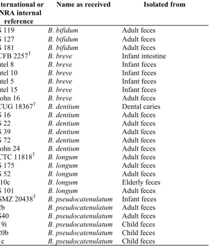
Table 1: References of the Bifidobacterium strains used for the validation of the PCR essay
International or INRA internal
reference
Name as received Isolated from
ATCC 27672 B. animatis Rat feces
P16 (Biavatia) B. animatis Chicken feces
F434 (Biavati) B. animatis Sewage
RA16 (Biavati) B. animatis Rabbit feces
RA20 (Biavati) B. animatis Rabbit feces
NCFB 2242T B. animatis Rat feces
DSM 20210T B. thermophilum Pig feces
Cheval 1/1 B. thermophilum Horse feces
Pigeon 1/2 B. thermophilum Pigeon feces
LC 403/1 B. thermophilum Raw milk
LC 458/3 B. thermophilum Raw milk
LC 294/2 B. thermophilum Raw milk
LC 103/1 B. thermophilum Raw milk
B 39/3 B. thermophilum Cow dung
B 105/5 B. thermophilum Cow dung
LC 288/1 B. thermophilum Raw milk
Porc 3/1 B. thermophilum Pig feces
B 42/1 B. thermophilum Cow dung
LC 110/1 B. thermophilum Raw milk
B 25/1 B. thermophilum Cow dung
T 585/1/2 B. thermophilum Raw milk
Pigeon 1/1 B. thermophilum Pigeon feces
Cheval 5/1 B. thermophilum Horse feces
T 528/4 B. thermophilum Raw milk
B 79/3 B. thermophilum Cow dung
LC 102/2 B. thermophilum Raw milk
LC 26/3 B. thermophilum Raw milk
LC 75/1 B. thermophilum Raw milk
F 38/3 B. thermophilum Raw milk cheese
B 25/2 B. thermophilum Cow dung
LC 205/1 B. thermophilum Raw milk
Pigeon 4/1 B. thermophilum Pigeon feces
Pigeon 4/3 B. thermophilum Pigeon feces
DSM 20434T B. choerinum Pig feces
Internal 1 B. pseudolongumb Unknown
Internal 2 B. pseudolongumb Unknown
RU 224 (Biavati) B. pseudolongum
subsp. Globosum Bovine rumen
Internal 3 B. pseudolongumb Unknown
Internal 4 B. pseudolongumb Unknown
MB7 (Biavati) B. pseudolongum
subsp. pseudolongum Pig feces
LC 287/2 B. pseudolongumb Raw milk
LC 289/2 B. pseudolongumb Raw milk
LC 302/2 B. pseudolongumb Raw milk
LC 407/1/1 B. pseudolongumb Raw milk
B 81/1 B. pseudolongumb Cow dung
LC 312/2 B. pseudolongumb Raw milk
LC 317/2 B. pseudolongumb Raw milk
LC 405/3 B. pseudolongumb Raw milk
Table 1 (continued) International or
INRA internal reference
Name as received Isolated from
LC 464/3 B. pseudolongumb Raw milk
LC 287/1 B. pseudolongumb Raw milk
LC 305/2 B. pseudolongumb Raw milk
Poule 1/2 B. pseudolongumb Chicken feces
B 86/1 B. pseudolongumb Cow dung
B 81/1 B. pseudolongumb Cow dung
LC 304/1 B. pseudolongumb Raw milk
LC 334/1 B. pseudolongumb Raw milk
LC 323/1 B. pseudolongumb Raw milk
LC 324/2 B. pseudolongumb Raw milk
LC 340/3 B. pseudolongumb Raw milk
LC 306/1 B. pseudolongumb Raw milk
Internal 5 B. pseudolongumb Unknown
LC 240/3 B. pseudolongumb Raw milk
LC 229/2 B. pseudolongumb Raw milk
LC 232/1 B. pseudolongumb Raw milk
LC 172/2 B. pseudolongumb Raw milk
LC 147/2 B. pseudolongumb Raw milk
LC 160/3 B. pseudolongumb Raw milk
LC 109/3 B. pseudolongumb Raw milk
LC 99/1 B. pseudolongumb Raw milk
LC 123/1 B. pseudolongumb Raw milk
LC 26/1 B. pseudolongumb Raw milk
LC 120/1 B. pseudolongumb Raw milk
B 121/1 B. pseudolongumb Cow dung
LC 700/2 B. pseudolongumb Raw milk
LC 697/3 B. pseudolongumb Raw milk
T 690/1/1 B. pseudolongumb Raw milk
T 702/2/2 B. pseudolongumb Raw milk
B 116/1/1 B. pseudolongumb Cow dung
B 117/1/3 B. pseudolongumb Cow dung
B 117/1/1 B. pseudolongumb Cow dung
LC 686/1 B. pseudolongumb Raw milk
LC 684/3 B. pseudolongumb Raw milk
LC 680/2 B. pseudolongumb Raw milk
LC 617/2 B. pseudolongumb Raw milk
RU 915 BT B. merycicum Bovine rumen
RU 687T B. ruminantium Bovine rumen
DSMZ 20102T B. minimum Sewage
LC 396/4 B. minimum Raw milk
LC 300/1 B. minimum Raw milk
Internal 6 B. cuniculi Unknown
Internal 7 B. adolescentis Unknown
BS3 B. adolescentis Adult feces
CCUG 18363T B. adolescentis Adult feces
206 la B. adolescentis Adult feces
503 le B. adolescentis Elderly feces
BS5 B. adolescentis Adult feces
BS50 B. adolescentis Adult feces
1604 3a B. adolescentis Elderly feces
DSMZ 20082 B. bifidum Adult feces
Table 1 (continued) International or
INRA internal reference
Name as received Isolated from
BS 119 B. bifidum Adult feces
BS 127 B. bifidum Adult feces
BS 181 B. bifidum Adult feces
NCFB 2257T B. breve Infant intestine
Butel 8 B. breve Infant feces
Butel 10 B. breve Infant feces
Butel 5 B. breve Infant feces
Butel 15 B. breve Infant feces
Crohn 16 B. breve Adult feces
CCUG 18367T B. dentium Dental caries
BS 16 B. dentium Adult feces
BS 22 B. dentium Adult feces
BS 39 B. dentium Adult feces
BS 72 B. dentium Adult feces
Crohn 24 B. dentium Adult feces
NCTC 11818T B. longum Adult feces
BS 175 B. longum Adult feces
BS 52 B. longum Adult feces
A 10c B. longum Elderly feces
BS 101 B. longum Adult feces
DSMZ 20438T B. pseudocatenulatum Infant feces B2b B. pseudocatenulatum Adult feces
BS40 B. pseudocatenulatum Adult feces
C19i B. pseudocatenulatum Child feces
C20b B. pseudocatenulatum Child feces
C1c B. pseudocatenulatum Child feces
ATCC: American Type Culture Collection, Rockville, MD, USA; CCUG: Culture Collection, University of Göteborg, Göteborg, Sweden; DSMZ: Deutsche Sammlung von Mikroorganismen und Zellkulturen, Göttingen, Germany; NCTC: National Collection of Type Cultures, Central Public Health Laboratory, London; England); NCFB: National Collection of Food Bacteria, Shinfield, Reading, Berks, England.
a Received from B. Biavati, Instituto di Microbiologia Agaria e Tecnica, Università degli Studi di Bologna, Bologna, Italy. b Subspecies not
determined.
2.2.3. PCR method for bifidobacteria detection 2.2.3.1. Target DNA preparation
Pure strains. One hundred and twenty-seven reference strains belonging to 14 Bifidobacterium species (Table 1)
and 37 non-Bifidobacterium strains belonging to species or genera often food-contaminating (5 Enterococcus spp., 5 Pseudomonas sp., 5 Staphylococcus aureus, 6 Lactobacillus, 4 Clostridium perfringens, 6 Bacillus
cereus, 5 E. coli and 1 Salmonella typhimurium) were tested for primers validation. Before testing, the
Bifidobacterium strains were withdrawn from frozen storage on Rosenow medium (Sanofi-synthelabo,
Marnes-la-Coquette, France) and subcultured on Brain Heart Infusion (BioRad, Marnes-Marnes-la-Coquette, France) at 37 °C for 48 to 72 h under anaerobic conditions.
One milliliter of bacterial cultures in BHI broth was centrifuged at 12,000 X g for 2 min using a bench-top centrifuge. The pellets were transferred in sterile, demineralized water, and the DNA was extracted using Wizard Genomic DNA purification kit (Prom-ega, Madison, WI, USA) with addition of lysozyme (10 mg/ml,
Eurogentec, Seraing, Belgium), as recommended for Gram-positive bacteria. DNA concentrations were spectrophotometrically estimated (GeneQuant pro, Amersham Pharmacia, Roosendaal, Netherlands). DNA samples were diluted with distilled water to obtain a concentration between 25 and 50 µg/ml.
Artificially contaminated samples. Artificially contaminated samples were prepared as follows: 40 ml of UHT
homogenizing, the resulting mixture was distributed in aliquots of 10 ml. They were inoculated with 100 µl of 10-fold serial dilutions of a 48 h culture of B. pseudolongum subsp. globosum (RU224) and B. thermophilum (DSM 20210T) in peptone sodium solution, and including a negative control without inoculation.
One milliliter of each aliquot was distributed on MRS medium (Oxoid) plates supplemented with mupirocin (50 mg/ml) for bifidobacteria counting after 72 h anaerobic incubation at 37 °C. The other part of aliquot (about 9 ml) was incubated during 24 h in anaerobic conditions at 37 °C. The same procedure was repeated with a 48 h incubation time. After this, 1 ml of each incubation broth was transferred into a microcentrifuge tube and centrifuged at 12,000 X g for 2 min using a bench-top centrifuge. The pellets were transferred in sterile, demineralized water, and the DNA extracted using Wizard Genomic DNA purification kit (Promega) as previously described for pure strains.
Raw milk and raw milk cheese samples. DNA was extracted from cultures obtained after the enrichment step of
the cultural-based method (from pure until 10-4 dilution for milk and until 10-6 dilution for raw milk cheese samples). One milliliter of each homogenized content was transferred in a microcentrifuge tube and centrifuged at 12,000 X g for 2 min using a bench-top centrifuge. The pellets were transferred in sterile, demineralized water, and the DNA extracted using Wizard Genomic DNA purification kit (Promega) as previously described for pure strains. In case of PCR inhibition, the DNA samples were diluted 10 fold.
2.2.3.2. Selection of primers. The sequences of the hsp60 gene are available on Genbank for several
representative Bifidobacterium species in human and animal feces (Accession number, B. adolescentis: AF210319, B. animalis: AY004287, B. cuniculi: AY004283, B. choerinum: AY013247, B. pseudolon-gum subsp. globosum: AF286736, B. pseudolongum subsp. pseudolongum: AF240573, B. merycicum: AY004277, B.
pseudocatenulatum: AY004274, B. ruminantium: AF240571, B. thermophilum: AF240567). These sequences
were aligned (ClustalW, http://www.ebi.ac.uk/clustalw/). From these sequence alignments, Bifidobacterium genus-specific degenerated primers were selected using Oligo software (Medprobe). Specificity of the primers for the Bifidobacterium-genus was checked realizing a Meg-ablast. Only Bifidobacterium DNA was fully complementary to the primers sequences (data not shown, www.ncbi.nlm.nih.gov/BLAST).
The genus-specific amplification of a 217 bp fragment of the hsp60 gene is generated using primers: B11 up: 5'-GTS CAY GAR GGY CTS AAG AA-3', B12 down: 5'-CCR TCC TGG CCR ACC TTG T-3' (Sigma Genosys, UK).
2.2.3.3. Controls. The following amplification controls were run with each series: positive, i.e. reaction mix
containing DNA extract from a positive strain of B. pseudolongum (B 116/1/1, Table 1), two reagent controls, i.e. mix containing all reagents without sample DNA and extraction control, i.e. 1000 µl of distilled water processed in the same manner as the samples.
Food can be a difficult matrix for PCR testing because it contains PCR inhibitors. So to be sure that a negative result is indeed due to absence of the target rather than to an inhibition of PCR reaction, we had to construct an internal PCR control.
An artificially created DNA fragment was used as an internal positive control in every reaction mixture, except for the other controls. The control DNA consisted of a fragment of 315 bp of the pGEMT vector, flanked by the target for the Bifidobacterium-genus PCR primers. This product was created by a two-step PCR as follows. Chimerical PCR primers flanked with the Bifidobacterium genus-specific primers were chosen: CI up: 5'-GTS CAY GAR GGY CTS AAG AAG CAG GAA AGA ACA TGT GAG CA-3' and CI down: 5'-CCR TCC TGG CCR ACC TTG TAC GAC CTA CAC CGA ACT GAG A-3'. The first step comprised amplification of DNA from the pGEMT vector using the chimerical primers by 45 cycles at the following PCR conditions. A 5 µl pGEMT (Eurogentec, Seraing, Belgium) DNA was introduced in a mix containing 0.2 mol 1-1 dNTPs, 400 pmol 1-1 of each chimerical PCR primers, 0.8 U of Dap Goldstar polymerase (Eurogentec), 1 x buffer: 20 mM Tris-HCl, pH 8.0, 100 mM KC1, 0.1 mM EDTA, 1 mM DTT, 50% glycerol, 0.5% Nonidet P-40 and 0.5% Tween-20 (Eurogentec).
The samples were subjected to an initial step of denaturation at 95 °C for 5 min, followed by 15 denaturation cycles at 95 °C for 30 s, annealing at 50°C for 30 s and extension at 72 °C for 30 s and 30 cycles of denaturation at 95 °C for 30 s, annealing at 60 °C for 30 s and extension at 72 °C for 30 s. In the second step, the amplicon of the first amplification was purified (QIAquick PCR Purification Kit, Qiagen, Westburg, The Netherlands), diluted 1/1000 in distilled water and used as a template to perform a second amplification using the
as internal control. As measured by optical density using a GeneQuant pro spectrophotometer UV (Amersham Pharmacia), the DNA concentration was 185 µg/ml. The final dilution in distilled water of the internal control target was established empirically to reduce competition with target DNA and corresponded to 1.1 µg/µl of DNA. The control DNA was used as a positive amplification control in all samples.
2.2.3.4. PCR conditions. PCR mix was composed of 0.2 mol 1-1 dNTPs, 400 pmol 1-1 of each primer, 1 U of FastStart TaqPolymerase (Roche), 1 x buffer: 500 mM Tris-HCl, 100 mM KC1, 50 mM (NH4)2SO4, pH 8.3/25 °C (Roche), 4 µl DNA (50-100 ng), 1 µl internal control and H2O in a total volume of 20 µl.
PCR was run using the following cycling conditions: 1 x 5 min at 95 °C, 40 x 95 °C for 30 s, 60 °C for 30 s and 72 °C for 30 s and a final extension (5 min at 72 °C). Samples were kept at 4 °C or stored at -20 °C before analyzing. A sample was considered as positive when the 217 bp amplicon was visible on 2% agarose gels after electrophoresis and ethidium bromide staining.
Two different protocols were tested: PCR from BH (PCR 1) and PCR from BHMup enrichment broth (PCR 2).
2.2.4. Statistical analysis
We chose the Mc Nemar test (Leroy and Farnir, 2000) to statistically evaluate the different methods (culture-based and PCR). Dilutions were tested as separate values. To compare results obtained at different steps of the raw milk cheese production, an ANOVA test (Dagnelie, 1975) was performed.
3. Results and discussion 3.1. Culture-based methods
Thirty-nine raw milk samples (195 dilutions) were analyzed by three culture-based methods using three combinations of enrichment and isolation media: Cultural 1 (BH/CMup), 2 (BHMup80/C) and 3
(BHMup/CMup). Table 2 presents the number of positive dilutions for each cultural method. The highest percentage of positives (95%) was detected for pure and for 10-1 dilutions with Cultural 3. Table 3 presents the comparison of the three methods by the Mc Nemar test (based on Chi-square table), which was calculated on the basis of the number of different results obtained on the 195 dilutions, with methods compared 2 by 2. In the Mc Nemar test, Cultural 3 was confirmed to be the best method. A statistical difference was observed between Cultural 2 and Cultural 3 in favor of Cultural 3 (χ2=5.56; P<0.025). A trend was noticed between Cultural 1 and Cultural 3 in favor of using mupirocin at both steps, enrichment and isolation (χ2 =2.91; P<0.l). No difference
was observed between methods using mupirocin only, either in the isolation medium or in the enrichment medium.
Many different selective agents were used for detection of bifidobacteria: lithium chloride, sodium propionate, nalidixic acid, neomycin sulphate, paromomycin sulphate, polymixin B sulphate (Payne et al., 1999). In the case of raw milk samples, an enrichment step was necessary because of the possible relatively low levels of
bifidobacteria (10 to 106 ml -1) compared to those in human or animal feces (107 to 10 g-1 ). Beerens (1998) recommended using at the enrichment step the BHI medium with addition of propionic acid, yeast extract, iron citrate, and at the isolation step, paromomycin as selective agent. However, the high number of lactobacilli not inhibited by paromomycin hid bifidobacteria at low dilutions.
Table 2: Number and percentage of positive raw milk samples analyzed by the three culture-based methods
Dilution/method Cultural 1 Cultural 2 Cultural 3
Pure 33/39 (85%) 33/39 (85%) 37/39 (95%)
-1 34/39 (87%) 31/39 (79%) 37/39 (95%)
-2 25/39 (64%) 23/39 (59%) 26/39 (67%)
-3 7/39 (18%) 8/39 (21%) 6/39 (15%)
-4 0/39 (0%) 1/39 (3%) 1/39 (3%)
Cultural 1: BH/CMup; Cultural 2: BHMup/C; Cultural 3: BHMup/CMup.
Pure: dilutions analyzed from pure enrichment broth; -1,-2, -3 and -4: dilutions, respectively, analyzed from 10, 102, 103 and 104 fold
Rada et al. (1997) and Rada and Petr (2000) showed that bifidobacteria were resistant to mupirocin when lactobacilli were susceptible. Mupirocin (pseudo-monic acid A) was originally isolated from Pseudo-monas
fluorescens and used as a topical antibiotic (Sutherland et al., 1985).
In raw milk samples, addition of mupirocin at the enrichment step can eliminate most of the lactobacilli strains present that could hide bifidobacteria in raw milk. If some lactobacilli strains were still present after the enrichment step, one might suppose that they would be eliminated during the isolation step by mupirocin, when present. Grand et al. (2003) also used mupirocin as selective agent for detection of bifidobacteria in probiotic milk products, as did Mikkelsen et al. (2003) in gastrointestinal samples from piglets and Simpson et al. (2004b) in probiotic animal feed.
The culture-based method presented in this study provides semiquantitative results. As none of culture-based methods are sufficiently selective to detect only bifidobacteria, the F6PPK test must be performed to confirm that isolated strains indeed belong to the genus Bifidobacterium. An alternative was to carry out the F6PPK test on the whole culture at the isolation step in order to more rapidly ascertain the contamination level of studied samples.
Table 3 : Comparison of the three culture-based methods by the Mc Nemar test based on numbers of different
results (+/- and - /+) obtained with methods compared 2 by 2 (195 dilutions/39 samples) Number of dilutions
(samples) analyzed by culture-based method
Compared methods Percentage of positive dilutions
+/- -/+ Statistical results
195 (39) Cultural 1/ Cultural 2 51/49 16 14 NS-χ2 = 0.06 P<0.8
195 (39) Cultural 1/ Cultural 3 51/55 7 15 NS-;χ2 = 2.91 P<0.l
195 (39) Cultural 2/ Cultural 3 49/55 4 14 S-;χ2 = 5.56 P< 0.025
Cultural 1: BH/CMup; Cultural 2: BHMup/C; Cultural 3: BHMup/CMup. +/-: Positive dilutions with the first method and negative with the second one. -/+: Negative dilutions with the first method and positive with the second one.
In favor of
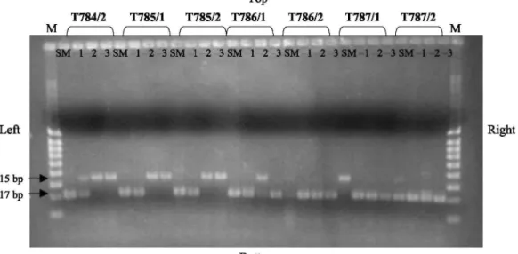
Fig. 1. PCR amplification of Bifidobacterium DNA from some raw milk samples and using an internal control.
Legend: T784/2, T785/1, T785/ 2, T786/1, T786/2, T787/1, T787/2: internal numbers of samples. M: 5 µl molecular weight marker (100-200-300-400-500-600-700-800-1000 bp). SM: PCR realized with DNA extracted from pure enrichment broth; -1, -2 and -3, dilutions, respectively, analyzed from 10-, 102-and 10 -fold dilutions
Table 4: Number and percentage of positive raw milk samples analyzed by the two PCR methods Dilution/method PCR 1 PCR 2 Pure 11/12 (92%) 12/12 (100%) -1 10/12 (83%) 11/12 (92%) -2 4/12 (33%) 7/12 (58%) -3 0/12 (0%) 3/12 (25%) -4 0/12 (0%) 1/12 (8%)
PCR 1: PCR realized from BH broth; PCR 2: PCR realized from BHMup broth.
Pure: dilutions analyzed from pure enrichment broth; -1, -2, -3 and -4: dilutions, respectively, analyzed from 10, 10 , 10 and 10 fold dilutions of the enrichment broth.
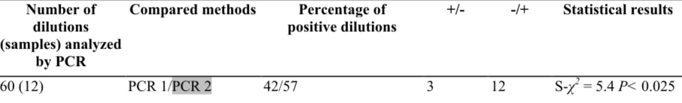
Table 5: Comparison of the 2 PCR methods by the Mc Nemar test based on numbers of different results (+/- and
-/+) obtained with methods compared 2 by 2 (60 dilutions/12 samples) Number of
dilutions (samples) analyzed
by PCR
Compared methods Percentage of positive dilutions
+/- -/+ Statistical results
60 (12) PCR 1/PCR 2 42/57 3 12 S-χ2 = 5.4 P< 0.025
PCR 1 : PCR realized from BH broth; PCR 2: PCR realized from BHMup broth. +/-: Positive dilutions with the first method and negative with the second one. -/+: Negative dilutions with the first method and positive with the second one.
In favor of
3.2. PCR methods
3.2.1. Validation of the primers on pure strains
Specificity of the primers was confirmed by PCR using chromosomal DNA extracted from 37
non-Bifidobacterium strains and from 127 non-Bifidobacterium strains. The primers were able to detect an expected 217
bp DNA fragment from all Bifidobacterium strains using the PCR described conditions. No amplification was obtained for strains of any of the other tested species (5 Enterococcus spp., 5 Pseudomonas sp., 5 S. aureus, 6 Lactobacillus, 4 C. perfringens, 6 B. cereus, 5 E. coli and 1 S. typhimurium). This validation was realized in triplicate.
3.2.2. Detection limit of the PCR method on artificially contaminated samples
PCR results obtained from enrichment media incubated for 24 and 48 h have been compared in relation with bifidobacteria counts on MRS plates (Oxoid) after 72 h anaerobic incubation at 37 °C. Depending on the incubation time of the enrichment media, the PCR method could detect DNA from 102 to 103 cfu ml-1 present in the sample when the incubation time of the enrichment medium was 24 h, and around 1-10 cfu ml-1 when it was 48 h.
3.2.3. Comparison of PCR methods (from BH and from BHMup80 enrichment media)
Bifidobacteria were detected in 12 samples (60 dilutions) by PCR from BH enrichment broth (PCR 1) and from BHMup broth (PCR 2) obtained after the enrichment step of the culture-based methods.
Four possibilities of results were observed, (i) Only the target was positive (217 bp fragment), (ii) The target (217 bp) and the internal control (315 bp fragment) were positive. In these two cases, PCR was considered as positive for bifidobacteria detection, (iii) Only the internal control was positive. In this case, the PCR was indeed negative for bifidobacteria detection, (iv) Finally, if the target and the internal control were negative, it signed PCR inhibition. In this case, it was necessary to do the PCR again on a diluted sample. Some of the results are presented in Fig. 1.
Table 4 presents the percentage of positive dilutions obtained with PCR 1 and PCR 2. The highest percentage of positive dilutions (100%) was detected for pure dilutions with PCR 2. Comparison between the two methods is presented in Table 5. A significant difference was observed between the two PCR methods in favor of PCR 2
(χ2=5.4; P<0.025). This showed that mupirocin in enrichment broth positively affected detection of
Bifidobacterium by PCR. It suggested that PCR sensitivity was better when a selective agent was used in an
enrichment broth. Elimination of most lactobacilli avoided competition with bifidobacteria that could be detected at higher dilutions.
Table 6: Comparison of different combinations of culture-based and PCR methods by the Mc Nemar test based
on numbers of different results (+/- and -/+) obtained with methods compared 2 by 2 (395 dilutions/79 samples, 255 dilutions/51 samples and 90 dilutions/18 samples)
Number of dilutions (samples) analyzed by
PCR and culture-based methods
Compared
methods Percentage of positive dilutions +/- -/+ Statistical results
395 (79) PCR 1/Cultural 1 51/43 49 23 S-;χ2 = 9.4 P< 0.005
255 (51) PCR 2/Cultural 2 56/53 21 13 NS-χ2=1.9 P<0.2
90(18) PCR 2/Cultural 3 61/55 4 2 NS - χ2 = 0.7 P<0.5
Cultural 1: BH/CMup; Cultural 2: BHMup/C, Cultural 3: BHMup/CMup, PCR 1: PCR realized from BH broth; PCR 2: PCR realized from BHMup broth.
+/- : Positive dilutions with the first method and negative with the second one. -/+: Negative dilutions with the first method and positive with the second one. In favor of
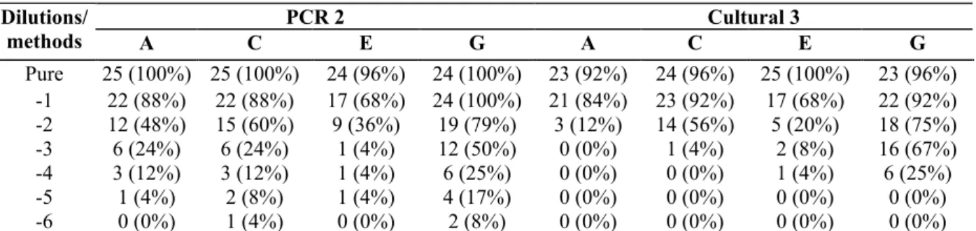
Table 7: Number of Bifidobacterium positive samples (percentage) of raw milk cheese at each step of production
(n=25 for A, C, E steps and n=24 for the step G)
PCR 2 Cultural 3 Dilutions/ methods A C E G A C E G Pure 25 (100%) 25 (100%) 24 (96%) 24 (100%) 23 (92%) 24 (96%) 25 (100%) 23 (96%) -1 22 (88%) 22 (88%) 17 (68%) 24 (100%) 21 (84%) 23 (92%) 17 (68%) 22 (92%) -2 12 (48%) 15 (60%) 9 (36%) 19 (79%) 3 (12%) 14 (56%) 5 (20%) 18 (75%) -3 6 (24%) 6 (24%) 1 (4%) 12 (50%) 0 (0%) 1 (4%) 2 (8%) 16 (67%) -4 3 (12%) 3 (12%) 1 (4%) 6 (25%) 0 (0%) 0 (0%) 1 (4%) 6 (25%) -5 1 (4%) 2 (8%) 1 (4%) 4 (17%) 0 (0%) 0 (0%) 0 (0%) 0 (0%) -6 0 (0%) 1 (4%) 0 (0%) 2 (8%) 0 (0%) 0 (0%) 0 (0%) 0 (0%)
PCR 2: PCR realized from BHMup enrichment broth; Cultural 3: BHMup/CMup.
Pure: dilutions analyzed from pure enrichment broth; -1, -2,-3, -4, -5 and -6: dilutions, respectively, analyzed from 10, 102, 103, 104, 105 and
10 fold dilutions of the enrichment broth.
3.3. Comparison of culture-based and PCR methods
PCR 1 and 2 have been compared to the culture-based methods 1, 2 and 3 (Table 6). Each comparison showed a greater number of positive results by PCR. However, the difference was only significant when PCR 1 was compared to Cultural 1 (χ2=9.4; P<0.005). No significant difference was observed when PCR 2 was compared to
Cultural 2 and 3. For each comparison test, a percentage of false negatives by PCR are present, respectively equal to 6%, 7% and 2%.
Even if the results were not always significant, a trend was observed in favor of PCR, suggesting that the PCR method is more sensitive than culture-based methods. This also indicates that inhibition phenomena apparently do not play a significant role in the given experimental settings. Although they cannot be ruled out entirely, we tried to minimize the effect by a dilution of the DNA extract.
3.4. Contamination of raw milk cheese samples along the production chain
Table 7 presents the number of positive dilutions with the two best methods (PCR 2 and Cultural 3). The highest percentage of positives (100%) was detected for pure (steps A, C and G) and for 10-1 dilutions (step G) with PCR 2. Cultural 3 detected
100% of positive dilutions for pure on step E, and respectively, 92%, 96% and 96% of positives for pure on steps A, C and G The two methods were compared on 693 dilutions of 25 samples. The significant difference
(χ2=20.04; P<0.0005) observed in favor of PCR 2 (Table 8) was not in agreement with previous results on raw
milk samples (no significant difference). However, it did correspond to the already observed trend in favor of PCR. The highest number of dilutions included in the test calculation on raw milk cheese samples would confirm that PCR 2 was a more sensitive method than the culture-based method.
Additionally, only 4% of false positives were obtained by PCR, which is in agreement with previous results obtained from raw milk samples.
The mean counts of bifidobacteria (Table 9) increased significantly (F=14.4; P<0.0005) from step A (milk) to step G (ripening at D+21), when studying the highest values obtained with the PCR or the culture-based method. The lowest mean level (2.52 log cfu g-1) of bifidobacteria was found on step E (after removal from the mold), where the pH decreased to 4.35 (on step C, pH was 6.45). This low pH can explain why E. coli disappeared from step E and why the level of bifidobacteria was still high, as these organisms can multiply at low pH (Biavati et al., 2000).
Table 8: Comparison between the PCR 2 and Cultural 3 methods by the Mc Nemar test based on numbers of
different results (+/- and -/+) obtained with methods compared 2 by 2 (693 dilutions/25 samples) Number of
dilutions (samples) analyzed by PCR
Compared methods Percentage of positive
dilutions +/- -/+ Statistical results
693 (25) PCR 2/Cultural 3 41/35 73 28 S -χ2 = 20.04 P< 0.0005
PCR 2: PCR realized from BHMup broth; Cultural 3: BHMup/CMup. +/- : Positive dilutions with the first method and negative with the second one. -/+: Negative dilutions with the first method and positive with the second one.
In favor of
Table 9: Mean counts (log cfu ml-1 or g-1 ! standard deviation) of bifidobacteria and E. coli in 25 raw milk
cheese samples at four production stens
Production stepsa Methods A C E G PCR 2 2.76 ! 1.3 2.96 1.46 2.20 1.12 3.79 1.53 Cultural 3 1.88 ! 0.73 2.48 0.82 2.00 1.00 3.54 1.38 PCR 2 or 2.80 ! 1.26 3.20 1.29 2.52 1.00 4.33 1.27 Cultural 3b E. coli 1.58 ! 1.52 1.98 1.34 0.73 1.07 0.18 0.50
PCR 2: PCR realized from BHMup broth; Cultural 3: BHMup/ CMup.
a Production steps: A, raw milk; C, after addition of rennet; E, after removal from the mold; G, ripening (Day 21). b Means calculated from the highest values obtained with either PCR 2 or Cultural 3.
4. Conclusion
The culture-based method BHMup/CMup was efficient since it showed that 95% of raw milk and more than 95% of raw milk cheese samples contained bifidobacteria. However, like all culture-based methods, this method is time-consuming and could not be easily applied to food industry controls. The PCR method performed from BHMup enrichment medium does not present this disadvantage and can be used effectively to detect
bifidobacteria as fecal indicators in raw milk cheese industries, instead of or with E. coli.
Lynch et al.(2002), Nebra et al. (2003) and Bonjoch et al. (2004) proposed the species B. adolescentis or B.
dentium as indicators of fecal pollution. As these species are dominant in human feces, they will indicate a
contamination of human origin. Moreover, Rhodes and Kator (1999) enumerated sorbitol-fermenting bifidobacteria to define human fecal pollution in estuarine watersheds. However, in raw milk cheese, the principal contamination was shown to be of animal origin (Beerens et al., 2000), most likely by cow dung on farm, since the same species, B. pseudolongum, was isolated from both kinds of samples. Therefore, in food industries, it seems important to define the human or animal origin of the contamination.
Further studies on the identification of bifidobacteria strains isolated from raw milk cheese samples should help to explain the increase of contamination level by bifidobacteria observed along the production chain.
Acknowledgements
We thank Glaxo SmithKline for providing the mupirocin used in the different media. This work was supported by the European Commission (Project QLK1-CT-2000-00805).
References
Beerens, H., 1990. An elective and selective isolation medium for Bifidobacterium spp.. Lett. Appl. Microbiol. 11, 155-157.
Beerens, H., 1998. Bifidobacteria as indicators of faecal contamination in meat and meat products. Detection, determination of origin and comparison with Escherichia coli. Int. J. Food Microbiol. 40,203-207.
Beerens, H., Hass Brae de la Perriere, B., Gavini, R, 2000. Evaluation of the hygienic quality of raw milk based on the presence of bifidobacteria: the cow as a source of faecal contamination. Int. J. Food Microbiol. 54, 163-169.
Bernhard, A., Field, K., 2000. Identification of nonpoint sources of fecal pollution in coastal waters by using host-specific 16S ribosomal DNA genetic markers from fecal anaerobes. Appl. Environ. Microbiol. 66, 1587-1594.
Biavati, B., Vescovo, M., Torriani, S., Bottazi, V, 2000. Bifidobacteria: history, ecology, physiology and applications. Ann. Microbiol. 50, 117-131.
Bonjoch, X., Balleste, E., Blanch, A., 2004. Multiplex PCR with 16S rRNA gene-targeted primers of Bifidobacterium spp. to identify sources of fecal pollution. Appl. Environ. Microbiol. 70,3171-3175.
Brigidi, P., Swennen, E., Vitali, B., Rossi, M., Matteuzzi, D., 2003. PCR detection of Bifidobacterium strains and Streptococcus
thermophilus in feces of human subjects after oral bacterio-therapy and yogurt consumption. Int. J. Food Microbiol. 81, 203-209.
Dagnelie, P., 1975. Analyse de la variance à un critère. In: Les Presses Agronomiques de Gembloux (Ed.) Théorie et Méthodes Statistiques, vol. 2, pp. 126-138.
Delcenserie, V, Bechoux, N., Leonard, T., China, B., Daube, G., 2004. Discrimination between Bifidobacterium species from human and animal origin by PCR-Restriction fragment length polymorphism. J. Food Prot. 67, 1284-1288.
Fasoli, S., Marzotto, M., Rizzotti, L., Rossi, F., Dellaglio, F., Torriani, S., 2003. Bacterial composition of commercial pro-biotic products as evaluated by PCR-DGGE analysis. Int. J. Food Microbiol. 82, 59-70.
Gavini, F., Beerens, H., 1999. Origin and identification of bifidobacteria strains isolated from meat and meat products. Int. J. Food Microbiol. 46, 81-85.
Gavini, F., Pourcher, AM., Neut, C, Monget, D., Romond, C, Oger, C, Izard, D., 1991. Phenotypic differentiation of bifidobacteria of human and animal origins. Int. J. Syst. Bacteriol. 41, 548-557.
Gilpin, B., James, T, Nourozi, F., Saunders, D., Scholes, P., Savill, M., 2003. The use of chemical and molecular microbial indicators for faecal source identification. Water Sci. Technol. 47, 39-43.
Grand, M., Küffer, M., Baumgartner, A., 2003. Quantitative analysis and molecular identification of bifidobacteria strains in probiotic milk products. Eur. Food Res. Technol. 217, 90-92.
Jian, W., Zhu, L., Dong, X., 2001. New approach to phylogenetic analysis of the genus Bifidobacterium based on partial hsp60 gene sequences. Int. J. Syst. Evol. Microbiol. 51, 1633-1638.
Jian, W., Dong, X., 2002. Amplification of bacterial heat shock protein 60 gene using inverse PCR method. Wei Sheng Wu Xue Bao 42, 56-61 (Chinese).
Leroy, P., Farnir, F., 2000. Le Chi-Carré dans les tests de conformité. In: Les Editions de l'Université de Liège (Ed.) Méthodes Statistiques en Médecine Vétérinaire, vol. 1, pp. 120-124.
Lynch, P., Gilpin, B., Sinton, L., Savill, M., 2002. The detection of Bifidobacterium adolescentis by colony hybridization as an indicator of human faecal pollution. J. Appl. Microbiol. 92, 526-533.
Malinen, E., Matto, J., Salmitie, M., Alander, M., Saarela, M., Palva, A., 2002. PCR-ELISA II: analysis of Bifidobacterium populations in human faecal samples from a consumption trial with Bifidobacterium lactis Bb-12 and a galacto-oligosaccharide preparation. Syst. Appl. Microbiol. 25, 249-258 (2003. Erratum in: Syst. Appl. Microbiol. 26, 154-155).
Martineau, B., 1999. Comparison of four media for the selection of bifidobacteria in dog fecal samples. Anaerobe 5, 123-127.
Matsuki, T., Watanabe, K., Fujimoto, J., Miyamoto, Y., Takada, T., Matsumoto, K., Oyaizu, H., Tanaka, R., 2002. Development of 16S rRNA-gene-targeted group-specific primers for the detection and identification of predominant bacteria in human feces. Appl. Environ. Microbiol. 68, 5445-5451.
Matsuki, T, Watanabe, K., Tanaka, R, Oyaizu, H, 1998. Rapid identification of human intestinal bifidobacteria by 16S rRNA-targeted species- and group-specific primers. FEMS Microbiol. Lett. 167, 113-121.
Matsuki, T., Watanabe, K., Tanaka, R., Fukuda, M., Oyaizu, H., 1999. Distribution of bifidobacterial species in human intestinal microflora examined with 16S rRNA-gene-targeted species-specific primers. Appl. Environ. Microbiol. 65, 4506-4512.
Mikkelsen, L., Bendixen, C, Jakobsen, M., Jensen, B., 2003. Enumeration of bifidobacteria in gastrointestinal samples from piglets. Appl. Environ. Microbiol. 69, 654-658.
Mullié, C, Odou, M., Singer, E., Romond, M., Izard, D., 2003. Multiplex PCR using 16S rRNA gene-targeted primers for the identification of bifidobacteria from human origin. FEMS Microbiol. Lett. 222, 129-136.
Nebra, Y., Blanch, R., 1999. A new selective Medium for Bifidobacterium spp. Appl. Environ. Microbiol. 65, 5173-5176.
Nebra, Y, Bonjoch, X., Blanch, A., 2003. Use of Bifidobacterium dentium as an indicator of the origin of fecal water pollution. Appl. Environ. Microbiol. 69, 2651-2656.
Pacher, B., Kneifel, W., 1996. Development of a culture medium for the detection and enumeration of bifidobacteria in fermented milk products. Int. Dairy J. 6, 43-64.
Payne, J., Morris, A., Beers, P., 1999. Note: evaluation of selective media for the enumeration of Bifidobacterium sp. in milk. J. Appl. Microbiol. 86, 353-358.
Petr, J., Rada, V, 2001. Bifidobacteria are obligate inhabitants of the crop of adult laying hens. J. Vet. Med., B. Infect. Dis. Vet. Public Health 48, 227-233.
Rada, V, Petr, J., 2000. A new selective medium for the isolation of glucose non-fermenting bifidobacteria from hen caeca. J. Microbiol. Methods 43, 127-132.
Rada, V, Sirotek, K., Petr, J., 1999. Evaluation of selective media for bifidobacteria in poultry and rabbit caecal samples. Zentralbl. Veterinarmed. Beih. 46, 369-373.
Requena, T, Burton, J., Matsuki, T, Munro, K., Simon, M.A., Tanaka, R., Watanabe, K., Tannock, G., 2002. Identification, detection and enumeration of human Bifidobacterium species by PCR targeting the transaldolase gene. Appl. Environ. Microbiol. 68, 2420-2427.
Rhodes, M., Kator, H., 1999. Sorbitol-fermenting bifidobacteria as indicators of diffuse human faecal pollution in estuarine watersheds. J. Appl. Microbiol. 87, 528-535.
Roy, D., Ward, P., Champagne, G., 1996. Differentiation of bifidobacteria by use of pulsed-field gel electrophoresis and polymerase chain reaction. Int. J. Food Microbiol. 29, 11-29.
Roy, D., Sirois, S., 2000. Molecular differentiation of Bifidobacterium species with amplified ribosomal DNA restriction analysis and alignment of short regions of the Idh gene. FEMS Microbiol. Lett. 191, 17-24.
Scardovi, V, 1986. Genus Bifidobacterium. In: Sneath, P., Mair, N, Sharpe, M., Holt, J. (Eds.), Bergey's manual of systematic bacteriology. Williams and Wilkins, Baltimore, pp. 1418-1434.
Simpson, P., Ross, R., Fitzgerald, G, Stanton, C, 2004a. Bifidobacterium psychraerophilum sp. nov. and Aeriscardovia aeriphila gen. nov.,
sp., nov., isolated from a porcine caecum. Int. J. Syst. Evol. Microbiol. 54, 401-406.
Simpson, P., Fitzgerald, G, Stanton, C, Ross, R., 2004b. The evaluation of a mupirocin-based selective medium for the enumeration of bifidobacteria from probiotic animal feed. J. Microbiol. Methods 57, 9-16.
Sueiro, R.A., Araujo, M., Santos, C.J., Gomes, M.J., Garrido, M.J., 2001. Evaluation of Coli-ID and MUG Plus media for recovering
Escherichia coli and other conform bacteria from groundwater samples. Water Sci. Technol. 43, 213-216.
Sutherland, R., Boon, R., Griffin, K., Masters, P., Slocombe, B., White, A., 1985. Antibacterial activity of mupirocin (pseudo-monic acid), a new antibiotic for topical use. Antimicrob. Agents Chemother. 27, 495-498.
Venema, K., Maathuis, A., 2003. A PCR-based method for identification of bifidobacteria from the human alimentary tract at the species level. FEMS Microbiol. Lett. 224, 143-149.