[Cell Cycle 3:12, 1498-1501; December 2004]; ©2004 Landes Bioscience
1498 Cell Cycle 2004; Vol. 3 Issue 12
Patrick Viatour
Marie-Paule Merville
Vincent Bours
Alain Chariot*
Laboratory of Medical Chemistry and Human Genetics; Center for Biomedical Integrated Genoproteomics; University of Liège; Sart-Tilman, Liège, Belgium *Correspondence to: Dr. Alain Chariot; Laboratory of Medical Chemistry; Pathology, +3 B23, Sart-Tilman; University of Liège; 4000 Liège, Belgium; Tel.: 32.0.4.366.24.86; Fax: 32 .0.4.366.45 34; Email : Alain.chariot@ulg.ac.be Received 10/20/04; Accepted 10/22/04
Previously published online as a Cell Cycle E-publication: http://www.landesbioscience.com/journals/cc/abstract.php?id=1328
KEY WORDS
BCL-3, NF-κB, phosphorylation
ACKNOWLEDGEMENTS
A.C. and M.P. M. are Research Associates at the Belgian National Funds for Research. A.C. is sup-ported by a grant from the Belgian Federation against Cancer and TELEVIE.
The members of the laboratory are also support-ed by grants from the Belgian Funds for Research (“FNRS”) and the “Centre anti-cancéreux”.
Extra Views
Protein Phosphorylation as a Key Mechanism for the Regulation
of BCL-3 Activity
ABSTRACT
Constitutive NF-κB activation, a hallmark of many human cancers, upregulates anti-apoptotic gene expression and therefore disrupts the balance between apoptosis and proliferation. In some lymphomas, this constitutive NF-κB activity is the result of point mutations or translocations of the genes coding for NF-κB inhibitors, namely IκBα or
p100. The BCL-3 protein is another member of the IκB family and is overexpressed in a subset of human B-cell chronic lymphocytic leukemias because of a chromosomal translo-cation. This oncoprotein is phosphorylated by multiple kinases including GSK3 and this phosphorylation regulates BCL-3 function by modulating its oncogenic potential and by regulating the expression of a subset of its target genes. Therefore, deciphering the NF-κB/IκB protein phosphorylations is critical in order to better understand the molecular mechanisms of NF-κB-mediated oncogenesis.
INTRODUCTION
NF-κB is a critical family of proteins that play crucial roles in inflammation, immunity, cell proliferation and apoptosis. NF-κB exists in a latent stage in the cytoplasm bound to inhibitory proteins collectively called IκBs.1A variety of extracellular stimuli, including
cytokines, pathogens or pathogens-related factors trigger signalling pathways that ultimately lead to the phosphorylation and subsequent proteasome-mediated degradation of the IκB.1Activated NF-κB subsequently migrate into the nucleus to regulate the expression
of multiple target genes. As an ankyrin repeats-containing protein, BCL-3 is a member of the IκB family but its function is not similar to the other IκBs.
BCL-3 was originally identified by molecular cloning of the breakpoint of the t(14 ;19) chromosomal translocation from a subset of human B-cell chronic lymphocytic leukemias.2This translocation causes BCL-3 overexpression and presumably dysregulation
of still mostly unknown downstream target genes involved in cell proliferation, apoptosis and differentiation.3 Enhanced BCL-3 expression due to gene amplification is also a
marker to differentiate t(2,5)-positive anaplastic large cell lymphoma from Hodgkin disease.4Moreover, high BCL-3 expression has also been described in solid tumors such
as nasopharyngeal carcinomas5and breast cancers.6
BCL-3 IS A TRANSCRIPTION FACTOR
Unlike the other members of the IκB family, BCL-3 is a nuclear protein and has been described as a transcriptional activator or repressor through formation of heterocomplexes with the NF-κB proteins p50 and p52.7-10Although the molecular mechanisms underlying
BCL-3-mediated transcription is unclear, BCL-3 harbors two transactivating domains located upstream and downstream the ankyrin repeats. BCL-3 activates transcription by removing inhibitory p50 homodimers from κB sites9 while in other circumstances, it
represses gene expression.11 BCL-3 physically interacts with coactivators such as JAB1,
Bard1 and Tip6012but also with transcriptional repressors such as HDAC1, -3 and -613,14
which could explain its dual role as a transcription activator or repressor. Although a clear demonstration of the physiological role of these interactions is still missing, a recent report demonstrated that the ability of BCL-3 to recruit HDAC1 is required to inhibit LPS-induced inflammatory responses in macrophages.13Another study even describes BCL-3
as a coactivator itself that is required for the expression of a subset of NF-κB target genes such as IP-10 in response to TNFα.15 How could all these interactions with NF-κB
of gene transcription? A study demonstrated that IL-1βstimulation triggers nuclear export of a specific corepressor complex and recruit-ment of a Tip60-containing activator complex by BCL-3 which is constitutively present within the promoter. As a result, a specific subset of NF-κB target genes are expressed upon cell stimulation.16
However, this experimental model does not explain why BCL-3 acti-vates but also represses gene transcription. This issue remains unclear to date but the answer may come from post-translational modifica-tion of BCL-3 itself. In any case, our preliminary results indeed indi-cate that, when overexpressed in NIH3T3 cells, BCL-3 induced the expression of about one hundred genes whereas twenty other candi-dates were repressed (unpublished results). Therefore, the dual role of BCL-3 regarding transcriptional activation or repression appears to be relevant.
BCL-3 ONCOGENICITY IS REGULATED BY PHOSPHORYLATION
Similarly to the NF-κB proteins, it is well accepted, although not yet proven, that the oncoprotein BCL-3 exerts its effect through
direct regulation of gene expression. In this context, BCL-3 onco-genic potential has been recently demonstrated in vitro and in vivo.14 Indeed, BCL-3 expressing NIH3T3 cells are transformed
and form tumors when injected in nude mice. Moreover, the BCL-3 target genes that mediate this oncogenic potential have been identi-fied14and include target genes such as SLPI which is known to
pro-mote the tumorigenic and metastatic potential of cancer cells.17
Interestingly, BCL-3 oncogenicity is attenuated by GSK3-dependent phosphorylation on its C-terminal domain and this phosphorylation triggers BCL-3 degradation through the proteasome pathway. Therefore, this mechanism physiologically limits intracellular BCL-3 levels and prevents BCL-3 oncogenicity. Although the consequences of BCL-3 phosphorylation on gene transcription remain largely unexplored, we demonstrated that GSK3-mediated BCL-3 phosphorylation modulates its interactions with the HDAC proteins and regulates BCL-3 ability to induce a subset of its target genes.14However, the expression of many BCL-3 target genes is not regulated by GSK3-mediated BCL-3 phosphorylation, which does not rule out the possibility that BCL-3 phosphorylation by other
www.landesbioscience.com Cell Cycle 1499
PHOSPHORYLATION OF THE ONCOPROTEIN BCL-3
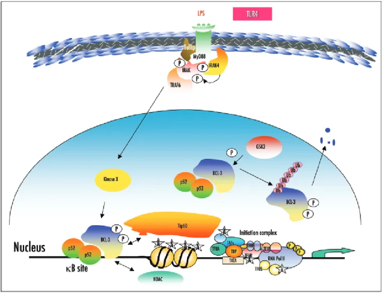
Figure 1. BCL-3 phosphorylation modulates its transcription potential. The kinase GSK3 phosphorylates BCL-3 on two residues within its C-terminal domain and this phosphorylation triggers ubiquitin linkage and proteasome-mediated BCL-3 degradation. In macrophages, LPS stimulation through the TLR4 triggers IRAK phosphorylation by the kinase IRAK4 and susbsequent recruitment of TRAF6 to the cell membrane. An LPS-inducible kinase subsequently phosphory-lates nuclear BCL-3 but this activated kinase remains to be identified. In the nucleus, BCL-3 recruits either some coactivators such as Tip60 or HDAC proteins in order to activate or repress transcription, respectively.
kinases may modulate BCL-3 transcriptional potential. Indeed, it has been known for many years that BCL-3 is heavily phosphorylated in several cell types including lymphoma cell lines.8,14,18,19Nuclear
BCL-3 is phosphorylated upon LPS treatment13and we have recently
identified a BCL-3 kinase distinct from GSK3 that is known to be activated by LPS (unpublished results). Therefore, at least two kinases phosphorylate BCL-3 in vivo. The biological consequences of this second phosphorylation on BCL-3 mediated gene transcription and on cellular transformation remains unknown but these issues are currently being addressed in our laboratory. It also remains to be determined whether BCL-3 is simultaneously phosphorylated by multiple kinases in any cell type or whether some cell specificity occurs.
Genetic disruption of the bcl-3 gene in mice indicated that BCL-3 is required for antigen-specific priming of T and B cells since mutant mice are impaired in germinal center reactions and T-depen-dent antibody responses to influenza virus and have a partial loss of
B cells which accounts for the immunological defects.20
Interestingly, many but not all defects observed in these mice were also reported for the p52 KO mice,21 therefore supporting the
hypothesis that p52 and BCL-3 form a functional, physiologically relevant transcriptional complex and induce the expression of many common target genes. Although BCL-3 is required for the formation of B cell follicles, it is not clear whether BCL-3 phosphorylation critically regulates this function. Indeed, chimeric mice that had been generated by injecting bone marrow precursor cells from BCL-3 KO mice expressing either wild type or a BCL-3 mutant no longer phosphorylated by GSK3 did not exhibit any defects in B and T cells development (unpublished results). However, because of the decreased transgene expression in the transduced cells, we could not evaluate the effects of GSK3-mediated BCL-3 phosphorylation over a long period of time (3–4 months). This issue could be elegantly elucidated through the generation of knockin mice. As overexpression of BCL-3 in B cells causes lymphoproliferation and splenomegaly,22 the knockin mice model could allow us to determine the exact role of BCL-3 in long term, sustained expression.
BCL-3 TARGET GENES
Very few BCL-3 target genes have been identified so far and include cyclin D1 in breast cancer cells.23 We recently identified about one hundred genes regulated by BCL-3 in NIH3T3 cells.14
Although the recruitment of this oncoprotein to the corresponding promoter sequences remains to be experimentally demonstrated, the expression of some of these BCL-3 target genes such as laminin-β2
chain (unpublished results) was indeed previously shown to be altered in Bcl-3 KO mice.24 Moreover, other candidates such as
Cxcl1 were previously described as NF-κB target genes. We are
cur-rently investigating whether the expression levels of BCL-3 and its target genes can be correlated in lymphomas and breast cancer sam-ples in order to identify genes that mediate BCL-3 oncogenic poten-tial in vivo.
LINKING BCL-3 FUNCTIONAL DOMAINS WITH TARGET GENE
EXPRESSION
As a member of the IκB family, BCL-3 harbors ankyrin repeats that mediate its interaction with p50 or p52. Moreover, these domains are required for interaction with the HDAC proteins. Whereas the C-terminal transactivating domain harbors all the
phosphorylated residues identified so far, the N-terminal domain displays a couple of lysine residues that are critical for BCL-3-medi-ated transcriptional activation.14 Indeed, a BCL-3 mutant lacking
these residues failed to activate most of the BCL-3 target genes. Why are these N-terminal lysine residues critical to mediate BCL-3 effects? Several hypothesis can be made but all of them require experimental validation. First, these lysine residues are targeted for ubiquitin link-age and several studies indeed demonstrated that some DNA-bound transactivation factors are targeted for ubiquitination within their transactivation domain, this signal being required for transcriptional activation.25,26Therefore, there may be a link between BCL-3
ubiq-uitination and activation. In any case, the molecular mechanism of BCL-3 ubiquitination and the identity of the ubiquitin E3 ligase remain unknown. Beside ubiquitination as a mechanism for tran-scriptional activation, the same lysine residues could also be targeted for acetylation, a post-translational modification associated with gene activation.26We are currently investigating whether BCL-3 is a
substrate of histone acetyltransferases. As a preliminary observation, some but not all histone acetyltransferases known to acetylate sever-al transcription factors, enhances BCL-3-mediated transcriptionsever-al activation (our unpublished results). Therefore, BCL-3 may recruit these proteins in order to activate transcription. However, direct acetylation of BCL-3 by histone acetyltransferases still needs to be experimentally demonstrated.
CONCLUSION
Although BCL-3 was identified more than fifteen years ago, most of its biological roles remains unexplored. The increasing interest for the role of NF-κB/IκB proteins in cancer will certainly help to better understand the BCL-3-dependent molecular mechanisms. It is also likely that phosphorylation will emerge as a critical mechanism for the regulation of BCL-3 activity.
References
1. Hayden MS, Ghosh S. Signaling to NF-κB. Genes Dev 2004; 18:2195-224.
2. McKeithan TW, Rowley JD, Shows TB, Diaz MO. Cloning of the chromosome transloca-tion breakpoint junctransloca-tion of the t(14;19) in chronic lymphocytic leukemia. Proc Natl Acad Sci USA 1987; 84:9257-60.
3. Ohno H, Takimoto G, McKeithan TW. The candidate proto-oncogene BCL-3 is related to genes implicated in cell lineage determination and cell cycle control. Cell 1990; 60:991-9. 4. Nishikori M, Maesako Y, Ueda C, Kurata M, Uchiyama T, Ohno H. High-level expression of BCL-3 differentiates t(2,5)(p23;q35)-positive anaplastic large cell lymphoma from hodgkin disease. Blood 2003; 101:2789-96.
5. Thornburg NJ, Pathmanathan R, Raab-Traub N. Activation of nuclear factor-κB p50 homodimer/Bcl-3 complexes in nasopharyngeal carcinoma. Cancer Res 2003; 63:8293-301.
6. Cogswell PC, Guttridge DC, Funkhouser WK. Baldwin AS Jr. Selective activation of NF-κ
B subunits in human breast cancer: Potential roles for NF-κB2/p52 and for Bcl-3. Oncogene 2000; 19:1123-31.
7. Bours V, Franzos, G, Azarenko V, Park S, Kanno T, Brown Siebenlist U. The oncoprotein BCL-3 directly transactivates through κB motifs via association with DNA-binding p50B homodimers. Cell 1993; 72:729-39.
8. Fujita T, Nolan GP, Liou HC, Scott ML, Baltimore D. The candidate proto-oncogene BCL-3 encodes a transcriptional coactivator that activates through NF-κB p50 homod-imers. Genes Dev 1993; 7:1354-63.
9. F9. Franzoso G, Bours V, Azarenko V, Park S, Tomita-Yamaguchi M, Kanno T, Brown K Siebenlist U. The oncoprotein BCL-3 can facilitate NF-κB-mediated transactivation by removing inhibiting p50 homodimers from select κB sites. EMBO J 1993; 12:3893-901. 10. Grundström S, Anderson P, Scheipers P, Sundstedt A. Bcl-3 and NFκB p50-p50 homod-imers act as transcriptional repressors in tolerant CD4+ T cells. J Biol Chem 2004; 279:8460-8.
11. 11. Kuwata H, Watanabe Y, Miyoshi H, Yamamoto M, Kaisho T, Takeda K, Akira S. IL-10-inducible Bcl-3 negatively regulates LPS-induced TNF-α production in macrophages. Blood 2003; 102:4123-9.
12. 12. Dechend R, Hirano F, Lehmann K, Heissmeyer V, Ansieau S, Wulczyn FG, Scheidereit C, Leutz A. The Bcl-3 oncoprotein acts as a bridging factor between NF-κB/Rel and nuclear coregulators. Oncogene 1999; 18:3316-23.
1500 Cell Cycle 2004; Vol. 3 Issue 12
13. 13. Wessells J, Baer M, Young HA, Claudio E, Brown K, Siebenlist U, Johnson P. BCL-3 and NF-κB p50 attenuate LPS-induced inflammatory responses in macrophages. J Biol Chem in press.
14. 14. Viatour P Dejardin E Warnier M, Lair F, Claudio E, Bureau F, Marine JC, Merville MP, Maurer U, Green D, Piette J, Siebenlist U, Bours V, Chariot A. GSK3-Mediated BCL-3 phosphorylation modulates its degradation and its oncogenicity. Mol Cell 2004; 16:35-45. 15. Leung TH, Hoffman A, Baltimore D. One nucleotide in a κB site can determine cofactor
specificity for NF-κB dimers. Cell 2004; 118:453-64.
16. Baek SH, Ohgi KA, Rose DW, Koo EH, Glass CK, Rosenfeld MG. Exchange of N-CoR corepressor and Tip60 coactivator complexes links gene expression by NF-κB and β -amy-loid precursor protein. Cell 2002; 110:55-67.
17. Devoogdt N, Hassanzadeh Ghassabeh G, Zhang J, Brys L, De Baetselier P, Revets H. Secretory leukocyte protease inhibitor promotes the tumorigenic and metastatic potential of cancer cells. Proc Natl Acad Sci USA 2003; 100:5778-82.
18. Bundy DL, McKeithan TW. Diverse effects of BCL3 phosphorylation on its modulation of NF-κB p52 homodimer binding to DNA. J Biol Chem 1997; 272:33132-9. 19. 19. Nolan GP, Fujita T, Bhatia K, Huppi C, Liou H-C, Scott ML, Baltimore D. The
BCL-3 proto-oncogene encodes a nuclear IκB-like molecule that preferentially interacts with NF-κB p50 and p52 in a phosphorylation-dependent manner. Mol Cell Biol 1993; 13:3557-66.
20. 20. Franzoso G, Carlson L, Scharton-Kersten T, Shores EW, Epstein S, Grinberg A, Tran T, Shacter E, Leonardi A, Anver M, Love P, Sher A, Siebenlist U. Critical roles for the Bcl-3 oncoprotein in T cell-mediated immunity, splenic microarchitecture, and germinal center reactions. Immunity 1997; 6:479-90.
21. 21. Franzoso G, Carlson L, Poljak L, Shores EW, Epstein S, Leonardi A, Grinberg A, Tran T, Scharton-Kersten T, Anver M, Love P, Brown K, Siebenlist U. Mice deficient in nuclear factor (NF)-κB/p52 present with defects in humoral responses, germinal center reactions, and splenic microarchitecture. J Exp Med 1998; 187:147-59.
22. Caamano JH, Perez P, Lira SA, Bravo R. Constitutive expression of Bc1-3 in thymocytes increases the DNA binding of NF-κB1 (p50) homodimers in vivo. Mol Cell Biol 1996; 16:1342-8.
23. Westerheide SD, Mayo MW, Anest V, Hanson JL, Baldwin Jr AS. The putative oncopro-tein Bcl-3 induces cyclin D1 to stimulate G(1) transition. Mol Cell Biol 2001; 21:8428-36. 24. Poljak L, Carlson L, Cunningham K, Kosco-Vilbois MH, Siebenlist U. Distinct activities of p52/NF-κ B required for proper secondary lymphoid organ microarchitecture: Functions enhanced by Bcl-3. J Immunol 1999; 163:6581-8.
25. Conaway RC, Brower CS, Conaway JW. Emerging roles of ubiquitin in transcription reg-ulation. Science 2002; 296:1254-8.
26. Freiman RN, Tjian R. Regulating the regulators: Lysine modifications make their mark. Cell 2003; 112:11-7.
www.landesbioscience.com Cell Cycle 1501