HAL Id: tel-01450519
https://pastel.archives-ouvertes.fr/tel-01450519
Submitted on 31 Jan 2017HAL is a multi-disciplinary open access archive for the deposit and dissemination of sci-entific research documents, whether they are pub-lished or not. The documents may come from teaching and research institutions in France or abroad, or from public or private research centers.
L’archive ouverte pluridisciplinaire HAL, est destinée au dépôt et à la diffusion de documents scientifiques de niveau recherche, publiés ou non, émanant des établissements d’enseignement et de recherche français ou étrangers, des laboratoires publics ou privés.
Design and characterization of hydrogel films and
hydrogel-ceramic composites for biomedical applications
David Moreau
To cite this version:
David Moreau. Design and characterization of hydrogel films and hydrogel-ceramic composites for biomedical applications. Materials. Université Paris sciences et lettres, 2016. English. �NNT : 2016PSLEM015�. �tel-01450519�
THÈSE DE DOCTORAT
de l’Université de recherche Paris Sciences et Lettres
PSL Research University
Préparée à MINES ParisTech
Elaboration et caractérisation de films d’hydrogel et de composites
hydrogel-céramique pour les applications biomédicales
Design and characterization of hydrogel films and
hydrogel-ceramic composites for biomedical applications
COMPOSITION DU JURY :
M. Philippe POULIN
CRPP, Université de Bordeaux, Rapporteur
M. André STUDART
Complex Materials, ETH-Zürich, Rapporteur
Mme. Ghislaine BERTRAND
CIRIMAT, ENSIACET-Toulouse, Examinateur
M. Pierre WEISS
LIOAD, Université de Nantes, Président du jury
M. Vincent GUIPONT
Centre des Matériaux, Mines Paristech, Paris Examinateur
M. Laurent CORTÉ
Centre des Matériaux, Mines Paristech, Paris Examinateur
Soutenue par David MOREAU
le 21 janvier 2016
h
Ecole doctorale n°432
SCIENCES DES METIERS DE L’INGENIEUR
Spécialité Sciences et Génie des Matériaux Spécialité Sciences et Génie des Matériaux
Dirigée par
Laurent CORT
É et Vincent GUIPONT
5 À mon grand-père.
7
Remerciements
Je tiens à remercier tout d’abord mes directeurs de thèse, Laurent Corté et Vincent Guipont. Laurent a été un encadrant fantastique au cours de ces quatre années passées au Centre des Matériaux (CDM) de l’Ecole des Mines et au laboratoire Matière Molle et Chimie (MMC) de l’ESPCI. Son enthousiasme, son savoir et sa curiosité scientifique, sa rigueur et sa bienveillance m’ont permis d’aller au bout de cette belle aventure. Je remercie Vincent pour ses conseils avisés et nos riches discussions. J’ai pu bénéficier de sa grande expertise sur la projection thermique et de ses excellents conseils en matière de présentation. A leurs côtés, j’ai fait de nombreuses découvertes scientifiques, parfois inattendues, et je les remercie pour leur soutien et leur engagement sans faille tout au long de ma thèse (et ce n’est pas terminé!).
J’adresse mes remerciements à Jacques Besson, directeur du CDM et à Ludwik Leibler, directeur du MMC, pour m’avoir accueilli au sein de leur laboratoire et pour m’avoir donné accès à toutes les ressources nécessaires pour réaliser mes travaux de thèse.
Je remercie également l’ensemble de mon jury de thèse. Je suis reconnaissant envers Monsieur Philippe Poulin pour avoir accepté avec enthousiasme de rapporter mon travail et pour m’avoir fait part de commentaires très constructifs pendant et après la soutenance. As well, I would like to acknowledge very much André Studart to have reviewed my thesis very carefully and to have discussed my results with passion. Je tiens à remercier Monsieur Pierre Weiss pour m’avoir fait l’honneur de présider mon jury de thèse et à Madame Ghislaine Bertrand d’y avoir participé avec beaucoup d’engouement. Vos questions et vos remarques ont contribué à rendre ma soutenance vivante et inoubliable.
Une thèse dans le domaine des biomatériaux nécessite de nombreux savoir-faire, techniques et connaissances dans des domaines scientifiques variés, allant de la biologie
8 cellulaire à la caractérisation aux rayons X sur ligne synchrotron en passant par la chirurgie animale. Ces travaux sont par conséquent le fruit de nombreuses collaborations enrichissantes à la fois sur le plan humain et scientifique. J’adresse mes plus sincères remerciements à Hervé Petite, Didier Hannouche, Manon Bachy, Arthur Vilain, Morad Bensidhoum et Frédéric Baudin du laboratoire de Bioingénierie et Bioimagerie Ostéo-Articulaire de l’Université Paris-Diderot, pour les études in vivo. Je remercie également François Rannou, Caroline Chauvet, François Etienne, Jean-Maurice Petit et Lydia Tsagris de l’unité Inserm UMR-S 1124 «Pharmacologie, Toxicologie et Signalisation cellulaire» de l’Université Paris-Descartes pour les expériences originales et intenses d’encapsulation cellulaire. Je remercie chaleureusement Henry Proudhon qui a participé activement à la caractérisation aux rayons X de nos systèmes à la fois au laboratoire Navier de l’Ecole Nationale des Ponts et Chaussées appuyé par Nicolas Lenoir et au synchrotron SOLEIL épaulé par Sylvie Giraud et Christian Mocuta.
En plus des collaborations extérieures, les travaux de cette thèse n’auraient jamais abouti sans l’aide précieuse de nombreuses personnes du CDM et du MMC à qui j’adresse toute ma gratitude et que je remercie. Yann Auriac, pour tout son soutien et son ingéniosité absolument remarquable; François Borit avec qui nous avons réalisé toute la campagne d’essais coldspray; Maria Betbeder, Fabrice Gaslain, Anne Laurent et Nicole Dedave, pour la microscopie électronique et, plus spécifiquement, en mode ‘low-vac’; Daniel Pachoutinsky, Sylvie Giraud, Henry Proudhon et Anthony Chesnaud pour les analyses aux rayons X; Jean-Christophe Tesseidre pour la corrélation d’image; Brigitte Raviard pour mes demandes de produits farfelus; Jean-Dominique Bartoux pour la granulométrie; Karine Vieillevigne pour les mesures ATG/ATD; Steve Duvinage pour ses conseils en photo; l’Atelier indispensable du CDM formé par René Cluzet, Jean-Pierre Etourneau, Christophe Meurin, Franck Bluzat et Georges Cassas pour leurs montages spécifiques et leurs coups de pouce divers et variés;
9 Corinne Soulié-Ziakovic pour les analyses IR; Gregory Sainte-Luce et Olivier Delhomme pour l’assistance informatique; Odile Adam pour la biblio et la documentation; Véronique Matos pour la logistique; Marie-France Boucher et Catherine Rouil pour leur assistance et le secrétariat chaleureux.
Une thèse c’est aussi des rencontres, des bons moments, des événements, des conférences. J’adresse un grand merci à: Nico la toupie, Victor, Erembert, Arina et Jia les occupants du bureau B108 pour l’avoir rendu aussi chaleureux; le bureau d’en face où règne une sacrée ambiance, grâce à une bande de joyeux lurons formée par Clém’, Gui’, Quentin, et Christophe (le grimpeur); David (aka DM, on finit ensemble!); François (stay cool ;)); Thibault (le perfectionniste); Samuel Forest et Anne-Françoise Gourgues pour leurs encouragements et leur bonne humeur; André Pineau et nos échanges sur les dispositifs médicaux dans les couloirs; PAP, mon partenaire de choc du trophée étudiant de golf; les doctorants et chercheurs du MMC qui m’ont accueilli à bras ouverts: Marie, Julien et André pour la bonne humeur quotidienne dans le bureau; le bureau (d’à côté) du bonheur formé par Coco, Technini, François la bonne affaire, Miki et Thibault (toujours le mot pour rire), auquel Jérémie se joignait presque quotidiennement; Lucie et Charlotte pour nos discussions tardives et le réconfort mutuel dans la rédaction; Raphael, Franz, Maddalena, Maïssa, Robi, Trystan, Max et Adrien pour l’ambiance du midi au PC café; Rémi et Michel pour nos discussions approfondies sur le PVA (entre autres); Fanny, Lise et Aurélie pour vos encouragements.
Et puis, il y a ceux qui m’ont toujours soutenu et encouragé dans la voie de la recherche. J’ai une pensée toute particulière pour Jean Woloszko, que je considère comme mon mentor depuis le début de mes études scientifiques. Je remercie également mes professeurs de l’Ecole Nationale d’Ingénieurs de Metz, Anne-Sophie Bonnet, Paul Lipinski, Emilie De-Brosses, Sylvain Philippon et Albert Tidu.
10 Enfin, je remercie ceux qui me soutiennent depuis longtemps et qui m’ont supporté pendant ces 3 années: mes parents, mon frère, mes grands-parents, Marine et mes amis de tous horizons. MERCI.
12
General introduction ... 16
Literature review ... 25
Section 1.
Chapter 1. Poly(vinyl alcohol) hydrogels ... 261.1 What is a hydrogel? ... 26
1.2 Poly (vinyl alcohol) chemical structure and synthesis ... 27
1.3 Crosslinking of PVA hydrogels ... 28
1.4 Physical properties of PVA hydrogels ... 36
1.5 Biological applications of PVA hydrogels ... 46
1.6 Clinical applications ... 48
1.7 Perspectives and challenges related to PVA hydrogels for biomedical application ... 49
Chapter 2. Hydroxyapatite ... 52
2.1 Chemical composition and preparation ... 52
2.2 Osteoconductive properties of HA ... 53
2.3 Biological response to HA particles ... 56
Chapter 3. Composites of PVA/HA ... 59
3.1 Fabrication processes and physical properties ... 59
3.2 Biological studies of PVA/HA composites ... 62
Conclusion ... 64
Materials ... 78
Section 2.
Chapter 4. Poly(vinyl alcohol) (PVA) ... 794.1 Commercial materials ... 79
4.2 PVA aqueous solution and substrates processing ... 79
4.3 Physicochemical characterization ... 81
Chapter 5. Hydroxyapatite (HA) ... 86
5.1 Manufacturing process of HA particles ... 86
5.2 Physicochemical characterization ... 87
5.3 Microstructure and physicochemical characterization ... 88
Hydrogel-ceramic composite coatings produced by
dip-Section 3.
coating for the osteointegration of hydrogel implants ... 96
Chapter 6. Poly(vinyl alcohol) hydrogel coatings with tunable surface exposure of hydroxyapatite ... 98
6.1 Introduction ... 100
13
6.3 Results ... 105
6.4 Discussion ... 117
6.5 Conclusion ... 121
Chapter 7. In vivo evaluation of the bone integration of coated poly(vinyl-alcohol) hydrogel fiber implants. ... 129
7.1 Introduction ... 129
7.2 Materials and methods ... 132
7.3 Results ... 138
7.4 Discussion ... 145
7.5 Conclusion ... 147
Hydrogel films by swelling induced gelation ... 154
Section 4.
Chapter 8. Hydrogel films by swelling induced gelation ... 1558.1 Introduction ... 155
8.2 Materials & Method ... 158
8.3 Results & Discussion ... 162
8.4 Conclusion ... 175
Chapter 9. Cell encapsulation in poly(vinyl alcohol) hydrogel films by swelling induced gelation ... 180
9.1 Introduction ... 180
9.2 Materials and methods ... 183
9.3 Results ... 189
9.4 Discussion ... 197
9.5 Conclusion ... 200
Section 5. Hydroxyapatite coatings on PVA hydrogels by cold spray
... 206
Chapter 10. Cold spray coating of submicronic hydroxyapatite on poly(vinyl alcohol) in dry and hydrogel states ... 207
10.1 Introduction ... 207
10.2 Materials & Methods ... 210
10.3 Results ... 214
10.4 Discussion ... 225
10.5 Conclusion ... 230
Chapter 11. Influence of the particle composition on the coating formation .. 236
11.1 Introduction ... 236
14
11.3 Results ... 239
11.4 Discussion ... 247
11.5 Conclusion ... 249
Appendix ... 252
Appendix 1. Diffusivity coefficients ... 252
Appendix 2. Encapsulation of chondrocyte cells in poly(vinyl alcohol) hydrogel films gelified at the surface of polyacrylamide swelling substrates. ... 254
16
General introduction
In this thesis, I explore a particular class of polymers which are called hydrogels. Basically, hydrogels are three-dimensional cross-linked networks of hydrophilic polymer chains. These polymer networks can contain a quantity of water of several tens to thousands times their dry mass. In 1960, Wichterle and Lim1 suggested that hydrogel-based materials could be of great interest for medical applications due to their water content and their structural, optical and mechanical properties that are close to those of living tissues. Since then, the progresses in medicine, biology and material sciences have led to multiple hydrogel-based inventions and to the development of commercial products for various biomedical applications, such as contact lenses,2 wound dressings,3,4 phantom tissues for medical imaging,5 drug delivery systems,6 tissue-engineered or non-degradable substitutes for organ and tissue repair,7,8 diagnostic devices9 or hygienic products.10
One particular application of hydrogels that could have a strong impact concerns the reconstruction of teared or diseased soft tissues of the osteoarticular system. Indeed, diseases and injuries of the osteo-articular system, such as osteoarthrisis (cartilage degeneration) or ligament tearing, have become today major health issues.11 The occurrence of these problems is mostly attributed to the aging and to the intensification of sport in the general population. For instance osteoarthritis concerns 45 million people in the United States of America and costs about 2,600 $ per person per year.12,13 Another example is the anterior cruciate ligament reconstruction, that is performed for instance on 35,000 people in France each year and represent a cost of about 7,000 € per patient.14 One particular complication for the treatment of most damaged osteoarticular soft tissues is that they do not heal properly and cannot be healed by primary repair. As a result, the treatment often requires surgical reconstruction
17 using an auto or allograft. Autografts, which are sample tissues from the patient itself, are the gold standard, because they do not suffer from immune rejection. However the side effect caused by the donor site morbidity can be limiting. Allografts that come from other patients are used sometimes, but their availability and cost can be prohibited. Moreover the immune rejection of the graft implies that the patient takes heavy immunosuppressive treatments or that the graft is decellularized by methods that can alter the properties of the tissue. In this context, hydrogel-based substitutes are drawing a lot of attention as they could provide off-the-shelf alternate solutions.15–18 Whether it be for ligament, cartilage or tendon replacement, such substitutes require a fine control of physicochemical properties to ensure the durability and the innocuousness, as well as an appropriate mechanical behavior that is appropriate to restore the physiological function. In particular, all these tissue substitutes are connected to bone. The anchoring is one of the major challenges in the design of these substitutes. Failures of anchoring can be dramatic and lead to failures or loosening of the implants. Indeed, anchoring points must resist to normal physiological cyclic loading, as well as punctual intense loads during the whole life of the patient. The general strategy to obtain a strong and durable anchoring to bone is to favor the creation of an intimate contact between the bone and the implant surface (osseointegration). This is usually achieved by functionalizing the implant surface. One functionalization strategy is to coat the surface of the hydrogel implant with a bioactive matter, such as bioceramics, drugs or even cells. The design and fabrication of such coatings is challenging: The coating process must produce adherent and cohesive coatings at the surface of the hydrogel substrates; It should preserve the properties of the substrate and also not compromise the bioactivity of the coating material; Lastly, it should be compatible with clinical practice requirements. In this work, I have studied processes to make ceramic coatings on hydrogel implants in the idea of improving the integration to bone tissue.
18 The hydrogels studied in this thesis are poly(vinyl alcohol) (PVA) hydrogels. PVA is a synthetic polymer which can form non-resorbable physically cross-linked hydrogels.19 These PVA hydrogels have a good history with biocompatibility in numerous in vivo studies for the reconstruction of various soft tissues.16 Nevertheless, cells adhere poorly to PVA hydrogels and a better control of the anchoring to bony tissues is still needed in several applications. According to the literature, one way to improve the osseointegration is the addition of a bio-ceramic coating, such as hydroxyapatite, might be an ideal option. Hydroxyapatite (HA) is the major mineral component of bone and has been shown to enhance osteoconduction.20 It is already used as a bone substitute and as a coating for metallic prosthesis (e.g. hip prosthesis). For this research, I investigate two approaches to produce hydroxyapatite coatings on PVA substrates. The first approach is a soft method, based on dip-coating and physical cross-linking, which does not require strong energetic input. The second is a more energetic approach, based on thermal projection of hydroxyapatite particles using the cold spray process.
In more details, in the first soft approach, we explore how to produce composite hydrogel-ceramic coatings on a hydrogel substrate. With a process combining dip-coating with physical cross-linking of PVA, coatings of hydroxyapatite particles embedded in a non-degradable PVA hydrogel matrix are formed on hydrogel substrates. The fabrication of these coatings necessitates an appropriate choice of the composition and of the processing parameters. It is essential to determine how the solution composition governs the thickness, adherence and cohesion of the coating. In addition the PVA hydrogel matrix, containing the HA particles, is not degradable. Therefore, the process needs to be adjusted to ensure that HA must sufficiently exposed and accessible by cells. This approach is applied to produce coatings on PVA hydrogel ligament substitutes that have been recently developed at the Centre des Matériaux (CNRS UMR 7633) (Mines ParisTech) in collaboration with the
19 laboratory of Prof. David N. Ku at the University of GeorgiaTech.21 Thanks to a close collaboration with the laboratory of “Bioingénierie et Bioimagerie Ostéo-Articulaire” (B2OA-UMR 7052) at Paris-Diderot University, we devised an in vivo study to assess their biocompatibility and their osteoconductivity. To carry out this study, we had to adapt the manufacturing process to sterile requirements and to produce coatings that are compatible with the animal model and surgical handling.
During the investigation of this first approach, I characterized in the parameters that govern the thickness of the coating and noted that it depends strongly on the swelling state of the hydrogel substrate. We studied how this phenomenon can be related to the peculiar ability of PVA solution to crosslink at high concentrations. This side study led to the discovery of a new method to produce hydrogel films, using the solvent depletion created at the surface of a swelling substrate. This swelling induced gelation method opens route to produce composite hydrogel-ceramic coatings without exterior action nor harsh treatment. We also wondered if sensitive matter, such as cells, can be encapsulated using this mild way to generate hydrogel films. In collaboration with the laboratory of “Toxicology Pharmacology and Cellular Signaling” (INSERM UMR-S 1124) at Paris-Descartes University, we investigated the process and devised encapsulation protocol that is compatible with cell culture requirements. With this first feasibility study, we aimed to understand how the cell viability depends on the different parameters of the process: the concentration of the solution, the swelling of the substrate and the temperature condition.
The second approach is a more energetic method, based on cold spray, which is a thermal spraying process. Usually thermal spraying is used, by way of plasma or high velocity oxy-fuel spraying, to coat metallic prosthesis with bioceramic particles. Here we exploit the relatively “mild” spraying conditions that cold spray process offers to produce hydroxyapatite coatings on thermo-sensitive PVA hydrogel substrates. Conversely to the first method, the
20 cold spray allows to form coatings of HA particles compactly stacked on hydrogel substrates without binder. However, the use of a thermal spraying process requires to define ranges of parameters for which it is possible to produce compact and adherent coatings of HA without deteriorating the hydrogel substrate. It is unlikely the PVA hydrogels can resist the temperature and pressure reached during spraying. However, an alternate approach consisting in firstly spraying onto dry PVA and secondly swelling the coated substrate might work. With a systematic study of the spraying parameters, we explore how to adapt the spraying process to produce a coating that is adherent and resist swelling. Microscopic investigations and temperature measurements during spraying help us understand what are the mechanisms involved in the coating formation. In a last very exploratory part of this work, I also studied how the composition and the microstructure of the sprayed particles influence coating thickness. Indeed, the temperatures used in cold spray do not melt the ceramic particles. Therefore, it is difficult to build a thick coating of pure ceramics. Here we approach this question by playing with the presence of synthesis residues that could act as a temporary binder to stack the sprayed particles.
This manuscript is organized in five sections as follows:
Section 1 is a non-exhaustive literature review describing general properties of poly(vinyl alcohol) hydrogels, of hydroxyapatite and of PVA/HA hydrogel composites.
Section 2 introduces the material used in this thesis, namely the PVA substrates and the hydroxyapatite particles.
Section 3 presents the manufacturing process of the first approach, based on dip-coating, and the in vivo evaluation of the PVA hydrogel and hydroxyapatite composite coatings using a rabbit model of bone tunnel healing.
21
Section 4 is dedicated to the understanding of the gelation mechanisms of PVA hydrogel film at the surface of a swelling polymer substrate and its application to cell encapsulation.
Section 5 presents the energetic approach, based on cold spray to produce HA coatings on hydrogel substrates, as well as the exploratory study of the effect of powder composition on coating thickness.
Each section is introduced by a short paragraph giving the interests and the motivations of the study. The sections are divided in several chapters. Chapters 6 to 11 have been written in the form of an article. I chose this type of writing format, because it makes the reading easier due to the various and numerous topics treated in this thesis work. In particular, each chapter contains its specific introduction of state of the art and “Materials&Methods”. In all of these chapters, a particular attention has been given not to repeat the same concepts and results. Some repetitions may still occur and I hope this will not make the reading too tedious.
22 References
1. Wichterle, O. & Lim, D. Hydrophilic gels for biological use. Nature 185, 117–118 (1960).
2. Maldonado-Codina, C. & Efron, N. Hydrogel Lenses-Material and Manufacture: A Review. Optom. Pract. 4, 101–115 (2003).
3. Intrasite®Gel. Smith&Nephew (2015). at <www.smith-nephew.com>
4. Aquasorb® Hydrogel Wound Dressing. DeRoyal (2015). at <www.deroyal.com>
5. Campbell, G. & Homblower, V. Tissue-mimicking phantom for prostate cancer brachytherapy. (2010) US 2010/0041005 A1
6. Chang, P. ., Meyrueix, R., Rivail, C. & Chateliier, J. Medusa: an Innovative Formulation Approach to Improve Pharmacokinetic & Safety Profiles of Biotherapeutics. ONdrugDelivery 4–6 (2011).
7. GelrinC. Regentis Biomaterials (2015). at <www.regentis.co.il>
8. Lifeline®. Cytograft tissue engineering (2015). at <www.cytograft.com>
9. Tokuda, T. et al. CMOS image sensor-based implantable glucose sensor using glucose-responsive fluorescent hydrogel. Biomed. Opt. Express 5, 3859 (2014).
10. Pampers. Procter&Gamble (2015). at <www.pampers.com>
11. Hochberg, M. C. Osteoarthritis: new approaches. Medicographia 35, 139 (2013).
12. Helmick, C. G. et al. Estimates of the prevalence of arthritis and other rheumatic conditions in the United States: Part I. Arthritis Rheum. 58, 15–25 (2008).
23 13. Gabriel, S. ., Crowson, C. ., Champion, M. . & O’Fallon, W. . Direct medical costs unique to people with arthritis. J. Rheumatol. 24, 719–725 (1997).
14. HAS. Critères de suivi en rééducation et d’orientation en ambulatoire ou en SSR Après ligamentoplastie du croisé antérieur du genou. (2008).
15. Sciarretta, F. V. 5 to 8 years follow-up of knee chondral defects treated by PVA-H hydrogel implants. Eur. Rev. Med. Pharmacol. Sci. 17, 3031–3038 (2013).
16. Baker, M. I., Walsh, S. P., Schwartz, Z. & Boyan, B. D. A review of polyvinyl alcohol and its uses in cartilage and orthopedic applications. J. Biomed. Mater. Res. B Appl. Biomater. 100B, 1451–1457 (2012).
17. Laurencin, C. T. & Freeman, J. W. Ligament tissue engineering: An evolutionary materials science approach. Biomaterials 26, 7530–7536 (2005).
18. Kobayashi, M., Chang, Y.-S. & Oka, M. A two year in vivo study of polyvinyl alcohol-hydrogel (PVA-H) artificial meniscus. Biomaterials 26, 3243–3248 (2005).
19. Hassan, C. M. & Peppas, N. A. in Biopolymers· PVA Hydrogels, Anionic Polymerisation Nanocomposites 37–65 (Springer, 2000).
20. Hench, L. L. Bioceramics: from concept to clinic. J. Am. Ceram. Soc. 74, 1487–1510 (1991).
21. Bach, J. S. et al. Hydrogel fibers for ACL prosthesis: Design and mechanical evaluation of PVA and PVA/UHMWPE fiber constructs. J. Biomech. 46, 1463–1470 (2013).
25
Literature review
Section 1.
With this PhD research, I explore different ways of producing composite hydrogels in the perspective of biomedical applications. The literature review given in this section focuses on the existing knowledge about the main components studied in this work. It summarizes some basic knowledge on polymer hydrogels, in particular poly(vinyl alcohol) (PVA) hydrogels, on hydroxyapatite and on composites of PVA hydrogels/hydroxyapatite. A very large body of literature exists on these topics and this review does not pretend to be exhaustive.
Chapter 1 is a general introduction about hydrogels and more specifically about hydrogels of poly(vinyl alcohol). We evoke their chemical structure and how they are synthesized. The different processes of cross-linking are summarized. Some specific properties of PVA hydrogels are also described, in particular their swelling and mechanical characteristics as well as their use in biomedical applications. In chapter 2, hydroxyapatite is introduced. First, we give some details about its physical-chemical properties and manufacturing processes. Then its role and mechanism in bone formation are presented. In chapter 3, we present how PVA and hydroxyapatite can be processed together to form composites and we discuss about the in vitro and in vivo response of such systems.
26
Chapter 1. Poly(vinyl alcohol) hydrogels
1.1 What is a hydrogel?
Hydrogels are hydrophilic polymeric networks able to swell in water. Depending on their physic-chemical properties, they can absorb a quantity of water equivalent to a few times to thousands times their dry mass. Hydrogels remain solid in water because the macromolecules of the network are cross-linked and cannot dissolve. Crosslinking can be of chemical or physical nature. Chemical crosslinking involves permanent covalent bonds between polymer chains. Physical crosslinking forms reversible bonds, which can be hydrogen bonds, Van der Waals interactions, molecular chain entanglements, or hydrophobic forces.1 The properties of hydrogels are defined by the polymer characteristics and the network structure. These properties can be tailored by the type of the polymer and the choice of the manufacturing process and its parameters.
Their high water content, their morphological characteristics, and mechanical properties close to that of biological tissues make them excellent candidates for biomedical uses.2 Hydrogels motivate active researches soft tissue prosthesis,3,4 tissue engineering scaffolds,1,4 drug delivery systems,5–7 contact lenses.8 They are currently used clinically in applications, such as cartilage substitutes,9 or contact lenses.8,10 Hydrogels for biomedical applications can be obtained from natural4 or synthetic polymers.11 Natural hydrogels include polysaccharides like agarose, alginate, or chitosan, and proteins such as collagen or gelatin. Synthetic hydrogels include poly(ethylene glycol) (PEG), poly(vinyl alcohol) (PVA), poly(acrylic acid) (PAA), poly(hydroxyl ethyl methacrylate) (polyHEMA), or poly(vinyl pyrrolidone) (PVP). In this thesis, we will mainly focus on hydrogels of PVA, which are intensively studied in the biomedical field.12
Chapter 1. Poly(vinyl alcohol) hydrogels
27 1.2 Poly (vinyl alcohol) chemical structure and synthesis
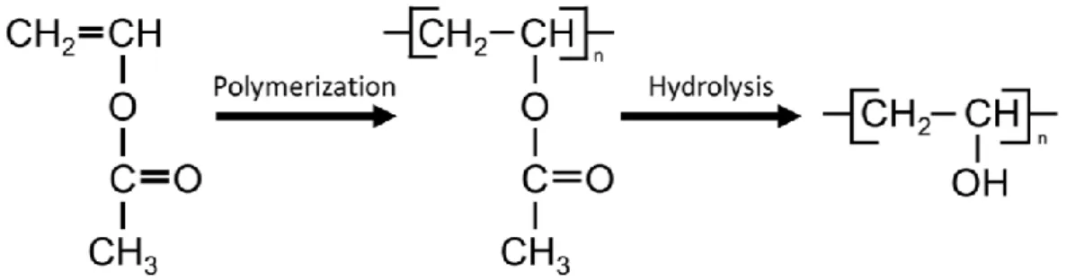
Poly (vinyl alcohol) has a hydrocarbon backbone structure with -OH hydroxyl pendant groups. The hydroxyl groups provide polarity to the PVA chains, which makes them soluble in water. The synthesis of PVA is not achieved directly by polymerizing the corresponding monomer of vinyl alcohol. Figure 1 illustrates the reactions of PVA synthesis. First, PVA is obtained from the free radical polymerization of vinyl acetate monomer, which gives poly (vinyl acetate) (PVAc) (C4H6O2).13 Then PVAc is hydrolyzed to produce PVA (C2H4O). This hydrolysis reaction is realized in aqueous media in presence of caustic or acidic catalysts.13
Figure 1 PVA synthesis: polymerization of vinyl acetate followed by a hydrolysis of poly(vinyl acetate).
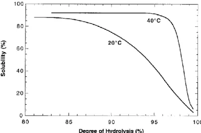
This reaction of hydrolysis is not always complete, and PVA chains always contain a fraction of residual acetate groups. Depending on the process, the degree of hydrolysis can range from partial (~70%) to almost full hydrolysis (>99%).14 It has been reported that the degree of hydrolysis has a strong impact on the solubility and crystallization of PVA. As shown in Figure 2, the solubility decreases strongly when the degree of hydrolysis reaches values superior to 97-98%.12,13 This sensitivity to the degree of hydrolysis is explained by the fact that residual acetate groups weaken the inter- and intra-molecular bonding between hydroxyl groups of the polymer, due a steric hindrance, and make defects that hinder crystallization, making PVA more soluble.
Chapter 1. Poly(vinyl alcohol) hydrogels
28
Figure 2 Solubility of PVA (Mw=77000 g/mol) as a function of degree of hydrolysis at dissolution
temperature of 20 and 40°C.13
1.3 Crosslinking of PVA hydrogels
Chemical crosslinking
Chemical crosslinking of PVA is achieved by using crosslinking agents or by irradiation. Common crosslinking agents for PVA are formaldehyde, gluraldehyde and others aldehydes.15 During chemical crosslinking, acetal bridges are formed between hydroxyl groups of pendant chains and the crosslinking agent in acidic environment, as shown in Figure 3.16
Chapter 1. Poly(vinyl alcohol) hydrogels
29 In the case of in vivo applications, these chemical crosslinking methods are usually not used because the cross-linking agents are often toxic and the processes require steps of washing to remove all unreacted crosslinking agents.17 However recently, it has been shown that PVA/chitosan hydrogel chemically crosslinked with glutaraldehyde could be biocompatible. In a study of Mansur et al.,18 cementoblast cells were seeded on PVA/chitosan hydrogel substrates with different glutaraldehyde content for 48 hours. Figure 4 presents MTT assays performed after 48 hours of culture. The absorbance relative values suggest a correct cellular viability on each substrate, containing 0%, 1% and 5% of glutaraldehyde.
Figure 4 Viability of cementoblast cells seeded on Chi/PVA (1:3) with different network crosslinking densities (without GA, 1.0% and 5.0% GA) via MTT biocompatibility assay; Cellular control (CC)
was used as reference.18
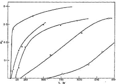
In other methods, chemical crosslinking is achieved by electron beam or gamma irradiation.12,19–21 These methods do not require cross-linkers or initiators, which can be toxic and difficult to remove. They also have the advantages of cross-linking and sterilizing the PVA hydrogels in one step.22 By varying the irradiation doses, hydrogel parameters (e.g. degree of crosslinking) can be tailored. Figure 5 shows the effect of irradiation dose on the crosslinking density for PVA aqueous solutions of 5, 10, 15 and 20 wt%.23
Chapter 1. Poly(vinyl alcohol) hydrogels
30
Figure 5 Variation of the crosslinking density p (mole/cms) of PVA networks with dose D, and initial concentration C, of aqueous PVA solutions. (Temperature of irradiation 30°C.) Bars represent
standard error of the mean.23
Physical cross-linking
Interestingly, physical cross-linking can be performed with PVA as an alternative method to chemical cross-linking. This physical cross-linking uses the ability of PVA to create non-covalent cross-links between chains above a critical concentration.24 Hydrogen bonds can formed between the hydroxyl pendant groups and several chains can assemble into crystallites, which act as cross-links to form three-dimensional network, as illustrated in Figure 6.25
Figure 6 Schematic representation of the network structure of PVA gels crosslinked by microcrystals.25
Chapter 1. Poly(vinyl alcohol) hydrogels
31 The melting temperature of PVA crystallites depends on the degree of hydrolysis of PVA chains.26 Figure 7 presents differential scanning calorimetry curves of PVA films produced by solvent casting of PVA solutions of different degree of hydrolysis (HD) and molecular weight (Mw).27 PVA with higher HD (>99%) exhibit a higher melting temperature than PVA with lower HD (87-89%), with a difference of temperature of about 25-30°C. For highly hydrolyzed PVA chains, used in in vivo applications, PVA crystals melt at about 225°C. When immersed in water, physical PVA networks swell and form insoluble and stable hydrogels up to temperature of about 70-80°C.12,28
Figure 7 Differential scanning calometry curves of PVA films cross-linked by solvent casting and of different molecular weight (Mw, g/mol) and hydrolysis degree (HD, %). PVA1: Mw=31-50k,
HD=87-89, PVA2: Mw=31-50k, HD=99, PVA1: Mw=146-186, HD=87-89, PVA1: Mw=146-186k, HD=99. 27
Several methods have been explored to induce and increase PVA crystallization.29 Certain methods require solvent casting,25,30 freezing/thawing.12,31 liquid-liquid phase separation (theta-gel formation),32,33 orheat treatment.34,35
Solvent casting:
PVA physical gels can be achieved from PVA solutions (aqueous or not) by slow dehydration.24,25 Packter et al. have produced PVA films slow dehydration of concentrated PVA solution (20 wt%) cast on glass plates maintained over different pre-equilibrated metal salt hydrates, or other drying agents to induce different rates of evaporation. They showed that
Chapter 1. Poly(vinyl alcohol) hydrogels
32 the degree of crystallinity depends on the drying conditions, as shown on Figure 8.24 Otsuka et al. produced dehydrated PVA films from 15 wt% PVA solution using the same process and obtained transparent films, as shown in Figure 9a. These films can be swollen in water and become stretchable hydrogels, as illustrated in Figure 9b.25
Figure 8 Degree of crystallinity (wx) of PVA gel in function of drying time for different rate of
evaporation depending on the nature of the drying agent (○ K2Cr2O7 hydrate; □ KBr ●Ca(N03)2
hydrate; ▲MgCl2 hydrate ∆SiO2 ).24
Figure 9 Pictures of the dehydrated sample (a) and the stretched swollen gel cut in a rectangle shape after repeated water exchange (b).25
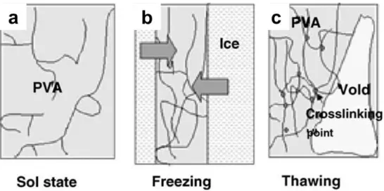
Freezing/Thawing gelation:
Another method is the freezing thawing technique. It has been introduced by Peppas in 1975.36 In this process, the reticulation of PVA network is induced by a combination of three principal mechanisms: liquid-liquid phase separation, hydrogen bonding and crystallite formation.37 During the freezing step of PVA aqueous solution (Figure 10a), phase separation occurs due to the formation of ice crystals which expel polymer PVA chains (Figure 10b),38
Chapter 1. Poly(vinyl alcohol) hydrogels
33 creating rich polymer and poor polymer regions by spinodal decomposition (Figure 11a).37 In rich polymer region, PVA chains are concentrated enough to trigger hydrogen bonds (Figure 11b) and PVA chains arrange to form crystallites (Figure 11c). Then hydrogel are allowed to thaw for certain period, letting voids between crosslinked PVA regions (Figure 10c). Crosslinks formed during the freezing remain insoluble and a permanent hydrogel network is formed. The number of freezing/thawing cycles and the duration of freezing and thawing strongly influence the microstructure, the swelling and mechanical characteristics of the network. This last point will be discussed in the following.
Figure 10 Schematic illustration of gel formation by freezing and thawing.38
Figure 11 Schematic representations of: a) liquid-liquid phase separation, resulting in the development of an interconnected structure of polymer-rich and polymer-poor regions, b) a network
Chapter 1. Poly(vinyl alcohol) hydrogels
34
Theta-gel formation of PVA network:
The gelation of PVA hydrogels can be also be achieved by liquid-liquid demixing, using gelling agent.32,33,39 The addition of a gelling agent like poly(ethylene glycol) (PEG) to PVA aqueous (or not) solution, decreases the solvent quality and imposes PVA to separate. This induces liquid-liquid demixing, creating PVA-rich phase, in which PVA chains crystallize, and PEG/water-rich phase, as illustrated in Figure 12.33
Figure 12 Schematic of a PVA theta-gel formation is shown here: a) PVA-PEG water mixture at 90 1C is a uniform solution; b) as the solution is cooled down phase separation begins and forces the PVA to form crystalline domains; c) with further cooling to near room temperature, phase separation
results in the formation of pores containing water surrounded by PVA rich regions.33
Annealing treatment:
Addition of an annealing treatment to PVA networks increase crystallization. Gonzalez and Alvarez have studied the effect of the annealing temperature on PVA hydrogels prepared by freezing/thawing. They showed that additional annealing treatment increase the crystallinity (Figure 13a) and reduce the water content Mt (Figure 13b) of the network. Mt was calculated as Mt=Mf-Mi/Mi, where Mf and Mi are the weights of the sample before and after immersion, respectively.
Chapter 1. Poly(vinyl alcohol) hydrogels
35
Figure 13 a) Crystallinity degree (Xcr%) and melting temperature (Tm) of hydrogels prepared from 10 wt.% PVA solution as a function of annealing temperature; b) Absorbed saline solution (%) at 37
◦C as a function of time for untreated and annealed 5 wt.% hydrogels.40
Kobayashi et al. have studied PVA hydrogels applied for artificial meniscus.35,41 They have produced PVA hydrogels by freezing/thawing followed by annealing treatment (Figure 14). Annealing treatment was performed to adjust the water content close to that of native meniscus. The final water content was 90 wt%.
Chapter 1. Poly(vinyl alcohol) hydrogels
36 1.4 Physical properties of PVA hydrogels
Microstructure of PVA hydrogels
The microstructure of PVA hydrogels depend on polymer characteristics (degree of polymerization, degree of hydrolysis, molecular weight), on the polymer concentration and on the type of gelation process and its parameters.
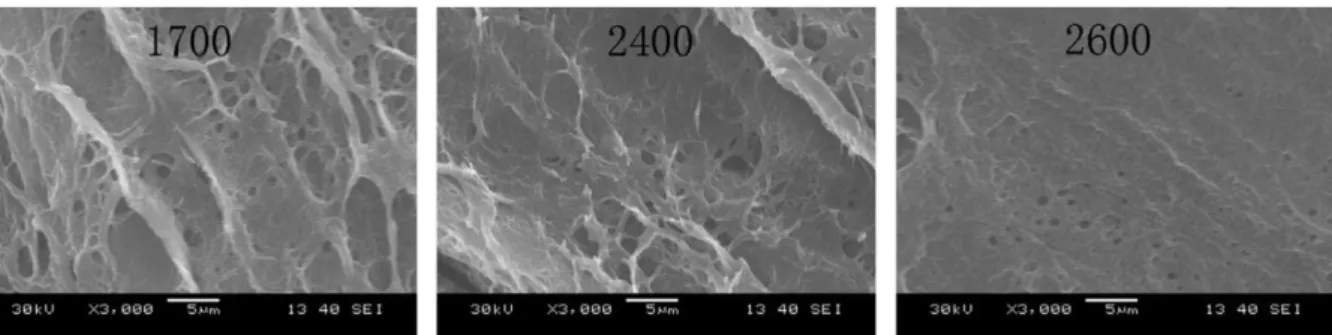
Different microstructures can be obtained depending on the properties of the initial polymer solution. Shi et al. have studied the microstructure of physical PVA/PVP (polyvinylpyrrolidone) hydrogels cross-linked by the freezing/thawing method. Here PVP was added to improve mechanical and tribological properties. They showed that the micro-pore size inside the hydrogel can be reduced by using PVA with a higher polymerization degree, as shown on Figure 15.42 This effect is due to length of the PVA chains, which governs the number of cross-linking points. They also suggested that an increase in polymer concentration in the PVA solution reduced the size of the pore, as shown on Figure 16. An increase of the polymer concentration induces denser packing of polymer chains in the hydrogel network.
Figure 15 SEM micrographs of the cross-sections of PVA/PVP hydrogels (10%PVA/PVP) with polymerization degree of 1700, 2400 and 2600.42
Chapter 1. Poly(vinyl alcohol) hydrogels
37
Figure 16 SEM micrographs of the cross-sections of PVA/PVP hydrogels, with polymer concentration solution of 10, 15 and 20 wt%.42
Different microstructures can be achieved by using various crosslinking processes. Holloway et al. have compared the microstructure of PVA hydrogel made through two different physical crosslinking processes: freezing/thawing (Figure 17) and aging (Figure 18).43,44 Briefly, PVA hydrogels were produced from PVA solution of 10, 20, 30 and 35 wt% by applying several cycles of freezing/thawing or by pouring the solutions in closed containers and let it aged for 31 days at room temperature.
Figure 17 Micrographs of freeze-thawed PVA (35 wt% of PVA). Scale bar is 80 µm.44
Figure 18 Micrographs of aged PVA hydrogels (35 wt% of PVA). Scale bar is 80 µm.44
They observed that the repetition of freezing thawing cycles tends to remodel the microstructure of the hydrogel network. An increase in the number of freezing thawing cycles induces an increase in the porosity of the network, as reported in Figure 19. As previously explained, during the freezing/thawing process phase separation occurs, which induces voids
Chapter 1. Poly(vinyl alcohol) hydrogels
38 filled with water and a small fraction of uncross-linked polymer (poor polymer regions). By repeating the number of cycles, the remaining uncross-linked polymer chains are expelled from these poor polymer regions to thicken the rich polymer regions. On the contrary, during aging process, no phase separation occurs. As a consequence, aged PVA hydrogels do not exhibit a high porosity level and do not show any microstructural changes with aging time, as shown in Figure 18. For instance, the porosity level was around 36% for a hydrogel obtained after 6 freezing/thawing cycles and 2.2% for 31 days aged hydrogel.44
Figure 19 Apparent porosity of PVA hydrogels in function of number of freezing thawing cycles.44
Swelling of PVA hydrogels
A deep understanding of the swelling behavior of PVA hydrogels is fundamental to produce artificial substitutes that are able to mimic the behavior of living tissues. The swelling of a hydrogel is generally characterized by the degree of swelling Q. Q is calculated as follows: Q=Veq/Vdry, where Veq is the volume of the hydrogel at equilibrium and Vdry is the volume of the dry hydrogel. The swelling can also be characterized by its water content WC. WC is calculated as follows: WC=(Meq-Mdry)/Meq*100, where Meq is the weight of the hydrogel at equilibrium and Mdry is the weight of the dry hydrogel.
Chapter 1. Poly(vinyl alcohol) hydrogels
39 Like for all polymer gels, the swelling of PVA hydrogels are directly related to the cross-linking degree of the PVA network. The formation of crosslinks decreases polymer chain mobility. As a consequence, highly cross-linked hydrogels have a tighter structure and will swell less compared to hydrogels with lower crosslinking ratios, as shown in Figure 20.45 This crosslinking degree is mostly governed by the on polymer characteristics, on the polymer concentration and on the type of gelation process and its parameters.
Figure 20 Swelling of chemically cross-linked PVA membrane as a function of the degree of crosslinking (● in water; ○ in ethanol).45
Swelling as a function of the polymer characteristics
The polymer characteristics affect the swelling behavior of the hydrogel. The degrees of hydrolysis and of polymerization have an influence on the degree of swelling. Shi et al. have studied the effect of the polymerization degree of PVA and the polymer concentration of the solution, from which hydrogel are produced, on swelling behavior of physical PVA/PVP hydrogels cross-linked by the freezing/thawing method. They showed that the swelling ratio (calculated as SR=Ws/Wd, where Ws and Wd are the swelling equilibrium and dry weight of the samples respectively) decreases significantly when the degree of polymerization increases, as shown in Figure 21.42 Longer PVA chains provide more cross-linking points that increases the crosslinking density of the hydrogel network, as previously mentioned.
Chapter 1. Poly(vinyl alcohol) hydrogels
40
Figure 21 Swelling ratio as a function of polymerization degree and of polymer concentration for PVA hydrogels in phosphate buffer solution (PBS).42
Swelling as a function of crosslinking process
For physically cross-linked PVA hydrogels, prepared by freezing/thawing method, the degree of swelling can be tuned by several parameter adjustments. Stauffer and Peppas have shown that the number of freezing/thawing cycles and the duration of the thawing time have an impact on the degree of swelling.46 An increase in the number of cycles has been shown to decrease significantly the water uptake (calculated as WU=W/W0, where W is the weight of the gel and W0 is the initial frozen weight after the first freezing process), as shown in Figure 22.46 Indeed, more and more crosslinks are created during each cycle, which is tightening the network and decreases the swelling ratio.46–48
Figure 22 Swelling ratio in function of the thawing time and of the number of freezing/thawing cycles: ■ 2 cycles; ● 3 cycles; □ 4 cycles; ○ 5 cycles.46
Chapter 1. Poly(vinyl alcohol) hydrogels
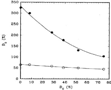
41 In addition, the swelling ratio decreases with increasing PVA content of the initial aqueous solution. Several studies have shown that increase of PVA content induces an increase of crosslinking density.21,42,46,47 This effect has been shown by Yang et al., where they produced chemical PVA hydrogel by γ-irradiation with addition of 1 or 2 freezing/thawing cycles. Figure 23 shows that the degree of swelling (calculated as DS=Ws/Wd, where Ws and Wd are the swelling equilibrium and dry weight of the samples respectively) decreases in function of the PVA content (wt%).21 Increasing of the PVA content improve the packing of the chains during the cross-linking.
Figure 23 The degree of swelling as a function of PVA content.21
Mechanical properties of PVA hydrogels
The mechanical response of PVA hydrogels is important for many biomedical applications, such as orthopaedic soft tissue grafts, vascular grafts, or wound dressings. These systems should sustain physiological solicitations and remain intact through the implantation duration. PVA hydrogels are characterized by an elastic rubbery behaviour.49 The mechanical behaviour of these hydrogels has been assessed by tensile, compressive, relaxation, creep test. Typical viscoelastic mechanical responses are shown by compressive stress-strain curves in Figure 24 and 25.50,51 The curve in Figure 24 shows the compressive response of a physical PVA hydrogel cross-linked by freezing/thawing method. Same behaviour (Figure 25) is obtained with chemical PVA hydrogels cross-linked by inverse suspension polymerization52
Chapter 1. Poly(vinyl alcohol) hydrogels
42 with epichlorohydrin as cross-linking agent. These two compressive experiments show that PVA hydrogels can support large deformations of more than 60 % with dissipation.
Figure 24 Compressive stress-strain curve of physically cross-linked PVA hydrogel.50
Figure 25 Typical force response profiles during a compression/release experiment performed on a PVA1 and PVA2 beads swollen in H2O. Beads were compressed to 20 (A), 40 (B), 60 (C), and 80%
(D) strain (applied at 4% s-1).51
This non-linear rubbery behaviour has also been confirmed by tensile tests, performed on physically cross-linked PVA hydrogels by freezing/thawing technique for the construction
Chapter 1. Poly(vinyl alcohol) hydrogels
43 of a bioprosthetic heart valve stent by Wan et al.53 Figure 26 presents the stress-strain curves for PVA hydrogels crosslinked by freezing/thawing cycles.
Figure 26 Stress–strain relationship of PVA hydrogel cross-linked by freezing/thawing method (1 to 6 cycles) compared to a porcine aortic root.53
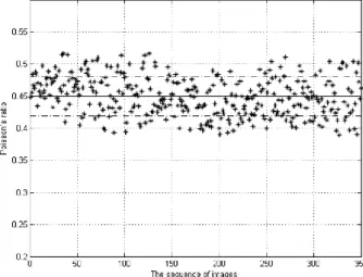
Poisson’s coefficient as also been evaluated. Since the living tissues are supposed to be uncompressible due to their high water content, it is important to obtain similar behaviour for PVA hydrogel.54,55 Recently, Chen et al. have proposed a new method, based on optical flow technique, to evaluate the Poisson coefficient of physical PVA hydrogels cross-linked by freezing/thawing technique (4 cycles).54 Figure 27 shows Poisson’s ratio values for a series of images of PVA hydrogels under tensile tests. They obtained reasonable values of Poisson ratio of PVA hydrogels compared to others studies.56 The mean value is 0.440.03.
Chapter 1. Poly(vinyl alcohol) hydrogels
44 Like for the swelling ratio, the mechanical properties of PVA hydrogels can be modified by changing their crosslinking degree, which can be achieved by controlling the composition and the crosslinking process. Conversely to the swelling properties, an increase in the degree of crosslinking will strengthen the mechanical properties (strength, stiffness) of hydrogel.57 For physical PVA hydrogels cross-linked by freezing thawing method, the PVA content, the molecular weight of the polymer, the number of freezing/thawing cycles and the duration and the rate of freezing and thawing are factors which affect the cross-linking density of PVA hydrogel networks and therefore have a significant effect on their mechanical properties.
It has been shown by Gupta et al. that the ultimate tensile strength of physical PVA hydrogels cross-linked by the freezing/thawing technique can be increased by increasing the polymer concentration, as shown on Figure 28a.58 They related this phenomenon to the degree of crystallinity, which was also increased by increasing the polymer concentration, as shown on Figure 28b.
Figure 28 a) Ultimate tensile strength as a function of PVA concentration; b) Degree of crystallinity as a function of PVA concentration.58
Furthermore, it has been shown that the number of freezing/thawing cycles improves mechanical property.59,60 In their experiments, Jiang et al. proceeded to uniaxial tensile strength tests on a cylindrical PVA hydrogel samples. They noted that the Young’s modulus
Chapter 1. Poly(vinyl alcohol) hydrogels
45 increased with the number of cycles, as shown in Figure 29.59 Previously, we evoked that the repetition of freezing/thawing cycle increases the formation of crystals between PVA chains. As a consequence, the increase of the crystallinity strengthens the hydrogel network.
Figure 29 Young’s modulus as a function of the number of Freezing/Thawing cycles.59
In their study, Stammen et al. explored the effect of water content on shear and compression properties of PVA hydrogels.50 For the network with the lowest water content (75%), the mechanical properties were higher than for the network with the highest water content (80%), as seen in Table 1. Compressive and shear modulus of both hydrogels are in the range of human cartilage.
Table 1 Comparison of compressive tangent modulus, failure stress and strain, and shear tangent modulus for both PVA hydrogel formulations and human articular cartilage.50
Chapter 1. Poly(vinyl alcohol) hydrogels
46 1.5 Biological applications of PVA hydrogels
In vivo studies
Thanks to their favourable characteristics such as high water content, mechanical strength, and a simple manufacturing process, PVA hydrogels have been intensively investigated for biomedical applications, in particular for soft tissue replacements. In several in vivo studies, PVA hydrogels have been shown to be biocompatible and well-tolerated. In particular, numerous systems based on PVA hydrogels have been suggested to restore articular cartilage defects.
In 1991, Noguchi et al. have investigated the biocompatibility of PVA hydrogels for the treatment of rabbit osteochondral defects in intra-articular capsule, as shown in Figure 30.61 They observed a slight inflammatory response in the early stage of implantation (after 2 weeks), which disappeared completely later (after 52 weeks). In addition, no disturbances of the surrounding articular cartilage were noticed.
Figure 30 A photomicrograph at low magnification of a horizontal section of the distal femur showing the implant in place.61
In another rabbit model, Maher et al. have implanted PVA-PVP (poly vinylpyrrolidone) hydrogels in osteochondrodral defects and followed-up for 6 months.62 No inflammation was reported as well as no bone osteolysis. However no intimate contact with
Chapter 1. Poly(vinyl alcohol) hydrogels
47 bone was noticed and implants were surrounded by a fibrous layer after 6 months of implantation, as shown in Figure 31.
Figure 31 At 24 weeks the implant is completely encapsulated in fibrous tissue.62
PVA hydrogels have been also investigated as a material for meniscus replacement. A two-year in vivo study made by Kobayashi et al. showed that PVA hydrogels might be suitable for meniscus replacement due its appropriate mechanical behaviour close to that of native meniscus (Figure 32) and because no surrounding cartilage osteoarthrisis arose.63
Figure 32 Mechanical properties of PVA-H artificial meniscus: (a) Stress–strain curves of the implanted and original PVA-H, and human natural meniscus in compression test. (b) Comparison of
the stress–relaxation curves of the implanted and original PVA-H, and human natural meniscus.63
Allen et al. have investigated novel PVA hydrogel implants for the replacement of diseased nucleus pulposus.64 Physical PVA hydrogel implants of 0.1 and 0.3 cm3 (Figure 33a), obtained by the freezing/thawing technique, were inserted in discectomy defects created in intervertebral disc of 20 male baboons over 24 months. After 24 months of implantation,
PVA implant Fibrous tissue
Chapter 1. Poly(vinyl alcohol) hydrogels
48 PVA implants located within the nuclear cavity were intact and were surrounded by loose connective tissue, as shown in Figure 33b.
Figure 33 a) PVA implants used in the study; b) PVA implants that were located within the nuclear cavity were intact and surrounded by loose connective tissue.64
1.6 Clinical applications
In a review, Baker et al. report a clinical study on PVA cartilage implants made by Carticept Medical in which 15 patients were followed up for 30 months and 13 patients presented successful outcomes.3 They indicate that no synovitis nor osteolysis in animal or clinical studies have been reported. In a follow-up review, it has been shown that PVA cartilage implants (Figure 34) could improve knee functionality and reduce pain for patients suffering from cartilage defects.9
Figure 34 10 and 15 mm synthetic cartilage implants.9
Several follow-ups have shown that those PVA implants are well anchored and have not been deteriorated after a long duration of implantation. This is suggested by MRI observations of PVA implants after 12 (Figure 35a) and 96 (Figure 35b) months of
Chapter 1. Poly(vinyl alcohol) hydrogels
49 implantation in a 50 year-old female patient. MRI at 96 months shows that the implant is fixed with no abnormalities. This patient recovered complete knee functionality and stability and was pain free.
Figure 35 50 year-old woman with 10 mm defect on the weight bearing area of the left medial central condyle at 12 (a) and at 96 (b) months post-implantation.9
1.7 Perspectives and challenges related to PVA hydrogels for biomedical application
Strong efforts are accomplished to develop efficient synthetic substitutes to replace other types of soft tissue of the osteo-articular system, in particular ligament and tendon. Recently, Bach et al. have developed artificial ligaments for anterior cruciate ligament replacement (ACL). These construct are made of assemblies of PVA hydrogel fibres. They have shown that these systems reproduced closely the tensile response of human ACL, as shown in Figure 36.65
Chapter 1. Poly(vinyl alcohol) hydrogels
50
Figure 36 Tensile force versus strain for different PVA hydrogel fibre constructs (red, blue and green curves). Shaded regions show the typical ranges for the native ACL and tendons used for autografts.65
Regarding their in vivo performance, PVA hydrogels have been shown to be bio-inert, with a very poor ability for cell adhesion.62,66,67 In particular, in the perspective of use for the replacement of soft osteoarticular tissues such as tendons, ligaments or cartilages, the implants need to be connected to surrounding bone tissue and a lack of affinity with the host bone tissue may lead to anchorage failures at the bone-implant interface. To address this question the surface of the implant can be functionalized by an osseoconductive coating. Such a coating can be obtained by the use of bioceramic particles like hydroxyapatite.
52
Chapter 2. Hydroxyapatite
2.1 Chemical composition and preparation
Hydroxyapatite (HA) is a mineral, which belongs to the calcium-phosphate materials. Its chemical formula is Ca10(PO4)6(OH)2. Hydroxyapatite crystallizes in the hexagonal system (Figure 37). Hydroxyapatite degrades at temperatures above 1400°C, where structural deterioration occurs, identified by a decrease in density and hardness.68
Figure 37 Hydroxyapatite structure [web.mit.edu]
This mineral is the main component of dentin and bone (65% of the mineral matter), and it has the closest Ca/P ratio to bone : Ca/P ratio of HA is 1.67 and bone ratio is 1.57-1.62.69 Because of its excellent biocompatibility, it is widely used for implant coating and as a mineral scaffold for bone substitution.69,70
Concerning HA synthesis, several methods can be used: wet methods such as precipitation and hydrothermal techniques, sol-gel methods with emulsion techniques, and dry methods. One of the most popular techniques is the precipitation method. It is based on a wet method and consists in a reaction of precipitation between calcium hydroxide (Ca(OH)2) and orthophosphoric acid (H3PO4) or ammonium phosphate ((NH4)2HPO4).71,72
Chapter 2. Hydroxyapatite
53 The size and the crystallinity of the particles created by this technique strongly depend on the pH of the solution and on the temperature. This technique is widely used because large amounts of HA can be produced at a reasonable cost and in absence of organic solvent.71 Another popular technique is a sol-gel method, which allows a homogeneous molecular mixing, a low processing temperature and ability to generate various form of HA, such as nanocrystalline powder and thin films).73 Dry methods of HA fabrication involve solid-state diffusion during calcination of mixtures containing appropriate amounts of Ca2+ and PO4 3-ions. The calcination temperature ranges between 900 and 1300°C.74 Hydroxyapatite can be available in different forms, like powders, rods, blocks, injectable pastes, as suggested in Figure 38.75
Figure 38 General appearance of various commercial calcium orthophosphate-based bone graft materials.75
2.2 Osteoconductive properties of HA
Hydroxyapatite is an osteoconductive material. Albrektsonn et al.76 give this definition: “an osteoconductive surface is one that permits bone growth on its surface or down into pores, channels or pipes”. Many in vitro studies have shown that implants containing HA particles enhance bone formation, including cell proliferation, adhesion, and an increase of osteocalcin level.77 Dalby et al. have shown that addition of hydroxyapatite improves cells adhesion. They used poly-methylmethacrylate (PMMA) cement coated with 0%, 4,6%, 8,8%
Chapter 2. Hydroxyapatite
54 of hydroxyapatite powder.78 They isolated human osteoblast cells (HOB) from femoral head of a patient undergoing total joint replacement. Then HOB cells were cultured on the material for 1, 3, 7, 14 and 28 days at 37°C. Morphological investigation by SEM showed that the anchorage of HOB looks qualitatively more efficient on HA coated PMMA than on bare PMMA, as shown in Figure 39.
Figure 39 SEM observations of PMMA/HA scaffold a)PMMA without HA; b) PMMA+4,4%HA; c)PMMA+8,8%HA.78
A higher number of focal adhesion plaques, viewed by vinculin staining, was observed as HA incorporation into the cements increased, as shown in Figure 40b and c. Actin cytoskeleton organization was improved and many stress fibres appeared.
Figure 40 Confocal observations a) PMMA without HA ; b) PMMA+4,4%HA ; c) PMMA+8,8%HA Fibronectin proliferation.78
Many in vivo studies and reviews have also reported the osseoconductive ability of HA, used for osteointegration of medical implant or as bone substitutes.70,77,79,80 In a study, Wang et al. have evaluated the osseointegration of Ti-6Al-4V implants with and without plasma sprayed HA coatings.80 After 4 and 12 weeks of implantation, the amount of new bone was more important with the coated implant than with the bare implant, as shown in Figure
Chapter 2. Hydroxyapatite
55 41a and b. In addition, a direct contact was noticed between the host tissue and the coated implant surface, whereas a fibrous tissue interface was found between the bare implant and the host bone tissue, as shown in Figure 41c and d.
Figure 41 Scanning electron microscopy observations using backscattered electron detector of histological sections in defective bone regions: a-b) HA coated implant face at 4 and 12 weeks; c-d)
Ti-6AI 4V at 4 and 12 weeks. O, original cortical bone; N, new bone; M, bone marrow; C, HA coating; T, Ti alloy.80
The interactions between HA and bone have not been clearly understood yet. Figure 42 shows a schematic representation of the mineralization process proposed by Bertazzo et al.81 It appears that ion transfers happen at the interface between hydroxyapatite coating and bone surface (1, 2 and 3), followed by proteins adsorption (4) and cells adhesion and proliferation (5 and 6), and finally new bone is formed (7) and natural metabolism starts again (8).
Chapter 2. Hydroxyapatite
56
Figure 42 Schematic diagram representing the phenomena that occur on the surface of hydroxyapatite after implantation.81
The bioactivity of a bioceramic material depends on its biodegradation in vivo. This mechanism occurs with physicochemical dissolution and cellular activity, namely the bioresorption. The kinetics of this biodegradation depends on multiple factors: Ca/P ratio, phase purity, crystalline size, porosity, surface area and solubility (Table 2). Compared to other phosphocalcic ceramics, HA particles have generally a slower degradation.75
Table 2 Existing calcium orthophosphates and their major properties.75
2.3 Biological response to HA particles
In this thesis, we mainly used HA micro-particles with spherical shape. Regarding the cellular inflammatory response to this type of HA particles, Laquerierre et al. have shown
Chapter 2. Hydroxyapatite
57 that spherical HA particles are expected to initiate less inflammatory response than irregular and needle-like particles.82 Other in vitro studies by Laquerriere et al. have shown that large non-phagocytable particles (10-70 µm) do not provoke a strong inflammatory response. For smaller sizes in the range 1-30 µm, they find that some cellular response and inflammatory activity occur and depend also on the shape of the particles.83,84 An in vitro and in vivo study by Malard et al. has shown that small particles with diameters ranging from 10 to 20 µm induce more inflammatory response than larger ones.85 These studies suggest that the in vivo inflammatory response of HA particles is mostly governed by their shape and their size. A proper combination of different sizes of HA particles could be used for the fabrication of composite implants. The efficiency of HA particles in terms of osteoconduction is also govern by the size of HA particles. Hulsart-Billström et al. have tested HA hydrogel composites with different sizes and shapes of HA particles. They implanted several compositions of such hydrogel compositesin the quadriceps of rats and observed the implant after 4 weeks following implantation. As shown in Figure 43, it appears that nanoHA particles having a mean size of 400 nm facilitate the formation of denser bone compare to other type of HA particles.86