médecine/sciences médecine/sciences
Knock-down of
filaggrin influences
the
mitogen-activated protein
kinases signaling
pathway in normal
human epidermal
keratinocytes
Shumei Wang1, Liyun Qiu2, Xianguang Meng3, Ningning Dang3*
>
Background: Filaggrin is an essential structural
protein of the stratum corneum binding to the
keratin intermediate filaments to form a dense
protein-lipid matrix. However, the function of
filaggrin in epidermal terminal differentiation is not
completely understood.
Aim: To evaluate the effects of filaggrin on normal
human epidermal keratinocytes (NHEKs) and to
investigate the relevant mechanisms.
Methods: Short hairpin RNA (shRNA) technology
was used to knock-down filaggrin in normal
human epidermal keratinocytes (NHEKs). Western
blot and real-time quantitative PCR (qRT-PCR)
were performed to detect expression of filaggrin,
differentiation-related proteins and MAPK-related
proteins.
Results: Filaggrin was successfully knocked down
in NHEKs (99% efficiency). We found that the lack
of filaggrin significantly decreased the expression
of some differentiation-related proteins, including
Cytokeratin 5 protein, Cytokeratin 14 protein, ST14
protein and SPRR3 protein (P<0.05). In addition,
filaggrin knock-down significantly decreased
expression of p-p38, p-ERK1/2, p-JNK, p-Akt, and
p-NF-kB in NHEKs.
Conclusion: Our study shows that filaggrin regulates
epidermal terminal differentiation and impairs
MAPK signaling pathway in normal human epidermal
keratinocytes.
<
Key words:
differentiation-related proteins,
epi-dermal terminal differentiation, keratinocyte
dif-ferentiation, mitogen-activated protein kinases,
normal human epidermal keratinocytes (NHEKs).
nutrient and entry of pathogens and allergens. The stratum corneum (SC) is the main component of the epidermal skin barrier, which is the final product of terminal differentiation of keratinocytes in epidermis.
The filaggrin gene (FLG) is mainly expressed in the granular layer of the skin, and the encoding profilaggrin protein is matured to filaggrin. Filaggrin is an important structural protein in the stratum corneum. It can enhance the terminal differentiation of the epi-dermis and the formation of skin barrier [1]. Mutations in FLG gene can result in reduced or complete loss of epidermal filaggrin and its degradation products [2]. The absence of filaggrin can lead to epi-thelial barrier defects, incomplete accumulation of keratin and loss of trans-epidermal water [3, 4]. FLG mutations are closely related to some common dermatological diseases, such as atopic dermatitis (AD) and ichthyosis vulgaris (IV) [5], but also to type 2 diabetes and cardiovascular and cerebrovascular diseases [6]. However, the roles of filaggrin in epidermal terminal differentiation is not fully understood.
In the present study, the function of filaggrin in human epidermal keratinocyte differentiation was studied. We used shRNA transfec-tion to knock down the filaggrin in normal human epidermal kerati-nocytes (NHEKs). RT-PCR and Western blot were performed to detect the expression of differentiation-related proteins.
1Department of Community
Medicine,
2Department of Pharmacy, 3Department of Dermatology,
Jinan Central Hospital affiliated to Shandong University, No.105 Jiefang Road, Jinan 250013, Shandong Province, China. Corresponding author: Ningning Dang
ningningdangxj@sina.com
Introduction
Skin is the first line of defense to protect the human body from environmental damage, loss of water and médecine/sciences 2018 ; 34 (focus issue, F1) : 94-8
SYNTHÈSE
REVUES
repeated at least three times. The expression levels of filaggrin were normalized to an endogenous control, GAPDH. PCR products were separated by electrophoresis on 2% agarose gels and stained with ethidium bromide for visualization.
Western blotting analysis
Western blotting was adopted to test the knock-down efficiency of filaggrin and the expression of diffe-rentiation-related proteins. Seventy-two hours after transfection, NHEKs cells were collected, proteins were extracted and quantified using the Mammalian Protein Extraction Reagent (M-PER, Pierce, Rockford, IL, USA). After 10% SDS-PAGE, proteins (20 μL) were transferred to PVDF membrane (Millipore, Bedford, MA, USA), and the non-specific binding sites of the membrane were blocked with 5% skim milk. Then the membrane was incubated with primary antibodies overnight at 4 ºC. After rinsing with TBST, the membranes were incubated with HRP-conjugated secondary antibody (rabbit IgG, 1:2000, Cell Signaling, Beverly, MA, USA) at RT for 1 h. Labeled pro-tein bands were detected using Gel-Pro Analyzer (Media Cybernetics, Silver Spring, MD, USA) after incubation with SuperSignal West Pico Chemiluminent Substrates (Pierce, Appleton, WI. USA) and exposed to X-ray film. The Wes-tern blotting was performed three times. The quantifica-tion analyses were performed using Image J. The relative expression level of proteins was calculated by determi-ning a ratio between their amount and that of GADPH. The rabbit polyclonal antibodies were : Filaggrin (1:250, Covance, Berkeley, CA, USA); Cytokeratin 5 (1:500,); Cytokeratin 10 (1:500); Cytokeratin 14 (1:1000); Loricrin (1:500); ST14 (1:500); SPRR3 (1:500), all from GeneTex (Irvine, CA, USA); p38 MAPK (1:250,); Phospho-ERK1/2 (1:250); Phospho-JNK (1:200); Phospho-NF-kB (1:200); Phospho-Akt (1:200); GAPDH (1:2000), all from Cell Signaling (Beverly, MA, USA) .
Statistical analysis
The SPSS11.0 software (SPSS Inc., Chicago, IL, USA) was used to analyze the experimental data by one-way ANOVA. Quantitative data are presented as the mean ± standard deviation (n=3). The Student’s t-test was used to analyze the difference between groups. P<0.05 was considered as statistically significant.
Results
Knock-down efficiency of filaggrin for different shRNAs
In order to detect the knock-down efficiency of filag-grin by shRNA transfection, filagfilag-grin-encoding mRNA
Materials and methods
Cell culture
NHEKs were obtained from Invitrogen (Carlsbad, CA, USA). They were cultured with EpiLife medium containing 10% fetal calf serum (FCS, Gibco, Carlsbad, CA, USA), 1.5 mM L-Glutamine, 100 IU/mL penicil-lin and 100 g/mL streptomycin (Gibco) in 6 cm dishes, at 37°C with 5% CO2. The medium was refreshed twice a week.
Knockdown of filaggrin using shRNA transfection
We designed and synthetized four sequences of filaggrin shRNA, as fol-lows: shRNA 1 (at nt-274, GTTGGCTCAAGCATATTATTT), shRNA 2 (at nt-769, CACCACTGATAGT CTATTATT), shRNA 3 (at nt-1627, CCACGAGCAATCGG-TAAATTT), shRNA 4 (at nt-4936, GTCCCATCAAGAAGATAGATT). BamHI and
EcoRI were used to cut the sequences of filaggrin shRNA, scramble oligos
and target vector pGLV-H1-GFP (GenePharma Co. Shanghai, China). T4 DNA Ligase was used to ligate the vector with insert after gel purifica-tion. The ligation products (pGLV-FLG-GFP and pGLV-scr-GFP) were then sequenced. 293T cells were used for transfection with pGLV-FLG-GFP or pGLV-scr-GFP to generate the corresponding viruses which were used to infect NHEKs. The plasmid pHelper 1.0 and pHelper 2.0 were packaged based on standard protocols. When NHEKs attained to 90% confluence, a mixture of 0.5 μL lipofectamine 2000 (Invitrogen), 10 pmol siRNA and 75 μl OPTI-MEM medium (Invitrogen) were added to each dish and incuba-ted for 20 min at room temperature (RT). After changing culture medium to serum-free DMEM in each dish, infected cells were cultured for 72 h and then collected for RT-PCR and Western blot analyses.
Real-time quantitative RT-PCR analysis
Real-time quantitative RT-PCR was performed to quantitatively esti-mate the mRNA expression of filaggrin and measure the filaggrin knock-down efficiency. Total RNA from cultured NHEKs was isolated using Trizol reagent (Invitrogen) according to the published method. The purified RNA was reverse transcribed to cDNA by using the MMLV Reverse Transcriptase system (Promega, Madison, WI, USA) according to the manufacturer’s instruction. Mx3000 Real-time PCR Instrument (Stra-tagen, La Jolla, CA, USA) was used to analyze quantitative RT-PCR of filaggrin according to the manufacturer’s instruction. Primer sequences were as follows: Filaggrin: forward: 5'-CACAAGATTCTGCGTATCAC TCAGG-3', reverse: 5'-GCCTTTCAGTGCCCTCAGATTG-3'; K5: forward: 5'-ATCGC CACT-TACCGCAAGCTGCTG-3', reverse: 5'-GAGGGAAACACTGCTTGTGACAACA GAG-3'[7]; K14: forward: 5'-CCGACACCTTCTCTTCACTCA-3', reverse: 5'-AGGAG CCCTTCATGGAGCTG-3'[8]; Loricrin: forward: 5'-ACGTCTCCTCGCAGCAGG-3', reverse: 5'-CTATTTGGACGGCCAGGT-3'[9]; ST14: forward: 5'-CACTG-GTGGTTCTA CTGAC-3', reverse: 5'-GTTT TCCAGGGTCCTCCGA-3'; SPRR3: forward: 5'-CTTCTC TGCACAGCAGGRCC-3', reverse: 5'-AG CAATTTAATGAG-GGAAGAGC-3'[10]; GAPDH: forward: 5'-CATGAGAAGTATGACAACAGC CT-3', reverse: 5'-AGTCCTTC CACGATACCAAAGT-3'. The reaction system was: 2×Real-time PCR Master Mix 10 μL, 20 μM primers 0.2 μL, cDNA template 2 μL, 5 U/μl rTaq DNA polymerase 0.4 μL and ddH2O 7.4 μL. The following PCR condition were used: 95 °C for 3 min, 95 ºC for 30 s, 62 ºC for 40 s, 40 cycles. This experiment was carried out in triplicate and independently
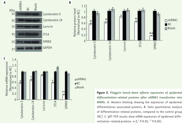
proteins by Western blotting, including cytokeratin 5 (K5), cytokeratin 14 (K14), ST14 (matriptase), SPRR3 (small proline-rich protein 3) and loricrin (Figure 2). Filaggrin knock-down resulted in a signi-ficant decrease in K5, K14, ST14 proteins (P<0.05) and especially in SPRR3 (P<0.01), while no significant changes in loricrin content was observed. The result suggested that filaggrin knock-down inhibits NHEKs differentiation. qRT-PCR analysis showed similar results, the filaggrin knock-down significantly redu-cing the NHEKs content in K5, K14, ST14, and SPRR3 mRNA (P<0.05, Figure 2C).
Filaggrin knock-down reduces MAPK pathway signaling activation in NHEKs
To further examine whether filaggrin knock-down affected the Akt, NF-kB and MAPK pathways in NHEKs, Western blotting was performed to analyze the phos-phorylation level of Akt, NF-kB, p38, ERK1/2 and JNK (Figure 3). The phosphorylation levels of p38 protein, ERK1/2 protein, JNK protein, Akt protein and NF-kB protein were decreased significantly (P<0.01) when filaggrin was knocked down, with the phosphorylation of Akt and JNK being almost completely blocked. These results indicate that filaggrin knock-down strongly impact the MAPK pathways and the Akt, and NF-kB signaling activation.
Discussion
Human skin consists of two different main layers, epidermis and subjacent dermis. The barrier of the epidermal skin-stratum corneum (SC) is the final product of the terminal differentiation of kerati-nocytes in epidermis. This terminal differentiation is strictly regulated by the sequential expression of various genes, a complicated multistep process [11]. Although studies have reported that many genes are involved in keratinocyte differentiation, there are also several important molecules that remain to be determined [12].
It is widely considered that filaggrin is one of the major markers for the epidermal terminal differentia-tion and formadifferentia-tion of SC. However, the relevant regu-lation mechanism of filaggrin in epidermal terminal differentiation is not completely clear. The epidermal proliferation of normal skin is limited to the basal layer and related to certain keratin proteins, such as K5 and K14 [13]. The K5/K14 pair is expressed in basal layer, maintains the cell proliferation potential, and its expression is related to cell differentiation [14, 15]. In addition, the small proline-rich protein and filaggrin were detected by RT-PCR analysis and Western blotting,
respectively. The four different shRNAs treatments both decreased mRNA expression, with a knock-down efficiency of 99% (P<0.01), 77% (P<0.01), 97% (P<0.01) and 33% compared with the NC group, res-pectively (Figure 1A). The knock-down efficiency of filaggrin protein was 88% (P<0.01), 69% (P<0.01), 84% (P<0.01) and 0%, respectively (Figure 1B, C). Therefore, we selected the shRNA1 which had the most knock-down efficiency for use in the subsequent experiments.
Filaggrin knock-down in NHEKs inhibits cell differentiation
To investigate the function of filaggrin in NHEKs differentiation, we evaluated the expression of epidermis differentiation-related
A B C 1.2 1 0.8 0.6 0.4 0.2 0 1.2 1 0.8 0.6 0.4 0.2 0 NC NC Blank Blank
shRNA1 shRNA2 shRNA3 shRNA4
NC Blank
shRNA1 shRNA2 shRNA3 shRNA4
Relative mRNA level of Filag
grin
(Normalized to NC)
Relative protein level of Filag
grin (Normalized to NC) shRNA 1 2 3 4 Filaggrin GAPDH 31 KDa 37 KDa
Figure 1. Four shRNAs inhibited the filaggrin expression both at the mRNA and
protein levels 72 h after shRNA transfection into NHEKs. A. Expression of
filag-grin mRNA after transfection with each kind of shRNA, compared to the control (NC). B. Western blotting of filaggrin after NHEKs transfection with each kind of shRNA. C. Semi-quantitative analysis of filaggrin protein levels for each kind of shRNA transfection into NHEKs, compared to the control group (NC). n=3, ** P<0.001.
SYNTHÈSE
REVUES
that NF-kB and Akt are intervening in epidermal dif-ferentiation in a way closely associated with the MAPK pathways. We found that the phosphorylation levels of p38, ERK1/2, JNK, Akt and NF-kB are inhibited signifi-cantly by the loss of filaggrin, indicating that filaggrin knock-down can inhibit the activation of MAPK, NF-kB and Akt signaling pathways (Figure 3).
Conclusion
In summary, this study demonstrates that filaggrin knock-down can inhibit the differentiation of NHEKs by down-regulating the expression of K5, K14, ST14 and SPRR3. In addition, it reduces the activation of mul-tiple signaling pathways, including MAPK, NF- kappa B and Akt pathways. However, whether the intermediate filament-related proteins are regulated by MAPK, the NF-kB or Akt pathways is unclear and requires further experiments. ‡
ACKNOWLEDGMENTS
This study was supported by China Postdoctoral Science Foundation (CPSF) (No.2014M550370, 2015T80740) and Shandong Provincial Natural Science Foundation, China: No. ZR2017MH074).
(SPRRs) are associated with increased epithelial proliferation, in particular a major cornified cell envelope (CE) protein, loricrin. The CE forms the outermost layer of the epidermis and maintains the epi-dermal barrier. Several studies have suggested that ST14 is involved in cell growth and differentiation and may be associated with the regulation of the suprabasal keratinocyte growth and differentia-tion [16]. In the present study, Western blotting results showed that filaggrin knock-down reduced K5, K14, ST14, loricrin, and SPRR3 expressions in NHEKs, indicating that filaggrin knock-down had an inhibitory effect on epidermal terminal differentiation of NHEKs through the down-regulation of differentiation-related protein expression (Figure 2).
The mitogen-activated protein kinases (MAPK) signaling pathway has been revealed to play critical roles in epidermal differentiation and skin barrier function. It involves p38 MAPK, ERK1/2 MAPK and JNK signaling pathways [17-20]. Lack of ERK1/2 caused a decrease in the expression of terminal keratinocyte differentiation genes [21-23]. Besides, another signaling pathway involving NF-kB and Akt has been also shown to play an important role in epidermal differentiation [24, 25]. These different signaling pathways are not independent in cell. Cellular stress can promote p38 phosphorylation, leading to the activation of the NF-kB pathway in human keratinocytes [26]. Moreover, it has been suggested that the NF-kB activation induced by TNF-a affect the activation of Akt [27]. Hence, our data suggest
A C B KDa 62 52 26 95 25 37 Cytokeratin 5 Cytokeratin 14 Loricrin ST14 SPRR3 GAPDH shRNA1 NC Blank
Relative protein level (Normalized to NC)
1.2 1 0.8 0.6 0.4 0.2 0 shRNA1 NC Blank Cytokeratin 5 Cytokeratin 14 Loricrin ST14 SPRR3 shRNA1 NC Blank Cytokeratin 5 Cytokeratin 14 Loricrin ST14 SPRR3 Relative mRNA expression (Normalized to NC)
1.2 1.4 1 0.8 0.6 0.4 0.2 0
Figure 2. Filaggrin knock-down affects expression of epidermal
differentiation-related proteins after shRNA1 transfection into
NHEKs. A. Western blotting showing the expression of epidermal
differentiation associated proteins. B. Semi-quantitative analysis of differentiation-related proteins, compared to the control group (NC). C. qRT-PCR results show mRNA expression of epidermal diffe-rentiation-related proteins. n=3,* P<0.05, ** P<0.001.
11. Popp T., Egea V., Kehe K., Steinritz D., Schmidt A., Jochum M., Ries C. 2011 Sulfur mustard induces differentiation in human primary keratinocytes: opposite roles of p38 and ERK1/2 MAPK. Toxicology Letters 2011;204(1), 43-51.
12. Yoon H.K., Sohn K.C., Lee J.S., Kim Y.J., Bhak J., Yang J.M., You K.H., Kim C.D., Lee J.H. 2008 Prediction and evaluation of protein-protein interaction in keratinocyte differentiation. Biochem Biophys Res Commun 2008;377(2), 662-667.
13. Ivanova P., Atanasova G., Poumay Y., Mitev V. 2008 Knockdown of PKD1 in normal human epidermal keratinocytes increases mRNA expression of keratin 10 and involucrin: early markers of keratinocyte differentiation.
Archives of Dermatological Research 2008;300(3), 139-145.
14. Alam H., Sehgal L., Kundu S.T., Dalal S.N., Vaidya M.M. Novel function of keratins 5 and 14 in proliferation and differentiation of stratified epithelial cells. Molecular Biology of the Cell 2011;22(21), 4068-4078.
15. Coulombe P.A., Kopan R., Fuchs E. Expression of Keratin K14 in the Epidermis and Hair Follicle: Insights into Complex Programs of Differentiation. Journal
of Cell Biology 1989;109(5), 2295-2312.
16. Chen Y.W., Wang J.K., Chou F.P., Wu B.Y., Hsiao H.C., Han C., Xu Z., Baksh A.N., Shi G., Kaul M. Matriptase regulates proliferation and early, but not terminal, differentiation of human keratinocytes. Journal of Investigative
Dermatology 2014;134(2), 405-414.
17. Raman M., Chen W., Mh. Differential regulation and properties of MAPKs.
Oncogene 2007;26(22), 3100.
18. Robinson M.J., Cobb M.H. Robinson MJ, Cobb MHMitogen-activated protein kinase pathways. Curr Opin Cell Biol 1997;9:180-186. 9(2), 180-186.
19. Jonak C., Mildner M., Klosner G., Paulitschke V., Kunstfeld R., Pehamberger H., Tschachler E., Trautinger F. The hsp27kD heat shock protein and p38-MAPK signaling are required for regular epidermal differentiation. Journal of
Dermatological Science 2011;61(1), 32.
20. Eckert R.L., Efimova T., Dashti S.R., Balasubramanian S., Deucher A., Crish J.F., Sturniolo M., Bone F. Keratinocyte survival, differentiation, and death: many roads lead to mitogen-activated protein kinase. The journal
of investigative dermatology Symposium proceedings/the Society for Investigative Dermatology, Inc [and] European Society for Dermatological Research 2002;7(1), 36.
21. Schmidt M., Goebeler M., Posern G., Feller S.M., Seitz C.S., Brocker E.B., Rapp U.R., Ludwig S. Ras-independent activation of the Raf/MEK/ERK pathway upon calcium-induced differentiation of keratinocytes. Journal of Biological
Chemistry 2000;275(52), 41011.
22. Seo H.R., Kwan Y.W., Cho C.K., Bae S., Lee S.J., Soh J.W., Chung H.Y., Lee Y.S. PKC|[alpha]| induces differentiation through ERK1/2 phosphorylation in mouse keratinocytes. Experimental & Molecular Medicine 2004;36(4), 292-299.
23. Stefanie W., Jisheng Z., Juan G.V., Fernando G., Javier M.O., Latifa B., Elena E., Wagner E.F. Terminal epidermal differentiation is regulated by the interaction of Fra-2/AP-1 with Ezh2 and ERK1/2. Genes & Development 2015;29(2), 144-156.
24. Calautti E., Li J., Saoncella S., Brissette J.L., Goetinck P.F. Calautti E, Li J, Saoncella S, Brissette JL, Goetinck PFPhosphoinositide 3-kinase signaling to Akt promotes keratinocyte differentiation versus death. J Biol Chem 280: 32856-32865. Journal of Biological Chemistry 2005;280(38), 32856-32865.
25. Lopez-Pajares V., Yan K., Zarnegar B.J., Jameson K.L., Khavari P.A. Genetic pathways in disorders of epidermal differentiation. Trends in Genetics Tig 2013;29(1), 31.
26. Connelly J.T., Mishra A., Gautrot J.E., Watt F.M. Shape-Induced Terminal Differentiation of Human Epidermal Stem Cells Requires p38 and Is Regulated by Histone Acetylation. Plos One 2011;6(11), e27259.
27. Oh J.H., Kwon T.K. Withaferin A inhibits tumor necrosis factor-alpha-induced expression of cell adhesion molecules by inactivation of Akt and NF-kappaB in human pulmonary epithelial cells. International Immunopharmacology 2009;9(5), 614-619.
CONFLICT OF INTEREST
All authors declare that they have no conflict of interest. REFERENCES
1. Osawa R., Akiyama M., Shimizu H. Filaggrin gene defects and the risk of developing allergic disorders. Allergology International Official Journal of the Japanese Society of Allergology 2011;60(1), 1-9.
2. Kezic S., O’Regan G.M., Yau N., Sandilands A., Chen H., Campbell L.E., Kroboth K., Watson R., Rowland M., Irwin M.W.H. Levels of filaggrin degradation products are influenced by both filaggrin genotype and atopic dermatitis severity. Allergy 2011;66(7), 934.
3. De D., Handa S. Filaggrin mutations and the skin. Indian Journal of Dermatology Venereology &
Leprology 2012;78(5), 545.
4. Eichenfield L.F., Ellis C.N., Mancini A.J., Paller A.S., Simpson E.L. Atopic Dermatitis: Epidemiology and Pathogenesis Update. Seminars in Cutaneous Medicine & Surgery 2012;31(3 Suppl), S3-5.
5. Thyssen J.P., Godoy-Gijon E., Elias P.M. 2013 Ichthyosis vulgaris: the filaggrin mutation disease.
British Journal of Dermatology 2013;168(6), 1155-1166.
6. Thyssen J.P., Linneberg A., Carlsen B.C., Johansen J.D., Engkilde K., Hansen T., Pociot F., Pedersen O., Meldgaard M., Szecsi P.B. A possible association between a dysfunctional skin barrier (filaggrin null-mutation status) and diabetes: a cross-sectional study. Bmj Open 2011;1(1), e000062.
7. Potemski P., Pluciennik E., Bednarek A.K., Kusinska R., Kubiak R., Kordek R. Evaluation of oestrogen receptor expression in breast cancer by quantification of mRNA. Histopathology 2007;51(6), 829-836.
8. Yoshida K., Sato K., Tonogi M., Tanaka Y., Yamane G.Y., Katakura A. Expression of Cytokeratin 14 and 19 in Process of Oral Carcinogenesis. Bulletin of Tokyo Dental College 2015;56(2), 105-111.
9. Lehr E., Jarnik M., Brown D.R. Human Papillomavirus Type 11 Alters the Transcription and Expression of Loricrin, the Major Cell Envelope Protein. Virology 2002;298(2), 240-247.
10. Chen B.S., Wang M.R., Cai Y., Xu X., Xu Z.X., Han Y.L., Wu M. Decreased expression of SPRR3 in Chinese human oesophageal cancer. Carcinogenesis 2000;21(12), 2147.
A B shRNA KDa 60 65 40 44 55 37 1 2 3 4 NC Blank P-AKT P-NF-kB P-P38 P-ERK1/2 P-JNK GAPDH
Relative phosphorylation level
(Normalized to NC) 1.2 1 0.8 0.6 0.4 0.2 0 p-AKT p-NF-kB p-P38 p-ERK1/2 p-JNK NC Blank
Figure 3. Filaggrin knock-down in NHEKs affects MAPK signaling pathways. A.
Expression of p-Akt, p-NF-kB, p-p38, p-ERK and p-JNK detected by Western blotting after shRNA1 transfection into NHEKs. B. Semi-quantitative analysis of p-Akt, p-NF-kB, p-p38, p-ERK and p-JNK, compared to the control group (NC). n=3, ** P<0.001.