Direction des bibliothèques
AVIS
Ce document a été numérisé par la Division de la gestion des documents et des archives de l’Université de Montréal.
L’auteur a autorisé l’Université de Montréal à reproduire et diffuser, en totalité ou en partie, par quelque moyen que ce soit et sur quelque support que ce soit, et exclusivement à des fins non lucratives d’enseignement et de recherche, des copies de ce mémoire ou de cette thèse.
L’auteur et les coauteurs le cas échéant conservent la propriété du droit d’auteur et des droits moraux qui protègent ce document. Ni la thèse ou le mémoire, ni des extraits substantiels de ce document, ne doivent être imprimés ou autrement reproduits sans l’autorisation de l’auteur.
Afin de se conformer à la Loi canadienne sur la protection des renseignements personnels, quelques formulaires secondaires, coordonnées ou signatures intégrées au texte ont pu être enlevés de ce document. Bien que cela ait pu affecter la pagination, il n’y a aucun contenu manquant.
NOTICE
This document was digitized by the Records Management & Archives Division of Université de Montréal.
The author of this thesis or dissertation has granted a nonexclusive license allowing Université de Montréal to reproduce and publish the document, in part or in whole, and in any format, solely for noncommercial educational and research purposes.
The author and co-authors if applicable retain copyright ownership and moral rights in this document. Neither the whole thesis or dissertation, nor substantial extracts from it, may be printed or otherwise reproduced without the author’s permission.
In compliance with the Canadian Privacy Act some supporting forms, contact information or signatures may have been removed from the document. While this may affect the document page count, it does not represent any loss of content from the document.
Molecules and Mechanisms of Glial
and Synaptic Plasticity
by
Keith Jeffrey T qdd
Départment de physiologie
Faculté de médecine
Thèse présentée à la Faculté des études supérieures
en vue de l'obtention du grade de PhD
en Sciences Neurologiques
juin, 2008
Université de Montréal
Faculté des études supérieures
Cette thèse intitulée
Molecules and Mechanisms of Glial and Synaptic Plasticity
Présenté par:
Keith Jeffrey Todd
a été évaluée par un jury composé des personnes suivantes :
Vincent Castellucci (président-rapporteur)
Richard Robitaille (directeur de recherche)
Adriana Di Polo (membre du jury)
Rita Balice-Gordon (examinateur externe)
dynamiques de la fonction synaptique au sein du système nerveux est grandissante. Cette évolution de pensée, vers un rôle actif des cellules gliales, a débuté dans les années 1990 avec des travaux pionniers
démontrant que la transmission synaptique induit des élévations calciques gliales .. De plus, nous savons maintenant que les cellules gliales ,ont la
capacité de moduler les fonctions neuronales et synaptiques sur des échelles de temps variables. Cette influence des cellules gliales sur les synapses contribue
à
l'une des propriétés synaptiques fondamentales, soit leur habilitéà
être modifiée ou leur plasticité. Les mécanismes gliaux influençant la direction de la plasticité de la synapse vers des états potentialisés ou déprimés ont récemment gagnés en compréhension. Toutefois, les mécanismes impliqués dans la plasticité des cellules gliaies elles-mêmes demeurent inconnus.Les neurotrophines font partie d'un groupe de facteurs contribuant aux mécanismes de plasticité dans le système nerveux et sont impliquées dans la fonction des cellules gliales. Ces observations suggèrent donc que les
neurotrophines puissent contribuer à la plasticité des cellules gliales. Ainsi. je propose dans cette thèse:
1) D'étudier l'un des aspects de base des interactions neurone-glie, soit, de déterminer comment s'explique la génération d'une action positive versus négative des cellules gliales sur la synapse.
2) Déterminer le rôle des neurotrophines sur la plasticité des cellules gliales. Dans la première section, nous avons démontré que les réponses calciques gliales sont différentes selon les paramètres de stimulation nerveuse et que ces dernières, exercent
à
leur tour un effet rétroactif différentiel sur les synapses. Cette étude démontre donc que les patrons d'activité neuronale dictent les patrons d'activité gliale menant ultimementà
des différences dans la contribution des cellules gliales à la plasticité. synaptique.
ii
Dans la deuxième section, l'implication des neurotrophines dans la plasticité gliale a été établie. En effet, l'application de différentes
neurotrophines, au cours de l'activité synaptique évoquée, a permis de
démontrer que ces facteurs altèrent les réponses calciques gliales .. Ainsi, ces résultats démontrent que les élévations calciques gliales sont plastiques et modifiables par les neurotrophines.
Les résultats présentés dans cette thèse démontrent l'importance des cellules gliales dans la régulation de la direction de la plasticité synaptique. D'autre part, ces études d~montrent l'importance des neurotrophines dans la communication neurone-glie. En effet, en considérant l'importance des' différences dans les réponses calciques gliales sur la plasticité synaptique, la capacité des neurotrophines à les moduler démontre bien leur rôle clé dans la fonction synaptique. Ces études révèlent finalement l'importance des
changements fins du signal calcique glial sur la fonction des synapses ainsi que l'importance des neurotrophines dans leur régulation.
ABSTRACT AND ENGLISH KEY WORDS
ln recent years, understanding of glial cell function has evolved from a merely supportive role to one of primary function in synaptic transmission. This shift -in thinking began around 1.990 with pioneering work demor;lstrating that glial cells in situ could actively respond to synaptic transmission with
elevations of intracellular calcium. We now know that glial cells also provide fee<;lback to neurons and synapses to modulate short-, and long-term
function. This influence of glial cells on synapses contributes to a fundamental property of synapses, their plasticity. However, little is known about what deterrnines whether glial cells influence synaptic plasticity in positive versus negative directions. Furthermore, although we have gained more information regarding the role of glial cells in synaptic plasticity, little is known about the plasticity of glial cells themselves. One group of factors that is implicated ina wide variety of plasticity mechanisms throughout the nervous system is the neurotrophins. We also know that these factors are involved in glial cell function, and this knowledge led me to become interested in their potential contribution to glial cell plasticity. This thesis addresses the fundamental questions of both glial contribution to synaptic plasticity and the plasticity of glial cells themselves. It will address the following specific questions: 1) what underlies the generation of positive, in comparison to negative feedback, to synapses by glial cells; and 2) the role of neurotrophins on glial cell plasticity. These studies will provide information on mechanisms of neuron-glial
interactions as weil as sorne molecules that could be important for these interactions.
ln the first study 1 found that modulation of nerve activity induced
distinct glial calcium responses. These different glial calcium elevations were shown to be involved in providing either positive or negative feedback to synapses. This study demonstrated that the pattern of neuronal activity dictates the pattern of glial activation, which ultimately leads to glial-mediated differences in synaptic plasticity.
Secondly, 1 investigated neurotrophin signalling to glial cells and the roles these factors have in glial cell plasticity itself. Application of different neurotrophins during nerve-evqked activity altered glial calcium responses. 1 found that glial cell calcium elevations are plastic and can be changed by neurotrophins.
iv
The results presented in this thesis demonstrate the crucial role of glial cells in regulating the direction of synaptic plasticity. Furthermore, 1 reveal the highly sensitive nature of glial cell calcium elevations, where small changes in glial calcium signais dramatically altersynaptic functiol1. In addition 1
demonstrate that glial cells are themselves plastic. When considering the observed importance of differences in glial calcium elevations on synaptic plasticity, neurotrophins may be vital for directing the outcome of neuron-glial communication.
Acknowledgements
1 would first and foremost like to thank my family for their continued support throughout my graduate program. My wife Mariko has been
particularly influential in keeping me grounded when 1 needed it and providing me with much needed motivation at critical times. In addition, my son Elliott who can always make me smile and forget my worries. Much gratitude goes to Richard for his guidance during my time in the lab. 1 would also Ii~e to express my sincere thanks to ail the members of the lab that made my time there enjoyableand profitable. Among them are Ëve-Lyne Bélair, Aude Panatier, Audrée Pinard, Isabelle Rousse, Alexandre Serrano, and Joanne Vallée, thank you. My thanks also go out to the members of mydefence committee and my comité de parrainage with special thanks to Dr. Rita Balice-Gordon my external examiner for her thoughtful and thorough contribution to my thesis.
LIST OF ABBREVIATIONS
ACh - acetylcholine
ATP - adenosine triphosphate
BDNF - brain derived neurotrophic factor CAPS - calcium activated protein for secretion CNS - central nervous system
EDL - extensor digitorum longus
mAChR - muscarinic acetylcholine receptor nAChR - nicotinic acetylcholine receptor NGF - nerve growth factor
NMJ - neuromùscular junction NO - nitric oxide
NOS - nitric oxide synthase NT -3 - neurotrophin 3 NT -4 - neurotrophin 4
PNS - peripheral nervous system PSC - perisynaptic Schwann cell PTP - post-tetanic potentiation ROS - reactive oxygen species
SNARE - soluble N-ethylmaleimide-sensitive-factor attachment protein receptor
Sol-soleus
TrkB - tropomyosin-related kinase receptor B TrkC - tropomyosin-related kinase receptor C
TABLE OF CONTENTS
Résumé et Les Mots Clés Français ... i
Abstract and English Key Words ... iii
List of abbreviations ... vi List of Figures ... x Introduction Figures ... x Chapter 1 Figures ... x Chapter 2 Figures· ... : ... x Appendix Figures ... , ... x 1 . Introduction ... 1 1.1., Preamble ... ,' ... 1 1.2. The Synapse ... :': ... 4 , 1 .3. Neuromuscular Junction ... 7
1.3.1. Organization and Function of the NMJ ... 8
1.3.1.1. Presynaptic Terminal of the NMJ ... 8
1.3.1.2. Properties of Pre$ynaptic Release ... 9
1.3.1.3. Synaptic Plasticity ... 10
1.3.1.3.1 Potentiation ... 10
1.3.1.3.2 Depression ... 11
1.3.1.4. Postsynaptic Muscle Fibres ... ,.~ ... 12
1.3.1.5. The End-Plate and Receptors ... 13
1.3.1.6. Perisynaptic Schwann Cells ... 14
1.4. Neuron-gliallnteractions in Synaptic Plasticity ... 16
1.4.1. A Brief History of Synapse-Glia Interactions ... 16
1.4.2; Calcium: A Breakthrough in the Study of Glial Cell Function ... 18
1.4.3. CNS Synaptic Function and the Implication of Glia ... : ... 19
1.4.3.1. Gliotransmission and Glial-to-Neuron Signalling ... 19
1.4.3.1.1 Chemical Transmitter Release ... 19
1.4.3.1,2 Gliotransmission and Synapse Development ... 20
1.4.3.1 .3 Physical Glial-to-Neuron Signalling ... 21
1.4.3.2. Waves of Activation ... 22
1.4.4. NMJ Modulation and the Involvement of PSCs ... 22
1.4.4.1. Acute Actions of PSCs ... 23
1.4.4.2. Long-term Actions of PSCs ...•... 24
1 .4.4.2.1 PSCs and Synapse Development ... 24
1.4.4.3. Review,Article ... ' ... 27
Introduction ... 28
Purine receptors and presynaptic modulation ... 28
Mechanisms of presynaptic modulation ... 32
Purinergic signalling to Schwann cells ... ~ ... 34
Modulation of PSC signalling ... 38
Purine-mediated synapse-glia interactions ... 41
viii
References ... 43
1.4.4.4. Conclusions and Perspectives ... 48
1.5. Neurotrophins ... 49
t.5.1. Neurotrophin Proteins ... 49
1.5.2. Regulation of Expression ... 50
1.5.2.1. Activity-Dependence of Expression ... : ... 50
1.5.2.2. Expression During Development and ln jury ... 50
1.5.3. Neurotrophin Release ... 51
1.5.3.1. The Release Process ... 51
1.5.3.2. Mechanisms and Effectors of Release ... 52
1.5.4. Neurotrophin Receptors ... 53
1.5.5. Neurotrophin-Mediated Signalling ... ; ... 56
1.5.5.1 . Functions of PLC-IP 3 ... 56
1.5.5.2. Pl3-Kinase Signalling and Its Functions ... 57
1.5.5.3. Results of MAP Kinase Signalling ... 58
1.5.6. Neurotrophins at the Neuromuscular Junction ... 58
1.5.6.1. Neurotrophins.in Neuromuscular Development ... , ... 59
1.5.6.2. Neurotrophins and Neuromuscular Plasticity ... 60
1.5.7. Neurotrophins and Glia ... 61
1.5.7.1. Neurotrophin-Mediated Signalling in Glia ... 61
1.5.7.2. Neurothophins and Myelination ... 62
1 .6. Goals of the Thesis ... 62
2. Chapter 2 (Research Article 1) ... , ... 64
2.1. Introduction to Chapter 2 ... : ... 64
2.2. Article: Glial Cells Decode Patterns of Neuronal Activity to Govern Synaptic Plasticity ... 65
2.2.1 . Abstract ... 66
2.2.2. Body ... 67
2.2.3. References and Notes ... 80
2.2.4. Supporting Online Material ... 82
2.3. Discussion to Chapter 2 ... 97
3. Chapter 3 (Research Article 2) ... 99
3.1. Introduction to Chapter 3 ... 99
3.2. Article: Neurotrophins modulate neuron-gUa interactions at a vertebrate synapse ... : ... 100
3.2.1. Abstract ... 101
3.2.2. Introduction ... 101
3.2.3. Materials and Methods ... 104
3.2.4. Results ... 106
3.2.5. Discussion ... 120
3.2.6. References ... 128
3.3. Discussion of Chapter 3 ... 134
4. General Discussion and Conclusion ... 136
4.1. Neuron-Glial Interactions in Synaptic Plasticity ... 136
4:1 .1 .1 . Detection of Patterns ... 137
4.1 .1.2. Synchrony ... 138
4.1.2. From Detection to Perception: Glia Solve for Differences in Neuronal Activity ... 140
4.1 .3. Glial Cells Bidirectionally Modulate Synaptic Function ... 142
4.1.3.1. Acute Feedback ... 142
4.1.3.2. Long-term Modulation ... ~ ... 143
4.1.4. Glial Cells Modulate Plasticity Through Purines ... 145
4.2. Neurotrophins and Plasticity ... 147
4.2.1. Modulation of Glial Calcium Responses ... 148
4.2.2. Crosstalk Between Signalling Cascades ... 150
4.2.3. Implications of Neurotrophins in the Tripartite Synapse ... 151
4.3. Conclusion ... ; ... ·152
. 5. Bibliography ... 154
6. Appendix 1 Review : Glial Cells in Synaptic Plasticity ... 1 6.1 . Abstract. ... Il 6.2. Introduction ... , ... III 6.3. Synaptic plasticity ... 1.11 6.4. Glia and synaptic plasticity ... IV
6.4.1. Glia, synaptogenesis and synaptic efficacy ... IV 6.4.2. Glia, Architectural Plasticityand Neurotransmitter Clearance ... VII 6.5. Glia and short-term synaptic plasticity ... IX 6.5.1. Short-term plasticity in the PNS ... IX 6.5.2. Short-term plasticity in the CNS ... XIII 6.5.2.1 . Hippocampal Plasticity ... XV 6.5.2.2. Retinal Modulation by Glial Calcium Waves ... XVII 6.5.2.3. Neuronal Synchrony ... XVIII 6.6. Further glial involvement.. ... XIX 6.6.1. Cerebellar Plasticity and Glia ... ; ... XX 6.6.2. Long term changes in the hippocampus ... XX 6.7. Conclusion ... XXI '6'.8. Referenœs ... XXIII 7. Appendix Il Copyright and Permissions ... XXIX
7.1. Review (Thesis pages 27-48) ... XXIX 7.2. Chapter 2 Article (Thesis pages 65-96) ... XXXI 7.3. Chapter 3 Article (Thesis pages 100-133) ... XXXII 7.4. Annex 1 Review (Thesis pages I-XXVIII) ... XXXV
LIST OF FIGURES
INTRODUCTION FIGURES
x
Figure 1. Classic view of the synapse ... 1
Figure 2. The tripartite synapse ... 2
Figure 3. Presynaptic and Postsynaptic Machinery ... ~ ... 6
Review Figure 1 . Schematic representation of the purinergic modulation of synaptic functions at the NMJ ... 31
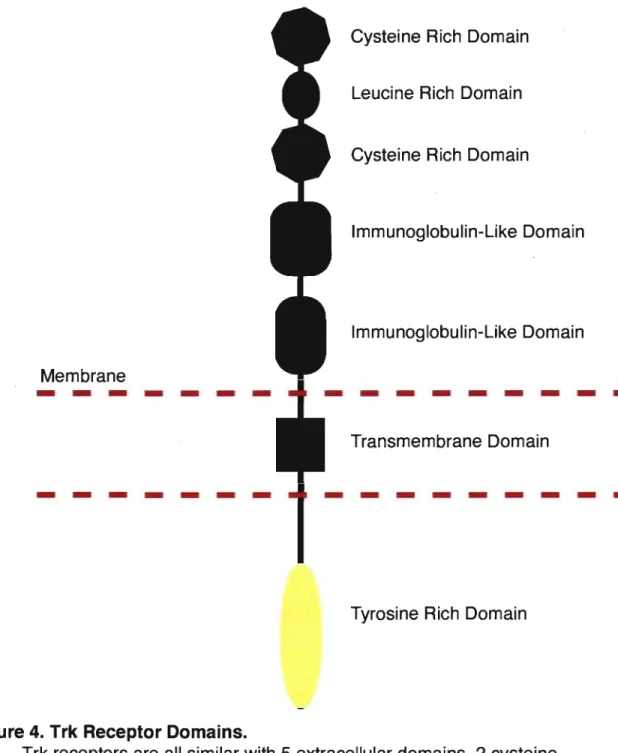
Figure 4. Trk Receptor Domains ... 55
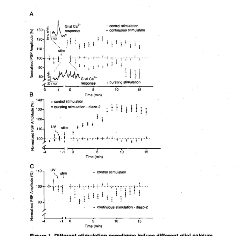
CHAPTER 1 FIGURES Figure 1. Different stimulation paradigms induce different glial calcium signais that determine post-tetanic plasticity ... : ... 70
Figure 2. Glial calcium and purine regulation determines the outcome of synaptic plasticity ... 74
Figure 3. Post-tetanicdepression is regulated by A 1 adenosine receptors ... 75
Figure 4. A2A receptors regulate post-tetanic potentiation ... 76
Supplemental Figure 1. Two patterns of stimulation used in this study ... 86
Supplemental Figure 2. UV photolysis alone does not affect glial activation or synaptic transmission ... 88
Supplemental Figure 3. Selectiye and precise activation of PSCs ... 89
Supplemental Figure 4. Postsynaptic muscle fibers have no effect on synaptic plasticity following photolysis on glial cells ... 91
Supplemental Figure 5. Specific glial photolysis of diazo-2 has no direct presynaptic effects and selectively blocks glial calcium elevations ... 92
Supplemental, Figure 6. Agonist application occludes synaptic plasticity ... 94
Supplemental Figure 7. Model of glial-mediated bidirectional modulation of synaptic plasticity ... '. ;' ... 96
CHAPTER 2 FIGURES Figure 1. Evoke Ca2+ responses in PSC are modulated by neurotrophins .. 107
Figure 2. BDNF and NT-3 differentially alter PSC receptor signalling ... 110
Figure 3. NT-3 and BDNF induce Ca2 + responses in PSCs ... 114
Figure 4. BDNF signais through activation of PLC-IP3 cascade ... 116
Figure 5. NT-3 requires extracellular Ca2+ ... ~ ... 118
Figure 6. NT -3 activates PSCs indirectly through purinergic receptors ... 120
Figure 7. Neurotrophins modulate PSC Ca2+ signalling and induce PSC , Ca2 + responses ... ; ... ; ... 121
)
ApPENDIX FIGURES
Figure 1. Bidirectional modulation of synaptic transmission by perisynaptic Schwann cells at NMJs ... XI Figure 2. Heterogeneity of hippocampal glial networks ... XIV
1.1.
PREAMBLEThe classical view of the synapse includes the presynaptic terminal and the postsynaptic neuron (Fig. 1). However, over the past two decades glial cells have emerged as a third cellular component important for
synaptic function. This development has lead to the use of the term "tripartite synapse" wh en referring to synapses to better reflect our new
"
understanding'of synapses as structures that not only include the pre- and postsynaptic neurons, but also the surrounding glia (Fig. 2).
Presynaptic ,'. ' Terminal '
Ca++ .
Figure 1. Classlc vlew of the synapse.
Communication between two neurons involves release of
neurotransmitter from the presynaptic 'terminal that binds to receptors on the postsynaptic œil.
Synapses are plastic, and therefore can be strengthened or ' weakened depending on the changing demands of the nervous system. Classically, plasticity was thought to involve the pre- and postsyn~ptic'
neurons of the synapse. However, glial cells surrounding the synapses have now been implicated in these phenomena throughout the nervous system (Allen and Barres, 2005; Todd et ak, 2006).
Presynaptic Terminal
Figure 2. The tripartite synapse.
The glial component is added in to the classical view of the synapse and is an activepartner in synaptic transmission both receiving input and providing feedback to neurons.
2
The study of communication between neurons and glia (ie neuron-glial interactions) began with the observation that glia could respond to neuronal activity with elevations of intracellular calcium (Dani et al., 1992; Jahromi et al., 1992; Reist and Smith, 1992). Since then, it has been
demonstrated that glial ceU responses to nerve activity are not simply on or off, but are graded and dependent on the frequency of stimulation (Pasti et al., 1997), and are also input specifie (Perea and Araque, 2005). This acute variability in glial calcium responses illustrates the dynamic nature of their interactions at the synapse. However, the contributions of distinct glial calcium signais on synaptic transmission remain unknown. One wonders if
glial calcium responses, as with synapses, are modifiable and plastic in a manner that would allow them to undergo lasting changes (minutes to hours) to their responsiveness.
Following the demonstration of glial cell responsiveness to synaptic activity, a number of laboratories investigated the capacity of glial cells to modify neuronal and synaptic function (Kang et aL, 1998; Newman and Zahs, 1998; Robitaille, 1998) through the release of transmitter substances from glial cells termed gliotransmitters. Since these first discoveries, a number of different examples have been published using different
preparations and approaches to demonstrate the involvement of glial cells in synaptic plasticity (Castonguay and Robitaille, 2001; Ullian et aL, 2001; Fellin et aL, 2004; Fiacco and McCarthy, 2004; Hama et aL, 2004; -Christopherson et aL, 2005; Pascual et aL, 2005; Ge et aL, 2006; Panatier et aL, 2006; Serrano et aL, 2006; Stellwagen and Malenka, 2006; Perea and Araque, 2007). Surprisingly, however, no one has yet demonstrated the ability of the same glial cell to induce both increases and decreases in synaptic function in the same neuron. Since synapses need to be able to be modified both positively and negatively, it seems likely that glial cells could be involved in both processes. Furthermore, if glial cells are to change their responsiveness under different synaptic situations, they too must be plastic.
ln this thesis 1 have provided information regarding the role of glial cells in different forms of plasticity and in glial plasticity itself. To investigate these unanswered problems, Istudied the involvement of glial cells in the generation of distinct forms of plasticity, specifically, their ability to detect dlfferent patterns of neuronal activity, and the resulting differences in their feedback to synapses. In addition, 1 have investigated the contribution of the neurotrophins, on the modulation of glial cell plasticity and neuron-glï'al interactions.
4 First, 1 demonstrate that glia from the same synapse could detect differences in the pattern,of endogenous neuronal activity, which is reflected in their calcium elevations. Second, activating the glial cells with distinct patterns of nerve activity resulted in feedback that was dependent on the initial pattern of activity. Finally, in Chapter 3 _1 demonstrate that glial cell calcium elevations are indeed plastic an9 that the neurotrophins are one group of molecules that are implicated in this plasticity.
The work contained in this thesis provides important insight into the mechanisms and sorne of the molecules involved in neuron-glial
interactions. These finding take steps towards explain the complex interactions between neurons and glia throughout the nervous system.
1.2.
THE SYNAPSESynapses are connections between two cells that allow the transfer and modulation of neuronal information. Most commonly, these are
connections between two neurons where one neuron, the presynaptic cell, releases a transmitter substance that will bind to receptors on the
postsynaptic cell. The binding of neurotransmitter to its receptors causes signalling to occur in the postsynaptic neuron either through the flux of ions through pore-forming receptors or through complex ,cascades of
intracellular messengers.
Transmitter release is a tightly regulated process for reliable communication in the nervous system. It occurs at active zones,
specialized regions of the presynaptic terminal where vesicle fusion occurs (Fig. 3) and it is regulated by many intracellular proteins as weil as sorne transmembrane proteins that span the synapse to physically join the pre-postsynaptic cells. Sorne of the most studied presynaptic proteins that are involved in vesicle trafficking and release make up a group known as the SNARE proteins. The SNARE proteins link together the nerve terminal
membrane, vesicles and calcium channels to tightly regulate vesicular release. They consist of SNAP-25, synaptobrevin, syntaxin and the
associated calcium-sensitive synaptotagmin (Bennett et al., 1992; Geppert et aL, 1994; Nishiki and Augustine, 2004). Calcium channels bind to the SNARE proteins SNAP-25 and syntaxin (Leveque et al., 1994; Sheng et aL, 1994; Martin-Moutot et al., 1996; Jarvis et al., 2002; Keith et al., 2007). Association with these presynaptic scaffolding proteins keeps calcium channels in close proximity to active zones, an organization that has been repeatedly suggested for over 30 years (Heuser et al., 1974; Robitaille et al., 1990). This close proximity between calcium channels and active zones (probably less than 50 nm) leads to high fidelity of transmitter release. In fact, following an action potential iUakes approximately 0.2 ms for vesicle fusion to occur. Presynaptic active zones allows for rapid and reliable vesicle release following arrivai of an action potential.
Postsynaptically, neurotransmitter binds to receptors to induce a
respon~e. Receptors are located across from active zones and held in
place by their interactions with scaffolding proteins that combine to make up the postsynaptic density (Fig. 3). The postsynaptic density contains receptors, scaffolding proteins and signalling proteins involved in the generation of the postsynaptic response.
Synaptic (Ieft • Neurotransmitter •
~
Receptor Potential / _ _ No· )Figure 3. Presynaptic and Postsynaptic Machinery.
6
At the presynaptic terminal vesicles fuse with the presynaptic membrane at active zones, regions specialized for the fusion of vesicles and the release of neurotransmitter upon arrivai of action potentials. Once neurotransmitter is released into the synaptic cleft is crosses to the postsynaptic cell where it binds to postsynaptic
receptors. In muscle, the postsynaptic membrane is composed of folds and valleys with nAChRs positioned at the tops of the folds and
voltage-gated sodium channels at the bottoms of the valleys. Activation of the nAChRs by neurotransmitter binding results in a receptor
potential that propagates passively down the membrane until it reaches the voltage-gated sodium channels where action potential initiation and ultimately muscle contraction occur.
Synaptic structures are modifiable both in morphology and in terms of the strength of the connection. Morphological adjustments can occur, for example, through actual changes in the size of the cellular elements or through their proximity to each other. Having said this, synapses are relatively stable structures once formed (Grutzendler et al., 2002). Aside from morphological alterations, synaptic strength can also be changed. Changes in the strength of synapses can be both increasing and
decreasing. These changes are generally referred to as synaptic plasticity. The mechanisms and processes involved will be discussed in more detail later. However, at a basic level these plasticity events can be the result of changes to the presynaptic terminal, the postsynaptic cell as weil as neighbouring glia.
This section has introduced general aspects of the chemical
synapse. Synapses include the presynaptic terminal, postsynaptic cell and surrounding glia. Synaptic transmission occurs through release of vesicles from the active zone, which binds to postsynaptic receptors. Most
commonly wethink of neuron-to-neuron synapses, however, one of the best-understood synapses in the nervous system is the neuromuscular junction (NMJ). The NMJ is the equivalent of neuron-to-neuron synapses, but is specialized to allow for synaptic transmission between neurons and muscles. Like central nervous system synapses the NMJ is surrounded by gUa that participate in its function (Auld et al., 2003). These properties make the NMJ an excellent model for studying neuron-glial interactions. In the sections to follow properties of the NMJ will be discussed in more detail.
1.3.
NEUROMUSCULAR JUNCTIONThe neuromuscular junction has been, studied for thelongest amount of time of any synapse in the nervous system. Chemical synaptic transmission has been best described in th"is system beginning in the 1950s with the work of Bernard Katz and colleagues (Del Castillo and Katz, 1954). 'Since then the NMJ has continued to be a useful model for
unders~anding mechanisms of release (poage and Meriney, 2002; Rizzoli
et al., 2003), postsynaptic mechanisms (Sanes and Lichtman, 2001) and more recently, for studying the tripartite synapse (Auld and RObitaille, 2003a).
(
1.3.1. ORGANIZATION AND FUNCTION OF THE NMJ
The NMJ, like other synapses, is composed of a presynaptic nerve terminal, a postsynaptic cell (muscle fibre) and surrounding glial cells (perisynaptic Schwann cells; PSCs). This is a relatively simple organization with only one innervating nerve terminal (at mature synapses), one
postsynaptic cell, and about 3-4 PSCs, with ail cellular compartments being visually identifiable.
1.3.1.1. Presynaptic Terminai of the NMJ
8
The presynaptic terminal of the mammalian NMJ is an elaborate structure that roughly takes on a pretzel-like shape. Release at the neuromuscular junction occurs upon the arrivai of an action potential and results in the liberations of tens to hundreds of vesicles. Calcium and ,presynaptic proteins associated with vesicular release play similar roles
at the NMJ as previously mentioned for synapses in general. Presynaptic terminais flJnctionally match the properties of their associated muscle. For example the presynapticterminal must be able to induce the form of contraction required by a given muscle, and sorne muscles are required to be active for brief periods of intense activity whereas others require more prolonged periods of activation. This is
achieved through a variety of presynaptic terminal properties such as the number of vesicles available and released (Reid et aL, 1999). These differences in properties give sorne terminais the ability to be active for longer periods of time, while others will fatigue more easily. Synapses that initially release fewer vesicles but can remain active for longer periods of time are called weak and fatigue resistant. Strong synapses fatigue more easily as they are normally only active for shorter periods of
time(Wood and Slater, 1997; Reid et al., 1999). Specifie properties will be discussed i!1 more detail in the following section.
1.3.1.2. Properties of Presynaptic Release
Although the structure of NMJs can vary widely amongst different species, the functional aspects seem to be weil conserved. For instance, frog NMJs, which can be hundreds of micrometers long, also release hundreds of vesicles for each action potential (Heuser et al., 1979; Katz and Miledi, 1979). However, rat terminais, which are around 50
micrometers in length also release around 50 vesicles per action potential (Wood and Slater, 1997). This suggests that a size/release relationship exists that is conserved at least amongst vertebrates.
Howeve~,.there are observable differences betwèen heuromuscular synapses. For instance, much interest has been focussed on the adaptive mechanisms that are associated with differences in weak and strong neuromuscular synapses. In the mammalian system, one
commonly used cornparison for weak and strong synapses is that of
so/eus (Sol; weak) and extensor digitorum fongus (EDL; strong)
muscles. At the weak Sol synapses, less fatigue is observed and release can be maintained for longer periodS of time at lower
frequencies (-20 Hz). However, the EDL displays rapid fatigue and is only active for short bursts of activity at high frequencies (-80 Hz) (Hennig and Lomo, 1985). Corresponding to the functional differences between these synapses are morphological ones, with the Sol
occupying larger synaptic areas than EDL NMJs (Waerhaug, 1992; Wood and Slater, 1997). In conjunction with occupancy of large synaptic areas, Sol nerve terminais also have a larger synaptic vesicle pool in comparison to those of EDL NMJs (Reid et al., 1999). These terminais also display differences in initial quantal release (Gertler and Robbins,
10
.1978; Wood and Slater, 1997; Reid et aL, 1999). However, the time required for vesicle recycling appears to be quite similar for both
terminais even across species, -115 sec (Betz and Bewick, 1992, 1993; Ryan et aL, 1993; Reid et aL, 1999). Since recycling time is similar, this suggests smaller release per stimulus and unit area along with the larger vesicle pool size contribute to the greater degree of fatigue resistance at Sol nerve terminais. Although there are sorne subtle differences in release properties and vesicle number, the same basic mechanisms apply to ail NMJs.
1.3.1.3. Synaptlc Plasticity
The amount of transmitter release that occurs is variable and changes in the amount of release are referred toas synaptic plasticity. Synaptic plasticity can occur over different time frames from
milliseconds to days and can be broadly broken up into short-, and long-term plasticity.
1.3.1.3.1 POTENTIATION
Enhancement of synaptic transmission can be divided temporally into facilitation, lasting tens of milliseconds, augmentation, lasting seconds, post-tetanic potentiation (PTP), lasting 10s of seconds and finally short-term plasticity, which lasts for minutes (Zucker and Regehr, 2002). Calcium plays importantroles in ail forms of synaptic
enhancement although the mechanisms are different. Facilitation is the result of residual calcium in the presynaptic terminal when a second impulse triggers release. This causes greater fusion of synaptic vesicles and a larger postsynaptic response. Katz and Miledi first suggested the . involvement of calcium in facilitation using the NMJ preparation (Katz
merely by the rate with which calcium diffuses away trom active zones (Cooperet aL, 1996). In comparison, augmentation, which is longer lasting, is mediated by the rate of calcium removal. It has been suggested for a number of years that the processes regulating
facilitation and augmentation are different (Landau et aL, 1973; Magleby and Zengel, 1982). There is evidence suggesting that calcium pumps and exchangers can be involved in regulating the duration of
augmentation and PTP (Parnas et aL, 1982; Wojtowicz and Atwood, 1985). This probably occurs through a build up of both sodium and ' calcium presynapticaUy. This build up then results in decreased removal by exchangers due to the increased intracellular sodium concentration (Birks and Cohen, 1968b, 1968a; Mulkey and Zucker, 1992). Another factor that can influences PTP is mitochondrial calcium loading. This
"
a/so occurs during periods of prolonged activity where mitochondria are loaded with calcium that is slowly released following cessation of activity (Tang and Zucker, 1997; David et al., 1998; David and Barrett, 2000). Ali of these different mechanisms lead to different forms of synaptic
enhancement, largely delineated by their duration.
1.3.1.3.2
DEPRESSIONAside from synaptic potentiation, depression also occurs. At the NMJ there are two main phases of depression that are often described. The first and shorter phenomenon occurs over seconds during
prolonged activity, while the other is a modification of longer duration. One of the main causes suggested to underlie the shorter depression mentioned above is depletion of the vesicle pool (Betz, 1970). This occurs during prolonged periods of activity, where vesicle release outpaces recovery and reéycling, such that fewer readily releasable vesicles are available. Once presynaptic activity stops, vesicle recycling
can catch up and return the vesicle pool to normal, and thus, no depression is apparent.
12
Other mechanisms of de pression that involve longer processes include modulation of presynaptic function by neuromodulators and postsynaptic desensitization (Zucker and Regehr, 2002). Some of these mechànisms have been described in our laboratory and include
neuromodulators such as nitric oxide (NO) and glutamate that can induce long lasting depression of neuromuscular transmission (Thomas and Robitaille, 2001; Pinard et aL, 2003). Interestingly, the depression induced by the se two transmitters appear to be quite similar. Recently, it has been demonstrated that in fact glutamate and NO are linked and cooperate to induce depression at the NMJ (Pinard and Robitaille, in press). This appears to be through presynaptic release of glutamate that 'binds to postsynaptic metabotropic glutamate receptors, which activate
NO synthase to produce NO. The NO is then able to diffuse back to the presynapse to cause depression.
The processes leading to synaptic plasticity are fundamental for proper synaptic functioning. Many of the early discoveries surrounding plasticity were made üsing NMJ preparations, discoveries that have proven to hold true throughout the nervous system.
1.3.1.4. Postsynaptic Muscle Fibres
The postsynaptic cell associated wittlNMJs is·the muscle fibre. There is only one presynaptic cell for each postsynaptic cell, unlike the organization of the CNS, however, during developmentmuscle fibres are innervated by multiple presynaptic terminais (Salice-Gordon and Lichtmàn, 1993).
Strong and weak synapses innervate different muscle fibre types. The differences in muscle fibres have been categorized a number of different ways based on myofibrillar ATPase isoform, twitch kinetics,
myosin heavy chain (MHC) isoform, and metabolic enzymes and processes used. This results in Type 1 fibres corresponding to slow twitch, and red fibres expressing MHCIf3 and being innervated by weak presynaptic terminais. These fibres are high in mitochondria, undergo
,
oxidative metabolism and are fatigue resistant (Peter etaI., 1972; Pétte and Staron, 2000). They also have a smaller diameter than other fibre types. Type liB fibres are larger in diameter, are one type of fast twitch, white fibre, that are low in mitochondria, run on glycolysis and express MHClib (Peter et al., 1972; Pette and Staron, 2000). These properties make white fibres more prone to fatigue and are innervated by the
strong presynaptic terminais. The other pure fast twitch 'fibres include type liA and 110. Finally there are mixed fibres that display a mixture of the above-mentioned properties(Pette and Staron, 2000). Most
mammalian muscles are composed of a mixture of fibre types rather than being purely one type or another.
1.3.1.5. The End-Plate and Receptors
Synaptic transmission occurs through the release of acetylcholine and activation of postsynaptic nicotinic acetylcholine receptors
(nAChRs). These receptors are pentameric in structure and are
p~rmeable to monovalent and divalent cations (Villarroel and Sakmann,
1996). As with ail postsynaptic cells, densities of receptor clusters and postsynaptic scaffolding proteins are present on muscle fibres in tfle
end-pla~e region. The clusters of nAChRs are located at the peaks of a number of folds in the postsynaptic membrane that make up the end-plate and are located directly across from active zones. In the troughs of these folds are located the voltage-gated sodium channels responsible for generating action potentials.
14
The NMJ has been an important model for studies of pre and
postsynaptic development, for example receptor clustering. Early studies observed sorne spontaneous clustering of nAChRs (Vogel et aL, 1972; Fischbach and Cohen, 1973), however, synapse formation and
. clustering associated with mature synapses requires activity (Anderson and Cohen, 1977; Frank and Fischbach, 1979), something that was also confirmed in vivo (Liu and Westerfield, 1992). Along with receptor
clustering, receptor synthesis is also increased with activity (Anderson and Cohen, 1977; Jessell et aL, 1979; Role et aL, 1985). Two factors
rele~sed in an activity-dependent manner that are important for the se
processes are neuregulin (Usdin and Fischbach, 1986; Falls et aL, 1993; Jo etaI., 1995), and agrin (Godfrey et aL, 1984; Meier et aL, 1997). Recent in vivo work has found that spontaneous nAChR clusters
are actually the site of presynaptic contact and that these clusters can be incorporated into maturing end-plates (Flanagan-Steet et aL, 2005; Panzer et aL, 2005).
1.3.1.6. Perisynaptic Schwann Cells
Perisynaptic Schwann cells (PSCs) are geno- and phenotypically distinct from myelinating Schwann cells. Whereas myelinating Schwann cells express high levels of proteins such as PO and myelin basic
protein, PSCs express low levels of these proteins and high levels of proteins such as NCAM and S100 (Mirsky and ~essen, 1996). In terms of phenotype, PSCs do not wrap axons, but rather surround nerve terminais and postsynaptic densities. In mammals PSCs arrive at NMJs starting in early postnatal life and increase in numbers to about three or four in adults (Love and Thompson, 1998). During development,
Schwann cells migrate slong axons rather than the opposite as previously thought (Gilmour et aL, 2002).
PSCs are found surrounding the synaptic cleft and can, in fact, participate in synaptic transmission. They express receptors for the
neurotransmitters of the NMJ and also transporters that could participate . in regulating plasticity events (Pinard et al., 2003). This will be discussed further in the following section (1.4.4). Aside from developmental and synaptic functions of PSCs, they are also important for synapse stability in mature animais (Reddy et al., 2003). PSCs are an integral part of neuromuscular synapses, participâting in development, maturation, stability and function.
16
1.4.
NEURON-GLIAL INTERACTIONS IN SVNAPTIC PLASTICITVGlial cells are the other major cell type in the nervous system aside from neurons. Like neurons·they are heterogeneous and fall into a nllmber of large categories;.1) microglia, the immune cells of the brain, 2)
myelinating glia, oligodendrocytes of the CNS and Schwann cells of the PNS, and 3) astroglia, which are the non-myelinating synaptic glial cells that are represented by PSCs at the NMJ.
Beginning in 1992 glial cells in situ were shown to actively respond to
synaptic transmission (Dani et aL, 1992; Jahromi et aL, 1992; Reist and Smith, 1992). It was another six years before it was demonstrated that glial cells could also provide modulation of synaptic function through feedback . (Kang et aL, 1998; Newman and Zahs, 1998; Robitaille, 1998). Now, it is widely accepted that glia are active partners in synaptic function that play a number of different roles in synaptic transmission~ This evidence has been derived from work at the NMJ and with CNS preparations .
. 1.4.1. A
BRIEF HISTORY OF SYNAPSE-GLIA INTERACTIONSAlthough it was not until relatively recently that glial cells were
demonstrated to actively participate in synaptic function, observations made by anatomists around the early 1900's suggested a variety of such functions (Somjen, 1988). For instance, Lugaro (Somjen, 1988) suggested that
astrocytes might be involved in neurotransmitter removal to regulate transmission, something that is now known to be true (Rothstein et aL, 1996; Bergles and Jahr, 1997, 1998; Huang and Bordey, 2004).
The first description of a dynamic glial response to synaptic transmission came from the work of Stephen Kuffler in the 60's. He and colleagues
showed that activity in neighbouring nerves could induce depolarization of glial ce Ils that was dependent on extracellular potassium (Kuffler et aL,
: ..
idea that glial cells buffered the extracellular milieu, however, a major
advancement in our ability to study glialcells came with the finding that they responded dynamically with calcium elevations. Since then a number'of different studies using isolated cell cultures, and a variety of semi-intact, and more recently, even
in vivo
studies, have revealed a vast number offunctions for glial cells in brain physiology (Volterra and Meldolesi, 2005; Haydon and Carmignoto, 2006).
ln terms of chemical activation of glial cells, the first studies to show this were done using cultured astrocytes and bulk application of transmitter substances (Cornell-Bell et aL, 1990). Following this, a number of studieS IJsed more intact preparations to demonstrate that this was not simply an artefact of culture (Da ni et al., '1992; Jahromi et al., 1992; Reist and Smith, 1992). Two of these studies, from Stephen Smith's laboratory, used different preparations. One was the hippocampal slice, and the other the NMJ
preparation and PSCs. These two studies arrived at the same basic
'conclusion, that glial cells respond with calcium elevations to nerve activity. Following the establishment of glia as active responders to synaptic transmission, the question turned to their involvement in providing
modulatory feedback tosynapses. This could occur in a nurnber of ways: regulated clearance of neurotransmitter, morphological changes affecting transmission, and release of chemical gliotransmitters. As already
mentioned, a major breakthrough occurred in 1998 when a number of 'papers from independent laboratories published results showing that glial
cells from both the CNS and PNS could indeed modulate neuronal function (Kang et al., 1998; Newman and Zahs, 1998; Robitaille, 1998). Since then, work has provided information on many of the finer points of neuron-glial communication and the man y and varied roles that glial cells play in synaptic function. '
1.4.2.
CALCIUM:A
BREAKTHROUGH IN THE STUDV OF GLIAL CELL FUNCTIONThe major acute response detectable in glial cells is the calcium response. This dynamic form of cellular response was discovered in the 1980'5 and has since been influential in studies of glial cell function and neuron-glial interactions (for example see Sugino et aL, 1984).
18
The intracellular signalling cascades responsible for generating glial calcium responses are the inositol1,4,5-triphosphate (IP3 ) and ryanodine receptor pathways (Haak et aL, 2001; Holtzclaw et aL, 2002). However, calcium responses can also be generated through influx of extracellular calcium (Verkhratsky and Kettenmann, 1996): The IP3 pathway is initiated through G-protein-mediated activation of phospholipase C (PLC), which cleaves phosphatidylinositol4,5-bisphosphate (PIP2) to f6-rm diacylglycerol and IP3 • The newly formed IP3 then activates IP3 receptors to release intracellular stores of calcium (Fig. 2). ,
Calcium elevations in glia can be generated spontaneously and through agonist-mediated activation. In term's of synaptic function, agonist-mediated activation of glial cells can be critically important for subsequent
modifications to that function. For instance, in the hippocampus, calcium signalling in astrocytes can be initiated by GABAs receptors that lead to potentiation of release by inhibitory interneurons (Kang et aL, 1998). This initially occurs through the presynaptic release of a neurotransmitter that then binds to receptors on the perisynaptic glial cell. This leads to a calcium elevation in the glial cell and subsequent release of gliotransmitter that acts through feedback on the presynaptic terminal or onto the postsynaptic cell. Interestingly, glial calcium responses are'not ail or none events. They are modifiable and dependent on the input used to actiyate them and the
intensity of stimulus (Pasti et aL', 1997; Perea and Araque, 2005). Glial cells, like neurons, express a number of different receptors and therefore, can be activated in a number of different ways. In neurons, different frequencies
and patterns of activity lead to different changes in synaptic plasticity. Although glial cells respond with different calcium elevations to different frequency input, it is unknown if this impacts any ongoing process.
Furtherlliore, nothing is known regarding the influence of different patterns of stimulation on glial activity. It is likely that differences in glial 'activation lead to differences in feedback to neurons, although this has never. specifically been tested.
1.4.3. CNS SVNAPTIC FUNCTION AND THE IMPLICATION OF GLIA
Due
to
issues with complexity and accessibility much of the early CNS glial work was performed in culture. However, it was soon realized that,
observations made in culture were not always representative of those occurring hi more intaèt preparations (Kimelberg et al., 1997). Now slice preparations pre commonly used and even sorne in vivo studies are
emerging (Wang et al., 2006). Despite early problems with accessibility, in"
situ
CNS-glial preparations have provided original pieces of information, and continue to improve our understanding of neuron-glial interactions.1.4.3.1. Gllotransmlssion and (,jlial-to-Neuron Signalling 1.4.3.1.1 CHEMICAL TRANSMITTER RELEASE
Gliotrarismission (the release of chemical transmitters from glia) is one discovery made through studies of CNS preparations. In fact, the release of gliotransmitters in response to activation has only definitively been
demonstrated in the CNS. There is also evidence for astrocytic vesicles
1
(Bezzi et al., 2004), and vesicular release machinery (Zhang et al., 2004). Furthermore, release of glutamate has been recorded in culture (Parpura et al., 1994), and suggested, somewhat controversially, to occur in slice
preparations (Angulo et al., 2004; Fellin et al., 2004; Fiacco and McCarthy, 2004). Although glutamate release by astrocytesleading to modulation of
CNS synapses is an attractive hypothesis, the evidence is currently under debate (Angulo et aL, 2004; Fellin et aL, 2004; Fiacco et aL, 2007).
20
Anot,her transmitter known to be released by glia is ATP (Fields and Burnstock, 2006; Zhang et aL, 2007). The release of ATP by glia is one of the major mechanisms used to propagate signais across glial networks (Newman and Zahs, 1997; Haas et aL, 2006). Another role for astrocyte purine signalling has recently emerged from two laboratories using
pharmacological and molecular approaches. These reports demonstrated the impact of such signalling in hippocampal plasticity where glial signalling and ATP release is fundamental for the generation of heterosynaptic
plasticity (Pascual et aL, 2005; Serrano et aL, 2006). Similarly, ATP release by astrocytes was earlier found to be involved in heterosynaptic
suppression, which occurs on a faster time scale (Zhang et aL, 2003). Purine release in neuron-glial interactions is a major factor in both the CNS· and PNS. The roles that purines play in neuron-glial interactions at the NMJ will be discussed in the following section.
1.4.3.1.2 GI-IOTRANSMISSION AND SYNAPSE DEVELOPMENT
Other important actions of factors released by glial cells have been shown in the context of development. For instance early studies in culture revealed the importance of glia for synapse development and synaptic transmission (Pfrieger and Barres, 1997; Ullian et al., 2001). A number of different factors have been shown to be important for these developmental changes such as cholesterol (Mauch et aL, 2001), thrombospondins
(Christopherson et aL, 2005), and TNFa (Beattie et al., 2002; Stellwagen and Malenka, 2006).
These studies often focus on excitatory synaptic transmission, however, roles for glia in inhibitory synapse formation have also recently come to light. For. instance, Balice-Gordon and colleagues demonstrated that
2005b). This appears to occur through two separate mechanisms that rely on the release of BDNF from neighbouring astrocytes. An interesting link with the previous discussion of purinergic signalling cornes from work in the PNS showing that axonal release of ATP cou Id initiate the release of BDNF from Schwann cells in a regulated manner (Verde rio et al., 2006). This study was performed in the context of regeneration; however, similar mechanisms may influence developmental progressions as weil. In addition to releasing neurotrophins, glial cells can also respond, to at least BDNF, with
intracellular calcium elevations (Rose et aL, 2003). This indicates that the neurotrophins are likely important molecules for neuron-to-glial
communication and glial-to-neuron signalling.
1.4.3'.1.3 PHYSICAL GLIAL-To-NEURON SIGNAL LING
Aside from factors that are released, glial cells can communicate with neurons through a wide variety of mechanisms. For example, one
1
mechanism of communication is the direct interaction of neuronal and glial cells, since glia enwrap synapses (Ventura and Harris, 1999; Murai et aL, 2003). These interactions are known to influence dendritic morphological plasticity (Murai et aL, 2003), and possibly synaptic development (Hama et aL, 2004). Interestingly, astocytes occupy distinct domains, do not overlap, but do contact synapses on multiple neurons (Bushong et al., 2002;
Halassa et aL, 2007). This provides the potential for inter-neuronal· interactions outside the normal axon pathways.
Aside from release and direct cell-to-cell contact, the morphological arrangement of a synapse can also affect its function. As mentioned, a phenomenon such as this was described in the hippocampus where glial contact increased dendritic stability (Murai et al., 2003). Another example of
22
the role that morphology could have on synaptic function has emerged from studies in the hypothalamus where lactation causes a retraction of glial processes from around synapses (Oliet et aL, 2001) and ultimately changes the plasticity expressed by these synapses (Panatier et aL, 2006). It is quite possible that differences in the anatomical arrangement of different
. synapses in other regions of the brain can have important effects on synaptic function, similar to what has been reported in the hypothalamus.
1.4.3.2. Waves of Activation
Glial calcium waves have been observed in a number of brain regions including the cortex (Haas et aL, 2006) and retina (Newman and Zahs, 1997). Glial calcium waves are proposed to have a number of roles in CNS function. For instance, they appear to be linked to metabolic functions to regulate glucose uptake over a larger a~ea (Bernardinelli et aL, 2004). Furthermore, these waves are likely involved in regulating neuro-vascular coupling (Metea and Newman, 2006; Takano et aL, 2006), since calcium signais in glial endfeet can induce changes in vascular.dilation (Zonta et aL, 2003; Mulligan and MacVicar, 2004). In terms of direct synaptic functioning, glial calcium waves may also be involved in long-distance communication and possibly even synchronization of multiple neurons through coherent release of gliotransmitter (Angulo et aL, 2004; Fellin et aL, 2004).
There are many molecules and mechanisms thatare implicated in the birdirectional communication between neurons and glial cells. It is becoming clear that the heterogeneity of glial cells is leading to an almost endless number of possibilities for the way they interact with neurons.
1.4.4. NMJ MODULATION AND THE INVOLVEMENT OF PSCs
The neuromuscular junction has proven to be a valuable model for investigating synapse-glia interactions. In fact, sorne of ·the pioneering
studies were performed on this preparation (Jahromi et aL, 1992; Reist and Smith, 1992; Robitaille, 1998). In the following sections neuron-glial
intractions involved in NMJ plasticity will be discussed. ,
1.4.4.1. Acute Actions of PSCs
Perisynaptic Schwann cells of the NMJ can be thought of as the peripheral nervous system equivalent to astrocytes of the CNS. They are non-myelinating, occupy distinct domains and surround the synapse. As mentioned, nerve activity induces PSC calcium responses through activation of a variety of receptors. PSCs are also involved in modulating synaptic function, through regulation of transmitter in the synapse (Smit et aL, 2001), signalling to the presynaptic terminal (Robitaille, 1998;
Castonguay and Robitaille, 2001) and providing overall stability to the synapse (Reddy et aL, 2003). These aspects of the NMJ-PSC preparation allow for its utility as a model for 'studies of neuron-glial interactions.
As with CNS astrocytes, PSCs are able to respond to a number of . different neurotransmitters. The most predominant are acetylcholine (ACh), which is n31eased from the nerve terminal, and the purines, which will be discussed in the review to follow. ACh is the primary neurotransmitter at the neuromuscular junction. In addition to activating the postsynaptic muscle fibre, it can also induce responses in PSCs through metabotropic
muscarinic acetylcholine receptors (mAChRs). At trog NMJs, mAChRs are suggested to be of an atypical form due to their unique pharmacological profile (Robitaille et aL, 1997).
The primary known and acute result of PSC activation is synaptic modulation. This can occur in the form of either potentiation (Castonguay and Robitaille, 2001) or de pression (Robitaille, 1998). The mechanisms of this modulation are presently unknown, however, the outcome of glial
24
modulation appears to be dependent on the intracellular signalling cascade that is recruited following nerve activity.
1.4.4.2.
Long-term Actions of PSCsSome of the most weil described PSC functions are those occurring over longer periods of time. Interestingly, results from the laboratory of Milton Charlton have suggested that mAChRs might be involved in regulating morphology of PSCs through, control of cytoskeletal elements. This was suggestèd through investigations showing that blockade of nerve activity resulted in the rapid upregulation of glial fibrillary acidic protein (GFAP) in PSCs (Georgiou et aL, 1994). This group went on to show that the receptors involved were mAChRs (Georgiou et aL, 1999). These results suggest that not only does neurotransmission have ac~te affects on PSCs, but that it likely also influences glial and synaptic stability. The reciprocal of this is true as weil, wherè removal of PSCs from the synapse results in synaptic
disassembly (Reddy et aL, 2003), suggesting that PSCs also provide stability signais to presynaptic terminais at NMJs.
1.4.4.2.1
PSCs AND SYNAPSE DEVELOPMENTIntestingly, PSCs, like astrocytes, also seem to release substances that encourage synaptic development. In cultur~, the addition of neurotrophic factors maintains the survival of neurons, but prevents synapse formation.
On the other hand, when Schwann cel! conditioned media is added, synaptogenesis can occur (Peng et al., 2003). Furthermore, evidence has recently emerged suggesting that factors released by glial cells can
modulate synaptic transmission during development, possibly to facilitate this process (Cao and Ko, 2007). This iIIustrates,the similarity in function between PSCs and astrocytes of the CNS.
Neurotrophins are also known to be important for Schwann cell
development and myelination. For instance, NT-3 can induce migration of Schwann cells prior to"myelination (Cosgaya et aL, 2002; Yamauchi et aL, 2003). Following migration, a developmental switch in neurotrophin release induces myelination (Chan et aL, 2001). Recently, an investigation of NT-3 expression and its effect on PSCs indicated that overexpression of NT -3 ' results in an increase in the number of PSCs at NMJs (Hess et al., 2007). In adults, however, TrkC expression is reduced, suggesting a primarily
developmental raie for NT -3-TrkC signalling in PSCs. Other more long-ter~
roles for neurotrophins may also exist as it is known that disruption of TrkB signalling results in disassembly of postsynaptic receptor clusters (Gonzalez et aL, 1999). This postsynaptic stabilizing influence of TrkB may be carried out by other neurotrophins or other factors acting at nerve terminais and PSCs.
One such molecule, w~lich is important for NMJ development, and negatively affects PSC stability, is neuregulin. Neuregulin expression can be regulatedby BDNF, GDNF, and NT-3 (Loeb et aL, 2002). Neuregulin is a potent factor for NMJ development and PSC survival during development (Trachtenberg and Thompson, 1996). It also known that following
de nervation PSCs send processes to neighbouring junctions that guide reinnervation (Son and Thompson, 1995b, 1995a). The elaboration of PSC sprouts is partially mimicked by induction of neuregulin signalling in PSCs (Hayworth et al., 2006), indicating its importance in a variety of pracesses.
The studies mentioned here demonstrate the complexity of interactions between axons, muscle fibres and PSCs. A number of different factors are implicated in any given process. However, the neurotrophins alone appear to have the potential for both direct and indirect actions on PSCs in
development and pathology.
One of the last major groups of molecules that 1 will discuss in the context of NMJ and PSC function are the purines. They are important for
26
. .
both modulation of synaptic activity and neuron-glial interactions and are discussed in the review to follow.
ln the followil1g pages readers will find the review article:
Todd KJ,Robitaille R (2006) Purinergic modulation of synaptic signalling at the neuromuscular junction. Pflugers Arch 452:608-614.
© Springer Publishing, 2006. Reproduced with permission, License number: 1787700253289 issued to Keith J. Todd
1.4.4.3. Review Article
Purinergic Signalling at the Neuromuscular Junction
Keith J. Todd and Richard Robitaille Département de physiologie
and
Centre de recherche en sciences neurologiques Université de Montréal
Address correspondence to:
Montréal, QC Canada, H7G 1 T7
Richard Robitaille
Département de physiologie Université de Montréal
P .
o.
box 6128, Station Centre-Ville Montréal, QuébecCanada, H3C 3J7 e-mail:
Acknowledgements: The authors thank Claude Gauthier for help with figure preparation. This work was supported by grants to RR from the Canadian Institutes for Health Research and by the National Science and Engineering research Council (NSERC) and by a group grant from Fonds de la recherché. en Santé du Québec. KJT was supported by a NSERC studentship.
28
INTRODUCTION
Adenosine triphosphate (ATP) was found to be co-released with acetylcholine (ACh) at neuromuscular junctions
(NMJs) over 30 years ago (Silinsky, 1975). Interestingly, purines can also be released 'from muscle fibres (Smith, 1991; Santos et aL, 2003) and Schwann cells (Liu et aL, 2005). Since then, the functions of purines at NMJs have been examined in a number of different preparations, revealing diverse functions. A number of different
. re~eptors are present on ail three elements of the NMJ: the
presynaptic nerve terminal, postsynaptic muscle and perisynaptic Schwann cells (PSCs). These receptors include P1 adenosine receptors on presynaptic terminais (Correia-de-Sa et aL, 1996; Silinsky, 2004; Baxter et aL, 2005) and PSCs (Robitaille, 1995; Rochon et al., 2001) and P2 ATP receptors on PSCs (Robitaille, 1995; Rochon et al., 2001), nerve terminais (Grishin et aL, 2005; Moores et aL, 2005) and muscle fibres (Collet et aL, 2002; Choi et aL, 2003; Santos et aL, 2003). Purinergic function in the peripheral nervous system and at the NMJ indicates roles during development (Fu and Poo, 1991), at mature NMJs (Redman and Silinsky, 1994; Correia-de-Sa et aL, 1996), in PNS myelination (Stevens and Fields, 2000) and neuron-glial interactions (Robitaille, 1995; Rochon et aL, 2001). This review will discuss the role of purines in modulation of NMJ function giving special focus to the involvement of purines in neuron-glia interactions. Sorne elements of this review have been published as part of a Novartis Foundation Symposium (Todd and Robitaille, 2006).
PURINE RECEPTORS AND PRESYNAPTIC MODULATION
Purinergic modulation of NMJ function can oœur in a bidirectional manner, as' either potentiation or depression of
. purinergic modulation depends on anumber of factors. For instance, the function of purinergic signalling changes with the developmental stage of the NMJ, where, during development of the tadpole NMJ, purines seem to potentiate neurotransmitter release (Fu and Poo, 1991) while in the adult frog the effect is opposite (Giniatullin and Sokolova, 1998). Dual effects of purines have also been reported at adult NMJs. For instance, using a rnammalian preparation, Correia-de-sa and colleagues (Correia-de-Sa et al., 1996) demonstrated purine-mediated potentiation and de pression of transmitter release depending on the frequency and pattern of stimulation. In this study, activation of P1 adenosine receptors of the A1 or A2Asubtypes were responsible for inducing the depressive and potentiating·effects respectively. This model suggests that the A1 receptor effect dominates during low-frequency activity, while higher levels of synaptic adenosine are required for activation of A2A receptors. These higher levels of adenosine are achieved when short-duration bursts of activity that cause the release of larger amounts of ATP that is then converted into adenosine by ectonucleotidases (Zimmermann et al., 1998). However, it is suggested that during prolonged high-frequency activation, release of ATP leads to
inhibition of ectonucleotidases such that the production of adenosine is reduced (Cunha, 2001). Correia-de- sa and colleagues suggested
~hat during hjgh-frequency bursts of nerve activity the accumulation of synaptic adenosine is greater and activation of adenosine
r~ceptor~ shifts such that transmitter release is potentiated through activation of A2A receptors (Correia-de-Sa et al., 1996; Oliveira et al., 2004). These phenomena are thought to function to preserve
transmitter during times of prolonged release and to potentiate release during times where run-down has already occurred so as to ensure muscle activation.
30
Other mechanisms of modulation can occur through direct activation of ATP receptors (Giniatullin and Sokolova, 1998; Giniatullin et aL, 2005; Grishin et aL, 2005; Moores et aL, 2005). These actions have also been shown to be potentiating or depressing depending on the receptors involved. When
metabotropic P2 receptors are·activated, they have an inhibitory effect similar to that of adenosine (Giniatullin and Sokolova, 1998). However, Morres et al. (Mocres et aL, 2005) found evidence for
presynaptic ionotropic P2X7 receptors that potentiate transmitter
release due to an increase in non-selective cation conductance. Therefore, it appears that multiple types of purinergic receptors may have sirnilar actions and can operate in parallel to modulate
MUSCLE
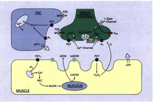
Review Figure 1. Schematic representation of the purinergic modulation of synaptic functions at the NMJ.
Diagram depicting the presynaptic nerve terminal (green), perisynaptic Schwann œil (PSC, blue) and postsynaptic muscle fibre (yellow). Sources of purines include presynaptic co-release with ACh, release from muscle fibres and possibly from PSCs. A first site of purines modulation is the presynaptic terminal where they regulate transmitter release. Both A, and P2Y receptors decrease transmitter release where P2Y receptors are thought to inhibit calcium channels through production of arachidonic acid (AA) (Grishin et aL, 2005). In the mouse A, receptors were shown to cause inhibition of calcium channels
through interaction of G~/y with the SNARE complex protein, syntaxin, which also interacts with the calcium channels (Silinsky, 2005).
Transmitter release can also be up-regulated through activation of P2X and A2A receptors. P2X, ionotropic receptors increase non-specifie
cation conductance leading to Ca2+ entry while A2A receptors are
involved in activation of L-type calcium channels (Oliveira et aL, 2004). Further inhibitory action can be derived from production of reactive oxygen species such as H2