HAL Id: hal-02631560
https://hal.inrae.fr/hal-02631560
Submitted on 27 May 2020
HAL is a multi-disciplinary open access
archive for the deposit and dissemination of
sci-entific research documents, whether they are
pub-lished or not. The documents may come from
teaching and research institutions in France or
abroad, or from public or private research centers.
L’archive ouverte pluridisciplinaire HAL, est
destinée au dépôt et à la diffusion de documents
scientifiques de niveau recherche, publiés ou non,
émanant des établissements d’enseignement et de
recherche français ou étrangers, des laboratoires
publics ou privés.
The effects of ginkgo biloba leaf extract on metabolic
disturbances associated to alloxan-induced diabetic rats
M. Naseem, M. Q. Zaman, H. Nazih, Khadija Ouguerram, I. Rabbani, H.
Zaneb, T. Yaqoob, H. U. Rehman, J. Michel, S. K. Tahir, et al.
To cite this version:
M. Naseem, M. Q. Zaman, H. Nazih, Khadija Ouguerram, I. Rabbani, et al.. The effects of ginkgo
biloba leaf extract on metabolic disturbances associated to alloxan-induced diabetic rats. Journal of
Animal and Plant Sciences (Pakistan), 2016, 26 (3), pp.627 - 635. �hal-02631560�
THE EFFECTS OF GINKGO BILOBA LEAF EXTRACT ON METABOLIC
DISTURBANCES ASSOCIATED TO ALLOXAN-INDUCED DIABETIC RATS
M. Naseem1*, M. Q. Zaman1, H. Nazih2, K. Ouguerram3, I. Rabbani1, H. Zaneb4, T. Yaqoob5, H. U. Rehman1, J. Michel2,
S. K. Tahir1, M. S. Yousaf1, K. A. Majeed1and M. S. Hussain1
1Department of Physiology, University of Veterinary and Animal Sciences, Lahore- Pakistan 2Faculty of Pharmacy, University of Nantes, Nantes- France
3UMR 1280 Phan, INRA, CRNH, West Human Nutrition Research Center, CHU, Nantes F-44093, France 4Department of Anatomy and Histology, University and Animal Sciences, Lahore- Pakistan. 5Department of Microbiology, University of Veterinary and Animal Sciences, Lahore- Pakistan
*Corresponding Author: mahrukhnaseem@rocketmail.com
ABSTRACT
Ginkgo biloba leaf extract (GBE) considered one of the most effective therapeutic herbs. We evaluated anti-diabetic
activities of GBE in rats. Rats were fed highly-fat diet for two weeks and divided into three groups (n=8): Non-diabetic control group (CG), Diabetic group (DG), Diabetic+100mg/kg GBE group (D+GBE). On 14thday, the rats were kept in
overnight-fasting and administered single intra-peritoneal injection of alloxan-monohydrate (120-130mg/Kg body weight (BW). BW and blood glucose were measured weekly up to 14thweek. Fasting/basal blood samples were collected for the
biochemical analyses. Liver, skeletal muscle and adipose tissue were also collected for mRNA gene expressions (GLUT-4, IRS-1, IR, PEPCK, SREBP-1c, FAS, PPAR-α, PPAR-γ, TNF-α). Significant reduction in BW was found in D+GBE group at week 14. Glycemia became normalized as an effect of treatment. GBE significantly decreased serum glucose, urea and ALT concentrations. We observed significant higher catalase and lower malondialdehyde levels in D+GBE group. GBE significantly increased HDL-Cholesterol and reduced triglycerides and VLDL-Triglycerides levels. GBE showed up-regulation for hepatic IRS-1 and down-regulation for PEPCK, however, up-regulation for GLUT-4 and PPAR-α found in muscle. Treatment decreased TNF-α expression in liver and adipose tissue. GBE has anti-diabetic mediated by a modulating effect on involved key genes.
Keywords: Alloxan, Carbohydrate and lipid metabolism, Diabetes mellitus, Ginkgo biloba leaf extract, Rats.
INTRODUCTION
Overwhelming evidence shows that diabetes mellitus is one of the major public health issue worldwide in the last few decades (Xia et al., 2011). This disease is a
cluster of metabolic alterations referred to as ‘chronic metabolic syndrome’ mainly associated with substantial
mortality and morbidity. The cause of diabetes mellitus is
insufficient insulin secretion from pancreatic β cells may
be due to defect in the insulin secretion, insulin action or both, which results in metabolic disorders syndrome mainly of carbohydrate, protein or fat (Nayak and Roberts, 2006). Oral hypoglycemic agents such as biguanides, thiazolidinediones and sulfonylurea are available along with insulin for the diabetic treatment but these products have adverse effect (Yeo et al., 2011).
Ginkgo biloba (GB) has been described as a
living fossil, being last remaining member of
Ginkgoaceae family commonly known as, ginkgo,
maidenhair tree, gymnosperm tree and dates back 250 million years in China (Maltas and Yaldiz, 2012). G.
biloba leaves are used as traditional Chinese herbal
medicine to treat asthma, cardiovascular diseases, aging, bronchitis, cancer, impair sexual dysfunction, cancer and
diabetes (Kudolo, 2001; Unger, 2013). The two major fractions present in standardized leaves extract of G.
biloba are: 6-7% terpenoids and 24-26% flavonoids
(Smith and Luo, 2003). Flavonoids present in G. biloba are: flavonol-gucoside of kaempferol, flavones, quercetin and isohamnetin with glucose or rhamnose. The terpenoids fraction consist of unique group of diterpenes (ginkgolide A, B, C, J and M) and sesquiterpene bilobalida (Smith and Luo, 2003; Malas and Yildiz, 2012). Very few data on the anti-diabetic molecular mechanism of G. biloba is available, so in the present study we investigated the anti-diabetic effect of G.
biloba, at molecular level in rat model.
MATERIALS AND METHODS
Experimental animals: A total of twenty-four normoglycemic male adult Wistar rats weighing 150-200 g, were selected. Rats were acclimated for a period of 12 days in the animal shed of University of Veterinary and Animal Sciences (UVAS), Lahore, Pakistan, prior to the experiment. All the animals were housed in stainless steel cages, 2 animals per cage, with wood litter bedding, which was changed on weekly basis The temperature of
Naseem et al., J. Anim. Plant Sci. 26(3):2016
animal shed was maintained at 24±5℃ with 12-h light: 12-h dark cycle and were given free access to water and food (standard rats chow) (El-Mesallamy et al., 2011). Body weight (BW) was recorded weekly and food intake was measured twice a week for each rat by determining the pre and post-weights of the food jars. The experiment was conducted for 14 weeks under the guidelines of ethical committee for welfare of laboratory animals of UVAS, Lahore, Pakistan.
Experimental Design: Rats were allowed to feed on high
fat diet fed (HFD: 12.7% maize starch, 6.5% dextrose, 6%, cellulose 3.9% sunflower oil, 31.3% beef tallow, 28.6% casein, 9.7% minerals and 1.3% vitamins by weight) for two weeks and then rats were divided randomly into following three groups (8 rats in each group).
Group I: Rats of this group were served as non-diabetic or negative control (CG) and given standard diet without any supplementation of G.
biloba leaves extract (GBE)
Group II: This group included diabetic control or positive control (DG) rats and given the standard diet without any supplementation of GBE.
Group III: This group was comprised of diabetic rats with standard diet supplemented with GBE at the dose of 100 mg/kg/day (D+GBE).
On 14thday, rats kept in fasting condition and
were administered a single dose of alloxan monohydrate (Sigma, USA) at the dose of 120-130 mg/Kg BW intra-peritoneally. After 6h of alloxan induction, 20% and 5% oral glucose solution was given to the rats to prevent hypoglycemia (Ebuehi et al., 2010). After 3 days of alloxan injection overnight fasting blood samples were collected from tip of tail and glycemia was measured (ACCU check, Germany). Only rats with fasting blood glucose levels higher than 250 mg/dl were considered diabetic and included in the study (Shankar et al., 2005; Cheng et al., 2013). Supplementation with the standardized ginkgo leaves extracts (terpenoids
6%/flavonoids 24%) was purchased from Hunan
Nutramax Inc (COCO CHEN, China) and treatment was started by mixing 100 mg/Kg GBE in the standard diet (40.7% maize starch, 20% dextrose, 5.8% sunflower oil and 22.5% casein, 9.7% minerals and 1.3% vitamins by weight) for 14 weeks.
Blood sampling and biochemical parameters: At the
end of 14th week; blood samples in fasting state, were
collected by cardiac puncture and serum was separated by centrifugation (Kumar, 2012). The biochemical analysis of fasting serum glucose (FSG), blood urea, creatinine,
alanine aminotransferase (ALT), asparatate
aminotransferase (AST), catalase (CAT) and
malondialdehyde (MDA) was done by using
commercially available kits (Randox, UK).
Serum total cholesterol (TC) and triglycerides (TG) were measured using enzymatic kits (Bio-Merieux, Marcy-l'Etoile, France). Cholesterol (VLDL-C, LDL-C and HDL-C) and TG (VLDL-TG) profiles were performed using fast protein liquid chromatography (FPLC) (AKTA FPLC SYSTEM, GE Healthcare, USA). Lipoprotein isolation was performed as described by Chetiveaux et al., (2002).
Tissue sampling and mRNA expression of genes in the liver, skeletal muscles and visceral adipose tissue:
Liver, skeletal muscle and adipose tissues were sampled and cleaned with normal saline solution and were stored immediately at -80 °C. RNA was extracted (Laboratoire de Biochimie, MMS, Nantes, France) using trizol reagent
(Ambion, USA), according to the manufacturer’s
instructions. Quantification of total RNA was measured at 260 nm. Total RNA (1 µg) was converted in to cDNA through the process of revers transcription using Super-ScriptIII Reverse Transcriptase (Invitrogen, France) in a 20 µl reaction volume. Quantitative PCR was performed using SYBR Green Supermix on MyiQ2 Real-Time PCR detecting system (Bio-Rad, Marnes-la-Coquette, France). Primers Sequence was determined by the primer3 website. The mRNA levels were normalized using GADPH as a housekeeping gene. Relative quantification was performed using the ∆∆CT method. Primers used for qRT-PCR are listed in Table 1
Statistical analysis: Results were expressed as mean ±
S.E.M. Data was analyzed statistically by One way repeated measure analyses of variance (ANOVA)
followed by PLSD Fisher’s test using Statview software
(SAS Institute Inc, SAS Campus Drive, Cary, NC, USA), to measure ginseng root extracts effects on all physiological and biochemical parameters except mRNA genes expression in studied organs, which were analyzed
by Kruskal Wallis, followed by Fisher’s test using
Stateview. P < 0.05 was taken as significant difference.
RESULTS
Body weight: The BW of rats in all the three groups was
monitored on 1st and 14th week of study (Table 2). The
BW of DG was found to be significantly (P<0.0001) decreased on week 14. D+GBE group also showed decrease of BW but this decrease was less than that recorded in DG group.
Blood and fasting serum glucose concentration: We
investigated whether GBE could lower the hyperglycemic activity in diabetic rats. Non-fasting blood glucose was measured at weekly basis throughout the study (data shown for the week1 and week 14). We found no significant differences in the blood glucose concentration between DG and D+GBE group during the first 2 weeks of treatment (data not shown). A significant increase
(P<0.0001) in blood glucose level was recorded in DG however, significant (P<0.0001) reduction for non-fasting blood glucose level was recorded with effect to drug between week 1 and week 14 (Table 2).
We further studied the FSG concentration in the alloxan induced diabetic rats at the end of our experimental trial is significantly (P<0.0001) higher in DG than in CG. However D+GBE rats showed significant (P<0.0001) decrease in FSG concentration (Table 3).
Serum Biochemical parameters: Serum concentrations
of creatinine, urea, AST and ALT in CG, DG and D+GBE groups were shown in Table 3. The GBE showed no change in creatinine and AST levels, but significant decrease in urea (P<0.001) and ALT (P<0.001) levels was observed (Table 3).
Determination of oxidative stress: In alloxan-induced
diabetic rats, we found a significantly (P<0.0001) decreased in serum CAT level and significant increase (P<0.001) for MDA level. However, D+GBE rats showed significant increase (P<0.05) for CAT and significant reduction (P<0.05) for MDA concentrations.
Serum lipid Profile: For lipid profile, GBE treated group
showed significant (P<0.0001) enhancement in HDL-C concentration and with no effect on TC, VLDL-C and LDL-C levels. However, D+GBE significantly decrease
TG (P<0.0001) and VLDL-TG (P<0.0001)
concentrations (Table 3).
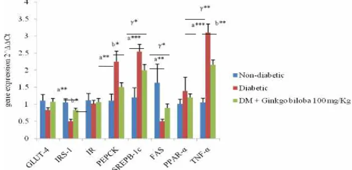
mRNA expression of genes: In order to understand the
underlying mechanisms that how GBE improved the
glucose tolerance in diabetic state, the mRNA expression of key genes were studied in liver, skeletal muscles and adipose tissues in rats. We studied glucose transporter-4 (GLUT-4) insulin receptor substrate-1 (IRS-1), Insulin receptor (IR), Phosphoenolpyrovate carboxykinase (PEPCK), Sterol regulatory element binding protein-1c (SREBP-1c), Fatty acid synthase (FAS), Peroxisome proliferator-activated receptor-α (PPAR-α), Peroxisome proliferator-activated receptor-γ (PPAR-γ) and tumor necrosis factor-α (TNF-α) in hepatic, skeletal muscle and adipose tissue.
mRNA genes expression in liver: For GLUT-4, IR,
PPAR-α, SREPB1-c and FAS no significant changes were observed in diabetic and GBE treated groups. In parallel, we found significant up-regulation (P<0.05) of IRS-1, and significant down-regulation of PEPCK (P<0.05) and TNF- α (P<0.001) in the D+GBE group (Fig 1).
mRNA gene expression in skeletal muscles: As shown
in Figure 2, significant up-regulation for GLUT-4 (P<0.001) and PPAR-α (P<0.001) in D+GBE treated group was recorded, whereas, same genes were down-regulated in diabetic rats.
mRNA gene expression in adipose tissues: As shown in
Fig 3, GLUT-4 and FAS were not affected in all the studied groups. However, significant decrease in IRS-1 (P<0.0001), IR (P<0.05) and PPAR-γ (P<0.001) in DG was found. The TNF-α showed, significant up-regulation (P<0.0001) in DG and significant (P<0.0001) down-regulation in D+GBE group.
Table 1. Genes and the primer sequences used.
Genes Forward primer (5'-3') Reverse primer (5'-3')
GAPDH TCCCATTCTTCCACCTTTGATGCT ACCCTGTTGCTGTAGCCATATTCAT
GLUT-4 GCTTCTGTTGCCCTTCTGTC TGGACGCTCTCTTTCCAACT
IRS-1 GCCAATCTTCATCCAGTTGC CATCGTGAAGAAGGCATAGG
IR GTGCTGCTCATGTCCTAAGA AATGGTCTGTGCTCTTCGTG
PEPCK AGTCACCATCACTTCCTGGAAGA GGTGCAGAATCGCGAGTT
SREBP-1c GGCATGAAACCTGAAGTGGT TGGGCTTTCACCTGGTTATC
FAS CGCCGTGGTGCTGGAGATTG CTTGCCGAGGTTGGTGAGGAAG
PPAR-α GAGACCCTCGGGGATCTTAG CGTCTTGTGTCCTGAGCTTG
PPAR-γ CTGACCCAATGGTTGCTGATTAC GGACGCAGGCTCTACTTTGATC
TNF-α GCAGAGCCTTCCAAGCCTACC GTTACCCAGCCCACCTCCTTTG
Abbreviations: GAPDH (Glyceraldehyde-3-phosphate dehydrogenase), GLUT-4 (Glucose transporter-4), IRS-1 (Insulin receptor
substrate-1), IR (Insulin receptor), PEPCK (Phosphoenolpyrovate carboxykinase), SREBP-1c (Sterol regulatory element binding protein-1c), FAS (Fatty acid sythase), PPAR-α (Peroxisome activated receptor-α), PPAR-γ (Peroxisome proliferator-activated receptor-γ), TNF-α (tumor necrosis factor-α).
Naseem et al., J. Anim. Plant Sci. 26(3):2016
Table 2. Effect of Ginkgo biloba Leaves extract on body weight, and blood glucose in non-diabetic (CG), diabetic Group(DG) and diabetic + Ginkgo biloba treated group (D+GBE) at week 1 and week 14 in rats.
Parameters CG DG D+GBE
Week 1 Week 14 Week 1 Week 14 Week 1 Week 14
BW (g) 165.36±1.51 206.20±2.36γ*** 165.93±1.41 143.75±2.20a*** 166.87±0.99 150.55±1.81b*** Blood glucose (mg/dl) 83.43±1.28 γ*** 83.68±1.35γ*** 417.07±25.43 a*** 485.58±25.52 a*** 410.5±19.65 227.38±10.89 b*** All values are presented as mean ± S.E.M.
γ***=P< 0.0001, Ginkgo biloba VS non-diabetic a***=P<0.001, diabetic vs non-diabetic b***=P<0.0001, ginkgo biloba vs diabetic Abbreviations: BW (Body weight)
Table 3. Effect of Ginkgo biloba Leaves extract on Fasting serum glucose, Creatinine, Urea, AST, ALT, anti-oxidative stress (CAT and MDA) and serum lipid profile (TC, C, LDL-C, HDL-C, TG and VLDL-TG) in non-diabetic (CG), diabetic Group(DG) and diabetic + Ginkgo biloba treated group (D+GBE) at week 14 in rats (n=8). Parameters CG DG D+GBE FSG (mg/dL) 83.68±1.35γ*** 485.58±25.52a*** 227.38±10.89b*** Creatinine (mg/dL) 1.63±0.12γ** 2.01±0.03a** 1.95±0.04 Urea (mg/dL) 18.70±0.37γ*** 173.8±3.98a*** 161.39±2.03b** AST (µ/L) 76.88±1.43γ*** 210.06±3.08a*** 209.43±1.56 ALT (µ/L) 36.23±0.72 40.9±1.68a** 36.88±0.19b**
CAT (KU/L) 20.83±0.26 18.60±0.65a*** 19.99±0.16b*
MDA (mmol/L) 6.54±0.23 7.55±0.28a** 6.95±0.22b*
TC (g/L) 0.82±0.01γ*** 1.34±0.01a*** 1.29±0.02 VLDL-C (g/L) 0.07±0.005γ*** 0.28±0.005a*** 0.29±0.005 LDL-C (g/L) 0.06±0.001γ*** 0.55±0.01a*** 0.54±0.01 HDL-C (g/L) 0.68±0.01γ*** 0.49±0.009a*** 0.54±0.008b** TG (g/L) 1.25±0.007γ*** 2.12±0.02a*** 1.56±0.01b*** VLDL-TG (g/L) 0.19±0.01γ*** 1.03±0.01a*** 0.75±0.006b***
All values are presented in mean±S.E.M
γ**=P< 0.001, Ginkgo biloba VS non-diabetic a*=P<0.05, diabetic vs nondiabetic b*=P<0.05, Ginkgo biloba vs diabetic
γ***=P< 0.0001, Ginkgo biloba VS non-diabetic a**=P<0.001, diabetic vs nondiabetic b**=P<0.001, Ginkgo biloba vs diabetic
a***=P<0.0001, diabetic vs non-diabetic b***=P<0.0001, Ginkgo biloba vs diabetic
Abbreviations: GBE (Ginkgo biloba extract), FSG (Fasting Serum Glucose), AST (Asparatate aminotransferase), ALT (Alanine aminotransferase), CAT (Catalase), MDA (Malondialdehyde), TC (Total Cholesterol), VLDL-C (Very Low Density Lipoprotein-Cholesterol), LDL (Low Density Lipoprotein-Lipoprotein-Cholesterol), HDL (High Density Lipoprotein-Lipoprotein-Cholesterol), TG (Triglyceride), VLDL-TG (Very Low Density Lipoprotein- triglyceride),
Fig 1. Effect of Ginkgo biloba Leaves extract (GBE) on mRNA gene expression of Glucose transporter-4 (GLUT-4), Insulin receptor substrate-1 (IRS-1), Insulin receptor (IR), Phosphoenolpyrovate carboxykinase (PEPCK), Sterol regulatory element binding protein-1c (SPEPB1-c), Fatty acid synthase (FAS), Peroxisome proliferator-activated receptor- α (PPAR-α), Peroxisome proliferator-activated receptor- γ (PPAR-γ) and tumor necrosis factor-α (TNF-α) in the liver of alloxan induced diabetic male Wistar rats. All values are mean ± S.E.M ; n=8. γ*P<0.05, γ**P< 0.001 non-diadetic vs Ginkgo biloba, a**P<0.001 and a***P<0.0001 diabetic vs non-diabetic, b*P<0.05 and b**P<0.001 Ginkgo biloba vs diabetic group.
Fig 2. Effect of Ginkgo biloba Leaves extract (GBE) on mRNA gene expression of GLUT-4 (Glucose transporter-4), IRS-1 (Insulin receptor substrate-1), IR (Insulin receptor), PPAR-α (Peroxisome proliferator-activated receptor- α), TNF-α (tumor necrosis factor-α), in the muscles of alloxan induced diabetic male Wistar rats. All values are mean ± S.E.M ; n=8. γ* P<0.05 and γ*** P<0.0001 non-diadetic vs Ginkgo
Naseem et al., J. Anim. Plant Sci. 26(3):2016
Fig 3. Effect of Ginkgo biloba Leaves extract (GBE) on mRNA gene expression of GLUT-4 (Glucose transporter-4), IRS-1 (Insulin receptor substrate-1), IR (Insulin receptor), Fatty acid synthase (FAS), PPAR-γ (Peroxisome proliferator-activated receptor- γ) and TNF-α (tumor necrosis factor-α) in the adipose tissues of alloxan induced diabetic male Wistar rats. All values are mean ± S.E.M ; n=8. γ**P<0.001 and
γ***P<0.0001 non-diadetic vs Ginkgo biloba, a* P<0.05, a** P<0.001 and a***P<0.0001 diabetic vs non-diabetic, b***P<0.0001 Ginkgo biloba vs diabetic group.
DISCUSSION
Diabetes is a global health problem, becoming more prevalent in the coming few decades (Wild et al., 2004). Diabetes is a pathological condition which is associated with oxidative stress (Ebuehi et al., 2010). In fruits, vegetables and plants phenolic compounds such as flavonoids and phenolic acid are widely distributes have the ability to scavenge active oxygen, hydroxyl radicals and superoxide by donating the electron. The flavonoids present in G. biloba leaves extract, exert their action by chelating or scavenging process in anti-oxidant mechanism and terpenes inhibit platelet activation factor (Maltas and Yildiz, 2012). These days, GBE gains attention of many researchers because of its beneficial anti-diabetic effects in diabetic subjects. Many authors reported a daily single dose of 120 mg of ginkgo leaf extracts in the clinical trials (Kudolo, 2001; Kudolo et al., 2006). The aim of the present research is to check the effect of G. biloba leaves extract, on alloxan induced diabetes and associated metabolic disturbances in rat.
We found significant reduction in the BW in diabetic rats, but although the BW was less diminished, it was normalized by GBE treatment. Our results for the reduction of BW in alloxan induced diabetic rats are in agreement with previous data (Ebuehi et al., 2010; Cheng
et al., 2013). Mechanisms involved in BW reduction in
diabetic condition were reviewed by Swanston-Flatt et
al., (1990) and Baynes (1991).
We found significant reduction in hyper-glycemic condition concentration in diabetic rats treated with GBE. Other researchers also showed reduction in glycemia with GBE treatment (Shankar et al., 2005; Zhou
et al., 2011; Cheng et al., 2013; Ren et al., 2013). This
blood glucose reduction could be related either to improvement in the plasma insulin concentration due to
positive influence of flavonoids on the pancreatic β-cells
or to enhancement in the blood glucose transport to the peripheral tissues (Cheng et al., 2013). To further understand the mechanisms that how GBE act as hypoglycemic agents, we investigated the mRNA gene expressions involved in the carbohydrate/glucose and lipid metabolic activities.
For GLUT-4 expression, significant up-regulation was found only in skeletal muscles after treatment with GBE. Since the expression of GLUT-4 in the skeletal muscles are regulated by metabolism and
insulin, thus any dysfunctioning of pancreatic β cells as
the case occur in diabetes also impaired the expression of GLUT-4 (Zorzano et al., 2005). Lee et al., (2010) found increase in GLUT-4 protein expression in whole cell lysates in adipose tissues and muscles in mice after treatment with nobiletin flavonoids.
Our data showed significant increase in the expression of IRS-1 only in hepatic tissue after GBE
treatment. Zhou et al., (2011) found significant increase in the expression of IRS-2 in hepatic cells but they did not measure the expression for IRS-1. The mRNA gene expression of IR in the liver did not vary among the groups as did the mRNA expression of IRS-1, indicating that insulin signaling seems little or no effect in liver, since we found no improvement in the expression of IR in any of the studied organ. Evidences show an increase in IR number and mRNA level of the IR in the liver, muscles and adipose tissue in the diabetic rats (Amessou
et al., 1999). Despite of increase in blood insulin
concentration, increased in PEPCK expression in diabetic animal models were found (Law et al., 2002) as also in our case. Other researchers also found down-regulation for PEPCK after treatment with different flavonoids (Cederroth et al., 2008; Herrera et al., 2013).
The dyslipidemia associated to diabetes condition; an effect of GBE treatment on dyslipidemia was also checked in this study. Although GBE treatment showed no effect on TC, VLDL-C and LDL-C, however,
GBE normalized HDL-C, TG and VLDL-TG
concentrations in diabetic rats. Dyslipidemia (high TG and Low HDL-C) is commonly observed in diabetes (Briones et al., 1984) and this appears directly correlated with an increased risk of coronary heart disease (Forte et
al., 1994). Ebuehi et al., (2010) found significant increase
in TC and TG concentration in alloxan induced diabetic rats. Kudolo (2001) found non-significant reduction in TC, TG and LDL-C; however they reported no change in the values of HDL-C after treatment with ginkgo in the non-insulin dependent diabetic mellitus (NIDDM) patients. We studied the genes involved in the lipid metabolism to better understand that how GBE plays a role to correct the dyslipidemia and hypertriglyceridemia in diabetic state.
SREBP-1c plays a pivotal role in lipolytic genes that regulated by insulin and glucose (Yuan et al., 2011). Lipid homeostasis is regulated by SREBPs and directly regulates the expression of more than 30 genes involved in the synthesis and uptake of the TC, TG, phospholipids and FAs (Horton et al., 2002). We reported no change in the expression of hepatic SREBP-1c in treated group and the results are not in agreement with Zhou et al., (2011). The mRNA expression of FAS is markedly down-regulated under metabolic complications, like insulin resistance and diabetes in liver (Real et al., 2010), as also reflected by our data. Flavonoids down-regulates the mRNA expression of FAS (Wu et al., 2011).
Our data showed that G. biloba improved lipid metabolism, through up-regulation of PPAR-α in muscles. PPAR-α also well characterized to increase the circulating amount of HDL level in accordance with
measured increase of HDL-C in our study.
Dysfunctioning of PPAR-α results in TC and TG metabolic abnormalities (Yoon et al., 2003). PPAR-γ is reported to control insulin sensitivity (Zhou et al., 2011);
they also reported up-regulation of PPAR-γ expression in hepatic cells after treatment with ginkgo leaves extract.
In the present study, we found the GBE helps the body to overcome the oxidative stress in diabetic state, by reducing MDA and enhancing CAT level in the body. MDA, the final product of lipid break down, is an important biomarker of oxidative stress in the body (Ren
et al., 2013) and one of the important indicators used for
the free radicals induced lipid peroxidation (El-Khayat et
al., 2011). Our data are in accordance with that reported
by Ren et al., (2013) and Cheng et al., (2013). This effect could be attributed to the anti-oxidant properties of flavonoids (Shankar et al., 2005; DeFeudis and Drieu, 2000) heavily present in ginkgo (Smith and Luo, 2003; Maltas and Yildiz, 2012).
Recent data showed that oxidative stress and inflammation play major roles in the onset and development of chronic diseases (Camps and Garcia-Heredia, 2014) and that inflammatory state exacerbate insulin resistance (Babish et al., 2010). We measured significant down-regulation for TNF-α only in liver and adipose tissue in effect with GBE treatment. There is no data for the tissues for TNF-α treated with G. biloba to compare with our work. Lee et al., (2010) found non-significant down regulation of TNF-α in adipose tissues after treatment with nobiletin: flavonoid found in citrus fruits.
There are strong evidences that flavonoids reduce the blood urea and serum creatinine level in diabetic state (Renno et al., 2008). In clinical trial on (NIDDM) patients, Kudolo, (2001) found no change in AST and ALT levels after 3 months treatment with G.
biloba.
In conclusion, the leaves extract of G. biloba have strong anti-diabetic and anti-oxidative effects. It also led to improve glucose and lipid metabolism by acting on the expression of key genes.
Acknowledgments: The work described in this paper
was supported by Higher Education Commission (HEC), Pakistan and West Human Nutrition Research Center, France.
REFERENCES
Amessou, M., S. Bortoli, V. Liemans, M. Collinet, B. Desbuquois, S. Brichard, and J. Girard (1999). Treatment off streptozotocin induced diabetic rats with Vanadate and Phlorizin prevents the over expression of liver insulin receptor gene. Eur J Endocrinol. 140(1): 79-86.
Baynes, J. (1991). Role of oxidative stress in development of complication. Diabetes. 40: 405-412.
Briones, E.R., S.J.T. Mao, P.J. Palumbo, W.M. O’Fallon,
Naseem et al., J. Anim. Plant Sci. 26(3):2016
Analysis of plasma lipids and apolipoproteins in insulin-dependent and noninsulin-dependent diabetics. Metabolism. 33: 42-49.
Babish, J.G., L.M. Pacioretty, J.S. Bland, D.M. Minich, J. Hu, and M.L. Tripp (2010). Antidiabetic screening of commercial botanical products in 3T3-L1 adipocytes and db/db mice. J Med food. 13(3): 535-547.
Camps, J. and A. García-Heredia (2014). Introduction: Oxidation and Inflammation, A Molecular Link Between Non-communicable Diseases. Adv Exp Med Biol. 824(2): 1-4.
Cederroth, C.R., M. Vinciguerra, A. Gjinovci, F. Kühne, M. Klein, M. Cederroth, D. Caille, M. Suter, D.
Neumann, R.W. James, D. Doerge, T.
Wallimann, P. Meda, M. Foti, F. Rohner-Jeanrenaud, J.D. Vassalli, and S. Nef (2008). Dietary phytoestrogens activate AMP-activated protein kinase with improvement in lipid and glucose metabolism. Diabetes. 57(5): 1176-1185.
Cheng, D., B. Liang, and L. Yunhui (2013).
Antihyperglycemic effect of Ginkgo biloba extract in Streptozotocin-induced diabetes in rats. BioMed Res Int. 2013:1-7.
Chetiveaux, M., H. Nazih, V. Ferchaud-Roucher, G. Lambert, Y. Zair, M. Masson, K. Ouguerram, D.
Bouhours, and M. Krempf (2002). The
differential apoA-I enrichment of pre1 and
HDL is detectable by gel filtration separation.
J Lipid Res. 43: 1986-1993.
DeFeudis, F.V. and K. Drieu (2000). Ginkgo biloba extract (EGb 761) and CNS functions: basic studies and clinical applications. Curr Drug targets. 1(1): 25-28.
Ebuehi, O.A.T., A.E. Ajuluchukwu, O.T. Afolabi, and A.I. Akinwande (2010). Oxidative stress in alloxan-induced diabetes in female and male rats. Adv Int Med Dent Sci. 3(3): 71-75.
El-Khayat, Z., J. Hussein, T. Ramzy, M. Ashour, and F. Oraby (2011). Protective effect of Panax ginseng against streptozotocin induced renal dysfunction in rats. J App Sci Res. 7(10): 1419-1423.
El-Mesallamy, H.O., N.S. Metwally, M.S. Soliman, K.A.
Ahmed, and M.M.A. Moaty (2011). The
chemopreventive effect of Ginkgo biloba and
Silybum marianum extracts on
hepatocarcinogenesis in rats. Cancer Cell Int. 11(1): 38- 49.
Forte, T.M. and M.R. Mc-Call (1994). The role of apolipoprotein AI-containing lipoproteins in atherosclerosis. Curr Opin Lipidol. 5: 354-364. Herrera, C.I., M.A. Martin, L. Bravo, L. Goya, and S.
Ramos (2013). Cocoa flavonoids improve
insulin signaling and modulate glucose
production via AKT and AMPK in HepG2 cells. Mol Nutr Food Res. 57(6): 974-85.
Horton, J.D., J.T. Goldstein, and M.S. Brown (2002). SREBPs: Activation of the complete program of cholesterol and fatty acid synthesis in the liver. J Clin Invest. 109(9): 1125-1131.
Kudolo, G.B. (2001). The effect of 3-month ingestion of
Ginkgo biloba extract (EGb 761) on pancreatic
β-cell function in response of glucose loading in
individuals with non-insulin dependent diabetes mellitus. J Clin Pharmacol. 41(6): 600-611. Kudolo, G.B., Wang W., Javors M., and Blodgett, J.
(2006). The effect of the ingestion of ginko
biloba extract (EGb761) on the
pharmacokinetics of metformin in non-diabetic and type 2 diabetic subjects: a double blind placebo controlled, crossover study. Clin Nutr. 25(4): 606-616.
Kumar, R (2012). Correlation of Selenium and other antioxidants in diabetic patients with and without complications. Free Rad Antiox. 2(1): 6-8.
Law, W.M.E., X.L. Wang, B.K. Law, R.K. Hall, M.
Nawano, and D.K. Granner (2002).
Epigallocatechin gallate, a constituent of green tea, represses hepatic glucose production. J Biol Chem. 277(38): 34933-34940.
Lee, Y.S., B.Y. Cha, K. Saito, H. Yamakwa, S.S. Choi, K. Yamaguchi, T. Yonezawa, T. Teruya, K. Nagai, and J.T. Woo (2010). Nobiletin improves hyperglycemia and insulin resistance in obese diabetic ob/ob mice. Biochem Pharm. 79(11): 1674-1683.
Maltas, E. and S. Yildiz (2012). Evaluation of phytochemicals and antioxidant activity of Ginkgo biloba from Turkey. Pharmacologia. 3(4): 113-120.
Nayak, B.S. and L. Roberts (2006). Relationship between
inflammatory markers, metabolic and
anthropometric variables in the Caribbean type 2 diabetic patients with and without microvascular complications. J Inflamm. 3: 17-23.
Real, J.M.F., J.A. Menendez, M.M. Navarrete, M. Bluher, A.V. Martin, M.J. Vazquez, F. Ortega, C. Dieguez, G. Fruhbeck, W. Ricart, and A.V. Puig (2010). Extracellular fatty acid synthase: A possible surrogate biomarker of insulin resistance. Diabetes. 59(6): 1506-1511.
Renno, W.M., S. Abdeen, M. Alkhalaf, and S. Asfar (2008). Effects of green tea on kidney tubules of diabetic rats. Br J Nutr. 100(3): 652-659. Ren, M., S. Yang, J. Li, Y. Hu, Z. Ren, and S. Ren
(2013). Ginkgo biloba L. extract enhances the effectiveness of syngeneic bone marrow mesenchymal stem cells in lowering blood
glucose levels and reversing oxidative stress. Endocrinology. 43(2): 360-369.
Swanston-Flatt, S.K., C. Day, C.J. Bailey, and P.R. Flatt (1990). Traditional plant treatments for diabetes. Study in normal and streptozotocin diabetic mice. Diabetologia. 33(8): 462-464.
Smith, J.V, and Y. Luo (2003). Elevation of oxidative free radicals in Alzheimer's disease models can be attenuated by Ginkgo biloba extract EGb 761. J. Alzheimer’s Dis. 5(4): 287-300.
Shankar, P.K., V. Kumar, and N. Rao (2005). Evaluation of antidiabetic activitiy of Ginkgo biloba in Streptozotocin induced diabetic rats. Iranian J Pharmacol Ther. 4(1): 16-19.
Unger, M. (2013). Pharmocokinetic drug interactions involving Ginkgo biloba. Drug Metab Rev. 45(3): 353-385.
Wild, S., G. Roglic, A. Green, R. Sicree, and H. King (2004). Global prevalence of diabetes: estimates for the year 2000 and projections for 2030. Diabetes Care. 27(5): 1047–1053.
Wu, C.H., M.C. Lin, H.C. Wang, M.Y. Yuang, M.J. Jou, and C.J. Wang (2011). Rutin inhibits oleic acid induced lipid accumulation via reducing
lipogenesis and oxidative stress in
hepatocarcinoma cells. J Food Sci. 76(2): 65-72. Xia, L.Y., L. Tong-hua, H. Zong-tao, E.L. Juan, and W.
Li-li (2011). Research progress on the
mechanism of single Chinese medicinal herbs in treating diabetes mellitus. Chin J Integr Med. 17(3): 235-240.
Yeo, J., Y.M. Kang, S.I. Cho, and M.H. Jung (2011). Effects of multi-herbal extract on type 2 diabetes. Chin Med. 6(10): 1-10.
Yoon, M., M. Lee, S. Jeong, J.J. Kim, C.J. Nicol, K.W. Nam, M. Kim, B.G. Cho, and G.T. Oh (2003). Peroxisome proliferator-activated receptor α is involved in the regulation of lipid metabolism by ginseng. Br. J. Pharmacol. 138(7): 1295-1302.
Yuan, H.D., S.J. Kim, and S.H. Chung (2011). Beneficial effects of IH-901 on glucose and lipid
metabolisms via activating adenosine
monophosphate activated protein kinase and phosphatidylinositol-3 kinase pathways. Metab Clin Exp. 60(1): 43-51.
Zorzano, A., M. Palacin, and A. Guma (2005).
Mechanism regulating GLUT-4 glucose
transporter expression and glucose transport in skeletal muscle. Acta Physiol. Scand. 183(1): 43-58.
Zhou, L., Q. Meng, T. Qian, and Z. Yang (2011). Ginkgo
biloba extract enhances glucose tolerance in
hyperinsulinisim induced hepatic cells. J Nat Med. 65(1): 50-56.