DOCTORAT DE L'UNIVERSITÉ DE TOULOUSE
Délivré par :Institut National Polytechnique de Toulouse (INP Toulouse) Discipline ou spécialité :
Développement des Plantes
Présentée et soutenue par :
M. GUOJIAN HU le mardi 4 juillet 2017
Titre :
Unité de recherche : Ecole doctorale :
Hormonal and epigenetic control of dependent and
pollination-independent fruit-setting in tomato
Sciences Ecologiques, Vétérinaires, Agronomiques et Bioingénieries (SEVAB) Laboratoire de Génomique et Biotechnologie des Fruits (G.B.F.)
Directeur(s) de Thèse : M. MONDHER BOUZAYEN
M. MOHAMED ZOUINE Rapporteurs :
M. ANTONIO GRANELL, UNIVERSITAT POLITECNICA DE VALENCE M. JEAN-MARC DERAGON, UNIVERSITE DE PERPIGNAN
M. JORDI GARCIA MAS, IRTA BARCELONA Membre(s) du jury :
M. JEAN-MARC DERAGON, UNIVERSITE DE PERPIGNAN, Président Mme JACQUELINE GRIMA-PETTENATI, UNIVERSITE TOULOUSE 3, Membre
M. MOHAMED ZOUINE, INP TOULOUSE, Membre M. MONDHER BOUZAYEN, INP TOULOUSE, Membre
Hormonal and epigenetic control of
pollination-dependent and -independent
Acknowledgment
This thesis represents not only my own work, it is also contributed by those colleagues who took lots of time and effort in epigenetic research in GBF laboratory. The fire burns better when everybody adds wood to it. Great thanks to those remarkable individuals who add “woods” to support my PhD study in the last four years. I wish to dedicate this thesis to all of them for their constant support and encouragement.
First and foremost I would like to thank my supervisor, Professor Mondher
BOUZAYEN, director of the GBF Laboratory, for his wonderful mentorship throughout my graduate career. He has taught me, both consciously and unconsciously, how good scientific thinking is performed. I appreciate all his contributions of time, energy, ideas, and one-year extended funding to make my PhD experience productive and stimulating. The enthusiasm he has for his research was contagious and motivational for me, even during tough times in my PhD pursuit. For everything he has done for me, my academic, and countless hours spent revising all my written works, I am forever grateful.
I would also like to thank my second supervisor, Dr. Mohamed ZOUINE,
for his long-term supervision at my PhD research project. It was an unforgettable experience to work with him. His wide expertise in bioinformatics and clever mind stimulated me, and his guidance, advice, and support helped me all the time. I am grateful, forever.
The members in GBF laboratory have contributed immensely to my personal and professional time in lab. I am especially grateful to: Pierre FRASSE, for his huge time and effort in establishing the ChIP procedures
together with me; Elie MAZA, for his expert advices in performing statistical
analysis and Anis DJARI, for his skillful manipulation for massive NGS reads.
I would like to acknowledge Lydie Tessarotto-Leronnier, Dominique
ii
and phenotyping of tomato plants. I also would like to thank Yanwei Hao, Jun
Hye SHIN, Baowen Huang, Yi Chen, Jing An, Meiying LIU, Julien Pirrello, Isabelle Mila, Benoit Van-Der-Rest, Jean-Marc Routaboul, Christian Chervin and Clementine Dumont for their kind help and support throughout my PhD studies.
A very special gratitude goes out to China Scholarship Council for helping and providing the funding for my PhD work.
In regards to 4-years life in Toulouse, I sincerely thank Madame Brigitte
Lafforgue offering me great help selflessly in and out of the lab. With a special
respect to Monsieur Gérard BAUDOT, I very much appreciate his enthusiasm
and willingness to teach me French language and culture. I also thank to all my friends in Toulouse, for their help, support and friendship.
Lastly, I am grateful to my parents for their dedication supporting my academic career. Even though they have limited education and cannot speak English, they understood the value of education and try their best in supporting my pursuits. Thank you.
谨以此篇献给在我博士学习阶段给予我巨大帮助和支持的老师、同事、家人 和朋友们,感谢你们长期的理解,支持和鼓励。谢谢!
Guojian HU
GBF, INP-ENSAT
Publications
ArticlesHu G, Frasse P, Maza E, AnisDjari, Benhamed M, Zouine M, Bouzayen M. 2017. Histone marks repositioning is the main driver of the transcriptomic reprogramming underlying the ovary to fruit transition in tomato. (Submitted) Hu G, Frasse P, Maza E, AnisDjari, Benhamed M, Zouine M, Bouzayen M. 2017. Auxin triggering gene reprogramming underlying fruit-setting mainly in a similar way to natural pollination in tomato. (In preparation)
Hu G, Fan J, Xian Z, Huang W, Lin D, Li Z. 2014. Overexpression of SlREV alters the development of the flower pedicel abscission zone and fruit formation in tomato. Plant Science 229, 86–95.
Hao Y, Hu G, Breitel D, Liu M, Mila I, Frasse P, Fu Y, Aharoni A, Bouzayen M,
Zouine M. 2015. Auxin Response Factor SlARF2 Is an Essential Component of the Regulatory Mechanism Controlling Fruit Ripening in Tomato. PLoS Genetics 11.
Oral communication
Hu G, Frasse P, Maza E, Anis Djari, Zouine M, Bouzayen M. Epigenetic regulation of the flower-to-fruit transition in tomato. Annual meeting QualityFruit, Porto, Portugal (2016.10.06)
Poster
Hu G, Mila E, Frasse P, Bouzayen M, Zouine M. Tomato SlARF5 is critical for flower development. The 12th Solanaceae International Conference, Bordeaux, France (2015.10.25)
iv
Résumé
La transition fleur-fruit, appelée nouaison, est déclenchée par la pollinisation des fleurs et ce processus est essentiel pour cycle reproducteur des plantes, la formation des semences et le rendement de production. Les
mécanismes moléculaires contrôlant cette importante transition
développementale ont été peu explorés. Les marques histones et la méthylation de l'ADN sont les deux principaux modes de régulation épigénétique, mais à ce jour, leurs contributions respectives à la reprogrammation transcriptionnelle qui est associée au programme d’initiation des fruits charnus n’ont pas fait l’objet d’aucune étude sur aucune espèce de plante. Afin d’explorer l’importance dans la transition fleur-fruit de ces deux types de régulation épigénétique, des approches de transcriptomique "genome-wide", de ChIP-seq se et de séquençage bisulfite d'ADN ont été mises en place chez la tomate, une espèce économique majeure et un modèle d’étude pour les fruits charnus. Les résultats révèlent une corrélation étroite entre le repositionnement des marques histones et les changements observés de l'expression génique globale. L’étude montre aussi que les marques H3K9ac et H3K4me3 agissent en synergie pour activer la transcription génique, alors que la marque H3K27me3 a un effet répressif. A l’inverse, il n’y a pas de corrélation entre les variations de la méthylation de la cytosine et l’évolution des profils transcriptomiques. Il ressort donc que ce sont les changements au niveau des marques histones plutôt que de la méthylation de l'ADN qui constituent le moteur principal de la reprogrammation génétique associée au processus de transition fleur-fruit chez la tomate. En concordance avec cette idée, le niveau d'expression des gènes associés à l’initiation du fruit, tels que ceux liés au métabolisme hormonal, à la division cellulaire ou au développement embryonnaire, est corrélé avec les modifications des marques H3K9ac ou H3K4me3, mais pas avec la méthylation de l'ADN. En outre, l'étude comparative des profils transcriptomiques associés à la formation du fruit
dépendant et indépendant de la pollinisation révèle l'intervention complexe de multiples voies de signalisation hormonales. Au total, notre étude présente un nouvel aperçu du contrôle de la reprogrammation génétique nécessaire à l’initiation du développement du fruit et révèle le rôle important du contrôle épigénétique dans ce processus de transition développementale. Dans le même temps, l’étude identifie un groupe de gènes impliqués dans la régulation épigénétique qui offrent des cibles potentielles pour les programmes d’amélioration de la nouaison des fruits, un processus majeur affectant le rendement de production.
vi
Abstract
The flower-to-fruit transition, so-called fruit setting, is triggered by flower pollination and this process is essential for plant reproductive success, seed formation and crop yield. The underlying molecular mechanisms controlling this developmental transition remain unclear. Histone marking and DNA methylation are the main epigenetic modes for genetic reprogramming, however, their respective contribution to the fruit set-associated transcriptomic reprogramming is also unknown. To address the contribution of the two types of epigenetic regulation to fruit set, genome-wide transcriptomic profiling, ChIP-sequencing and DNA bisulfite sequencing were applied to tomato, a major economic crop and a model system for fleshy fruit. The study emphasizes the tight correlation between histone repositioning and gene expression changes revealing that H3K9ac and H3K4me3 histone marks synergistically promote gene transcription, whereas H3K27me3 marking has a repressive effect. We concluded that changes in histone marks rather than in DNA methylation are the main drivers of genetic reprogramming associated with the fruit set transition in tomato, and H3K9ac and H3K4me3 marking is the primary players in this control mechanism. Consistently, the expression level of fruit set-associated genes such as those related to hormone metabolism, cell division, and embryo development correlated with changes in H3K9ac or H3K4me3 marking, but not with DNA methylation. In addition, comparative study of transcriptomic profiling between pollinationdependent and -independent fruit set, uncovered the complex intervention of multiple hormone signaling pathways involved in the flower-to-fruit transition. Auxin appears as the central hormone triggering the extensive transcriptomic reprogramming associated with the initiation of early fruit growth. Altogether, the study provides new insight into the control of gene reprogramming underlying fruit the shift from flower to fruit and uncovers a set of genes encoding modifiers of epigenetic marks which may provide new targets for breeding programs aiming to improve fruit setting, a major process impacting crop yield.
摘 要
坐果是植物从花到果实的一个转变过程,它依赖于正常的授粉受精,同时也对种子的形 成和果实的产量等起决定作用,然而对调控该转变过程的分子机理却并不清楚。组蛋白修饰 和 DNA 甲基化是表观遗传调控基因重编程的两个主要方面,它们各自对坐果过程中的贡献 程度知之甚少。为了揭示该问题,我们选取果实模式作物--番茄作为研究对象,采用全基因 组水平上的转录组测序,组蛋白修饰的染色质免疫共沉淀测序 (ChIP-seq) 以及甲基化测序 (BS-seq)来进行系统分析研究。本论文印证了组蛋白修饰和基因表达之间的紧密联系,即 H3K9ac 和 H3K4me3 修饰能协同促进基因的转录,而 H3K27me3 修饰具有转录抑制作用。相 比于 DNA 甲基化修饰,组蛋白修饰是番茄坐果过程中调控基因重编程的主要驱动力,其中 H3K9ac 和 H3K4me3 在此方面尤为突出。同样地,与坐果紧密联系的基因比如激素代谢类, 细胞分裂类以及胚和胚乳发育类,都显现出是 H3K9ac 或 H3K4me3 修饰而不是 DNA 甲基化 修饰参与了基因表达的主要调控作用。此外,生长素能独立于授粉而触发坐果,本论文也通 过比较授粉坐果和生长素诱导坐果过程中的转录组变化来研究其内在的基因调控网络。结果 显示多种激素信号转导途径共同参与了两类主要坐果过程,而其中生长素是调节整个坐果过 程中基因重编程的主要调控信号。综上所述,本论文对番茄坐果过程中表观调控基因重编程 这一调节机制提出了更深的见解,同时此研究也揭示了潜在的表观修饰基因可能参与坐果形 成,这也为育种及提高作物产量提供了新的方向。viii
Abbreviations
ABA: Abscisic acidAFLP: cDNA–amplified fragment length polymorphism
AG: AGAMOUS
ARF: AUXIN RESPONSE FACTOR ATX1: ARABIDOPSIS TRITHORAX 1 Aux/IAA: Auxin/INDOLE-3-ACETIC ACID
BR: Brassinosteroid
CBF1: C-repeat/DRE binding factor 1 CDK: Cell Division protein Kinase ChIP-seq: Chromatin Immuno-Precipitation assays coupled to deep sequencing
CK: Cytokinine CLF: CURLY LEAF
CMT3: CHROMOMETHYLASE3 CNR: COLORLESS NON-RIPENING DA: Differentially Associated (histone marks)
DAO: Dioxygenase for Auxin Oxidation DDM1:
DECREASE-IN-DNA-METHYLATION1
DE: Differentially Expressed (genes) DMCs: Differentially Methylated sites DML: DEMETER-LIKE
DMR: Differentially Methylated Regions DPA: Days Post Anthesis
DRM1/2: DOMAINS REARRANGED METHYLTRANSFERASE1/2
EMF2: EMBRYONIC FLOWER2 ERF: Ethylene Response Factor FDR: False Discovery Rate
FIE: FERTILIZATION-INDEPENDENT ENDOSPERM
FIS2: FERTILIZATIONINDEPENDENT SEED2
GAs: Gibberellins
GID1: GA INSENSITIVE DWARF1 GO: Gene Ontology
GRF2: GROWTH REGULATING FACTOR 2
HAT: Histone acetyltransferases HDA1: Histone deacetylase 1 HDAC: Histone deacetylases HDMs: Histone demethylases HKMTs: Histone lysine
methyltransferases IAA: Indole-3-acetic acid IAM: Indole-3-acetamide IAMT: IAA-methyltransferase IAN: Indole-3-acetonitrile IAOx: Indole-3-acetaldoxime IPyA: Indole-3-pyruvic acid JA: Jasmonate acid
JHDMs: JmjC domain–containing histone demethylases
KAO: ent-kaurenoic acid synthase KO: ent-kaurene oxidase
LTRs: Long Terminal Repeat Retrotransposons
MEA: MEDEA
MET1: METHYLTRAFERASE1
MSI: MULTICOPY SUPPRESSOR OF IRA
MTases: Methyltransferases NOR: NON-RIPENING
NPA: 1-N-naphtylphtalamic acid PCR: Polymerase chain reaction PI: PISTILLATA
PIN: PIN-FORMED PM: Plasma Membrane
PRC2: Polycomb Repressive Complex 2
PRMTs: Arginine methyltransferases PSY1: PHYTOENE SYNTHASE PTMs: Posttranscriptional histone modifications
RIN: RIPENING INHIBITOR
ROS1: REPRESSOR OF SILENCING 1 RPD3: Reduced Potassium
Dependency protein 3 SA: Salicylic acid
SBP-box/SPL: SQUAMOSA Promoter
SEP: SEPALLATA
SIR2: Silent Information Regulator protein
SlIAA9: AUXIN/INDOLE3-ACETIC
ACID 9
SlTIR1: TRANSPORT INHIBITOR
RESPONSE 1
SOL: The International Solanaceae Genomics Project
SWN: SWINGER TAA: TRYPTOPHAN AMINOTRANSFERASE OF ARABIDOPSIS
TAG1: Tomato agamous 1 TES: Transcription End Site TEs: Transposable elements TFs: Transcription Factors
TIR1/AFB: TRANSPORT INHIBITOR RESPONSE 1/AUXIN SIGNALING F-BOX PROTEIN
TPL: TOPLESS
TSS: Transcription Start Sites UGT: UDP glucosyltransferase VRN2: VERNALIZATION2 WGBS: Whole Genome Bisulfite Sequencing
x
Table of Contents
Acknowledgment ... i Publications ... iii Résumé ... iv Abstract ... vi 摘 要 ... vii Abbreviations ... viii Chapter I ... 1I.1 Bibliographic review ... 2
1. The fruit set process ... 2
1.1 Tomato, a fruit crop with worldwide increasing importance ... 2
1.2 Tomato as a model plant for fleshy fruit research ... 4
1.3 Genomic resources on the tomato model ... 5
1.4 Micro-Tom: a miniature tomato with rapid life cycle ... 6
1.5 Tomato flower anatomy ... 7
1.6 Tomato fruit setting: a transition from flower to fruit. ... 8
1.7 Hormone signaling, a complex regulation network for fruit setting ... 11
2. Epigenetics ... 24
2.1 General introduction to chromatin dynamics ... 25
2.2 Posttranscriptional histone modifications in plant ... 27
2.3 DNA methylation in plant ... 44
2.3.1 RNA-directed DNA methylation (RdDM) ... 45
2.3.2 DNA methylation maintenance and dynamics ... 52
2.3.3 Reader of DNA methylation ... 56
I.2 Objective of the study ... 58
Chapter II Results and Discussion ... 63
Transcriptomic reprogramming underlying tomato fruit setting is associated with repositioning of histone marks rather than change in cytosine methylation. ... 64
Abstract ... 65
Results ... 69
Genome-wide transcriptomic profiling of the flower-to-fruit transition ... 69
A number of epigenetic regulation genes are differentially expressed during fruit set ... 72
Distribution of H3K9ac, H3K4me3 and H3K27me3 histone marks among tomato genes ... 74
H3K9ac and H3K4me3 marks positively correlate with actively transcribed genes, and H3K27me3 is associated with repressed genes ... 79
H3K9ac and H3K4me3 have an additive effect on gene expression ... 81
Change in histone marks underlying the flower-to-fruit transition ... 85
Gain or loss of histone marks correlates with changes in the expression of important fruit setting-related genes ... 86
Dynamics of DNA methylation during the flower-to-fruit transition ... 100
Discussion ... 103
Materials and Methods ... 107
Plant materials and sampling ... 107
Chromatin immunoprecipitation and sequencing... 107
Genome bisulfite treatment and sequencing ... 109
RNA Sample preparation and sequencing ... 109
RNA-seq data processing... 109
ChIP-seq data processing ... 110
BS-seq data processing ... 111
Identification of methylated cytosine sites and regions ... 111
Gene ontology analysis ... 112
Identification of putative orthologs in tomato genome ... 112
Accession Numbers ... 112
Supplemental Data ... 113
Acknowledgements ... 114
Author Contributions ... 114
Chapter II ... 115
Pollination-triggered and auxin-induced fruit setting in tomato involve similar transcriptomic reprogramming and epigenetic regulation ... 116
xii
Introduction ... 118
Result ... 121
Auxin induces pollination-independent fruit-setting ... 121
Both pollination and auxin induce massive changes in the expression of hormone-related genes. ... 128
Auxin induces significant change in histone marking similar to pollination induced fruit set. ... 133
Common set of differentially expressed genes regulated by pollination and auxin are prominently associated with changes in histone marking. ... 139
Multiple hormone signals associated with differential gene expression and epigenetic changes underlying the flower-to-fruit transition. ... 142
Transcriptional factors potentially for flower-to-fruit transition undergo expression change and epigenetic change. ... 149
Discussion ... 156
Material and Methods ... 159
Plant materials and sampling ... 159
Chromatin immunoprecipitation and sequencing... 159
RNA Sample preparation and sequencing ... 161
RNA-seq data processing... 161
ChIP-seq data processing ... 162
Gene ontology analysis ... 162
Identification of putative orthologs in tomato genome ... 162
Accession Numbers ... 163
Supplemental Data ... 163
Acknowledgements ... 164
Author Contributions ... 164
Chapter III ... 165
Changes in histone marking rather than in DNA methylation are the main drivers of the genetic reprogramming associated with the flower-to-fruit developmental transition ... 166
Auxin-induced fruit setting involves similar gene reprogramming than natural pollination in tomato ... 171
Main trends to follow ... 172
1
Chapter I
Bibliographic review
&
I.1 Bibliographic review
1. The fruit set process
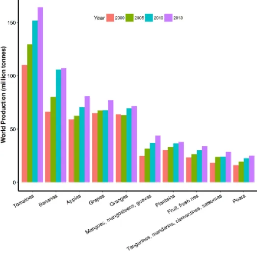
1.1 Tomato, a fruit crop with worldwide increasing importance The tomato is a fleshy fruit commonly considered as major vegetable crop that has achieved tremendous popularity over the last century. It is now widely grown in every country of the world - in outdoor fields, greenhouses and gardens. As shown in Figure 1, among all fruits, tomatoes represent the most important world production from 2000 to 2013.
3
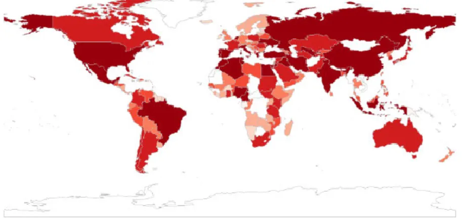
In 2013, more than 150 million tons of tomatoes were produced, far before bananas (~100 million tons) which is the second fruit crop. Asia produced 99.21 million tons in 2013, which accounts for 60% of world production (Table 1). The global distribution of tomato productions in the world (Figure 2) Shows that China is the biggest producer of tomatoes (51 million tons), followed by India with 18 million tons and United States of America with 13 million tons. The tomato can be divided into two categories: fresh market tomatoes, and processing tomatoes which are grown outdoors for the canning industry. In both cases, world production and consumption has grown quite rapidly over the past 25 years. In summary, the rapid increase of tomato production and consumption in the world points out to the economic importance of tomatoes.
In addition to meet the consumer preference in terms of taste, tomatoes contribute to human healthy diet as they are a good source of vitamins A and C, and anti-oxidant compounds like lycopene. Both vitamins are important for bone growth, immune system and blood vessels. Lycopene is also a very powerful antioxidant which can help prevent the development of many forms of cancer. Currently, tomato has a higher consumption rate in more developed countries although in developing countries the tomato is becoming a more important part of the food basket. Therefore, to match the need of the expanding market and the consumers, improving tomato yield and fruit quantity is becoming a major topic in scientific research.
Table1 World tomato production (million tons)
Area 2010 2011 2012 2013 Africa 18.24 17.31 19.12 19.18 Americas 24.45 24.04 24.96 24.59 Asia 87.13 94.84 96.32 99.21 Europe 21.71 21.65 20.99 20.97 Oceania 0.55 0.37 0.47 0.56 Total 152.08 158.21 161.86 164.49
Figure 2. Distribution of tomato production in global world in 2013. Unit: million tons
1.2 Tomato as a model plant for fleshy fruit research
Solanum is one of the largest angiosperm genera that includes annual and
perennial plants from diverse habitats. The tomato (Solanum lycopersicum) besides being an important Solanum crop, is widely used as a model system for studying flesh fruit development including fruit formation, fruit ripening, as well as metabolic pathways (Emmanuel and Levy, 2002; Carrari and Fernie, 2006; Sun et al., 2006; Egea et al., 2010; Mochida and Shinozaki, 2010; Pineda et al., 2010; Yin et al., 2010). Tomato is also a reference species for the Solanaceae family research, as it has a relatively small genome and contains a same haploid chromosome number and conserved genome organization with other Solanaceous plants, such as pepper, eggplant, potato and Nicotiana, (Rick and Yoder, 1988; Hille et al., 1989; Tanksley, 2004; Tomato_Genome_Consortium, 2012). Besides, the tomato genome has been sequenced by The International Solanaceae Genomics consortium (SOL) and large genetic resources are available. Moreover, a high number of studies on abiotic and biotic stress responses have been widely carried out in the tomato (Matsui et al., 2010; Rellán-Álvarez et al., 2010; Rivero et al., 2010; Uehara et
5
al., 2010). Therefore, the tomato has been widely selected as a core model system for fruit study.
1.3 Genomic resources on the tomato model
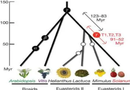
In 2012, a high-quality genome sequence of domesticated tomato ‘Heinz 1706‘ was produced (Tomato_Genome_Consortium, 2012). The predicted tomato genome size is approximately 900 megabases (Mb), of which 760 Mb were assembled and aligned to 12 chromosomes. Tomato chromosomes consist of pericentric heterochromatin and distal euchromatin. Most majority of repeats are located within and around centromeres, in chromomeres and telomeres, whereas genes are concentrated in euchromatin. Tomato genome is quite different from those of Arabidopsis and Sorghum since tomato has fewer high-copy, full-length long terminal repeat (LTR) retrotransposons with older average insertion ages (Figure 3, 2.8 versus 0.8 million years ago).
Figure 3. Speciation and polyploidization in eudicot lineages. (From
Tomato_Genome_Consortium, 2012)
The ITAG2.3 annotation predicted 34,727 protein-coding genes in tomato. Among these, 30,855 are detected by RNA sequencing (RNA-Seq) approaches from various tissues, and 31,741 show high similarity with Arabidopsis genes.
Furthermore, a comprehensive database named SGN (https://solgenomics.net/) was dedicated to all kind of tomato genomics tools and resources.
1.4 Micro-Tom: a miniature tomato with rapid life cycle
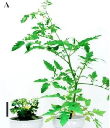
Micro-Tom is a miniature dwarf determinate tomato cultivar, released in 1989 by J. Scott and B. Harbaugh for gardening purposes and first used as a genetic tool 1997 (Reference by the Avraham Levi). Considering the relatively small genome of the tomato, Micro-Tom shares many features of Arabidopsis that make it a suitable model system, such as small size (8 cm when grown in 50-100 mL pots, Figure 4) and short life cycle (70-90 days from sowing to fruit-ripening).
Figure 4. Comparison of growth criteria in Micro-Tom and M82. Appearance of
80-day-old Micro-Tom (left) and M82 (right) plants from a side view. (Saito et al., 2011)
Thus, it is possible to grow a large population of micro-Tom lines using reduced spaces and to obtain more generations in less time. Besides, micro-Tom is also easily transformed with T-DNA by Agrobacterium-mediated
7
transformation of cotyledons and hypocotyls. Therefore, together with these features, it allows the use of micro-Tom for large scale mutagenesis.
1.5 Tomato flower anatomy
Tomato plant is day neutral and set flowers under conditions of either short or long days, which promotes fruit setting at multiple latitudes. Generally, 4-7 weeks after sowing, tomato plant grows into reproductive stage. Flowering is the period between floral initiation and production of mature flower. This process takes about 2 weeks, during which the number of carpels and the shape of the fruit are determined. The tomato flower is organized in four whorls of organs which are showed below (Figure 5).
Figure 5. Tomato flowers typically consist of six stamens attached to the corolla
tube, with the anthers partly fused to form a cone like structure surrounding the female pistil.
The peduncle (also called pedicel) is the stem that supports the flower. The outermost whorl consists of the sepals. Collectively, the sepals are called the calyx. The next whorl, the bright yellow petals, serves to attract pollinators. The male reproductive organs, the stamens, which bear pollens, sit inside the petals. A single tomato stamen consists of two elongated compartments. The individual stamens are fused together to form a yellow cylinder that surrounds the carpels.
The tomato carpels are green. They vary in number from cultivar to cultivar, but they are invariably fused together into a single bulb-like structure—ovary at very early stage of floral development. Thus, the number of carpels in the tomato flower determines the number of locules in the fruits. When the ovary is fertilized by pollen, the ovules develop into seeds.
1.6 Tomato fruit setting: a transition from flower to fruit. 1.6.1 Preparation phase
The formation of tomato fruit occurs upon a proper development of reproductive organ after floral bud initiation. At eight days after floral bud initiation, reproductive development occurs with microspoangia (for male part) and placental development (for female part). Afterwards, the style and the ovary grow nearly equally in length, and ovule primordia are emerging from the placental tissues. In the following 3~4 days, the megasoprocyte undergo two meiotic divisions, representing the first stage of megagametogenesis. Fourteen days after floral bud initiation, the integument enveloped the nucellus completely and the micropyle is well defined. At the same time, the embryo sac development is taking place. The presence of the megaspore at the chalaza end of the ovule indicates the development of the egg apparatus. In general, approximately sixteen to nineteen days after bud initiation, the flower is ready for fertilization, which triggers the completion of ovule development and the maturation of the well distinguished vegetative and generative cells (Xiao et al., 2009).
1.6.2 Phase I: Fertilization and fruit setting
Flower opening marks the start of the first fruit landmark. At the time of anthesis, the anther lobes undergo dehiscence to release the pollen, which germinates soon after landing on the receptive stigma. Pollen tubes grow close
9
to the base of the style 6 hours after pollination, and reach the ovules approximately 2 hours later. Ten to twelve hours after pollination, the pollen tubes had released their content resulting in fertilization of the ovules which represents fruit development landmark 2. Upon pollen tube reaching the embryo sac, it delivers one sperm to the egg cell to form a diploid embryo and another sperm to central cells to form a triploid endosperm. The fertilized ovule will become a seed later on. Nearly two days after anthesis, senescence of petals, stamens and style is associated with successful fertilization. If the flower is not fertilized successfully, it separates from the plant at the abscission zone, this causes the flower to wither and die (Gillaspy, 1993).
1.6.3 Phase II: Cell division, embryo and pericarp development Following successful fertilization, cell division is activated in the ovary (Figure 6). This phase lasts 3~6 days after fertilization, at which it represents 4-16 embryo cell stage (the third landmark of fruit, Xiao et al., 2009). During this phase, the first embryo division occurs approximately 4 days post-anthesis. In the following, the embryo develops to globular embryo stage (4th landmark,
6~10 dpa), heart shape embryo stage (5th landmark, 10~12dpa), torpedo stage
(6th landmark, 13~16dpa) and coil stage (7th landmark, ~20dpa). At the same
time of phase II, cell division also occurs in pericarp and other tissues. In the very early stage of phase II, cell division activity is highest in pericarp and placental tissues (Spurr, 1959; Gillaspy, 1993). In pericarp, the width doubled from 0dpa to 2dpa, and then further doubled at 5dpa and again at 10dpa. Across the pericarp, the numbers of cell layers increase from 10 at 0dpa to 17 at 2dpa, while at the later stage, cell division is mainly confined to the outer layer. And at 5dpa, cell number reaches the final number of 19-21, suggesting that pericarp cell division may finish at or before 5dpa. High mitotic activity was also observed in the cells at peripheral integument layers but not in the fertilized embryos at early stage of phase II, and within the columellar and placental
tissues as well. These vascular tissues and developing embryo show mitotic activity which continues till the end of phase II.
Cell expansion occurs concurrently with cell division in the pericarp of the early developing fruit. Comparing to the short stage of cell division (5dpa in LA1589), cell expansion and elongation span fruit development from 2dpa until mature green stage. At phase II, the dividing pericarp cells in the developing fruits are small, condensed, and abundant in cytoplasmic substances and contain small vacuoles. As cell enlarges, the primary cell wall and the cytoplasmic layer become relatively thinner, and vacuoles occupy a greater proportion of the cell volume.
As indicated above, embryo continues to divide and differentiate. In normal fruit, generally, the developing embryo controls the rate and sustenance of cell division in the surrounding fruit tissues. Because number of fertilized ovules generally determines the initial growth rate of the ovary, i.e. the rate of cell division (Gillaspy, 1993). When ovules do not develop normally and equably, lopsided fruits will form with presence or absence of seeds at different locules. It also indicates the positive correlation between the number of the developing seeds and sustained fruit growth.
1.6.4 Phase III: Cell expansion and embryo maturation
After the period of cell division, fruit growth is due mostly to an increase in cell volume. This phase lasts six to thirty days after fertilization until the fruit grows to the mature green stage. Cell expansion (cell volume) makes the greatest contribution to the final size of the fruit. In tomato, cell size can increase by more than 10 times in placenta, locular tissue and mesocarp after fertilization. This cell expansion in the fruit tissues does not keep the same rate as the developing of embryo, which does mainly divide and differentiate into seed tissues. During this phase, the embryo develops into a bilateral embryo that has well-formed cotyledons and an established root-shoot axis.
11
Figure 6. Overview of tomato fruit development. The first stage is fruit set, the initiation
of fruit growth after the flower has been successfully pollinated and fertilized. After fertilization, cell division takes place, which lasts up to 14 d. This period is followed by 6–7 weeks (wk) of mainly cell expansion, during which the volume of the fruit rapidly increases. Once the fruit has reached its final size it starts to ripen. (A, E) Flower and micrograph of an ovary at anthesis, awaiting pollination. Bar=200 μm. (B, F) Fruit of 0.8 mm in diameter, 10 d after pollination, and a micrograph of its pericarp. Bar=200 μm. (C, G) Fruit of 3 cm in diameter, 5 weeks after pollination, and a micrograph of its pericarp. Bar=200 μm. (D) Ripe tomato fruit. P, pericarp; op, outer pericarp; ip, inner pericarp; pl, placenta; o, ovules. (de Jong et al., 2009a)
1.7 Hormone signaling, a complex regulation network for fruit setting
The flower-to-fruit transition, fruit set, is very sensitive to environmental conditions. Thus, understanding the underlying mechanisms that regulate this process is crucial for maintaining yield production of fruit crops. Fruit set depends on the successful pollination and fertilization of the flower (Gillaspy, 1993). After double fertilization, the embryo and surrounding tissues generate the first signals that trigger fruit growth. In the early 20th century, phytohormone
such as auxin and gibberellin have been found to be critical signals that can induce the fruit set and growth independently from pollination (Gustafson, 1936; S. H et al., 1957). In recent years, several regulators involved in these hormone pathways were identified as major players of fruit setting.
1.7.1 Auxin and fruit setting
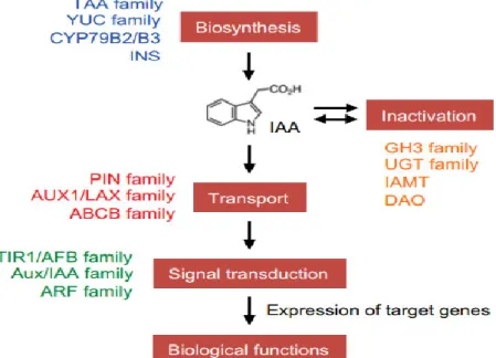
Gustafson (1936) firstly demonstrated that application of substances similar to auxin onto the stigma of the flower in tomato and in several other species can stimulate ovary development into seedless (parthenocarpic) fruit. The application of pollen extracts to the surface of the ovary also showed similar results, thus, it is hypothesized that pollen grains contain substance similar to the plant hormone auxin and that after pollination, the pollen may transfer this substance to the ovary to trigger fruit growth (Gustafson, 1937). In the following years, the role of auxin in fruit development has been firmly established by many researchers (Jones et al., 2002; Srivastava and Handa, 2005; Wang et al., 2005; Goetz et al., 2006; Serrani et al., 2008; Sorefan et al., 2009; de Jong et al., 2009b). Direct evidences demonstrating that auxin triggers fruit set at the physiological, biochemical and molecular levels have accumulated in the last decade. These studies addressed all aspects of auxin activity, including auxin biosynthesis and metabolism, polar auxin transport, perception and signal transduction, and auxin response (Figure 7).
Figure 7. A schematic model of auxin biosynthesis, inactivation, transport, and signal
13
glucosyltransferase, IAMT: IAA-methyltransferase, and DAO: dioxygenase for auxin oxidation (Kasahara, 2015).
1.7.2 Auxin biosynthesis and metabolism
It has hypothesized that the seeds are a probable source of de novo auxin biosynthesis during fruit growth based on the observation that in the majority of angiosperm species, the formation of seeds is closely linked to fruit growth. It was shown in diverse range of species that higher concentrations of indole-3-acetic acid (IAA) was detected in the seeds compared to other fruit tissues (Varga and Bruinsma, 1976). The natural auxin indole-3-acetic acid (IAA) is synthesized through both tryptophan (Trp)-dependent and Trp-independent pathways (reviewed by Pencík et al., 2013; Tivendale et al., 2014). Five major naturally occurring compounds are recognized as IAA precursors (Figure 8), including pyruvic acid (IPyA), acetaldoxime (IAOx), indole-3-acetonitrile (IAN), indole-3-acetamide (IAM), and indole-3-acetaldehyde (IAAld). The IPyA pathway is considered as the “main” auxin biosynthetic pathway (reviewed by Zhao, 2012), in which tryptophan is converted to IPyA by TAA enzymes and then to IAA by YUCCA enzymes (Cheng et al., 2006). Several studies in distinct species confirmed that high expression of TAA and YUCCA genes in seed tissues is correlated with auxin accumulation, suggesting that this pathway is also likely dominant in fruit. In Arabidopsis embryos, TAA1 and some YUCCA genes showed overlapping expression profiles, and loss of TAA1 and YUCCA lead to developmental defects (Cheng et al., 2006; Cheng et al., 2007; Stepanova et al., 2008). In tomato, YUCCA family (known in tomato as toFZY), particulary toFZY6, is preferentially expressed in seed tissues, suggesting similar dominant TAA-YUCCA pathway for tomato fruit growth (Expósito-Rodríguez et al., 2007; Expósito-Rodríguez et al., 2011).
Figure 8. Auxin biosynthesis and storage forms in higher plants.
Possible pathways for plant auxin biosynthesis. Solid arrows indicate those steps for which enzymes are known. Dashed arrows indicate those steps for which no enzyme has been identified or the enzyme identity is in question (Enders and Strader, 2015).
An additional mechanism in the control of auxin homeostasis is conjugation of IAA to amino acids by the auxin-inducible GH3 amido synthetases (Staswick et al., 2005). Although the mechanism is not clear, it seems to play complex roles in diverse plant developmental processes. GH3 expression is detected in many tissues, and also in some ripening fruit such as pepper, grape berries, apple and tomato. It has been suggested that IAA conjugation contributes to keep low concentration of free IAA during fruit development and ripening.
15 1.7.3 Polar auxin transport
Polar auxin transport is critical for auxin distribution and gradient formation in various tissues and organs. Generally, formation of this local auxin maxima and minima is important to control various aspects of plant growth and development (Grieneisen et al., 2007; Ljung, 2013). Auxin transport is controlled by AUX/LAX and PIN-FORMED (PIN) family proteins that direct cellular auxin influx and efflux, respectively (Figure 9).
Several studies in Arabidopsis suggest the critical role of auxin transport for reproductive development, especially that of PIN genes. Firstly, application of chemical inhibition of polar auxin transport through 1-N-naphtylphtalamic acid (NPA), severely affects the apical-basal patterning of the gynoecium. Benkova et al. (2003) suggested that early gynoecial primordium development is, similar to all other aboveground developing primordia, dependent on PIN1 activity (Benková et al., 2003). In support of this, Alvarez et al. (2009) showed that PIN1
is strongly expressed instage-7 and stage-8 gynoecia (Larsson et al., 2013).
The pin1 mutant (Okada et al., 1991) as well as pin3 pin7 (Benková et al., 2003) mutants have severely distorted gynoecia. The valve-length of these mutants is reduced concomitantly with enlarged style, stigma, and gynophore (Bennett et al., 1995; Sohlberg et al., 2006). Similar gynoecia defects have also been described when PINOID, which encodes an auxin-inducible serine–threonine protein kinase regulating the polarity of PIN1 localization, is compromised (Christensen et al., 2000; Benjamins et al., 2001; Friml, 2004; Sohlberg et al., 2006). In addition, PIN3 is expressed during fruit development and its localization is regulated at the valve margin to create a local auxin minium (Sorefan et al., 2009). Therefore, altogether, this suggests that auxin accumulation is critical for gynoecium development.
Figure 9. Auxin transport proteins regulate intracellular and cell to cell auxin fluxes
(Balzan et al., 2014). Auxin (IAA) crosses the plasma membrane (PM) through passive diffusion, as protonated form, or through PM transporters, as deprotonated form. PINs are efflux carriers located at the PM and ER and can be re-inserted in the lipid bilayer by recycling via the endocytic pathway. AUX/LAXs and PILs are influx carriers located at PM and ER, respectively. ABCBs are located at the PM and use energy from ATP to translocate IAA. The coordinated localization of the different transporters determines the overall directionality of the auxin flux and contributes to the regulation of intracellular auxin levels.
In tomato, gynoecium gives rise to the fleshy fruit organ. There are 10 PIN and 5 AUX/LAX genes in the tomato (Nishio et al., 2010; Mounet et al., 2012; Pattison and Catalá, 2012) and the investigation of their function during fruit development is starting to be investigated. The majority of PIN and AUX/LAX genes in tomato are expressed primarily in immature fruit and predominantly in the internal tissues between seed and pericarp (Nishio et al., 2010; Mounet et al., 2012; Pattison and Catalá, 2012; Pattison et al., 2015), however, their
17
specific function in fruit development still remains unclear. Transcriptomic profiling indicated that PIN1, PIN4, PIN7, PIN9, LAX1 and LAX2 genes are expressed mainly in the placenta tissues during fruit set (Pattison and Catalá, 2012; Pattison et al., 2015). Application of NPA in this placental tissues, results in increased auxin activity in the internal placental tissues where PIN expression is the highest. These data stresses the importance of the placental tissue for auxin transport in tomato fruit. Besides, RNAi silencing of tomato PIN4 in this tissue leads to parthenocarpic fruit formation, probably due to the accumulation of an excess of auxin in the ovary (Mounet et al., 2012). Except in placental tissues, PIN5 transcripts accumulated the highest in tomato embryo, endosperm and septum of 4DPA ovaries as shown by tissue-specific transcriptomic data. Considering the hypothesis that seed is the source of auxin synthesis, PIN5 might be an important efflux carrier for fruit set. In addition, within the same datasets, auxin influx carriers LAX1 and LAX2 are also relatively highly expressed in inner tissues. This different expression pattern of auxin transport genes suggests their indispensable roles for auxin accumulation and gradient formation for fruit formation.
1.7.4 Auxin signal transduction
Upon perception in the nucleus, auxin can trigger broad and specific transcriptional responses. The core components of the auxin signaling machinery belong to four protein families: the F-box TRANSPORT INHIBITOR RESPONSE 1/AUXIN SIGNALING F-BOX PROTEIN (TIR1/AFB) auxin co-receptors, the Auxin/INDOLE-3-ACETIC ACID (Aux/IAA) transcriptional repressors, the TOPLESS (TPL) co-repressors and the AUXIN RESPONSE FACTOR (ARF) transcription factors. Auxin promotes an interaction between TIR1/AFB and Aux/IAA proteins, resulting in degradation of the Aux/IAAs and the release of ARF repression (Wang and Estelle, 2014; Salehin et al., 2015). Gene expression associated with ARF activation has been implicated in diverse
processes in land plants, including tropic responses and the establishment of polarity, as well as embryogenesis and organogenesis in flowering plants, and both gametophyte and sporophyte development in nonflowering plants (Smet and Jürgens, 2007; Möller and Weijers, 2009; Prigge et al., 2010; Vernoux et al., 2010; Bennett, 2015; Flores-Sandoval et al., 2015; Kato et al., 2015). At the cellular level, auxin affects all aspects of cellular growth, including cell elongation, cell division and differentiation (Perrot-Rechenmann, 2010; Takatsuka and Umeda, 2014).
Auxin functions by triggering genome-wide transcriptional responses via its effects on ARF activity (Figure 10). At low auxin levels, Aux/IAA transcriptional repressors interact with ARFs and recruit TPLs to repress their activity. However, in the presence of auxin the TIR1/AFB proteins bind to Aux/IAA transcriptional repressors and mediate their polyubiquitylation and subsequent proteasomal degradation. Another important output of the pathway is the rapid induction of auxin-responsive genes, including Aux/IAAs and the GH3 family of auxin homeostasis modulators, triggering negative-feedback loops (Benjamins and Scheres, 2008).
In tomato, IAA9 is the first auxin signaling component identified that is required for fruit set (Wang et al., 2005). In situ hybridization, showed that IAA9 mRNA was detected across flower tissues, peaked at anthesis, and then its levels decrease in 3DPA ovary tissues. At anthesis, IAA9 transcript formed a gradient wherein the signal is strongest in ovule, sporogenous tissue, tapetum, petals, vascular bundles, developing style, placenta, and funiculus, but, relatively weak in sepals, ovary wall and the columella. Once the flower is fertilized, the IAA9 transcript gradient is dissipated, which leads to a decrease of transcript abundance in the placenta, funiculus and inner integument of embryonic sac. The down-regulation of IAA9 transcript levels in tomato plants resulted in a pleiotropic phenotype. The IAA9-antisense lines formed simple leaves instead of wild-type compound leaves, and fruit initiation occurs prior to pollination and fertilization. These phenotypes suggest that IAA9 acts as a
19
transcriptional repressor of auxin signaling for fruit set (Wang et al., 2005). Consistently with this, transcriptomic profiling of tomato ovary showed the down-regulation of IAA9 at three days post-pollination (Wang et al., 2009a). Besides, some other Aux/IAAs also showed changes after pollination, such as IAA2, IAA5, IAA14 and IAA18 (named by Vriezen et al., 2008). Among those, IAA2 and IAA14 were also found to increase in the pollinated ovary, specifically in the placenta and ovular tissues. This suggests that the newly synthesized Aux/IAAs are rapidly degraded via the SCFTIR1-mediated ubiquitination and
implies the wide activity of auxin after pollination.
In addition of Aux/IAA genes, ARF genes also play critical roles for fruit set. SlARF7, the homologue of Arabidopsis ARF7, is highly expressed in the placental tissues of the flower, and then rapidly decreases after pollination (de Jong et al., 2009b). Down-regulation of SlARF7 by RNAi strategy leads to parthenocarpic fruit formation, suggesting it is a negative regulator of fruit set. In Arabidopsis, ARF8 was detected in sepals, anthers and carpels prior to anthesis. At anthesis, expression of ARF8 was strong within the mesocarp layers of the fruit, and in the septum and ovules. After fertilization, ARF8 protein declined in embryo and endosperm compartments (Goetz et al., 2006). This expression pattern is similar to that of SlARF7 in tomato, and interestingly, parthenocarpic phenotype was also observed in arf8 mutant, suggesting that SlARF7 and Arabidopsis ARF8 may shares analogous function in fruit setting. However, arf8 mutant allele contains a mutation in the putative translation initiation codon, which may result in a truncated protein. Introduction of this aberrant form of ARF8 in tomato also results in parthenocarpic fruit set (Goetz et al., 2007), indicating that SlARF8 may compete with endogenous SlARF8 or interfere with SlARF7 protein in the formation of protein complexes. However, overexpression of SlARF8 in tomato also leads to parthenocarpic fruit formation, suggesting that SlARF8 may function in a different way than SlARF7. In addition, overexpression of the stable form of SlARF10 (35S:mSlARF10) that escapes miR160-targeted degradation, also leads to seedless fruit (Hendelman et al.,
2012). Moreover, instead of large functional seeds, the transgenic fruit contained extremely tiny and soft seed-like structures that could not germinate. Further inspection suggests that this impaired seed development is probably due to perturbation of a post-fertilization process. This study indicates that ARF repressors also play critical roles in fruit setting.
Besides, parthenocarpy has also been observed when auxin perception is affected. Overexpression of the tomato SlTIR1 altered the content of numerous auxin-responsive genes and resulted in parthenocarpy, demonstrating the importance of auxin perception in tomato fruit set (Ren et al., 2011).
Figure 10. A model for the TIR1/AFB-mediated auxin signaling pathway. (A) DIII/IV
regions of Aux/IAAs and ARFs are homologous and the DIII/IV of most Aux/IAAs and ARFs form a PB1 domain that comprises both an acidic (+) and a basic (-) face. (B) When auxin level is low, Aux/IAA proteins and ARFs form multimers through directional interaction between the acidic and basic interfaces of their DIII/IV regions. Aux/IAAs in the multimers
21
recruit co-repressor complexes, which consist of TPLs and HDACs and repress transcription of target promoter through removing acetyls (Ac) from local chromatin. In addition, Aux/IAAs in the multimers may block ARFs from efficient binding to AuxREs in their target promoters. When auxin level is high, auxin promotes ubiquitination and degradation of Aux/IAAs through a SCFTIR1/AFB-proteosome module, and the released ARFs form dimers and even higher-order complexes that activate expression of target genes. (Wang and Estelle, 2014)
1.7.5 Gibberellin signal pathway for fruit set
Wittwer et al (1957) showed that a second type of growth substance, gibberellins (GAs), can also trigger parthenocarpic fruit formation. Right after this finding, several gibberellin-like plant hormones were identified in different families of flowering plant, leading to the assumption that GAs are also involved in early stages of fruit development. So far, remarkable advances have been made in the past few years with regard to how GA biosynthesis, response and signaling pathway influences fruit initiation (Hu et al., 2008; Rieu et al., 2008; Dorcey et al., 2009; Mariotti et al., 2011; Carrera et al., 2012; Fuentes et al., 2012; Garcia-Hurtado et al., 2012). Application of active gibberellins (GA1 and GA3) in the absence of pollination can induce fruit set in several horticultural species and in Arabidopsis (Gillaspy, 1993; Dorcey et al., 2009). While application of GA inhibitors resulted to restricted fruit growth (Serrani et al., 2008). The increased hormonal content in parthenocarpic plants suggests that endogenous GA concentration in developing ovaries is the limited factor controlling fruit development. Further, blocking GA inactivation by knocking-out of the five GA inactivating enzymes, GA 2-oxidases, also leads to the formation of parthenocarpic fruits (Rieu et al., 2008). GA biosynthesis enzymes (GA 20-oxidase and GA 3-20-oxidase) are required for silique elongation (Hu et al., 2008; Rieu et al., 2008) and alternation of active GA form (GA4) in tomato by
overexpression of citrus CcGA20ox1 also triggers fruit growth without pollination (Garcia-Hurtado et al., 2012). However, in tomato plants with constitutive repression of SlGA20ox1, the ovaries remained fertile and developed normally after cross-pollination with wild-type pollen, but exhibited
parthenocarpic trait (Olimpieri et al., 2011). Moreover, the individual silencing of SlGA20ox1, SlGA20ox2, and SlGA20ox3 genes still have no effects on fruit set (Xiao et al., 2006), suggesting that more than one GA20ox gene is required for controlling fruit set in tomato. Taken together, these studies indicate that gibberellin accumulation is critical for fruit set.
The GA signal transduction pathway (Figure 11) requires the recognition of GA by its receptor called GA INSENSITIVE DWARF1 (GID1). The GID1–GA complex interacts with the nuclear repressor DELLA leading to its ubiquitin-dependent proteolytic degradation through the 26S proteasome. When there is no GA, DELLA proteins repress the gibberellin responsive genes. While the presence of GA3 stimulates the degradation of DELLA, and then, releases the repression from DELLA to initiate GA signal transduction. Reducing the mRNA level of SlDELLA gene in tomato induced very similar phenotypes to gibberellin-induced fruit and CcGA20ox1-overexpressing fruits (Martí et al., 2007), as fruits were facultative parthenocarpic, smaller in size, and elongated in shape. This indicates that reducing DELLA mRNA levels of may release the repression of downstream proliferating factors involved in the gibberellin signal pathway, which are normally induced after successful pollination and fertilization. Therefore, SlDELLA is a negative regulator for fruit set by restraining the gibberellin signal and thereby preventing ovary growth prior to pollination and fertilization.
23
Figure 11. A model for the gibberellin signaling pathway (Lor and Olszewski, 2015).
In tomato, fruit set is partly mediated by GAs following a complex but yet unknown hormonal cross-talk with auxin. However, in despite of the advances that shed some light on the regulation of fruit setting, to date, the molecular mechanisms by which these hormones regulate fruit set and development are still poorly understood. Transcriptomic analysis showed that in GA-induced ovaries, some auxin signalling genes are affected (Vriezen et al., 2008), suggesting that auxin may act prior to or independently of GA. In support of this hypothesis, Serrani et al., (2008) showed that application of IAA or 2,4-D together with GA synthesis inhibitors (PCB) can significantly reduce parthenocarpic fruit setting and growth, suggesting that the effect of auxin was mediated by GA. Indeed, parthenocarpic fruits induced by 2, 4-D had higher levels of active GA1 than unpollinated ovaries. Also, 2,4-D can alter GA metabolism in vivo, and the transcript levels of genes encoding copalyldiphosphate synthase (SlCPS), SlGA20ox1, SlGA20ox2, and SlGA20ox3, and SlGA3ox1 are higher in 2, 4-D treated ovaries, while transcript levels of GA-inactivating enzyme SlGA2ox2 are lower. These results suggest that auxin-induced fruit-set in tomato is mediated partially by gibberellins.
Furthermore, the silencing of SlARF7 in tomato affected part of the auxin signaling response pathway, and resulted in enhanced GA signaling. However, the levels of GA were strongly reduced, suggesting that SlARF7 also acts as a modifier of the GA response during the early stages of tomato fruit development.
2. Epigenetics
The term epigenesis means "extra growth", taken directly from Koine Greek ἐπιγέννησις, used in English since the 17th century. Later, "epigenetics" is coined by C. H. Waddington in 1942 as a portmanteau of the words epigenesis and genetics. Figure 12 describe the major events in epigenetics in the last three centuries. In 2008, a consensus definition of the epigenetic trait, "stably heritable phenotype resulting from changes in a chromosome without alterations in the DNA sequence", was made at a Cold Spring Harbor meeting (Berger et al., 2009). Since then, many words were generated in parallel to "genetics". The "epigenome" is a parallel to the word "genome", referring to the overall epigenetic state of a cell, and epigenomics refers to more global analyses of epigenetic changes across the entire genome. The phrase "genetic code" has also been adapted—the "epigenetic code" has been used to describe the set of epigenetic features that create different phenotypes in different cells. It could also represent the total state of the cells, with the position of each molecule accounted for in an epigenomic map, a diagrammatic representation of the gene expression, DNA methylation and histone modification status of a particular genomic region. In brief, epigenetics is a frontier field in the genetic science.
25
Figure 12. Timeline of epigenetics study (http://www.epigenetic.us/disco.htm)
2.1 General introduction to chromatin dynamics
In eukaryotic cells, the DNA is organized in a dynamic polymeric complex called chromatin. The fundamental repeating unit of the chromatin polymer is the nucleosome. The nucleosome contains a nucleosome core with 145-147 base pairs (bp) of DNA wrapped around an octamer of histone proteins, composed of two copies of four types of histones, including histone H2A, histone H2B, histone H3, and histone H4 (Figure 13). All nucleosome cores are connected with a linker DNA. Approximately 20bp of this linker DNA is typically found in association with the linker histone H1. The nucleosome core together with the linker histone is called the chromatosome. Adding the remaining linker DNA to the chromatosome completes the nucleosome.
Chromatin is composed of long arrays of nucleosomes (Figure 13). These arrays are progressively condensed through a hierarchy of higher-order structures, starting with an extended conformation and ultimately generating
two distinct cell-cycle-specific forms, interphase chromatin and metaphase chromosomes. Importantly, chromatin is not simply a scaffold for DNA. On the contrary, it is an active signaling hub in all the genome-templated processes, from gene expression to DNA replication and DNA damage repair. Chromatin assembly pathways and nucleosome remodeling complexes control nucleosome composition, occupancy, and positioning throughout the genome. The chemical landscape of nucleosomes is varied through an extensive network of histone posttranslational modifications (PTMs) and the incorporation of histone sequence variants, which carry variant-specific modifications. Besides, DNA also harbors chemical modifications on its own. Together, this allows for specific recruitment and exclusion of downstream effectors, leading to direct and indirect control of chromatin structure and function. The complex and dynamic nature of chromatin is exemplified in the cell cycle regulated condensation of interphase chromatin into mitotic chromosomes, which following mitosis then redistributes throughout the nucleus.
Figure 13. Nucleosome structure. Left: The crystal structure of the nucleosome core
particle consisting of H2A (yellow), H2B (red), H3 (blue) and H4 (green) core histones and DNA. Middle: The major structure in DNA compaction: nucleosome, 30 nm fiber and chromosome. Right: Nucleosomes composed to chromatin.
27
2.2 Posttranscriptional histone modifications in plant
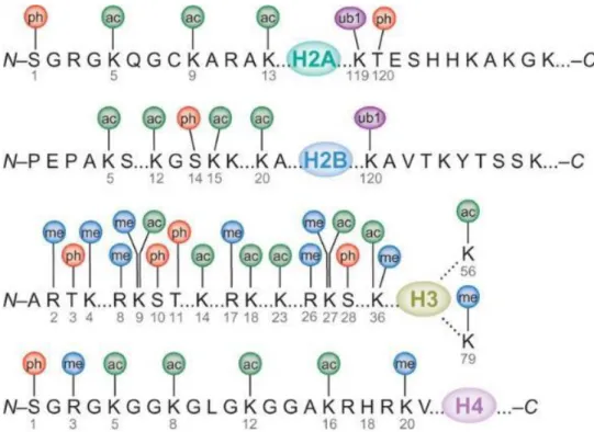
As the exposing of the N-terminal tails of histone proteins from the nucleosome core, the most majority of histone PTMs occur on these tail regions. Histones are subjected to different kinds of PTMs such as methylation, acetylation, phosphorylation, ubiquitination, and sumoylation. The interaction between negatively charged DNA phosphate backbone and the positively charged lysine and arginine-rich histone proteins is key to the dynamic of nucleosome. The “histone code” hypothesis proposes that the combination of different covalent modification states of these lysine and/or arginine residues on histone tails (Figure 14, 15) can provide signals for recruitment of specific chromatin-associated proteins, which in turn alter chromatin states and affect transcriptional regulation (Jenuwein and Allis, 2001). Among the PTMs, acetylation and methylation are the two most commonly studied forms.
Figure 14. Diverse post-translational modifications present in Histones.
Figure 15. The role of chromatin during transcription (Li et al., 2007)
2.2.1 Histone acetylation for plant development
Histone acetylation is carried out by histone acetyltransferases (HATs). These enzymes catalyze the transfer of an acetyl group from acetyl-coenzyme A (acetyl-coA) to an amino group of a histone lysine residue. This transfer can neutralize the positively charged lysine residue, thus weakening the interaction with negatively charged nucleosomal DNA and neighboring nucleosomes, and resulting in a more open chromatin structure (Figure 16, reviewed in Shahbazian and Grunstein, 2007; Li and Reinberg, 2011). Besides, lysine acetylation is a reversible modification, which can be removed by histone deacetylases (HDAC).
29
Figure 16. Acetylation targets Lys residues in the amino-terminal tails of core histone proteins. A string of nucleosomes is shown with the tails protruding when
acetylated. Acetylation of the tail domains inhibits the folding of nucleosome arrays into secondary and tertiary chromatin structures, with acetylation of histones H2B and H4 having the greatest effect on tertiary structure formation. Thus, histone tail acetylation results in chromatin decondensation, thereby allowing access to transcription factors and other transcription co-activators. (Verdin and Ott, 2015)
Most of the studies on plant histones were carried out in the model organisms Arabidopsis. One type of HATs, AtGCN5, has already been well characterized both in vitro and in vivo (Srivastava et al., 2015). In yeast, GCN5 protein is a component of several multi-subunit protein complexes, such as ADA and SAGA (Grant et al., 1997). In each complex, GCN5 cooperates with other common adaptors such as the ADA2 protein to stimulate transcriptional activation (Balasubramanian et al., 2002). Similarly in Arabidopsis, AtGCN5 can interact with adaptor proteins AtADA2a and AtADA2b. And this complex can then be recruited by other transcription factors (e.g., C-repeat/DRE binding factor 1, CBF1). Both ada2b and gcn5 mutants can induce various pleiotropic defects in Arabidopsis, including dwarfism, aberrant root development, short petals and stamens, and reduced expression of cold-regulated genes in cold acclimation (Vlachonasios et al., 2003). These studies suggest that histone acetylation plays an important role in plant gene expression in response to
environmental changes. AtGCN5 has also been isolated in the suppressor screen for a topless-1 mutation (tpl-1) that transforms the shoot pole into a second root pole during embryogenesis (Long et al., 2006). When mutating
AtGCN5 in the tpl-1 background, the shoot pole was recovered with proper
differentiation (Long et al., 2006), suggesting AtGCN5 might repress the transcription of meristem controlling genes (e.g. WUS) through activation of their upstream repressors.
Currently, lysine acetylation has been found to occur on H3 (K4, K9, K14, K18, K23, K27, K36 and K56), H4 (K5, K8, K12, K16, K20 and K91), H2A (K5 and K9) and H2B (K5, K12, K15, K16, K20 and K120). In general, this PTM is largely associated with a transcriptionally active state. So far, three histone effectors capable of reading acetylated lysine have been identified (Musselman et al., 2012). The bromodomain is the most thoroughly characterized acetyllysine reader found in various types of nuclear proteins including histone acetyltransferases, transcriptional coactivators and chromatin-remodelling factors (Filippakopoulos et al., 2012). Despite little sequence similarity between family members, bromodomains fold into a highly conserved four-helix bundle structure to ensure the insertion of of actyllysine (Figure 17). All known AcK binding pockets are hydrophobic with hydrogen bond capacity at the bottom. AcK intercalates into the pocket mainly through a hydrogen bond and the interaction is stabilized by a network of water-mediated intermolecular hydrogen bonds (Zeng et al., 2010). Besides, the double PHD finger (DPF) of human Dpf3b protein and the double pleckstrin homology (PH) domain of the histone chaperone Rtt106 have been found to associate with acetylated histone peptides (Zeng et al., 2010; Su et al., 2012). But they bind to different types of histone acetylation marks, with PHD finger module interact with H3K14ac and Rtt106 interact with H3K56ac. Recognition of acetyllysines shows significant impact on transcription control. As the BRD family of proteins is composed of transcription regulators, generally they are specific for singly or multiply acetylated histone peptides. Members of an important subgroup of
31
bromodomain proteins contain an extra-terminal domain and act as transcription factors in Arabidopsis. Three of these transcription factors, GTE1/IMB1, GTE4 and GTE6, have been functionally characterized and are involved in seed germination, cell division and leaf development, respectively (Duque and Chua, 2003; Yii et al., 2005; Airoldi et al., 2010). Moreover, other proteins containing bromodomain module, such as BRAHMA (BRM), an SNF2 chromatin-remodelling ATPase, can interact with SWI3C and forms an ATP-dependent chromatin-remodelling complex involved in development, phytohormone signalling and stress response (Wu et al., 2012b; Efroni et al., 2013; Yang et al., 2015).
Figure 17. Structures of the readers bound to histone peptides that are acetylated at lysine residues (Musselman et al., 2012).
The histone deacetylase contains four types of enzymes (I, II, III and IV). With the exception of class IV that is plant specific (the HD2-like proteins), others are homologous to yeast RPD3 (reduced potassium dependency protein 3), HDA1 (histone deacetylase 1 protein), and SIR2 (silent information regulator protein 2) proteins, respectively. Studies in yeast and plant have shown that different histone deacetylases are involved in distinct biological processes but may also have some overlapping functions. In Arabidopsis, AtHD1 and AtHDA6 are best characterized RPD3-like HDACs and exhibit divergent and overlapping