Université de Sherbrooke
Dysregulated microRNA expression in human osteoclasts and Paget’s disease of bone
Par
Elizabeth Stephens
Département de médicine, service de rhumatologie (Programme de immunologie)
Mémoire présentée à la Faculté de médecine et des sciences de la santé en vue de l’obtention du grade de maitre ès sciences (M. Sc)
en immunologie
Sherbrooke, Québec, Canada Juin, 2020
Membres du jury d’évaluation
Pre Sophie Roux, M.D., PhD. département de médecine, service de rhumatologie Pre Lee-Hwa Tai, PhD, département de médecine, service de immunologie Pr Luigi Bouchard, T.M., PhD., MBA, Département de biochimie et de génomique
fonctionelle
RÉSUMÉ
Dérégulation de l’expression des microRNAs dans les ostéoclastes pagétiques Par
Elizabeth Stephens Programme d’Immunologie
Mémoire présenté à la Faculté de médecine et des sciences de la santé en vue de l’obtention du diplôme de maitre ès sciences (M.Sc.) en Immunologie, Faculté de médecine et des sciences de la santé, Université de Sherbrooke, Sherbrooke, Québec, Canada, J1H 5N4 L'épigénétique comprend des mécanismes par lesquels le génome intègre des signaux intrinsèques et environnementaux. Les microARN (miARN) sont de petits brins d’ARN non codant qui régulent la traduction des gènes après leur transcription, et affectent
également les processus épigénétiques. Des profils dérégulés de miARN ont été associés à diverses pathologies, dont les maladies osseuses, faisant des miARN une cible attrayante comme biomarqueurs ou cibles thérapeutiques. Nous avons émis l'hypothèse que les profils de miARN étaient dérégulés dans les ostéoclastes (OC) dérivés de monocytes
périphériques (PBMCs) de patients atteints de maladie osseuse de Paget, et qu’ils pourraient contribuer à sa pathogénie et au phénotype des OCs pagétiques.
Une analyse par qPCR a permis d’identifier 2 miARNs sur les 14 testés, dont l’expression était significativement diminuée dans les OCs pagétiques comparativement aux OCs de sujets sains appariés pour l'âge et le sexe: 146a-3p et 155-5p; miR-133a-3p a également été évalué, miR connu dans les OCs dont l’expression n’est pas modifiée dans la maladie de Paget (contrôle du modèle). Dans un modèle de différenciation OC in vitro à partir de monocytes fœtaux humains, nous avons étudié l’impact d’une inhibition (antagomiR) ou d’une surexpression (mimics) de ces miRs sur la formation de cellules multinucléées, la résorption osseuse et la signalisation intracellulaire des
ostéoclastes, en évaluant des voies OC importantes connues pour être modulées dans d’autres systèmes.
Résultats : miR-133a 3p stimulait la résorption osseuse sans effet sur la formation de cellules multinucléées, alors que miR-146a-3p et miR-155-5p diminuaient la formation des cellules multinucléées et la résorption osseuse. Les brins opposés (146a-5p et miR-155-3p) n’avaient en revanche aucun effet sur ces paramètres. Par ailleurs, l’inhibition de miR-133a-3p diminuait la phosphorylation de p38 (sans affecter les autres MAP kinases), mais l’expression des kinases classiques de p38 (TAK1, MEK3/6) n’était pas affectée; miR-146a-3p affectait l’activation de NF-κB possiblement en diminuant l’expression de TRAF6; enfin, miR-155 5p, diminuait l’expression du facteur de transcription MITF.
Les profils des miARN des ostéoclastes pourraient avoir une valeur importante pour fournir de nouvelles informations significatives sur le phénotype des ostéoclastes dans la maladie de Paget et dans d'autres maladies osseuses avec des ostéoclastes hyperactifs. Mots-clés: ostéoclastes humains, microARN, profil de microARN, maladies osseuses
SUMMARY
Dysregulated microRNA expression in human osteoclasts and Paget’s disease of bone By
Elizabeth Stephens Immunology Program
Thesis presented at the Faculty of medicine and health sciences for the obtention of Master’s degree diploma (maitre ès sciences (M.Sc.)) in Immunology, Faculty of medicine
and health sciences, Université de Sherbrooke, Sherbrooke, Québec, Canada, J1H 5N4 MicroRNA (miRNA) are small, non-coding pieces of RNA that
post-transcriptionally regulate gene expression. Because of the epigenetic role of miRNA, dysregulated miRNA profiles have been associated with diseases, including bone diseases. Recently, miRNA have become an attractive target for disease biomarkers or therapeutic targets. We hypothesized that dysregulated miRNA profiles in osteoclasts (OCs) from patients with Paget’s disease of bone (PDB) may be contributing to disease pathogenesis and may play a role in the pathogenic phenotype of PDB OCs.
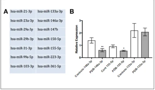
A qPCR analysis revealed that the expression of 2 miRs out of the 14 tested, miR-146a 3p and miR-155 5p, were significantly reduced in PDB OCs versus those of age-and-sex matched healthy controls. Both miRNA are known to be expressed in OC precursors and involved in OC-important signalling pathways. This lead to this in vitro study of the effects of these miRNA on OCs in terms of their effects on bone resorbing capabilities, OC formation and effects on kinases and OC-important signalling pathways. We also examined miR-133a 3p in vitro, which is known to be important in OCs but remained stable in PDB, to serve as a control for the model.
For miR-133a 3p, we determined that inhibition has significant effects on bone resorption and p38 activation, while OC formation was not affected. Mimicking miR-133a 3p had no significant effects on the in vitro OCs. We confirmed that miRNA-146a 3p, when mimicked, has negative effects on OC formation, bone resorption capabilities and NFκB activation via TRAF6 disruptions. Contrarily, when miR-146a 3p is inhibited, we observed overall increases in OC formation, bone resorption capabilities and NFκB activation as well as increased TRAF6 expression. And finally, we demonstrated that miR-155 5p, when mimicked, negatively affects bone resorbing capabilities and MITF levels within the OC and is detrimental to overall OC formation. Meanwhile, inhibition of miR-155 5p resulted in increased levels of bone resorption and MITF expression, while having no significant impact on levels of OC formation.
So, through this study we determined the effects that the selected miRNA have on in vitro human OCs in order to gain a preliminary understanding of their roles in human OC to aid in prediction of how they may be contributing to disease pathogenesis. This research can serve a platform for further research into how these changes can affect the phenotype of PDB OCs and, in the future, this understanding could lead to prospective therapeutic targets.
TABLE OF CONTENTS
Résumé... i
Summary ... ii
Table of Contents ... iii
List of Figures ... vi
List of Tables ... vii
List of Abbreviations ... viii
Introduction ... 1
1.1 Bone Tissue ... 1
1.1.1 The skeletal system ... 1
1.1.2 Composition of bones ... 1 1.1.3 Bone Remodelling ... 2 1.2 Osteoblasts ... 5 1.2.1 Differentiation of osteoblasts ... 5 1.2.2 Function of Osteoblasts ... 7 1.2.3 Osteocytes ... 8 1.3 Osteoclasts ... 9 1.3.1 Osteoclastogenesis ... 9 1.3.1.1 M-CSF & RANK-L ... 10
1.3.1.2 MAP kinases and Akt signalling pathways ... 12
p38α signalling ... 12
ERK signalling ... 13
JNK signalling ... 13
Akt signalling ... 13
1.3.1.3 NFκB: a major OC transcription factor ... 15
1.3.2 Bone resorbing process ... 17
1.4 Paget’s Disease Bone ... 19
1.4.1 General features ... 19
1.4.3 Pagetic Osteoclasts ... 21
1.5 MicroRNA ... 23
1.5.1 Generation and function ... 23
1.5.2 MicroRNA in bone and bone diseases ... 26
1.5.3 miRNA clinical applications ... 27
1.6 miR-133a in bone cells ... 29
1.7 miR-146a in cells of the osteoclast lineage ... 30
1.8 miR-155 in osteoclasts ... 33
1.9 Hypothesis and Objectives ... 36
1.9.1 Hypothesis ... 36
1.9.2 Objectives ... 36
Materials and Methods ... 37
2.1 Cell Culture ... 37
2.2 Transfection ... 39
2.3 Analysis of Protein Expression ... 41
2.4 qPCR validation of transfection ... 45
2.5 Bone Resorption Assays ... 45
2.6 Cell Multinucleation Assays ... 46
2.7 Statistical Analysis ... 47
Results ... 48
3.1 Quantitative PCR & choice of miRNA ... 48
3.2 The effects hsa-miR-133a on humans osteoclasts in vitro ... 49
3.2.1 Formation of multinucleated cells ... 51
3.2.2 Bone Resorption ... 52
3.2.3 Kinase activation ... 55
3.3 The effects of hsa-miR-146a on human OCs in vitro ... 59
3.3.1 Formation of multinucleated cells ... 60
3.3.2 Bone Resorption ... 62
3.3.3 NFκB pathway ... 64
3.4: The effects of hsa-miR-155 on human osteoclasts in vitro ... 68
3.4.2 Bone Resorption ... 70 3.4.3 MITF activation ... 72 Discussion ... 74 4.1 hsa-miR-133a-3p ... 74 4.2 hsa-miR-146a-3p ... 76 4.3 hsa-miR-155-5p ... 78
4.4 Limits of the in vitro study ... 80
4.5 Overall conclusions and perspectives ... 81
Acknowledgements ... 85
LIST OF FIGURES
Figure 1: Bone Remodelling ... 5
Figure 2: Osteoblast development ... 8
Figure 3: Osteoclast development. ... 12
Figure 4: Osteoclast Signalling Pathways ... 15
Figure 5: NFκB Canonical Signalling Pathway ... 16
Figure 6: Osteoclastic Bone Resorption ... 19
Figure 7: MicroRNA Generation ... 25
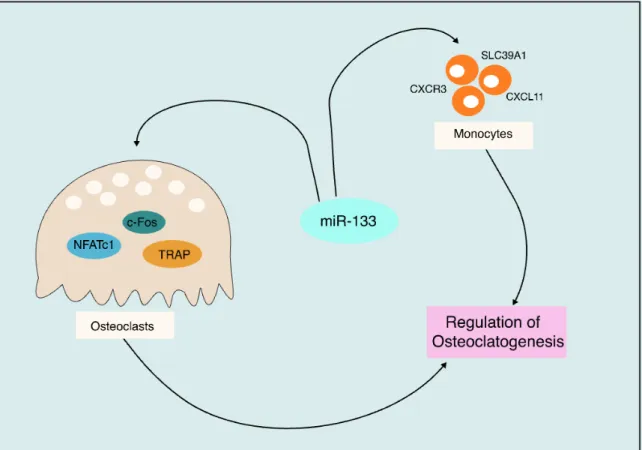
Figure 8: miR-133a mechanistic ... 30
Figure 9: miR-146a mechanistic ... 33
Figure 10: miR-155 mechanistic ... 35
Figure 11: Chosen miRNA for study ... 49
Figure 12: miRNA qPCR Validation ... 50
Figure 13: Multinucleated cell generation and effects of hsa-miR-133a-3p inhibition ... 51
Figure 14: Multinucleated cell generation and effects of hsa-miR-133a-3p mimics ... 52
Figure 15: The effects of inhibiting hsa-miR-133a on bone resorption ... 53
Figure 16: The effects of mimicking hsa-miR-133a-3p on bone resorption ... 54
Figure 17: The effects of inhibiting and mimicking hsa-miR-133a-3p on activation of OC important kinases ... 56
Figure 18: Effects of hsa-miR-133a-3p inhibitors upstream and downstream of p38 ... 57
Figure 19: Effects of hsa-miR-133a-3p mimics at varied concentration on p38 phosphorylation and levels of NFATc1 ... 59
Figure 20: Multinucleated cell generation and effects of hsa-miR-146a-3p and -5p inhibitors ... 61
Figure 21: Multinucleated cell generation and effects of hsa-miR-146a-3p and -5p mimics ... 62
Figure 22: The effects of inhibiting hsa-miR-146a-3p and -5p on bone resorption ... 63
Figure 23: The effects of mimicking hsa-miR-146a-3p and -5p on bone resorption ... 64
Figure 24: IκB levels following RANK-L stimulation (200 μg) at various time intervals . 65 Figure 25: The effects of hsa-miR-146a-3p and -5p inhibitors on the NFκB pathway ... 66
Figure 26: The effects of hsa-miR-146a-3p and -5p mimics on the NFκB pathway ... 68
Figure 27: Multinucleated cell generation and effects of hsa-miR-155 inhibitors ... 69
Figure 28: Multinucleated cell generation and effects of hsa-miR-155 mimics ... 70
Figure 29: The effects of inhibiting hsa-miR-155a-3p and -5p on bone resorption ... 71
Figure 30: The effects of mimicking hsa-miR-155-3p and -5p on bone resorption ... 72
Figure 31: The effects of inhibiting hsa-miR-155-3p and -5p on MITF levels ... 73
Figure 32: The effects of mimicking hsa-miR-155-3p and -5p on MITF levels ... 73
LIST OF TABLES
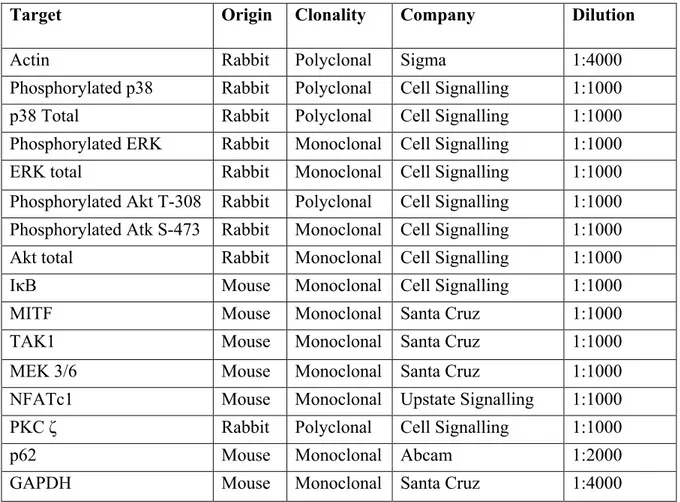
Table 1: miRNA sequences ... 39 Table 2: Transfection concentrations ... 40 Table 3: Primary Antibodies ... 44
LIST OF ABBREVIATIONS
Akt Protein kinase B
ANOVA Analysis of variance
ATF Activating transcription factor
Bcl-2 B-cell lymphoma 2
BMP Bone morphogenetic protein
BMU Basic multicellular unit
BSA Bovine serum albumin
c-Fms Macrophage-colony stimulating factor 1 receptor c-Src Proto-oncogene tyrosine-protein kinase
CBM Cord blood monocyte
CTR Calcitonin receptor
DMP Dentin Matrix Acidic Phosphoprotein ERK Extracellular signalling-related kinase
FBS Foetal bovine serum
Hh Hedge hog signalling
HSC Hematopoietic stem cell IGF Insulin-like growth factor
IKK IκB kinase
IFN Interferon
IL-6 Interleukin
IRAK1 Interleukin-1 receptor-associated kinase
IκB Inhibitor of κB
JNK c-Jun N-terminal kinase
LRP LDL receptor-related protein
M-CSF Macrophage colony-stimulating factor
Mac-1 Macrophage-1 antigen
MAPK Mitogen-activated protein kinase
MAPKK MAPK kinase
MEK Mitogen-activated protein kinase kinase
MEKK MEK kinase
MITF Micropthalmia-associated transcription factor
MMP9 Matrix metallopeptidase 9
MSC Mesenchymal stem cell
mTOR Mechanistic target of rapamycin
MV Measles virus
MVNP Measles virus nucleocapsid protein
NEMO NFκB essential modulator
NFAT Nuclear factor of activated T cells
NFκB Nuclear factor kappa-light-chain-enhancer of activated B-cells
NSE Neuron-specific enolase
OC Osteoclast
OPG Osteoprotegerin
OSCAR Osteoclast-associated receptor
PBMC Peripheral blood mononuclear cells
PDB Paget’s Disease of Bone
PDK Phospho-Inositide Dependent Kinase
PFA Paraformaldehyde
PI3K Phosphoinositide 3-kinase
PKC Protein kinase C
PTH Parathyroid hormone
qPCR Quantitative polymerase chain reaction Raf Rapidly accelerated fibrosarcoma
RANK Receptor activator of nuclear factor kappa-B
RANK-L Receptor activator of nuclear factor kappa-B ligand
Rho Ras homologous proteins
RISC RNA-induced silencing complex
RNA Ribonucleic acid
RSV Respiratory syncytial virus
Runx2 Runt-related transcription factor 2
SD Standard deviation
siRNA Small interfering RNA
snU6 Small nuclear RNA u6
SOCS Suppressor of cytokine signalling
TAK Transforming growth factor beta-activated kinase TGF-β Transforming growth factor beta
TNF Tumour necrosis factor
TRAF TNF receptor associated factor TRAP Tartrate-resistant acid phosphatase
UBD Ubiquitin-binding domain
INTRODUCTION 1.1 Bone Tissue
1.1.1 The skeletal system
The skeletal system is a vitally important component of the human body, having many important metabolic as well as mechanical functions (Xu and Teitelbaum, 2013). Past the evident roles of structural support and importance in locomotion, the skeleton is fundamentally important for a multitude of functions within the body, including mineral homeostasis, protection of vital organs such as the brain and heart, acid-base homeostasis, a reservoir for growth factors and cytokines as well as providing an environment for hematopoiesis (Xu and Teitelbaum, 2013) (Clarke, 2008). The bones that make up the skeleton all have unique shapes, sizes and characteristics, depending on their intended role. There are long bones, for example the ulna and femur, which are important for weight bearing and facilitating movement. Generally, due to the size of the long bones, they encase a majority of the body’s bone marrow, which is vital for hematopoiesis (White, Black and Folkens, 2012). Short bones, such as the carpal and tarsal bones, are important for stability as well as aiding in movement (White, Black and Folkens, 2012). There are flat bones, such as the skull and the pelvis that are vital for organ protection due to their unique shapes (White, Black and Folkens, 2012). And lastly, irregular bones and sesamoid bones such as the vertebrae and knee cap respectively, which do not fit into any other categories and have various specific functions depending on their unique shapes (White, Black and Folkens, 2012). Together, all of these bones types work collectively to form the skeleton and elicit all of the important functions that bones and the skeletal system have within the human body.
1.1.2 Composition of bones
The bone mass in the body is made up of about 80% cortical bone and 20% trabecular bone (Clarke, 2008). Cortical bone is characterized by its dense and solid nature while trabecular bone is more mesh-like, spongy bone. Cortical bone makes up the solid protective cavity around the marrow space while the trabecular bone is interspersed throughout these cavities (Clarke, 2008). Bone matter is made up of both organic and
inorganic compounds. About 50-70% of bone mass is composed of minerals, 20-40% organic matrix, 5-10% water and >1% lipids (Clarke, 2008). The organic compound is made up of about 90% Type I collagen fibres with the remaining 10% being other types of collagen or non-collegenic proteins (e.g. bone sialoprotein, osteonectin, osteocalcin). The inorganic portion of bone, or the mineral portion, is largely crystalline hydroxyapatite (White, Black and Folkens, 2012). Typically, the organic collagen fibres of both cortical and trabecular bone are laid in a lamellar pattern, which provides optimal strength to the bones. Alternatively, bones subjected to pathological conditions can deviate from this lamellar pattern and the bone can be laid in a woven pattern, which occurs following high turnover rates and provides a weaker structure (Clarke, 2008). In order to keep the skeletal system functionally optimally, bones are remodelled throughout one’s life in order to keep bones healthy and strong.
1.1.3 Bone Remodelling
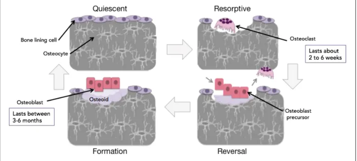
Thus, due to the many important roles of bone within the body, bone is constantly being remodelling to maintain health and strength and avoid disruptions to function. Bones are continually remodelled throughout one’s life for many reasons including bone health maintenance, growth, and in response to physiologic influences (Clarke, 2008). The human body is ever-changing and adapting and our bones are no exception. Bone remodelling takes place within an anatomic structure known as a basic multicellular unit (BMU) in order to keep the remodelling process tightly regulated (Xu and Teitelbaum, 2013). BMUs consist of 3 major types of cells including osteoclasts (OCs), osteoblasts and osteocytes (Xu and Teitelbaum, 2013). OCs are the only bone resorbing cells in the body. OCs are multinucleated cells that arise from a hematopoietic monocyte/macrophage lineage (Yavropoulou and Yavos, 2008). Osteoblasts are the mononucleated, bone forming cells that arise from a mesenchymal stem cell (MSC) lineage. Bone-lining cells are derived from osteoblasts and form a monolayer that covers the bone and keeps it in a quiescent state (Rosenburg, Rosenburg and Soudry, 2012). Osteocytes are the most abundant bone cell and are also derived from osteoblasts. They are embedded within the bone and maintain the bone integrity (Xu and Teitelbaum, 2013). Within the BMU, these 3 types of cells work together to carry out bone remodelling, and in adults, thousands of BMUs operate at any time moment. Bone remodelling takes place via 4 distinct stages.
The first two stages of the bone remodelling process are the activation and resorptive stage. Generally, bone-lining cells maintain the bone in a quiescent state but when remodelling is signalled, OCs are recruited to the intended site of resorption (Xu and Teitelbaum, 2013). Bone remodelling can be signalled spontaneously as general bone maintenance, in response to an area in need of repair, or to regulate calcium levels (Clarke, 2008) (Kenkre and Bassett 2018). Both local and systemic factors regulate bone remodelling. On a local scale, osteocytes play a key role in OC recruitment and activation at the intended resorptive site through the release of the cytokine receptor activator of nuclear factor kappa-B ligand (RANK-L), which is vital for OC activation and bone resorbing capabilities (Kenkre and Bassett, 2018). Other factors, such as macrophage colony stimulating factor (M-CSF), interleukin 6 (IL-6) and tumour necrosis factor alpha (TNF-α), also locally contribute to triggering the bone to exit a quiescent state as they contribute to the local generation and activation of OCs (Siddiqui and Partridge, 2016). In terms of systemic factors that regulate bone remodelling there are calcitonin, parathyroid hormone (PTH), vitamin D3 and estrogen (Siddiqui and Partridge, 2016). Calcium homeostasis is a key player in the regulation of bone remodelling as bones hold the body’s calcium stores, so PTH and calcitonin are important factors in the regulation and release of these stores (Kenkre and Bassett, 2018). Therefore, whether locally or systematically triggered, the resorptive stage of bone remodelling takes place through the generation and activation of OCs and, once activated, the OCs resorb the bones in a process that can take anywhere from 2 to 6 weeks to complete (Xu and Teitelbaum, 2013) (Figure 1).
Once the resorptive stage has been completed, the reversal phase commences to stop the OCs from resorbing and to recruit bone-building osteoblasts to the resorbed site. The reversal phase couples bone resorption to the formation of new bone, and is characterized by the presence of macrophages that clean the resorption zone and prime it for new bone. During this time OCs are signalled to undergo apoptosis by factors such as transforming growth factor beta (TGF-β), bone morphogenetic proteins (BMPs) and insulin-like growth factor (IGFs) which all contribute to the reversal phase of bone remodelling (Siddiqui and Partridge, 2016). As well, cytokines and growth factors that promote the differentiation of MSCs into osteoblasts, as well as the recruitment of osteoblasts to the resorbed area, are upregulated to begin the osteoblast differentiation
process in order to prepare for the next stage of the bone remodelling cycle (Rosenburg, Rosenbury and Soudry, 2012) (Figure 1).
Following osteoblast recruitment to the resorbed site, an increase in growth hormones and factors will result in the maturation of the osteoblasts in preparation for the rebuilding of the bone matrix (Siddiqui and Partridge, 2016). The secretion of IL-6, from OCs and factors such as BMP-2, wingless-INT proteins (Wnts) and IGF-1 may all contribute to the recruitment and activation of osteoblast in order to build bone (Kenkre and Bassett, 2018) (Siddiqui and Patridge, 2016). It can take anywhere between 3 to 6 months to complete the bone rebuilding process (Clarke, 2008). During this process the osteoblasts proliferate and deposit osteoid. Osteoid is an un-mineralized bone matrix that will fill the resorbed area, which will be followed by mineralization via the deposition of inorganic hydroxyapatite (Raggatt and Partridge, 2010). When the area has been sufficiently rebuilt, the osteoblasts either become embedded in the osteoid and differentiate into osteocytes, rest on top of the bone and differentiate into bone lining cells, or undergo apoptosis (Raggatt and Partridge, 2010). Osteocytes play a key role in the completion of bone formation through the secretion on Wnt inhibitors, which will ultimately cause a halt in osteogenesis (Kendre and Bassett 2018) (Figure 1).
Following these 4 phases stages, the bone remodelling cycle has been completed and the bone can enter into a resting or quiescent state. The bone lining cells derived from osteoblasts will cover the surface of the bone, and will keep the bone in this quiescent state (Xu and Teitelbaum, 2013). Bone remodelling is tightly coupled within the BMU to ensure that the rate of resorption by the OCs does not exceed the rates of formation by the osteoblasts in order to maintain bone homeostasis. Excessive resorption or formation can cause pathological condition to arise, as seen in many bone diseases such as Paget’s disease of bone and osteoporosis, which reiterates why tight coupling of degradation and rebuilding of bone is imperative during bone remodelling (Raggatt and Partridge, 2010) (Reddy, 2004).
Figure 1: Bone Remodelling
The process of bone remodelling takes place in 4 stages after quiescence: activation, resorption, reversal and formation. The bone resorbing cells are the OCs and the bones building cells are the osteoblasts. Osteoblasts then differentiate into osteocytes or bone lining cells that keep the bone in a quiescent state.
1.2 Osteoblasts
Osteoblasts arise from the mesenchymal lineage and are the bone building cells of the body. Osteoblasts work closely coupled with the OCs in order to maintain bone homeostasis. They are the key player in the formation stage of bone remodelling as they work to deposit osteoid and rebuild bone, as well as playing an important role in the regulation of bone remodelling through excretion of regulatory cytokines and signals (Rosenburg, Rosenburg and Soudry, 2012). Following completion of bone formation, osteoblasts can further differentiate into osteocytes or bone lining cells for the bone to enter into a quiescent state.
1.2.1 Differentiation of osteoblasts
Osteoblasts are specialized, mononuclear cells that arise from bone marrow produced mesenchymal stem cells (MSCs) (Rosenburg, Rosenburg and Soudry, 2012). There are multiple factors, such as hormones, cytokines and growth factors that trigger the differentiation of MSCs into osteoblasts (Rosenburg, Rosenburg and Soudry, 2012). MSCs have the potential to become osteoblasts, chondrocytes or adipocytes, so the first phase in osteoblast differentiation is the commitment to the osteoblast lineage (Capulli, Paone and
Rucci, 2014). To commit the MSCs to the osteoblast lineage, the hedgehog (Hh) signalling pathway is important (Rosenburg, Rosenburg and Soudry 2012). The Hh signalling pathway activates the runt-related transcription factor 2 (Runx2). Runx2 positive cells are of bipotent and can either enter into the osteoblastic lineage or chondrocyte lineage (Capulli, Paone and Rucci, 2014). The activation of the Runx2 gene is vital for osteoblastic differentiation and will remain expressed throughout the development of a mature osteoblast (Zhang, 2010) (Figure 2).
Following the commitment of MSCs to osteoblast precursors, the Wnt signalling pathway becomes critical for differentiation into pre-osteoblasts (Catano-Lopes, Canhão and Fonseca, 2007). The Wnts are secreted glycoproteins that activate multiple cascades that influence a multitude of cellular functions. The Wnt/β-catenin pathway, also called the canonical pathway, is activated through the binding of a Wnt protein to the Wnt receptor Frizzled and co-receptors LDL receptor-related protein 5/6 (LRP 5/6) (Eriksen, 2010). This cascade is important in early differentiation and development of osteoblasts, as well as survival. It acts by triggering an increase in cytoplasmic β-catenin, which promotes osteoblast-important transcription of target genes (Rosenburg, Rosenburg and Soudry 2012). The Wnt pathway is also important in the promotion of proliferation of osteoblast precursors. When there is a defect seen in co-receptor LRP 5 (e.g. lrp5-null mice), thus a lack of Wnt β-catenin activation, low bone mass is observed as a result of insufficient osteoblast proliferation (Zhang, 2010) (Figure 2).
Following this, at the time of osteoblast development, insulin-like growth factor 1 (IGF-1) causes an increase in the activation of the mitogen-activated protein kinases (MAPKs) p38 and extracellular signalling-related kinase (ERK) as well as the protein kinase B (Akt) pathway (Zhang, 2010). These activations will become important for the maturation of the pre-osteoblasts into mature osteoblasts. These pathways will work to upregulate the transcription factor osterix (Osx), which is vital for osteoblast maturation (Zhang, 2010). As well, BMPs, specifically BMP-2, will contribute to the upregulation of Osx (Zhang, 2010). This transcription factor has inhibitory effects on the Wnt/β-catenin pathway, which, as mentioned, is vital for proliferation and differentiation. This inhibition drives pre-osteoblasts from a proliferation phase to shift into a maturation phase (Zhang, 2010) (Rosenburg, Rosenburg and Soudry 2012). Once the osteoblasts have matured,
Runx2, Osx and activating transcription factor 4 (ATF4) are all important in osteoblastic function. ATF4 plays an important role in collagen synthesis, which is vital in a mature and functioning osteoblast (Capulli, Paone and Rucci, 2014). Thus, from commitment to maturation, multiple pathways are activated for successful differentiation of MSCs into mature osteoblasts, and the upregulation of the genes Runx2 and Osx are especially vital in this process (Figure 2).
1.2.2 Function of Osteoblasts
Once the MCSs have undergone differentiation into the large, mononucleated, cuboidal, mature osteoblast, these cells can secrete bone matrix and modulate bone remodelling through secretion of regulatory molecules (Eriksen, 2010). Mature osteoblasts will work to deposit osteoid, and eventually undergo apoptosis or re-differentiate into osteocytes or bone lining cells, which are both important and abundant bone cells.
The process of bone building takes place in two stages: the deposition of the organic matrix called osteoid, followed by mineralization. The organic matrix that is deposited by the osteoblasts is largely made up of collagen type I proteins, but also includes other types of proteins such as other types of collagen, non-collagen proteins and proteoglycans. Together, these proteins make up the organic portion of the bone, which, as mentioned, is deposited as osteoid (Capulli, Paone and Rucci, 2014). Following the deposition of the osteoid, an inorganic matrix, composed of hydroxyapatite crystals, is generated by the osteoblasts in order to mineralize the newly deposited osteoid. Mineralization takes place to give the new bone structure and acts as a mineral store for the body (Murshed, 2018). Mineralization is carried out through the release of hydroxyapatite crystals from extracellular matrix vesicles, that derive from osteoblast membranes by polarized budding (Capulli, Paone and Rucci, 2014). To form matrix vesicles, calcium binding proteins, calcium binding phospholipids and calcium channel forming annexins all must be present to create an environment that will form tri-calcium phosphate (Ca3(PO4)2). The newly formed calcium triphosphate then undergoes hydroxylation, which results in hydroxyapatite (Capulli, Paone and Rucci, 2014). This inorganic, calcium rich substance is then released from the matrix vesicles and it fills the spaces between the organic, collagen rich osteoid in order to mineralize the newly synthesized bone matrix.
Once the osteoblasts have laid the osteoid and carried out the mineralization process to adequate levels, the osteoblasts have completed their function. They then undergo re-differentiation into bone lining cells or become embedded in the bone matrix and differentiate into osteocytes (Erikson, 2010). If neither of these re-differentiations occurs, the osteoblast will undergo apoptosis (Figure 2).
Figure 2: Osteoblast development
Osteoblastogenesis, or, the developmental process mesenchymal stem cells undergo in order to become mature osteoblasts. There are many important signalling pathways, such as Hh and Wnt, as well as multiple important transcription factors such as Runx2 and Osx. Following maturation and completion of bone deposition, osteoblasts can re-differentiate into osteocytes, bone lining cells or undergo apoptosis.
1.2.3 Osteocytes
As mentioned, when osteoblasts have completed building new bone matrix, they undergo apoptosis, differentiate into bone lining cells or they differentiate into osteocytes. Osteocytes are the most numerous of the bone cells, accounting for approximately 90-95% of the bone cells (Capulli, Paone and Rucci, 2014). As the osteoblasts differentiate into osteocytes they develop many arms projections (dendritic processes) as they lose their cuboidal shape as they enclose themselves within the deposited osteoid. Osteocytes are
important for the maintenance of healthy bone, and it has been determined that they contribute to the regulation of the bone remodelling cycle through cross-talk with both OCs and osteoblasts (Capulli, Paone and Rucci, 2014). Osteocytes play a crucial role in the regulation of bone remodelling, and they do this through the expression of molecules such as dentin matrix acidic phosphoprotein 1 (DMP1), which regulates bone formation (Erikson, 2010). As well, depending on the situation, they secrete RANK-L or Wnt antagonists, which, when needed, can trigger OC formation or inhibit osteoblasts respectively (Kenkre and Bassett 2018). Osteocytes are important and abundant bone cells that maintain the bone and also play a key role in the regulation of remodelling.
1.3 Osteoclasts
OCs are the unique, bone resorbing cells in the body. OCs are large, multinucleated cells derived from the hematopoietic stem cell (HSC) monocyte/macrophage lineage. They have the unique ability to break down mineralized bone matrix, which makes them a key player in the bone remodelling process (Xu and Tietelbaum, 2010). Signalization for the production of OCs, starting from HSCs, involves a variety of signals both local and systematic. Osteoclastogenesis, which is the process of the formation and maturation of OCs, is a tightly regulated process involving signalling pathways, kinases and transcription factors.
After a first phase of proliferation, essential for differentiation to occur, the mononuclear osteoclastic precursors merge together, and gradually acquire the characteristics of multinucleated OCs. The osteoclastic markers appear— such as Tartrate-resistant acid phosphatase (TRAP), calcitonin receptor (CTR) and integrin αvβ3— and the macrophagic markers disappear— such as neuron-specific enolase (NSE), macrophage-1 antigen (Mac-1). Osteoclastogenesis is schematized in 4 stages, those being: commitment, differentiation, multinucleation and maturation (Yavropolou and Yavos, 2008).
1.3.1 Osteoclastogenesis
In order to stimulate hematopoietic stem cells (HSCs) to undergo commitment to the OC lineage and, further, differentiation into OCs, there are a multitude of important signalling factors including cytokines, hormones and growth factors involved. The most
important of these signals are the cytokines M-CSF and RANK-L (Yavropolou and Yavos, 2008).
1.3.1.1 M-CSF & RANK-L
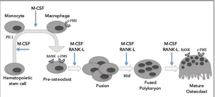
M-CSF is a crucial cytokine involved in OC commitment and generation. M-CSF binds to macrophage-colony stimulating factor 1 receptor (c-FMS), in order to stimulate OC precursors to commit to the OC lineage, and further, development into pre-OCs (Hershey and Fisher, 2004). Stimulation by M-CSF is critical for early stage OC development as it is the causal factor in the upregulation of OC-important transcription factors such as PU.1, microphthalmia-associated transcription factor (MITF) and c-Fos (Yavropoulour and Yovos, 2008). PU.1 is vital for the commitment of HSCs to the monocyte/macrophage lineage and development into OCs. PU.1 targets OC specific genes, such as the receptor activator of nuclear factor kappa-B (RANK) gene (TNFRSF11A), which results in the expression of the RANK receptor: a necessary receptor for further differentiation into OCs (Zhao, 2007). MITF is a leucine zipper transcription factor that is essential to OC development and maturation (Hershey and Fisher, 2004). Interactions between MITF and PU.1 target OC specific genes, such as cathepsin K, TRAP and OC-associated receptor (OSCAR) and expression of these genes is imperative for OC maturation (Yavropoulour and Yovos, 2008). In addition, c-Fos is an important transcription factor for the expression of nuclear factor of activated T cells 1 (NFATc1), and the presence of c-Fos has been associated with proliferation, differentiation and survival –important processes in OC generation (Zhao, 2007). Studies have shown that the lack of c-Fos results in osteopetrosis due to the inability of HSCs to differentiate past macrophages to enter the OC lineage (Grigoriadis et al. 1994). Without M-CSF stimulation, the expression of these OC-important transcription factors would be lost and the pre-OC would lack the expression of the RANK receptor, TRAP, NFATc1, OSCAR and cathepsin K, which would result in failure to produce mature OCs (Dougall et al. 1999). Following commitment, M-CSF also stimulates proliferation of the precursors, as well as works to prevent apoptosis of pre-OC (Figure 3).
RANK ligand, or RANK-L, is the second fundamentally necessary cytokine in the process of osteoclastogenesis and it initiates its signalling effects by binding to the RANK receptor. A defect of the RANK receptor, which would disrupt RANK-L binding, is seen to
results in the development of osteopetrosis and defects in tooth eruption due to the lack of or absence of OC formation, highlighting how vital RANK-L is to the formation of OCs (Dougall et al. 1999). RANK-L plays an important role throughout the lifespan of an OC, from formation, following the M-CSF induced commitment and proliferation, to fusion and maturation (Dougall et al., 1999). The binding of RANK-L to the RANK receptor activates the signal transducer TNF receptor associated factor 6 (TRAF6), which then acts to activate multiple OC-important signalling pathways (Armstrong et al. 2002). The p62 scaffolding protein, encoded by SQSTM1, is one of the functional links reported between RANK-L and TRAF6-mediated signals. Once bound to TRAF6, the complex p62-TRAF6 interacts with kinases to form multimeric protein complex with atypical protein kinase C (e.g. ζPKC), or with TGFβ-activated kinase 1 (TAK1) and adaptor proteins TAB1 and TAB2, that regulate NF-κB activation, as well as the activation of MAPK or Akt pathways (McManus and Roux, 2012).
RANK-L binding is important for the upregulation of multiple OC-important transcription factors such as nuclear factor kappa-light-chain-enhancer of activated B-cells (NFκB), NFATc1, c-Fos and MITF and OC specific genes, OSCAR and TRAP (Feng, 2005) (Yavropoulou and Yavos, 2008) (Figure 3). OSCAR is a receptor that is important in the crosstalk of the immune system and the skeletal system, which is important in the regulation of bone metabolism (Nemeth et al. 2011). The OSCAR gene is expressed following the RANK-L induced up regulation of NFATc1 and NFκB, and is an important differentiation factor in OC development (Nemeth et al. 2011). Tartrate-resistant acid phosphatase, TRAP, is an enzyme that is produced in active OCs (Blumer et al. 2012). As the committed precursors proliferate and move forward towards maturation, they will undergo fusion with other RANK presenting cells to grow in size and this process is not possible in the absence of RANK-L (Yavropoulou and Yvos, 2008). It is also noteworthy that osteoprotegerin (OPG), a secreted decoy for the RANK receptor, competes with RANK-L for the RANK receptor (Feng, 2005). This occurs to modulate osteoclastogenesis to keep OC levels regulated and homeostatic. This is exemplified through murine models, where OPG knockout mice develop early osteoporosis due to the excessive levels of osteoclastogenesis and, further, the overexpression of OPG results in osteopetrosis, due to lack of OC formation (Feng, 2005).
Therefore, for HSCs to be committed to the OC lineage and undergo differentiation and multinucleation, the cytokines M-CSF and RANK-L are important to upregulate the necessary transcription factors and OC-important genes. In order to do this, M-CSF and RANK-L must activate multiple signalling pathways, including the MAPK pathways. The MAPK pathways including p38α, c-Jun N-terminal kinase (JNK) and ERK are the most central kinase pathways in osteoclastogenesis and they all contribute to the development and activation of functioning OCs (Lee et al. 2018). As well as the MAPKs, the Akt pathway and the NFκB pathway both have important and necessary contributors to successful osteoclastogenesis and OC survival (Feng, 2005).
Figure 3: Osteoclast development.
The development of OCs begins with hematopoietic stem cells. Commitment and differentiation require signalling from the cytokines M-CSF and RANK-L. Committed pre-OCs will proliferate and then fuse to create a fused polykarion, which will then mature into a bone resorbing, multinucleated OC.
1.3.1.2 MAP kinases and Akt signalling pathways p38α signalling
As mentioned, the p38α pathway is a key player in osteoclastogenesis, and is considered to be one of the key MAPKs in osteoclastogenesis. Activation of this MAPK contributes to OC formation and maturation, and thus affects the overall bone resorption capacities of the OC (Lee et al. 2018). Lack of p38α, in p38α knockout mice, has been observed to result in an increased overall bone mass due to the decreased resorption caused
by the lack of p38α (Cong et al. 2017). The p38α pathway is activated through both M-CSF as well as RANK-L binding. Upstream of p38α, transforming growth factor beta-activated kinase 1 (TAK1) activates the MAPK kinase (MAPKK) mitogen-beta-activated protein kinase kinase 3/6 (MEK3/6), which will phosphorylate p38α (Lee et al. 2018). The activation of p38α results in increased levels of NFATc1, MITF, cathepsin K, and TRAP which are all important in OC development (Lee et al. 2018) (Figure 4).
ERK signalling
The ERK signalling pathway is also central to OC development. It is activated through both RANK-L and M-CSF stimulation. The activation of ERK1/2 via M-CSF is mediated though rapidly accelerated fibrosarcoma (Raf) and MEK1/2 and plays an important role in early osteoclastogenesis by contributing to precursor proliferation and survival (Xu and Teitelbaum, 2013). Via RANK-L stimulation, ERK1/2 is activated by upstream TRAF6. This activation contributes to OC formation, differentiation and maturation from committed precursors (Xu and Teitelbaum, 2013). The activation of ERK1/2 stimulates the expression of multiple important factors such as c-Fos, NFATc1 and MITF, which are all important to OC development (Lee et al. 2018). ERK 1/2, through both M-CSF and RANK-L stimulation, is highly important for osteoclastogenesis, throughout the entire process, from commitment to maturation (Lee et al. 2018) (Figure 4). JNK signalling
An additional MAPK that is central to OC development is JNK. JNK is activated through both M-CSF and RANK-L signalling and results in an upregulation of OC important factors such as c-Jun, c-Fos and NFATc1 (Lee et al. 2018). Upon binding of M-CSF, MEK kinase 1 (MEKK1) and TAK1 are both activated which contribute to the phosphorylation of JNK. As well, RANK-L binding activates TRAF6, which contributes to the activation of JNK. The activated JNK pathway is important in the differentiation of OCs, as well as their resistance to apoptosis during development (Lee et al. 2018) (Figure 4).
Akt signalling
Akt is a serine/threonine-specific protein kinase and is another important signalling pathway involved in OC differentiation (Moon et al. 2012) (Kawamura et al. 2007). Akt
signalling can be activated through both binding of M-CSF and RANK-L. Through ligand binding to either of these receptors, activation of proto-oncogene tyrosine-protein kinase (c-Src) will activate phospho-inositide dependent kinase 1 (PDK1). This results in the activation of phosphoinositide 3-kinase (PI3K) and Akt at the Serine473 residue (Sarbassov et al. 2005). Alternatively, mTOR can be activated, which will phosphorylate Akt at the Thronine308 residue (Sarbassov et al. 2005). Phosphorylation of Akt at either, or both, residues results in activation and upregulation of multiple OC specific genes including NFATc1, c-Fos, OSCAR and TRAP (Moon et al. 2012). When there is a disruption of the Akt pathway, in Akt knockout mice, it is observed that there is a decrease in OC generation as well as disruptions in OC survival (Kawamura et al. 2007). This reiterates the importance of Akt signalling in OCs, and this pathway’s essential role in formation (Figure 4).
Figure 4: Osteoclast Signalling Pathways
The most important pathways (via RANK-L and M-SCF binding) for OC differentiation and maturation include the Akt pathway, the ERK pathway, the p38 pathway, JNK pathway and the NFκB pathway. The up regulation of PU.1, NFATc1, c-Fos and MITF are all important transcription factors for lineage commitment, OC differentiation and survival following activation.
1.3.1.3 NFκB: a major OC transcription factor
Canonical NFκB activation plays an extremely important role in OC formation and is central to cytokine induced OC formation as well as the survival of OCs (Boyce et al. 2015). NFκB p50/p52 double knockout mice have been found to develop osteopetrosis, due to the disruption and lack of OC formation (Zhao, 2007). NFκB is held in an inactive state by inhibitory NFκB proteins that are called inhibitors of κB (IκBs). There are 3 isoforms including IκBα, IκBβ and IκBε, but IκBα is the interacting factor with the NFκB (Boyce et al. 2015). Upon activation of the RANK receptor via RANK-L, the NFκB pathway is called into action. This begins with the recruitment of the signal transducer TRAF6, which interacts with p62 as well PKCζ. Then, an IκB kinase (IKK) complex, which includes
IKKα, IKKβ and NFκB essential modulator (NEMO), is activated which phosphorylates IκB (Boyce et al. 2015). Phosphorylated IκB then releases the NFκB complex that was being held inactive, thus allowing for the activation of NFκB and translocation to the nucleus. The NFκB complex is made up of multiple proteins, including the heterodimer of p50 and p65, one of whose major impact is the upregulation of the NFATc1 gene, a gene central to OC formation (Boyce et al. 2015) (Xu and Teitelbaum, 2013). NFATc1 is a transcription factor that is necessary for OC formation and survival; thus, activation of this transcription factor via the NFκB pathway plays a central role in successful OC formation, and aids in the survival of the OC (Boyce et al. 2015) (Xu and Teitelbaum, 2013) (Figure 4 & 5).
Figure 5: Canonical NFκB Signalling Pathway
NFκB is transcription factor that is activated following RANK-L binding to the RANK receptor. Following activation, NFκB (composed of the heterodimer of p50 and p65) will translocate to the nucleus and result in an upregulation of OC important genes such as NFATc1.
1.3.2 Bone resorbing process
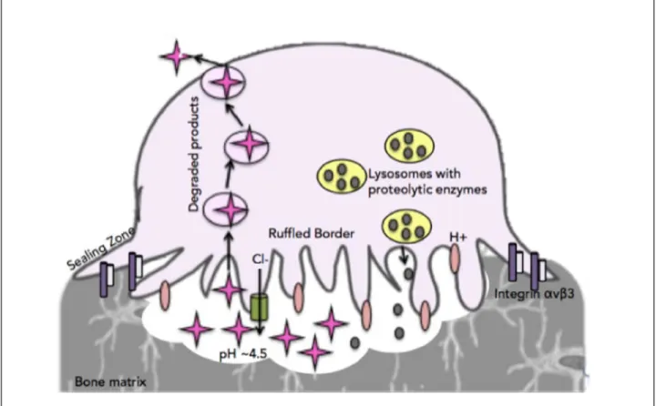
OCs have the exclusive ability to resorb mineralized bone matrix due to the generation of specialized structures and distinct proteolytic enzymes (Xu and Teitelbaum, 2013). The first step in bone resorption is for the OC to recognize the mineralized bone matrix, which will cause the OC to adhere to the bone and trigger morphological changes of the OC’s structure (Soysa and Alles, 2016). The αvβ3 integrins are the key player in the initiation of functional changes of the OC as it prepares to be activated and begin resorbing bone (Xu and Teitelbaum, 2013). When the αvβ3 integrins detect the bone matrix the OC will become polarized to the bone, cytoskeleton reorganization will be triggered and the formation of the actin ring and sealing zone will take place (Soysa and Alles, 2016). The integrins will signal c-Src that will activate Ras homologous proteins (Rho), which are regulatory GTPases, which will drive cytoskeletal alterations and generation of the actin ring (Xu and Teitelbaum, 2013). The actin ring is an important structure in osteoclastic bone resorption as it allows for the OC to form an isolated area that will house specialized resorbing machineries: the resorptive compartment and the ruffled border (Xu and Teitelbaum, 2013).
The resorptive compartment is the enclosed space within the sealed zone that will undergo acidification and will house the ruffled border; a ruffled membrane will arise due to the fusion of secretory lysosomal vesicles inserting many vacuolar H+ ATPases into the membrane as the components for acidification as well as proteolytic enzymes are delivered (Xu and Teitelbaum, 2013). The resorptive compartment will be acidified to a pH to approximately 4.5 in order to break down the mineralized bone matrix (Xu and Teitelbaum, 2013). The OC pumps hydrogen ions (H+) into the resorptive compartment to achieve acidification and chloride ions are also pumped into the resorptive compartment to maintain electroneutrality of the area (Raggatt and Partridge, 2010) (Kenkre, 2017). The acidic environment that is achieved in the resorptive compartment breaks down the inorganic, mineralized component of the bone matrix and it is released as calcium and phosphorus (Raggatt and Partridge, 2010) (Kenkre, 2017). After the inorganic, mineralized component has been dissolved, the organic matrix must be broken down. A unique characteristic of OC is that they contain many specialized acidic vesicles that carry the proteolytic enzymes that are needed to degrade the organic bone matrix (Xu and
Teitelbaum, 2013). The key degradation component that is carried in the OC in acidic vesicles is the lysosomal proteolytic enzyme cathepsin K. Cathepsin K is an OC-specific enzyme that relies on M-SCF and RANK-L signalling in order for the cathepsin K gene to be upregulated in the osteoclastogenesis process (Yavropoulour and Yovos, 2008). Without cathepsin K, the degradation of the bone matrix is altered, as seen in patients with cathepsin K mutation who develop pycnodysostosis, a bone disease characterized by facial dysmorphia and diffuse osteosclerosis (Xu and Teitelbaum, 2013). The dissolution of the organic bone matrix is achieved largely by cathepsin K but other proteolytic enzymes are present, such as matrix metallopeptidase 9 (MMP9) or TRAP, and aid in the break down as well (Raggatt and Partridge, 2010). So, between acidification via hydrogen ion and the release of proteolytic enzymes, the resorptive compartment achieves the acidity necessary to degrade the bone matrix (Figure 6).
Once the organic matrix has been broken down and degraded, the remaining products are translocated from the resorptive compartment to the non-resorptive side of the OC (Xu and Teitelbaum, 2013) (Figure 6). This continues until the target bone has been sufficiently degraded, at which time the OCs are signalled to undergo apoptosis. Therefore, between the formation of the actin ring, or sealing zone, the resorptive compartment and the ruffled border, it is clear that bone resorption requires multiple morphological changes in order to successfully activate into a mature, bone resorbing OC. Following this process, the resorptive stage of the bone remodelling cycle is complete and osteoblasts can be recruited to the site to rebuild fresh bone matrix.
Figure 6: Osteoclastic Bone Resorption
OCs recognize mineralized bone matrix through the intergrins αvβ3, which triggers a conformational change to create an actin ring, or sealing zone, that will house the ruffled border and resorptive compartment. These conformational changes are vital for successful bone resorption. Within the resorptive compartment, hydrogen ions create an acidic environment (pH 4.5) and proteolytic enzymes break down the organic bone matrix, which is then transcytosed to the non-resorbing side of the OC.
1.4 Paget’s Disease Bone
1.4.1 General features
Paget’s Disease of Bone (PDB) was first reported by Sir James Paget in 1877 as Osteitis Deformans – a chronic multifocal skeletal disease (Reddy, 2004). Paget’s Disease is characterized by bone remodelling that is excessive and disorganized (Reddy, 2004). In other words, at pagetic lesions, the OCs are overactive in their bone resorbing capabilities, which is followed by osteoblastic bone repair that is rushed which results in poor quality bone formation (Roodman and Windle, 2005). PDB may be monostotic, meaning that the pagetic lesions stay localized to a single bone – or polyostotic – affecting multiple bones (Shaker, 2009). It is very uncommon for new pagetic lesions to develop throughout the
course of the disease. It has also been observed that pagetic patients have an increased risk of osteosarcoma in the affected bone, though this is relatively rare with an incidence of less than 1% (Roodman and Windle, 2005). PDB can cause many symptoms including bone pain, fracture, bone deformity and neurological problems or deafness due to compression from enlarged bones (Shaker, 2009) (Reddy 2004).
1.4.2 Etiology: genetics and environmental factors
Paget’s Disease of Bone is the second most frequent metabolic bone disorder, following osteoporosis, affecting about 1-2% of the population over 55 (Shaker, 2019). PDB is a late onset disease, being rare under the age of 25, infrequent under the age of 40 and mostly affecting patients over the age of 55 (Shaker, 2009). The prevalence of PDB is varying depending on ethnic and geographic groups, having a higher prevalence in Caucasians of Western European descent and a very low incidence in those of Asian descent (Reddy, 2004).
PDB has a strong genetic component as about 40% of Pagetic patients have a first degree relative that is also affected (Roodman and Windle 2004). The most common mutation associated with PDB is a mutation affecting the SQSTM1 gene. SQSTM1 encodes the ubiquitin-binding protein sequestosome1, also known as p62 (Chamoux et al. 2013). This gene is affected in approximately 50% of familial cases and about 10% of sporadic cases. Mutations affecting the SQSTM1 gene cluster around the ubiquitin-binding domain (UBD) domain of p62, the most common being is P392L, though there have been about 30 other mutations reported (Chamoux et al. 2013) (Zach et al. 2018). P62, which is an important regulatory molecule linking ubiquitinated protein to autophagy, is also central to many signalling pathways and is involved in regular maintenance and basic functioning of cells. Dysfunction or loss of the p62 protein can result in obstructed signalling pathways, and abnormal aggregations of proteins, which can lead to the development of disease such as PDB (Chamoux et al. 2009). PDB has been seen to be of the autosomal dominant nature of inheritance, with genetic heterogeneity and incomplete penetrance (Reddy, 2004) but high penetrance (about 80%) in patients over the age of 60 (Chamoux et al. 2009). In addition, it has been noted in a p62 knockout murine model that the dysfunction of p62 results in osteolytic lesions that resemble those observed in pagetic patients (Zach et al. 2018). Knocked-in mice expressing the p62P394L gene (the murine equivalent of human
p62P392L) develop focal osteolytic lesions, some of them resembling PDB lesions (Daroszewska et al. 2011), although in another study co-expression of the measles virus nucleocapsid protein gene (MVNP) in the OC lineage was required (Kurihara et al. 2011). This reiterates the importance of p62 mutation to the development of PDB and its role in the pathogenic phenotype.
There is also thought to be an environmental component of the development of Paget’s disease, such as diet, toxins, and potential viral etiologic affect – the latter having undergone extensive research (Zach et al. 2018). Pagetic OCs are observed to have inclusion bodies found in their nuclei and cytoplasm, which resemble viral nucleocapsids (Reddy, 2004). There have been many studies performed to examine these nucleocapsids in terms of their biological, immunohistochemical, and ultrastructural components, but no specific virus has been determined to have causal effects (Roodman and Windle, 2005). The two viral antibodies that have been determined to cross-react with the inclusion bodies in PDB have been found to be measles virus (MV) and respiratory syncytial virus (RSV) (Mills et al. 1984). As well, RT-PCR and in situ hybridization determined the presence of MV RNA in pagetic OCs (Reddy, 2004), while other studies found no evidence of such RNA. Another study, interestingly, demonstrated that transfecting MV into OCs resulted is OCs with a pagetic phenotype (Kurihara et al. 2000). Though there have been many studies and hypotheses on the subject, there has been no causal evidence between viral infection and the development of Paget’s disease, though, as mentioned, some studies suggest a possible link. Though the environmental effects are not as well understood and the genetic effects in terms of contributions to the development of PDB phenotype, environmental effects are still considered to play a role in disease pathology.
1.4.3 Pagetic Osteoclasts
OCs are a key player contributing to the pathological conditions that arise in PDB. As mentioned before, in Paget’s disease, the OCs are overactive in their bone resorbing capabilities, which results in bone formation that is hurried and disorganized, resulting in bone fragility, enlargement and fracture, among other symptoms. In this disease, the OCs are found in an increased number as well as enlarged in size (Shaker, 2009). A healthy OC will generally have between 3 and 20 nuclei, but in pagetic patients, OCs have been observed up to contain up to 100 nuclei (in vitro) (Roodman and Windle, 2005). Pagetic
OCs are also observed to have inclusion bodies in their cytoplasm and nuclei that have been hypothesized to arise from a viral origin, but it has more recently been notes that these inclusion bodies resemble aggregations of misfolded or ubiquitinated proteins (Reddy, 2004) (McManus et al. 2016). Because of this, it is hypothesized that perhaps the inclusions bodies could arise from defective autophagy, a process which has not been found influenced by the SQSTM1 mutation (McManus et al. 2016). Defective autophagy could contribute to the alterative state of bone resorbing activity that is seen in PDB (Alosono et al. 2017). It has also been determined that patients with PDB have increased levels of circulating IL-6 and RANK-L, which both contribute to the activation of osteoclastogenesis (Shaker, 2009). IL-6 and RANK-L both play critical roles in the signalling for activation and bone resorbing capabilities, and the increased levels could be contributing to the over-activity of OCs that is observed in PDB. As well, the OCs precursors within pagetic patients are hypersensitive to differentiation factors such as RANK-L and 1,25 dihydroxy-vitamin D3 which, again, could contribute to the increased number and activation state of pagetic OCs (Shaker, 2009).
It has also been determined that OCs in PDB are resistant to apoptosis, which could be a contributing factor to the bone destruction that is characteristic of this disease (McManus et al. 2016) (Chamoux et al. 2009). Pagetic OCs have also been observed to have a basal increase in kinase activation, which also could contribute to the resistance to apoptosis as well as the over activation that is observed. It is seen that the Akt pathway and the ERK pathway have increased basal phosphorylation levels in PDB, and as described above, these pathways are both important in osteoclastogenesis and survival (McManus et al. 2016). With a basal increase in these important pathways, there is an increased activation of pagetic OCs, which ultimately contributes to the excessive bone resorption that is characteristic of this disease.
As mentioned, following the excessive osteoclastic bone resorption in PDB, there is disorganized bone reformation by the osteoblasts. The osteoblasts of patients with PDB are increased in number at pagetic lesion, but their morphology remains normal (Reddy, 2004). Osteoblasts themselves are do not appear to be pathogenic in PDB, and their increase in number and over-activity at pagetic lesion is likely due to the increase in cytokines, such as RANK-L, that is associated with PDB. Because of the cross-talk and dependence between
the osteoblasts and OCs in the bone remodelling process, it is important to consider osteoblasts in PDB bone destruction and pathogenesis, but, OC are the primary affected cell. Therefore, OCs are the key focus of our research and a key player in PDB pathology, though their dysregulation does affects other bone-important cells such as osteoblasts. Understanding OC remains the first step to better our understanding of PDB. As seen in PDB there are multiple alterations to the state of the OCs that contribute to the disease pathogenesis, which is why research to further understand OCs in PDB is important. As there are many signalling pathways that are central to OC development and activation, mechanisms of regulating these pathways may aid in the understanding of pagetic OCs.
.
1.5 MicroRNA
Epigenetics includes mechanisms by which the genome integrates intrinsic and environmental signals, which can occur in 3 ways: DNA methylation, histone modifications and chromatin remodelling or through miRNA. MicroRNA (miRNA) are small non-coding pieces of RNA, generally around 22 nucleotides long, that regulate gene expression post-transcriptionally, and also affect epigenetic processes. They are epigenetic regulators that most often silence genes by acting on the transcribed mRNA, which blocks translation into protein. It is thought that about 60% of protein-coding genes are regulated by microRNA (Esteller, 2011). MicroRNA have regulatory roles in many functions of many different types of cells, including proliferation, apoptosis, differentiation and development (Bernado et al. 2011). The function of a given miRNA can be species and tissue dependant, so a single miRNA may have varied functions and targets depending on where it is found (Bernado et al. 2011).
1.5.1 Generation and function
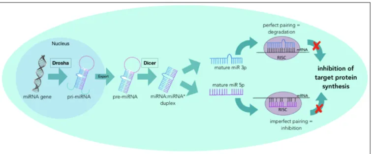
MiRNAs are transcribed by the cell as a necessary regulator to ensure optimal cellular function (MacFarlene and Murphy, 2010). As such, miRNA are encoded into the genome. The process to generate a mature and functioning miRNA molecule starts in the nucleus with the miRNA gene that is transcribed into primary microRNA, or “pri-miRNA”, that is about 1000 base pairs long, in a hairpin loop structure (Bernado et al. 2012). Following transcription into pri-miRNA, the enzyme Drosha cleaves the pri-miRNA
into a precursor miRNA, or “pre-miRNA”, which at this time is about 60-100 base pairs long in a hairpin loop formation (Bernardo et al. 2012). Following this cleavage, the pre-miRNA is exported out of the nucleus into the cytoplasm via transporter that containing Exportin 5 and Ran-GTP. Once in the cytoplasm, the enzyme Dicer will cleave the pre-miRNA to the mature size of about 22 nucleotides long. At this point the pre-miRNA is still in a duplex of the 3p and 5p strands, also called the miRNA:miRNA* duplex (MacFarlene and Murphy, 2010). The mature duplex then unwinds and becomes 2 separate miRNA. In some cases only one miRNA has a function in the cell, so the other strand will be degraded and in other cases both strands have their own unique function (Guo et al. 2014). It is not fully understood what influences the decision of which arm, if only one, will become dominant, but it has been proposed to be influenced through hydrogen-bonding selection (Griffiths-Jones et al. 2011). Further, it has been proposed that the dominant arm could change due to the type of tissue or the time of development, but this is also not well understood (Guo et al. 2014). Once the dominant arm has been established and has matured, it is now single stranded miRNA that can be loaded into Argonaute proteins to form a RNA-induced silencing complex (RISC)– a complex that will facilitate the miR’s action on the target gene (Bernardo et al. 2012) (Figure 7). There are two outcomes of miRNA gene silencing: inhibition or degradation of the target mRNA. When the miRNA is perfectly complementary to the target mRNA, called a perfect pairing, then the mRNA is degraded (Bernardo et al. 2012). When the miRNA has an imperfect pairing with the mRNA, the mRNA is not degraded, but rather silenced through miRNA binding and holding in a non-translatable state (MacFarlene and Murphy, 2010). Through these 2 mechanisms, miRNA can inhibit the protein synthesis of the target protein, thus post-transcriptionally regulating mRNA translation. As well, some miRNAs can also upregulate gene expression under specific conditions (O’Brien et al. 2018). Though cells will make the miRNAs that they require for gene regulation, cells can also export the miRNA:RISC complex through exosomes or microvesicles. These mature miRNA can either remain in the blood where they could be used as biomarkers for disease, or they may be taken up by recipient cells where they can elicit epigenetic effects (Loranzo et al. 2019).
Extracellular miRNAs are present in most biological fluid due to the selective export from the cell that generates them into membrane-derived exosomes and out of the
cell. Once exported, miRNA are relatively stable, which contributes to their attractive potential to be used as biomarker for disease (Boon and Vickers, 2014). It has been shown that when the body is faced with pathological conditions, different exosomal miRNA signatures arise that can be associated with said disease (Li, Jiang and Wang, 2019). Once exported, miRNA play a role in cell-to-cell communication as they are delivered to new cells, acting in a way similar to steroid hormones. Exosomal miRNA can have influential effects on the mRNA translation within the new cell it enters by via endocytosis, where the miRNA can then act as an endogenous miRNA and bind their target mRNA (Bhome et al. 2018). The exact mechanisms of intracellular transport of miRNA and which miRNA are exported remains somewhat elusive. In a similar way, weather exosomal miRNA enter target cells in a random or non-random nature remains unknown (Bhome et al. 2018). Despite the uncertainties, it can be concluded that exosomal miRNA can play a key role in disease pathogenesis through cell-to-cell communications and remain an important target for disease biomarkers (Bayraktar, Van Roosbroeck and Calin, 2017).
Figure 7: MicroRNA Generation
The generation and processing of any miRNA starts in the nucleus with the translation of the intended miRNA gene. From here, it is processed through a series of cleavages until it reaches the mature size of approximately 22 base pairs. It is loaded into a RISC complex to bind its target mRNA and inhibit target protein synthesis.
1.5.2 MicroRNA in bone and bone diseases
As mentioned, miRNA contribute to the regulation of protein expression post-transcriptionally. Because of this influential role in gene translation, miRNA have been found to regulate the etiology of many types of disease, and bone disease is no exception (Moore and Xiao, 2013). Within many bone diseases such as osteoporosis, rheumatoid arthritis and malignant bone disease, dysregulated miRNA profiles in a variety of bone cells contribute to disease pathogenesis. Differentiated miRNA profiles within osteoblasts, OCs, osteocytes or even immune regulators can all contribute to the occurrence of bone disease (Moore and Xiao, 2013). Because bone remodelling is such a tightly regulated process, when any of the involved bone cells are effects, the vital homeostasis that the bone remodelling process can be disrupted and pathological conditions can arise (Tang et al. 2014).
Within OCs there have been studies to classify the importance of specific miRNA within the OC life span – from commitment to differentiation to survival. Between the early stages of an OC, to the maturation, there are many systems that are vital to the maintenance of healthy OC function (Lozano et al. 2019). During precursor commitment to the OC lineage, miRNAs such as miR-21, hsa-miR-144, miR-124 and mmu-miR-218 are important regulators in differentiation, motility and proliferation of precursors (Tang et al. 2014) (Lozano et al. 2019). In the early stage of the pre-OCs, miRNAs such as mmu-miR-29 and hsa-miR-503 play a role in regulation of the necessary processes for OC precursors to develop (Tang et al. 2014) (Lozano et al. 2019). Next, as osteoclastogenesis progresses and the precursors undergo fusion, mmu-miR-7b and hsa-miR-142 play important roles in regulation of this process (Tang et al. 2014) (Lozano et al. 2019). As the actin ring is formed and the OC undergoes extensive conformational changes mmu-miR-31 and mmu-miR-26 become important (Tang et al. 2014) (Lozano et al. 2019). And lastly, the survival of the mature, bone resorbing OC is regulated in part by hsa-miR-142 and mmu-miR-34 (Tang et al. 2014) (Lozano et al. 2019). This is not a comprehensive list of all of the miRNA involved in the osteoclastogenesis process, and many miRNA are known to be important in OCs but the stage in which they are necessary has not been determined: such as hsa-miR-146a, hsa-miR-125a, mmu-miR-155, mmu-miR-218, mmu-miR-124 and hsa-miR-133, to mention a few (Tang et al. 2014) (Lozano et al. 2019). In addition, many