Design of a novel covert SSVEP-based BCI
D. Lesenfants
1, N. Partoune
1, A. Soddu
1, R. Lehembre
1, G. M¨
uller-Putz
2, S.
Laureys
1, Q. Noirhomme
11Coma Science Group, Cyclotron Research Centre and Neurology department, University of
Li`ege, Li`ege, Belgium
2Laboratory of Brain–Computer Interfaces, Institute for Computer Graphics and Vision, Graz
University of Technology, Graz, Austria
Abstract
Brain computer interfaces (BCI) employing steady-state visually evoked potential (SSVEP) modulations have been investigated increasingly in the last years because of their high signal-to-noise ratio and information transfer rate. However, independent SSVEP BCI based on covert attention show a drop in robustness which makes it difficult to use on patients with impaired or nonexistent ocular motor control. In the present paper, offline analysis is aimed at investigating the influence of three important parameters on the performance of covert SSVEP BCI : feature extraction algorithms, window length and number of harmonics. We also proposed a new ”checkerboard” pattern and compared its performance with lines pattern. We have shown that the use of this pattern and only one harmonic yielded an average accu-racy of approximately 79% across five subjects (with four subjects at more than 81%) with 6s-window length and feature extraction algorithm based on canonical correlation analysis or lock-in analyzer system. The short 5 or 6s-concentration time, the absence of training due to the use of only one harmonic, the robustness make this method very well suited for detecting command following and testing communication in unresponsive post-comatose patients.
1
Introduction
Current brain-computer interfaces (BCI) [1] relying on steady-state visually evoked potentials (SSVEP), while demonstrating high information transfer rates and considerable robustness, de-pend on gaze control [2,3]. This rules out applicability to those whose severe disabilities extend to impaired or nonexistent ocular motor control and prevents the use as a diagnostic tool for post-comatose patients. Independent SSVEP BCI based on covert attention have been proposed but have shown a drop in robustness. First covert SSVEP BCI were based on block pattern [4]. While it works on healthy controls who can move their gaze away of both pattern, it is not suit-able for patients without gaze control. They could unintentionally move their gaze on the wrong stimulation or move their gaze away of both patterns preventing detection of their wishes. Other stimulation patterns take the advantage of the ability to covertly attend to one of two overlapping stimuli by presenting either mixed vertical and horizontal lines or moving dots. We here propose a new covert stimulation pattern which enables a better discrimination between two stimuli. In the following, we will compare the results obtained with this new pattern with the results of a lines pattern. We will also study the influence of different feature extraction algorithms, the duration of the stimulation and the number of harmonics. Use of higher harmonics has been shown to positively influence classification in overt SSVEP [2] but has never been tested in covert SSVEP.
2
Materials and methods
Five healthy subjects (two males) aged between 19 and 43 years old participated in the study. All had normal or corrected-to-normal vision. Subjects were seated about 1m from a 17”CRT monitor with a refresh rate of 85 Hz on which was displayed three different stimulation patterns. Each pattern contained a red and a yellow stimulus flashing at f1 = 858 Hz ≈ 10.625Hz and f2
= 85
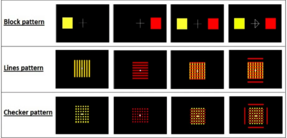
6 ≈ 14.167 Hz respectively. First, a block pattern [4, 5] with two 8 by 8 cm squares, one of
each color, separated by 12 cm with a white fixation cross in between (Fig.1, first row). Second, a lines pattern [6,7] with 7 vertical yellow 13 by 1 cm strips separated from each other by 1 cm, and 7 horizontal red strips with a white fixation square in the middle (Fig.1, second row). Third, a “checkerboard” pattern where all overlapping segments of the lines pattern were removed. This pattern is a 13 by 13 cm “checkerboard” made of red, yellow and black 1 by 1 cm squares with a white fixation square in the middle (Fig.1, third row). Subjects were instructed on which stimulus to attend by either an arrow indicating the left or the right stimulus (block pattern) or a matched color strips surrounding the pattern (Fig. 1, last column). EEG signals were recorded from 12 Ag/AgCl rings electrodes at location P3, P1, P2, P4, P O7, P O3, P Oz, P O4, P O8, O1, Ozand O2,
referenced to Pz based on the international 10-20 electrode system, with a BrainVision V-Amp
amplifier (band pass filter between 0.01 and 250 Hz with a sampling frequency of 1kHz). Eye movements were monitored with four electrodes: two on the left and right temples; the other two over and under the supra-orbital ridge.
Figure 1: The different patterns used. Columns 1 to 3 shows the two different stimuli separately and together, and column 4 illustrates how a stimulus is indicated to the subject.
Each subject underwent a total of 5 runs, each lasting around 6 min. Each run contained 30 7s-trials separated by 5s rest period. One of the three patterns was continuously flashing at the screen during a whole run. During a run, an equal number of both stimuli was presented in random order. The first run consisted of an overt block pattern where the subject was instructed to direct his/her gaze to the stimulus. This run ensured subject response to SSVEP stimuli. Next four runs alternated between covert lines and checkerboard pattern. The subject was instructed to fix his/her gaze on the white square in the middle and to concentrate on one of the stimulus. Half of the subjects started with the lines pattern. Instruction stayed on the screen for the whole trial duration. Inter-run periods were at the discretion of the subject and lasted 2 to 5 minutes. Runs from the same pattern were concatenated before analysis and trials order was randomized to remove all time effects.
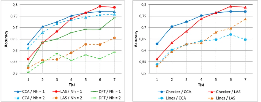
For each trial, the signal at EEG channels were individually decomposed in overlapping windows of length T with T ranging from 1 to 7s: seven 1s-windows starting from 0 to 6s, six 2s-windows starting from 0 to 5s, ..., one 7s-window starting at time 0. Frequency features were extracted from each window with three feature extraction algorithms proposed in the literature: discrete-time
Fourier transform (DFT), canonical correlation analysis (CCA) [8,9] and lock-in analyzer system (LAS) [3]. First and second harmonics of each stimulation frequency were extracted. Features set included either both harmonics or just the first. Classification performance were computed with a linear discriminant analysis and assessed with a 10-folds cross validation. The cross validation was repeated 5 times, then the results were averaged first for each time window, second for all subjects and time window of the same length. Analysis was done with Matlab and Cogent 2000 toolbox developed by the Cogent 2000 team at the FIL and the ICN and Cogent Graphics developed by John Romaya at the LON at the Wellcome Department of Imaging Neuroscience.
3
Results
Overt block pattern data from 2-s-window could be classified with more than 99% of accuracy for all subjects. Visual inspection of EOG showed no eye movement during the covert trials. The checkerboard pattern outperformed the lines pattern for every feature extraction algorithms and time windows (Fig.2, right). Classification accuracy increased with window length until reaching a plateau at 5s for CCA and 6s for LAS. DFT accuracy increased constantly. CCA has better accuracy than LAS and DFT for window smaller than 5s. CCA accuracy was 70 ± 3% and 75 ± 3% at 2 and 4s respectively. For 6s and 7s windows, LAS gave the best results reaching 79 ± 7% in average with 4 subjects at more than 81% (Fig.2, left). Using the first two harmonics instead of only the first one decreased the classification (Fig.2, left).
0,5 0,55 0,6 0,65 0,7 0,75 0,8 1 2 3 4 5 6 7 A ccur acy T(s) CCA / Nh = 1 LAS / Nh = 1 DFT / Nh = 1 CCA / Nh = 2 LAS / Nh = 2 DFT / Nh = 2 0,5 0,55 0,6 0,65 0,7 0,75 0,8 1 2 3 4 5 6 7 A ccur acy T(s)
Checker / CCA Checker / LAS Lines / CCA Lines / LAS
Figure 2: Relationship between recognition accuracy with the epoch length T for different number of harmonic Nh and feature extraction algorithms (left) and different patterns with a
single harmonic (right).
4
Conclusion
In the present paper, we studied the design of independent BCI based on covert SSVEP and showed that we can get around 80% accuracy. Due to the small number of subjects, all these results should be seen as preliminary. We proposed a new stimulation pattern which increased the classification of nearly 10% in comparison to a lines pattern [6,7] and seems to do better than moving dots pattern [9] but with different conditions. The new pattern has no overlap between stimuli which facilitates the concentration as reported by 4 out of 5 subjects; the last one being neutral. We also showed that CCA is the most suitable method for features extraction with window smaller or equal to 5s while LAS may be preferred for longer time window. These two algorithms gave better results than the classical DFT. Surprisingly and contrary to results from overt SSVEP [2], the use of the second harmonic leads to a decrease of classification accuracy. This could be due to over training of the classifier or to the use of a CRT screen instead of LEDs. However, a power spectrum analysis did not show any peak besides the ones of the first harmonics. No influence of higher harmonics has also been reported in overt SSVEP analysis with CCA [8].
These results acquired with only the first harmonic and advanced frequency feature extraction algorithms demonstrate that SSVEP BCI could be used independently of gaze control but also independently of training. Indeed, using the first harmonic and the CCA method give only two features for each stimuli. The maximum of the features gives the user command [8].
The short concentration time, 5s or 6s, the absence of training, the robustness make this method very well suited for detecting command following and testing communication in unresponsive post-comatose patients. Futures studies will concentrate on implementing an online version of the BCI and on testing on more subjects. The use of a restricted set of electrodes will also be tested.
5
Acknowledgment
Steven Laureys is Senior Research Associate; Andrea Soddu and Quentin Noirhomme are Post-doctoral Fellows at the Fonds de la Recherche Scientifique (FRS). This work is supported by the European ICT Programme Projects FP7-247919 DECODER, DISCOS, Mindbridge, COST, Mc-Donnell Foundation, Mind Science Foundation, FRS, Reine Elisabeth Medical Foundation and University and University Hospital of Li`ege. The text reflects solely the views of its authors. The European Commission is not liable for any use that may be made of the information contained therein.
References
[1] J.R. Wolpaw, N. Birbaumer, D.J. McFarlandm, G. Pfurtscheller, and T.M. Vaughan. Brain-computer interfaces for communication and control. Clinical Neurophysiology, 113:767–791, 2002.
[2] G.R. M¨uller-Putz, R. Schere, C. Brauneis, and G. Pfurtscheller. Steady – state visual evoked potential (ssvep)– based communication : impact of harmonic frequency components. Journal of Neural Engineering, 2:123–130, 2005.
[3] G.R. M¨uller-Putz, E. Eder, S.C. Wriessnegger, and G. Pfurtscheller. Comparison of dft and lock-in amplifier features and search for optimal electrode positions in ssvep-based bci. Journal of Neuroscience Methods, 168:174–181, 2008.
[4] S.P. Kelly, E.C. Lalor, B. Reilly, and J.J. Foxe. Visual spatial attention tracking using high–density ssvep data for independent brain–computer communication. IEEE Transactions On Neural Systems And Rehabilitation Engineering, 13(2):172–178, June 2005.
[5] S.P. Kelly, E.C. Lalor, C. Finucane, G. McDarby, and B. Reilly. Visual spatial attention control in an independent brain-computer interface. IEEE Transactions on Biomedical Engineering, 52(9):1588–1596, September 2005.
[6] Y. Chen, A.K. Seth, J.A. Gally, and G. M. Edelman. The power of human brain magnetoen-cephalographic signals can be modulated up or down by changes in an attentive visual task. Proceedings of the National Academy of Sciences, 6:3501–3506, March 2003.
[7] Z. Allison, D.J. McFarland, G. Schalk, S.D. Zheng, M. Moore Jackson, and J.R. Wolpaw. Towards an independent brain–computer interface using steady state visual evoked potentials. Clinical Neurophysiology, 119:399–408, 2008.
[8] G. Bin, X. Gao, Z. Yan, B. Hong, and S. Gao. An online multi-channel ssvep-based brain-computer interface using a canonical correlation analysis method. Journal of Neural Engineer-ing, 6(4):46002–46008, June 2009.
[9] D. Zhang, A. Maye, X. Gao, B. Hong, A.K. Engel, and S. Gao. An independent brain-computer interface using covert non-spatial visual selective attention. Journal of Neural Engineering, 7(1):16010–16021, 2010.