PROTEIN CRYSTALLOGENESIS OF THE ARROWHEAD
PROTEASE INHIBITOR-A
Mé moire
LE YI LIN
Maîtrise en biochimie
Maître è s sciences (M.Sc.)
Qué bec, Canada
© Le Yi Lin, 2015
Résumé
Les études sur la structure atomique des macromolécules biologiques ont été d'une grande importance en révélant des relations structurales et fonctionnelles d'enzymes, d'acides nucléiques et d'autres macromolécules dans divers systèmes biologiques. Dans la présente étude, la cristallogenèse et la cristallographie aux rayons X ont été utilisés pour déterminer la structure d'inhibiteur de protéinase A à une résolution atomique.
Les inhibiteurs de protéinase A et B (API-A,-B) de la arrowhead, sont les deux principaux composés inhibiteurs qui sont purifiés à partir de la arrowhead (Sagittaria sagittifolia,
Linn.). Ils sont des inhibiteurs à double tête multifonctionnels de diverses protéases. La structure du complexe ternaire de l'inhibiteur API lié à deux trypsines a été déterminée. Cependant, la structure tridimensionnelle de l'apoenzyme API-A est encore inconnue. A cet effet, la cristallogenèse de l’apoenzyme API-A a été réalisée. Après des étapes de purification et de cristallisation appropriées, des cristaux de l’apoenzyme API-A d’une qualité permettant la diffraction des rayons-X à haute résolution ont été obtenus en utilisant la méthode de diffusion de vapeur dans une goutte suspendue. Un ensemble complet de données de diffraction des rayons-X jusqu’à une résolution de 3,10 Å a été obtenu avec un crystal en forme de diamant, dont, le groupe d'espace a été déterminé comme étant P1. Ces données seront importantes pour comprendre la fonction d'API-A dans des systèmes biologiques.
Abstract
Structural studies of biological macromolecules at atomic resolution have revealed many specific aspects of the structure/function relationships of the enzymes, nucleic acids and other macromolecules in biological systems. In the present study, protein crystallogenesis and X-ray crystallography were used to determine the structure of the protease inhibitor A at atomic resolution.
The arrowhead protease inhibitors A and B (API-A, -B), are the two major inhibitor components which are purified from the tubes of arrowhead (Sagittaria sagittifolia, Linn.). Both API-A and API-B are double-headed multifunctional protease inhibitors. The ternary structure of the inhibitor API-A complex with two trypsins has been reported. However, the three-dimensional structure of the apoenzyme API-A is still unknown. In this regard, crystallogenesis of apoenzyme API-A was studied. After proper purification and crystallization steps, diffraction-quality crystals of the apoenzyme API-A were obtained, using the hanging drop vapor diffusion method. Moreover, a complete set of X-ray diffraction data was collected up to a resolution of 3.10 Å, using a diamond-shaped crystal whose space group was determined to be P1. These data will be important for understanding the function of API-A in biological systems.
Table of Contents
Résumé... iii
Abstract ... v
Table of Contents ... vii
List of figures ... ix
List of tables ... xi
List of abbreviations ... xiii
Acknowledgements ... xv
Chapter 1Introduction ... 1
1.1 Protease ... 2
1.2 Serine protease ... 5
1.3 Serine protease inhibitor ... 10
1.4 Arrowhead protease inhibitor A ... 17
1.5 Problematics ... 22
1.6 Research objective ... 23
Chapter 2Materials and methods ... 25
2.1 Methods in protein preparation ... 26
2.1.1 Cell culture ... 26
2.1.2 Protein purification ... 27
2.1.3 Measurement of protein concentration ... 29
2.1.4 Mass spectrometer ... 29
2.2 Methods used for protein crystallization ... 32
2.2.1 Preparation of protein solutions before crystallization ... 34
2.2.2 Vapor diffusion methods ... 34
2.2.3 Other methods for crystallization ... 36
2.2.4 New ideas in crystallization ... 39
2.2.5 Preparation of protein crystals before structure determination ... 46
2.3 Methods in protein structure determination ... 47
2.3.1 Nuclear magnetic resonance and X-ray crystallography ... 47
2.3.2 X-ray data collection ... 50
Chapter 3Results and discussion ... 53
3.1 Practice in crystallization methods ... 54
3.1.2 The effect of composition modification ... 56
3.2 Crystallogenesis study of API-A ... 59
3.2.1 Expression of API-A ... 59
3.2.2 Purification of API-A ... 62
3.2.3 Crystallization of API-A ... 65
3.3 X-ray data collection ... 67
Chapter 4Conclusion ... 71
Chapter 5Perspectives ... 75
Reference ... 77
List of figures
Figure 1. Schematic representation of a protein substrate binding to a protease. ... 2
Figure 2. Schematic representation of two possible enzyme-substrate complexes of papain with a hexapeptide. ... 3
Figure 3. The X-ray crystal structure of the archetypal serine protease chymotrypsin. ... 7
Figure 4. The catalytic triad in chymotrypsin complexes. ... 8
Figure 5. The generally accepted mechanism for serine proteases. ... 10
Figure 6. Possible mechanisms of proteinase inhibition. ... 16
Figure 7. Overall structure of API-A and its complex. ... 19
Figure 8. The views of the two interfaces. ... 21
Figure 9. Sequence display of API-A, from Protein Data Bank. ... 27
Figure 10. Mass spectrometry protocol. ... 31
Figure 11. Equilibration pathways in various protein crystallization methods. ... 33
Figure 12. Schematic representation of hanging drop, sitting drop and sandwich drop vapor diffusion method. ... 35
Figure 13. Classification scheme for the results of a crystallization screen. ... 38
Figure 14. Two-dimensional-phase diagrams of protein concentration versus PEG concentration. ... 40
Figure 15. Drop volume ratio effect on supersaturation. ... 42
Figure 16. Two-dimensional-phase diagrams of thaumatin (A) and proteinase K (B), and prediction of trajectories of varying composition modifications at 300 K and 295 K, respectively. ... 44
Figure 19. Agarose gel electrophoresis of DNA extracted from cultured cells. ... 59
Figure 20. SDS-PAGE image of bacterial supernatant and Ni-NTA column fractions. ... 62
Figure 21. SDS-PAGE image of size exclusion chromatography fractions. ... 63
Figure 22. SDS-PAGE image of blue-sepharose affinity chromatography. ... 64
Figure 23. Peptide analysis of the two bands API-A (above is upper band and below is lower band) by the method of mass spectrometry. ... 65
Figure 24. Main shapes of crystals obtained. ... 66
List of tables
Table 1. Families of peptidase inhibitors... 13
Table 2. “Hits” and “Hits Increase” obtained by conventional screening and composition modification. ... 45
Table 3. The summary table of released entries from Protein Data Bank (PDB). ... 50
Table 4. The effect of composition modification to the crystallization of glucose isomerase. ... 57
Table 5. The effect of composition modification to the crystallization of β-amylase. ... 58
Table 6. Result of the pET28a-derived target gene sequencing. ... 61
Table 7. The summary data collection. ... 68
List of abbreviations
Å: angstrom Aa: amino acids
API: Arrowhead Protease Inhibitors bp: base pair
BSA: bovine serum albumin
℃: Degree Celsius
ESI:electrospray ionization g: gram
HPLC:high-performance liquid chromatography
IPTG: isopropyl-β-D-thiogalactopyranoside kDa: kilodalton
L: liter M: molar
MALDI: matrix-assisted laser desorption/ionization β-Me: β-mecaptoethanol mg: milligram min: minute ml: milliliter mM: millimolar MPD: 2-Methyl-2, 4-Pentanediol MS: mass spectrometry
MS/MS: tandem mass spectrometry m/z:mass-to-charge ratio
ng: Nanogram O.D.: optical density
PAGE: polyacrylamide gel electrophoresis PCG: Protein Crystal Growth
PDB: Protein Data Bank PEG: polyethylene glycol PIs: protease inhibitors
PMSF: phenylmethanesulfonyl fluoride rpm: revolutions per minute
SDS: sodium dodecyl sulfate sec: second
serpins: serine protease inhibitors Tris: tris(hydroxymethyl)aminomethane µg: microgram
Acknowledgements
To undertake and finish this master degree is not easy, fortunately, with the help from my supervisors, my family members, the committee members, the dean and secretary of department, I got more than that I could imagine, the master degree in biochemistry.
My supervisors professor Jacques Lapointe and professor Sheng-Xiang Lin, thank you for giving me the experience of being your student, your gentleness and patience will be remembered through all my life. During more than two years of study, I met too many problems both in study and in daily life, your kindness and patience helped me to get over those difficulties, and helped me to succeed.
I would thank my wife Zhi Qian Zhou, my parents and parents in law, they supported me all the time, gave me courage to conquer the difficulties.
My committee members, Dr. Manon Couture and Dr. Patrick Lagüe, thank you for your acknowledgements in my exams, with your great encouragement, I took back my confidence. And especially thank you for your approvals in my decision, I appreciate that a lot.
Dr. Louise Brisson, Dr. Lisa Topolnik and Mme. Véronique Bédard, I had so many problems to ask for your favor, and you all helped me dealing with them, then gave me the best results, thank you for your understanding as well as your great help.
Great thanks to Dr. Rong Shi, for offering me help in editing my memory and for those useful suggestions regarding my research.
My colleagues in CHUL, Dr. Dao-Wei Zhu, Dr. Ming Zhou, Bo Zhang, Chenyan Zhang, Xiao-Qiang Wang, Dan Xu, Jean-François Thériault, Mouna Zerradi, also very excellent colleagues in Laval University, Jonathan Huot, Sébastien Blais, Van-Hau Pham, Guillaume Bonnaure and Imane Fiher. Thank you all for your help during my study and experiments. Thanks to professor Stéphane Gagné, for his kindness on offering me the course on protein structure based on my special situation.
Chapter 1
Introduction
1.1 Protease
A protease (also termed peptidase or proteinase) is defined as an enzyme that performs proteolysis. Proteases can be found in animals, plants, bacteria, archaea and viruses. Sequencing of the human genome revealed that more than 2% of our genes encode proteases (1-5).
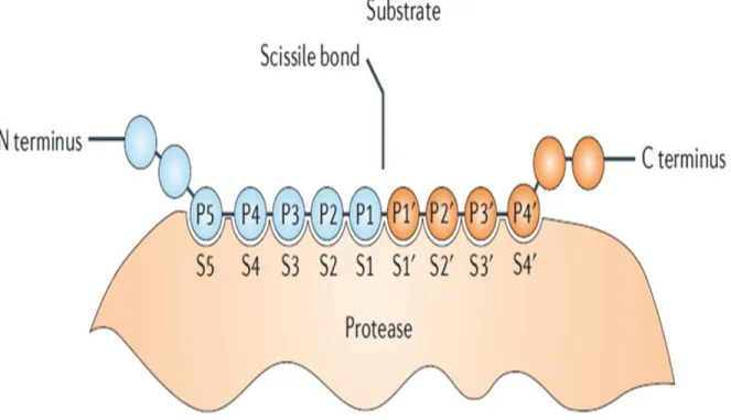
The protein catabolism by a protease results from the hydrolysis of the peptide bonds that link amino acids together in polypeptide chain (Figure 1). Most proteases are relatively nonspecific for their substrates, and target multiple substrates in an indiscriminate manner, whereas some proteases exhibit an exquisite specificity toward a unique peptide bond of a single protein (1).
Figure 1. Schematic representation of a protein substrate binding to a protease.The surface of the protease that is able to accommodate a single side chain of a substrate residue is called the subsite. Subsites are numbered S1-Sn upwards towards the N terminus of the substrate, and S1’-Sn’ towards the C terminus, beginning from the sites on each side of the scissile bond. The substrate residues they accommodate are numbered P1-Pn, and P1’-Pn’, respectively (5).
Generally, the active site of an enzyme performs two functions, which are binding the substrate and catalyzing the reaction. For example, Papain is an endopeptidase (which cleaves protein substrates in the middle of the molecule) which has a large active site that extends over about 25 Å and can be divided into 7 “subsites” (Figure 2). The subsites are located on both sides of the catalytic site, 4 on the one side and 3 on the other. Each subsite accommodates one amino acid residue of the peptide substrate. The “positions” of the residues in the substrate peptide were numbered according to the “subsites” they occupy, and also depending on which bond is split. So S1’ and S1 are flanking the catalytic site, whereas hydrolysis will occur between residues P1 and P1’ (6, 7).
Figure 2. Schematic representation of two possible enzyme-substrate complexes of papain with a hexapeptide. The active site of the enzyme is composed of 7 ‘subsites’ (S1-S4 and Sl’-S3’) located on both sides of the catalytic site C. The positions, P, on the hexapeptide substrate are counted from the point of cleavage and thus have the same numbering as the subsites they occupy. Complex A will yield as products two molecules of tripeptide, B one molecule of tetrapeptide and one of dipeptide (6).
Besides the generalized protein digestion, proteases possess complex functions. For example, proteases regulate growth factors, cytokines, chemokines and cellular receptors, both through activation and inactivation, which can lead to intracellular signaling, gene regulation, inflammation, apoptosis, blood coagulation, and embryogenesis (3-5, 8-10). Proteases are accordingly used widely in medical fields; the predominant use of proteases has been in treating cardiovascular diseases, but they are also emerging as useful agents in the treatment of sepsis, digestive disorders, inflammation, cystic fibrosis, retinal disorders, psoriasis and other diseases (4). Up-regulation of proteolysis is associated commonly with different types of cancer and is linked to tumor metastasis, invasion and growth (9). Proteases also play key roles in plants, where they contribute to the processing, maturation, or destruction of specific sets of proteins in response to developmental cues or to variations in environmental conditions (11). Likewise, many infectious microorganisms require proteases for replication or use proteases as virulence factors, which has facilitated the development of protease-targeted therapies for diseases of great relevance to human life such as AIDS (5). Proteases are also important tools of the biotechnological industry where they are used as biochemical reagents or in the manufacture of numerous products (11).
Proteases have evolved multiple times. The comparisons of the amino acid sequences, three-dimensional structures, and enzymatic reaction mechanisms of proteases, have revealed the existence of distinct families, each of them performing the same proteolytic reactions by completely different catalytic mechanisms (1, 2, 5, 8).
Proteases were initially classified into endopeptidases, which cleave protein substrates in the middle of the molecule, and exopeptidases, which cleave protein from the N or C termini (aminopeptidases and carboxypeptidases, respectively), the action of which is directed by the
NH2 and COOH termini of their corresponding substrates. However, new classification schemes
were made according to the availability of structural and mechanistic information on these enzymes. Based on the mechanism of catalysis, proteases are classified into six distinct classes:
aspartic, glutamic, and metalloproteases, cysteine, serine, and threonine proteases. Glutamic proteases have not been found in mammals so far. The first three classes, aspartic, glutamic, and metalloproteases, utilize an activated water molecule as a nucleophile, to attack the peptide bond of the substrate, whereas for cysteine, serine, and threonine proteases, the nucleophile is an amino acid residue (Cys, Ser, or Thr, respectively) located in the active site. By amino acid sequence comparison, proteases of the different classes can be further grouped into families, and families can be assembled into clans based on similarities in their three-dimensional structures. Metalloproteases and serine proteases are the most densely populated classes, with 194 and 176 members, respectively, followed by 150 cysteine proteases, whereas threonine and aspartic proteases contain only 28 and 21 members, respectively (1, 5).
1.2 Serine protease
The serine proteases, also called serine endopeptidases, are characterized by a uniquely reactive serine side chain. The serine proteases are widely distributed and of diverse functions, and are found in both eukaryotes and prokaryotes. Almost one-third of all proteases can be classified as serine proteases (12, 13).
Serine proteases can be classified into two broad categories based on their structure: chymotrypsin-like (trypsin-like) and subtilisin-like (14). In humans, they are responsible for co-ordinating various physiological functions, including digestion, immune response, blood coagulation and reproduction (13).
Chymotrypsin-like serine proteases are the most abundant in nature. They have a distinctive structure, which consists of two β-barrel domains that converge at the catalytic active site. According to the substrate specificity, chymotrypsin-like serine proteases can be further
chymotrypsin-like (large hydrophobic residues Phe/Tyr/Leu at P1) or elastase-like (small hydrophobic residues Ala/Val at P1) (15).
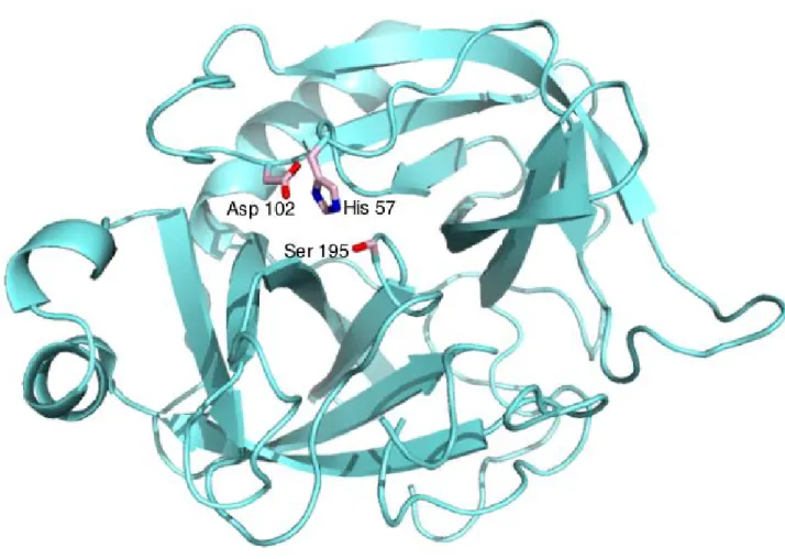
A good example of this category is chymotrypsin. It has 245 residues, arranged in two six-stranded beta barrels (Figure 3). The active site cleft is located between the two barrels. This structure is generally divided into catalytic, substrate recognition and zymogen (an inactive enzyme precursor which requires a biochemical change to become an active enzyme) activation domain components. These three processes involve many of the same structural features and are intricately intertwined. Moreover, these domains are common to all chymotrypsin-like serine proteases (13).
Subtilisin, a serine protease found in prokaryotes, is evolutionarily not related to the chymotrypsin-clan, but share the same catalytic mechanism, which is using a catalytic triad to create a nucleophilic serine. The comparison of chymotrypsin and subtilisin is the classical example used to illustrate convergent evolution, since the same mechanism evolved twice independently in topologically different proteins during evolution (12, 13).
Figure 3. The X-ray crystal structure of the archetypal serine protease chymotrypsin (16).
The three catalytic residues (His 57, Asp 102 and Ser 195) are labeled.PDB number: P00766
(CTRA_BOVIN).
The main function for all proteases is to hydrolyze peptide bonds. To achieve this, there are three obstacles to overcome: (a) amide bonds are very stable due to electron donation from the amide nitrogen to the carbonyl group. Proteases usually use a general acid to interact with the carbonyl oxygen, to activate an amide bond of the substrate; (b) water is a poor nucleophile; proteases always activate water, usually via a general base; and (c) amines are poor leaving groups; proteases protonate the amine prior to expulsion. These mechanisms can also be used to hydrolyze other acyl compounds, including amides, anilides, esters, and thioesters (13). Before carrying out the proteolysis reactions, the proteases must establish themselves in appropriate
most cases, they are present in complex networks, which include other proteases, substrates, cofactors, inhibitors, adaptors, receptors, and binding proteins (1).
The reaction mechanism of the serine proteases was originally revealed by the presence of the Asp-His-Ser “charge relay” system or “catalytic triad”, in which serine serves as the nucleophilic amino acid at the active site (Figure 4). More recently, serine proteases with novel catalytic triads and dyads have been discovered, including Ser-His-Glu, Ser-Lys/His, His-Ser-His, and N-terminal Ser (16, 17).
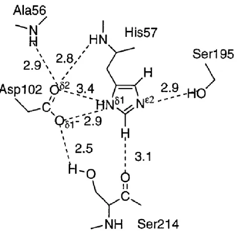
Figure 4. The catalytic triad in chymotrypsin complexes.The hydrogen bonding network of the catalytic triad is depicted for the complex with the protein inhibitor eglin C. The dotted lines represent potential hydrogen bonds (13).
The catalytic triad is part of an extensive hydrogen bonding network. It spans the active site cleft, with Ser195 on one side and Asp102 and His57 on the other (Figure 3). Generally, hydrogen bonds are shown between the Nδ1-H of His57 and Oδ1 of Asp102, also between the OH of Ser195 and the Nε2-H of His57, although the latter hydrogen bond is lost when His57 is protonated. The complexes of chymotrypsin and protein inhibitor eglin C are discussed to explain the mechanism in serine protease (Figure 4). The hydrogen bonds are observed throughout the catalytic cycle. Moreover, the OH of Ser214 forms a hydrogen bond with Oδ1 of Asp102, this hydrogen bond can be found in almost all chymotrypsin-like proteases. Accordingly, Ser214 was once considered the fourth member of the catalytic triad, although more recent evidence indicates that it is dispensable. Hydrogen bonds are also observed between the Oδ2 of Asp102 and the main chain NHs of Ala56 and His57. These hydrogen bonds are believed to orient Asp102 and His57. Additionally, a novel hydrogen bond is observed between the Cε1-H of His57 and the main chain carbonyl of Ser214, the carbonyl oxygen of Ser214 also plays a part in the polypeptide binding site, so it was considered to mediate the communication between substrate and the catalytic triad. Similar hydrogen bonds are also observed in subtilisin and other classes of serine hydrolases, which suggests that this interaction may be important for catalytic triad function (13, 18).
An oxyanion hole (a pocket in the structure of an enzyme which stabilizes a deprotonated oxygen or alkoxide, often by placing it close to positively charged residues) was believed to exist and it is formed by the backbone NHs of Gly193 and Ser195 (Figure 5). These atoms form a pocket of positive charge, and the pocket can activate the carbonyl of the scissile peptide bond and stabilize the negatively charged oxyanion of the tetrahedral intermediate. The oxyanion hole is structurally linked to the catalytic triad and the Ile16-Asp194 salt bridge via Ser195. Subtilisin also contains an oxyanion hole, formed by the side chains of Asn155, Thr220, and the backbone NH of Ser221 (13, 19).
Figure 5. The generally accepted mechanism for serine proteases (13).
The generally accepted mechanism for chymotrypsin-like serine proteases is shown (Figure 5). In the acylation half of the reaction, Ser195 attacks the carbonyl of the peptide substrate, with the assistance by His57 who acts as a general base, to yield a tetrahedral intermediate. The resulting His57-H+ is stabilized by the hydrogen bond to Asp102. The oxyanion of the tetrahedral intermediate is stabilized by interaction with the main chain NHs of the oxyanion hole. Then the tetrahedral intermediate collapses with expulsion of the leaving group, assisted by His57-H+ acting as a general acid, to yield the acylenzyme intermediate. The deacylation half of the reaction repeats the above sequence: water attacks the acylenzyme, assisted by His57, yielding a second tetrahedral intermediate. Finally this intermediate collapses, expelling Ser195 and carboxylic acid product (12, 13).
1.3 Serine protease inhibitor
As they catalyze the cleavage of peptide bonds, the proteases can be potentially very damaging in living systems, so their activities need to be kept strictly under control. The action of proteases
can be controlled in vivo by several mechanisms: regulation of gene expression; activation of their inactive zymogens; blockade by endogenous inhibitors; targeting to specific compartments such as lysosomes, mitochondria, and specific apical membranes; and post-translational modifications such as glycosylation, metal binding, S–S bridging, proteolysis, and degradation (1). Among these mechanisms, the interactions between proteases and their inhibitors can be very efficient and important (20, 21).
Protease inhibitors (PIs) can regulate their corresponding proteases in a very significant way. They are widely distributed in all organisms, including microorganisms, plants and animals. Many of these inhibitors have a much larger size than their target proteinases. Only some microorganisms produce and secrete small non-proteinaceous inhibitors, which impair the proteolytic activity of host proteinases (21).
Protease inhibitors are widely used for clinical applications. Some inherited diseases attributable to abnormalities of proteases were proved to be susceptible to treatment with the inhibitors administered as drugs, with synthetic inhibitors that take over their function or with the natural inhibitors made available by gene therapy (20). There is great interest in developing more potent therapeutic PIs for treating human diseases related to cancer (22), pancreatitis (23), thrombosis (24), and AIDS (25).
The number of proteinaceous protease inhibitors isolated and identified so far is extremely large. These inhibitors can have very different polypeptide scaffolds, and some of them are bis- or multi-headed tandem proteins (20, 21, 26). They have been classified into families of related structures or according to the catalytic class of their target proteases. However, this classification is hampered by the occurrence of both compound inhibitors and pan-inhibitors; the compound inhibitors contain inhibitor units of different protease classes, whereas the pan-inhibitors target enzymes of different classes through a trapping reaction induced after inhibitor cleavage by the targeted protease (1, 27).
Protease inhibitors can be classified functionally as single-headed (if they have only one active reactive site), double-headed (if they have two) and so on (28). They can also be classified according to their mechanism of inhibition into four groups: the canonical inhibitors using substrate-like binding mode; exosite-binding inhibitors (exosites are the regions outside of the active site that influences catalysis), which bind a region adjacent to the active site; a third group of protease inhibitors use an intermediate mechanism based on a combination of the canonical and exosite-binding mechanisms; finally, allosteric inhibitors bind a region that is distantly located from the active site (1, 21).
In the paper of Laskowski and Kato (28), protease inhibitors could best be classified in their homologous families, but at that time the available sequence information was very limited. In recent years, a system was created, in which the inhibitor units of the protease inhibitors are assigned to 48 families on the basis of similarities detectable at the level of amino acid sequence. Then, on the basis of their three-dimensional structures, 31 of the families are assigned to 26 clans (20).
In most cases, all members of a specific inhibitor family are directed against the target proteinases of the same mechanistic class; only a very few protein inhibitors exhibit ‘dual’ activity simultaneously exerted towards proteinases from different mechanistic classes.
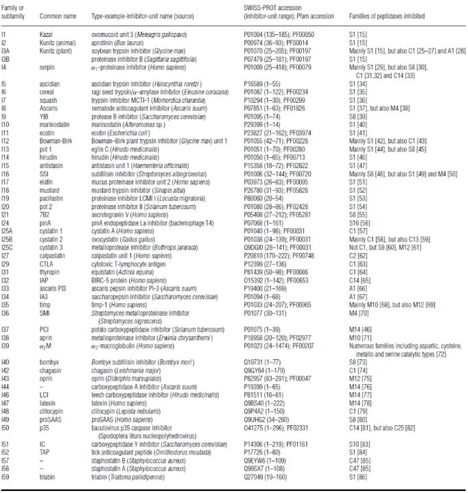
Table 1. Families of peptidase inhibitors (20).
The ways by which the inhibitors interact with their target enzymes vary enormously. Concerning the interactions between the inhibitors and their target proteases, two general types
can be recognized: irreversible ‘trapping’ reactions, and reversible tight binding interactions (20). The irreversible trapping reaction depends most directly on the peptidase activity of the target protease; it is covalently modified and thereby inhibited by its proteolytic activation of the protease inhibitors. This kind of inhibitor acts as a substrate, utilizes the enzymes catalytic machinery to trap and inhibit the protease. This kind of reaction is specific for endopeptidases because it depends upon the cleavage of an internal peptide bond within the inhibitor, cleavage that triggers a conformational change. If the target protease is in its catalytically inactive form, it will fail to enter into a trapping reaction, no matter whether it may well bind tightly to an inhibitor from one of the reversible classes. Trapping reactions are never truly reversible because the inhibitors are modified and reformed, which can also be described as a suicide inhibitor or suicide substrates.
For the reversible tight-binding interactions, the inhibitor makes high-affinity interactions with the active site of the target enzyme. The mechanism here is termed the ‘standard’ mechanism. In a standard mechanism inhibitor, the inhibitory unit has a single reactive-site peptide bond, and inhibition is caused by the binding of the inhibitor to the target enzyme in a substrate-like fashion.
The reversible tight binding interactions mechanisms between proteases inhibitors and their target proteases can be classified in four kinds (Figure 6). Firstly, for the canonical inhibitors, they bind the active site of their target proteases and block directly the active site in a virtually substrate-like manner. Secondly, the exosite-binding inhibitors, do not bind the active site of target proteases to directly block the catalytic residues, but they bind a region adjacent to the
active site, thereby preventing substrate access to this center.The exosite binding provides two
major benefits: it increases the surface area of the protein-protein interaction, leading to a greater affinity, and it can have a significant effect on the specificity of the inhibitor. The third kind of mechanism is carried out by a group of protease inhibitors using an intermediate mechanism
based on a combination of the canonical and exosite-binding mechanisms. Finally, the mechanism of allosteric inhibitors includes the binding of a region that is distantly located from the active site, but this binding prevents dimerization of the target protease and can effectively block its activity (21).
1.4 Arrowhead protease inhibitor A
The inhibitors of soybean Kunitz-type serine proteases generally contain 170–200 residues and have two disulfide bonds. Most members have only one reactive site, normally located in the region of residues 60–70 (29-31). However, a few members termed double-headed inhibitors have two reactive sites, which can simultaneously bind two protease molecules; these inhibitors are classified into family I3 of peptidase inhibitors (Table 1) (20). Most members of this type are grouped into subfamily I3A. However, owing to their very low sequence similarity to other members, the double-headed arrowhead protease inhibitors API-A and API-B, purified from arrowhead Sagittaria sagittifolia, Linn., are grouped in subfamily I3B (20). API-A and API-B, consist of 179 residues and three disulfide bonds. They can inhibit various serine proteases, including trypsin, chymotrypsin, and porcine tissue kallikrein (32, 33).The API-A and API-B share 91% of sequence identity, though their inhibitory specificities differ. API-A can bind one molecule of trypsin and another molecule of chymotrypsin, whereas the API-B can bind two molecules of trypsin simultaneously (34).
The overall fold of API-A belongs to the β-trefoil fold with a core of six antiparallel β-strands (β1 to β12) surrounded by 13 loops, which resembles that of the Kunitz-type trypsin inhibitors. The hydrophobic core consists of a shallow β-barrel formed by three β-ribbons (β1- β12, β4- β5
and β8- β9) and a cap of three hairpins (β2- β3, β6- β7 and β10- β11). A 310 helix is located
between loop 8 and loop 9. In addition, API-A contains three disulfide bonds. The first bond (Cys43–Cys89) cross-links loop 3 and loop 6 of reactive site 1 (RS1), whereas the other two bonds
are located in RS2, the one of (Cys141–Cys144) forms an intraloop disulfide bond in loop 10, the
other one of (Cys139–Cys148) stabilizes loop 10 with strand β9. These disulfide bonds were
considered to play a role in stabilizing the conformation of the reactive sites (35) (Figure 7A). The reactive sites 1 (RS1) and 2 (RS2) of API-A were identified as Leu87 and Lys145, respectively, and both reactive sites adopt a typical noncovalent “lock and key” inhibitory mechanism in a
Reactive site 1, composed of residues P5 Met83 to P5’ Ala92, has a novel conformation with the
Leu87, which is embedded in the S1 pocket completely, even though it is not a favorable P1 residue for trypsin. Reactive site 2, composed of residues P5 Cys141 to P5’ Glu150, the P1 residue
Lys145 adopts a classic mode binding with trypsin, using a two-disulfide-bonded loop (Figure 7,
Figure 7. Overall structure of API-A and its complex. A, overall structure of API-A, with β-strands and loops numbered sequentially. Two P1 residues and six cysteine residues are highlighted as yellow sticks. B, overall structures of the API-A ternary complex with trypsin. API-A is colored by secondary structure assignment: 310 helix inred, strands inblue, and loops
The crystal structure of API-A in complex with two molecules of bovine trypsin (Figure 7B), was the first report on the three-dimensional structure of this double-headed Kunitz-type trypsin inhibitor in complex with two molecules of protease. It shows a ternary complex of the double-headed inhibitor API-A simultaneously bound to two trypsin molecules, where the two reactive sites of API-A adopt different conformations (35).
At the reactive site 1, residues P1’ to P3’ of API-A adopt a novel conformation, whereas a hydrogen bond network bridges loops 6 and 8 (Ile88-O-Phe115-N, Asp90-N-Asp113-O, and
Asp90-Oδ2-Asp113-N); such a conformation has not been reported for other Kunitz-type inhibitors.
Loop 6 becomes more rigid because of the intraloop hydrogen bond between Pro86-O and
Cys89-N. Moreover, the interloop disulfide bond Cys43-Cys89 between loops 6 and 3 further
stabilizes the first reactive site loop and thus also contributes to the novel conformation of RS1. The reactive site 2 adopts a canonical conformation even though the presence of two disulfide bonds (Cys139-Cys148 and Cys141-Cys144) makes it different from other protease inhibitors that
contain only one disulfide bond.
Notably, the three important spacer residues Arg76, Glu124, and Arg126 adopt a unique interaction pattern that is different from the patterns in other Kunitz-type inhibitors. Arg76 forms two direct hydrogen bonds with P2 (Arg76-Nη2-Cys144-O) and P4 (Arg76-Nε-Glu142-O) and one indirect
hydrogen bond with P4 (Arg76-O-Glu142-Oε2) that is mediated by a water molecular, Wat67. The
hydrogen bonds contributed by Arg76 are important for stabilizing RS 2, thus making it
indispensable for maintaining the inhibitory activity.
The two other spacer residues Glu124 and Arg126 also participate in two salt bridges and one
hydrogen bond (Glu124-Oε1-Arg76-Nη1, Glu124-Oε2-Arg126-Nε, and Arg126-Nη2-Cys148-O). In
addition, P4 Glu142 and P4’ Pro149 form hydrogen bonds with the neighboring residues Ser64
(Ser64-N-Glu142-Oε2) and Ala138 (Ala138-N-Pro149-O), respectively. This interaction network in
addition to two disulfide bonds gives loop 10 a well defined conformation that aids in the inhibitory activity.
Figure 8. The views of the two interfaces. A and B, represent interfaces 1 and 2 between reactive sites of API-A (in green) and trypsin (in magenta) (35). The residues oftrypsin-1 and -2 are labeled with a prime and a double prime, respectively. All hydrogen bonds are shown as dashedlines. The water molecules (Wat3, Wat6, and Wat99) are shown as red spheres.
For the interface 1, the P1 residue Leu87 snugly fits into the S1 pocket of trypsin-1. The formation of a typical intermolecular antiparallel β-sheet, which is stabilized by three main chain hydrogen bonds, also helps the binding conformation at interface 1. Moreover, the residues surrounding RS1 also contribute to the enzyme-inhibitor interaction at interface 1, via hydrophobic and hydrophilic interactions. For the interface 2, there are classic protease-inhibitor interactions presented at the interface between RS2 and trypsin-2. In accordance with the
canonical binding mode, the carbonyl group of the scissile peptide bond between P1 Lys145 and
P1’ Ile146, is snugly embedded in the oxyanion hole. The main chain hydrogen bonds are mainly
contributed by P1 Lys145 (Figure 8) (36). In addition to direct hydrogen bonds, the Nζ atom of P1
Lys145 forms five indirect hydrogen bonds mediated by two water molecules Wat3 and Wat6.
1.5 Problematics
We have mentioned the essential role of proteases in cell physiology, and its relationship with several pathological conditions such as cancer, neurodegenerative disorders, and inflammatory and cardiovascular diseases. Many proteases are a major focus of attention for the pharmaceutical industry as potential drug targets against these diseases, or as diagnostic and prognostic biomarkers (1, 5).
In the clinical applications against diseases caused by proteases, protease inhibitors are often utilized as treatments to bind the target protease in a substrate-like manner, which can inactivate or control the proteases. Many protease inhibitors have been proved to have the potential of treating human diseases (22-25). It is well known that many of the protease inhibitors contain multiple homologous inhibitor domains in a single polypeptide chain. These domains are not identical to each other, but are functional inhibitors in their own right (20). Accordingly, the identification of the possible inhibitor units (domains) in a certain inhibitor can help finding out its potential target proteases. This would have a significant impact in the clinical application of this inhibitor.
As one of the Kunitz-type serine protease inhibitors, the sequence of API-A was determined in 1992 (32). Until the year of 2009, the structural data of API-A was obtained by the method of X-ray crystallography (35), which is the only three-dimensional structure of API-A being published. However, in this report, the API-A was crystallized with trypsins, the structure obtained is the ternary structure of the API-A complex with two molecules of trypsin. It is known that the conformation of many proteins changes upon binding to a ligand (37), so we may think that because of binding to trypsins, the conformation of API-A was changed by the interactions at the active sites between API-A and trypsins. Owing to the conformational changes, it could be difficult to recognize the other possible inhibitor units of API-A. Because of this, it will be very interesting to know the structural conformation of API-A in its apoenzyme form. Maybe some other possible active sites will be found, which could help in finding other potential target proteases or some other possible substrate binding patterns.
Secondly, after knowing the structural conformation of API-A in its apoenzyme form, it will be interesting to compare the conformations both in its apo-protein form and in complex. Knowing the conformational changes of API-A when it binds to the proteases can be helpful to the studies of protein-protein interactions, and also be useful in designing and developing other therapeutic inhibitors of the same catalytic mechanism.
1.6 Research objective
To better understand the principle of protein crystallization, as well as to improve the crystallization success, different protein crystallization methods may be used, such as the methods of relative crystallizability and composition modification.
In order to obtain the conformational information of the apo form of API-A, we have chosen X-ray crystallography to determine its structure. To produce a crystal of apo API-A with adequate quality for X-ray crystallography, various methods of protein crystallogenesis were
collected. Finally, the data processing steps such as the phase determination, model building and refinement, are used to obtain the three-dimensional structure of apo form of API-A.
Chapter 2
2.1 Methods in protein preparation
The first step in a project of protein structure determination by X-ray diffraction is to obtain a highly pure and homogeneous sample of a solution of that protein. As relatively large quantities, normally a few milligrams of proteins, are needed to form crystals, and a proper concentration of the protein solution is important to produce crystals, that protein is usually overexpressed before its purification.
2.1.1 Cell culture
The cells expressing API-A in Escherichia coli BL21 (DE3) were provided by the laboratory of professor Cheng-Wu Chi, Shanghai Institutes for Biological Sciences (Figure 9). The target gene of API-A was cloned into a pET28a-derived expression vector encoding 6-histidine residues (His6 tag at the N-terminus) after the start codon. The constructs were co-transformed with
PKY206, a plasmid containing the groESL genes of E. coli, encoding the chaperones GroEL and GroES (35), in order to optimize the correct folding of API-A.
The cultured cells were grown at 310 K, in 2×YT medium (5 g of NaCl, 16 g of bactotryptone, 10 g of yeast extract in 1 liter of H2O), containing kanamycin (SIGMA) and tetracycline
(SIGMA) that added at a final concentration of 30 µg/ml and 10 µg/ml respectively. When the culture had reached an OD600 nm of 0.7, protein expression was induced by adding isopropyl
1-thio-β-D-galactopyranoside to a final concentration of 0.2 mM, and the culture was then incubated at 16 °C for 20 hours (35).
Figure 9. Sequence display of API-A, from Protein Data Bank (35).
2.1.2 Protein purification
All the procedures were carried out at 4 °C or on ice unless otherwise specified. Cells (8g) were collected from cell culture, centrifuged at 3,400 rpm, 4 °C for 20 min, (Thermo Scientific HLR-6 rotor, Sorvall RC-3B Plus Refrigerated Centrifuge) and re-suspended in the buffer A containing 300 mM NaCl, 50 mM Tris-HCl, at pH 7.5, 10% glycerol, 0.5 mM PMSF, 10 mM β-Me. Cell
lysis was carried out as follows: after adding 1 mg/ml lysozyme (from chicken egg white, Sigma-Aldrich), 20 mM imidazole and 1 µg/ml of each of the following protease inhibitors (leapetin, chymostain, antipan, aprotinin and pepstatine A), the mixture was left on ice for 15 min before sonication. The sonication was carried on ice with ten 8-s bursts separated by 10-s intervals at output 2.5, using a Sonic Dismembrator (Model F550, Fisher Scientific).
The homogenates after sonication were centrifuged for 45 min at 42,000 rpm (70 Ti rotor, Beckman), at 4 °C. The supernatants were collected and were filtered through syringe filter (0.2 µm pore size, Filtropur S), and then mixed with 5 ml of the nickel-nitrilotriacetic acid affinity resin (Qiagen) pre-equilibrated with buffer B (buffer A containing 20 mM imidazole), and incubated by rotating for 4 h at 4 °C. Then the mixture was loaded onto a column and the flowthrough was collected. The column was washed with 10 column volumes of buffer B, and 10 column volumes of buffer C (buffer A containing 30 mM imidazole). Bound proteins were eluted with buffer D (buffer A containing 300 mM imidazole).
The fractions of high protein concentration were collected into a total volume of 1 ml, using an Amicon Ultra 10-kDa cut-off concentrator (Millipore). This small volume is suitable for the next purification step by gel filtration chromatography. The Hiload 16/60 Superdex 75 (Amersham Biosciences) column was chosen. Before use, the column was equilibrated with buffer E (containing 50 mM Tris-HCl, at pH 7.5, 10% glycerol, 0.5 mM PMSF, 10 mM β-Me). A flow rate of 1 ml/min was used for both sample loading and washing.
Blue sepharose was chosen as the last purification step. After equilibration in buffer E, the sample protein was loaded at the flow rate of 1 ml/min and the column was washed with the same buffer until the optical density baseline was stable. After the unbound proteins were washed out, the bound sample was eluted using an increasing linear salt gradient, from 100% buffer E to 100% eluent buffer F (buffer E containing 500 mM NaCl).
2.1.3 Measurement of protein concentration
The Bradford assay was chosen to determine the protein concentration. The Bradford assay is a colorimetric protein assay based on an absorbance shift of the dye Coomassie Brilliant Blue G-250. By binding the sample protein, the dye of red form is converted into its blue form. During the formation of this complex, two types of bond interactions take place: the red form of Coomassie dye first donates its free electron to the ionizable groups on the protein, which disrupt the protein’s native state, consequently exposing its hydrophobic pockets. These pockets in the protein’s tertiary structure bind non-covalently to the non-polar region of the dye via van der Waals forces, positioning the positive amine groups close to the negative charge of the dye. The bond is further strengthened by the ionic interaction between the two. The binding of the protein stabilizes the blue form of the Coomassie dye. Thus the amount of the complex present in solution is a measure for the protein concentration, and can be estimated by use of an absorbance reader. The bound form of the dye has an absorption spectrum maximum at 595 nm. The increase of absorbance at 595 nm is proportional to the amount of bound dye, and thus to the amount (concentration) of protein present in the sample.
The Bradford protein assay is less susceptible to interference by various chemicals that may be present in protein samples. However, detergents such as sodium dodecyl sulfate and triton x-100 can interfere with the assay, as well as strongly alkaline solutions (38).
In this study, all the procedures were almost free of the use of detergents. A Beckman DU-70 spectrophotometer was used for the measurements of protein concentration. BSA was used as the standard.
2.1.4 Mass spectrometer
As a method for protein identification, we used the tandem mass spectrometry (MS/MS) identification, which is nowadays a well established method (39) (Figure 10). Being widely used to measure the characteristics of molecules, it converts them to ions so that they can be moved
about and manipulated by external electric and magnetic fields.
The mass spectrometer is composed of three elements: firstly, an ion source, such as a high energy electron beam, which causes the sample protein ionized to cations by loss of an electron; two techniques are commonly used in this step, the electrospray ionization (ESI) or matrix-assisted laser desorption/ionization (MALDI). Then through the mass analyzer, the ions are sorted and separated according to their mass and charge. This step is achieved by accelerating and focusing the ions in a beam, which is then bent by an external magnetic field. Two mass analyses are used, either ‘‘in space’’ or ‘‘in time.’’ For ‘‘in space’’ configurations, such as triple quadrupole (TQ), quadrupole/time-of-flight (Q-TOF), or time-of-flight/time-of-flight (TOF–TOF), the primary and secondary analyses are performed sequentially as ions travel through the instrument. For ‘‘in time’’ configurations, such as quadrupole ion trap (Q-IT), they are performed consecutively within the same analyzer. Finally the separated ions are measured by a detector, and a chart displaying the results is obtained (40).
A MS/MS spectrum is usually presented as a vertical bar graph, each bar represents an ion which has a specific mass-to-charge ratio (m/z), and the length of the bar indicates the relative abundance of the ion. Most of the ions formed in a mass spectrometer have a single charge, so the m/z value is equivalent to mass itself.
The spectrum can then be interpreted and correlated with theoretical peptide sequences from protein or genomic databases. The last step is to combine the peptide identification results into a list of proteins that are most likely present in the sample.
For the preparation of the sample protein API-A, the first step in this study was to reduce the complexity of the sample protein by applying protein separation techniques. The system of Fast-Performance Liquid Chromatography (FPLC) was combined with three steps of purification: by affinity with a nickel-nitrilotriacetate resin; by size-exclusion chromatography and blue sepharose chromatography. The purified proteins were sent to the “Plate-forme protéomique du Centre de génomique de Québec”, where they were cleaved into peptides using trypsin as the
Figure 10. Mass spectrometry protocol. (Emmanuel Barillot, Laurence Calzone, Philippe Hupé, Jean-Philippe Vert, Andrei Zinovyev, Computational Systems Biology of Cancer Chapman & Hall/CRC Mathematical & Computational Biology , 2012)
2.2 Methods used for protein crystallization
In this and the following (2.3) sections, I will review all methodology available today for protein crystallization and then provide the details on methodology used in my study in the Results section (Chapter 3).
In order to produce good diffraction-quality crystals, the techniques are different for small molecules and macromolecules, owing to the differences in conformational freedom degree. For small molecules, as they tend to have few degrees of conformational freedom, they are normally crystallized by a few methods such as vapor deposition and recrystallization. On the contrary, macromolecules tend to have more degrees of conformational freedom; accordingly, the crystallization methods for macromolecules must also maintain their structural stability, for example by using crystallization conditions that will keep the tertiary structure of the macromolecules intact.
For protein crystallization, the procedures are often carried out in solutions; by lowering the solubility of target macromolecules very gradually, the production of crystallites can befavored. However precipitation is likely to happen when the process is too quick, yielding useless dust or amorphous gel. The purpose of any crystallization method is to drive the macromolecule solution from its undersaturation status to a high supersaturation status; as a result, crystal nucleation happens (Figure 11). The crystal growth can be characterized into two steps, the nucleation of crystallites, followed by the growth of the crystallites. Normally the solution condition which favors the nucleation step does not favor the subsequent growth. In the aspect of X-ray crystallography, as our goal is to obtain a single crystal, well ordered and large enough, starting with a limited amount of protein molecules, the ideal condition will be the one that favors the crystallite growth but does not favor the nucleation. If we can produce fewer crystal nuclei, we will have better chances to obtain a larger size crystal. By using an adequate crystallization method under the suitable condition, the crystallite will form first, then, along with the crystals growth, both the concentration of soluble macromolecules and the supersaturation will start to
decrease. Finally until the equilibrium has been reached, the concentration of soluble macromolecules will decrease and reach the solubility curve, thus preventing further growth of the crystals. If the method and condition are well suitable, diffraction-quality crystals can be obtained.
Figure 11. Equilibration pathways in various protein crystallization methods. This theoretical two-dimensional phase diagram displays how supersaturation is reached to trigger crystallization. These are based on the batch, dialysis, free-interface diffusion or vapor diffusion
The crystallization of biological macromolecules can be considered as a two-stage process. The first stage is called “screening”, which identifies certain chemical and physical conditions, under which the sample protein has a propensity to crystallize. This can be judged by the appearance of small crystallites. The second stage is called “optimization”, which is about to refine the chemical and physical parameters by different crystallization methods to produce well qualified crystals suitable for analysis by X-ray diffraction.
2.2.1 Preparation of protein solutions before crystallization
Before beginning crystallization, the sample protein needs to be concentrated and transferred to dilute buffer containing little or no salt depending on the nature of the protein. This is achieved using centrifugal concentrators. In order to screen a reasonable number of conditions, at least 200 µl of protein at adequate concentration is needed. Most proteins can be crystallized at the concentration of 10 mg/ml, so normally a sample protein that is ready to be crystalized will be concentrated to 10 mg/ml; however the sample protein concentration can also be adjusted according to the protein nature. The sample protein solution needs to be centrifuged before use, to remove the precipitated protein and leave the soluble protein molecules.
2.2.2 Vapor diffusion methods
Among the crystallization methods that are still widely used today, probably the vapour diffusion techniques are the most popular throughout the world (43, 44).
Following the same crystallization principle, three simple and practical vapour diffusion methods are nowadays widely applied (Figure 12). They were named the hanging drop, sitting drop and sandwich drop vapour diffusion method.
Figure 12. Schematic representation of hanging drop, sitting drop and sandwich drop vapor diffusion method. (Cited from: Methods of Protein Crystallization, 1999-2005 Airlie J McCoy, University of Cambridge)
When using vapor diffusion methods, they are usually carried out in a 24-well tissue culture plate sealed with siliconized glass cover slips. In each well, there is normally 1 ml of reservoir solution, which contains buffer, salt, crystallizing agent and additives. Very small amount of protein solution and reservoir solution are mixed by gently drawing and expelling by a pipette to form a crystallization droplet; for hanging drop diffusion method, the ideal droplet is smaller than 5 μl (45). Then, each well is closed by sealing the glass slips onto the wells using petroleum jelly. The plate is left undisturbed at operating temperature so that vapor diffusion may start to take place. The culture plate can be checked after certain periods to see how the crystals grow. The trays are placed against a dark background, and illuminated from the side, using a microscope for visual examination.
Both the crystallization droplet containing the macromolecule and the reservoir solution are at the concentration lower than that are required for the formation of crystals. In such close environment, this droplet is equilibrated against the reservoir solution, in which the crystallizing agent is at a higher concentration than it is in the droplet. Under such a condition, the equilibration proceeds slowly until the vapor pressure in the droplet equals the pressure of the reservoir solution. Along with the process, the volume of droplet decreases, as a result, the
concentration of all constituents in this drop increase to reach supersaturation (43). Moreover, during the process of equilibration, the evaporation rates from hanging drops have been experimentally determined by using crystallizing or dehydrating agents, such as ammonium sulfate, sodium chloride, PEG or MPD (46, 47). The main parameters determining the water equilibration rate are initial drop volume, water pressure of the reservoir, temperature and the chemical nature of the crystallizing agent (43).
In some studies, it is reported that crystal growth may be favoured in the slow process that permits attain the supersaturation without causing precipitation. This could be a good reason for the crystallization successes with the widely used PEG owing to its particularly slow equilibration rates (48).
2.2.3 Other methods for crystallization
There are several physical and chemical parameters that can greatly influence the results of crystallization, such as temperature, electric or magnetic field, pH, type of buffer, additives and precipitants. Owing to the fact that parameters influence the appearance and growth of protein crystals, and that every protein has its own distinctive chemical properties, the process of protein crystallization cannot be linear. After getting information from many of the steps, constant re-evaluations are required for this procedure in an iterative manner.
Methods that control or alter parameters as a function of time, pH or temperature are considered to be effective options (45, 49), in addition to the basic method of sparse matrix screening for crystallization of proteins. The latter is an approach using a set of solutions (cocktails) to screen and identify crystallization conditions. These solutions are readily available in commercial kits. These cocktails cover a range of pH, chemical species, and concentrations by varying buffers, salts, precipitant and detergents in the reservoir solution (50, 51). A sparse matrix design is used to formulate a reasonable number of cocktails (reservoir solutions) to survey the vast chemical landscape.
Protein solubility screens may be carried out first, in order to select the appropriate protein concentration for crystallization. They can probe the solubility of the proteins at different concentrations, in different buffers conditions, with different additives or stabilizers, and with different precipitating agents. Usually, proteins do not crystallize because of the lack of conformational homogeneity of the purified protein. Protein solubility screens can function as the optimum solubility screen, to obtain the most homogeneous and monodisperse protein conditions; it is helpful for proteins that usually aggregate and cannot be concentrated before setting up crystallization screens. In most cases, crystals appear in the precipitate cloud, because crystals can only grow from supersaturated solutions; in other words, the solution that contains precipitates can probably bear crystals.
After protein solubility screen, fast screening of crystallization conditions can be carried out. These commercial available sparse matrix or grid screen solution kits can be obtained from many companies, such as sparse matrix, matrix screen PEG/ions, grid screen ammonium sulfate, grid screen PEGs, grid screen PEG/LiCl, grid screen alcohols, grid screen salts.
A grid or a matrix screen for protein solubility can also be designed using commercial kits. By observing the precipitant situation, the modification trend can be judged considering that protein solubility decreases proportionally as the polymer concentration increases. In the case of clear drops, the concentration of precipitant agent can be increased to produce a stronger precipitating environment. When using PEG as precipitant agent, changing to larger molecular weight can be advisable. Reversely, in the case of heavy precipitate, lower concentration and smaller molecular weight precipitant agent can be adopted. Salt and pH can be adjusted to reach a condition which produces the environment suitable to make the switch between clear drop and heavy precipitate. Normally in this step, at least some microcrystals, polycrystalline aggregates or thin plates can be observed (Figure 13).
Detergents are often used as additives in crystallization trials. In the protein solutions, the hydrophobic interactions may lead to nonspecific aggregation and consequently restrain
dioxane, 1,6-hexanediol, ethylene glycol and butyl ether, for their ability to influence micelle stability (52). Considering that the length of the detergent chain may influence the micelle size, the amount of precipitant can be decreased by increasing the length of the detergent chain. The advantages of adding additives is that they can form hydrogen bonds or electrostatic, reversible crosslinks between proteins in the crystal lattice, to cause consequent favorable changes in its physico-chemical properties or conformation (45). In the experiment shown in Figure 13, 72 additive conditions of Additive Screen (Hampton Research) were applied in the crystallization drop to improve the crystal growth and quality.
Figure 13. Classification scheme for the results of a crystallization screen (53, 54). (a) Heavy brown or flocculent precipitate, (b) clear drop, (c) phase separation or light precipitate, (d) granular precipitate, (e) microcrystalline precipitate, (f) spherulites, (g) micro-crystals, (h) multiple crystals, (i) small crystals (needles, thin plates), (j) well shaped single crystals.
Other methods can be used supplementarily in different cases, such as crystallization in electric or magnetic fields (53, 54), under pressure (55), under micro- or super gravity (41).
2.2.4 New ideas in crystallization
2.2.4.1 Relative crystallizability
“Relative Crystallizability” was used as a new measurable parameter to semi-quantitatively evaluate the possibility of protein crystal growth (PCG) under certain conditions, such as temperature, pH and salts (56, 57). It is defined as the percentage of the nucleation zone over the phase area delineated by the experimental protein and precipitating agent concentration ranges in the two-dimensional-phase diagram, since spontaneous nucleation occurs in the area of nucleation zone. Crystal-solution phase diagrams are designed to plot the initial concentration of protein versus the concentration of precipitating agent, under certain crystallization condition. The solutions were placed in incubators at different temperatures (288, 295, 300, and 303K). By confirming the crystal growth situation in Sparse Matrix Screen experiments, which use PEGs as precipitating agents, the proteins crystal growth is in excellent agreement with the relative crystallizability. The relationship between solubility dependence, relative crystallizability and crystallization success has been demonstrated, which could be used to identify efficient crystallization regions and to provide an efficient approach to PCG (Figure 14).
In this study, by examining a target protein and drawing out the nucleation zone area (to which the modification can be realized by modifying temperatures or other parameters in order to affect the zone), the authors (Zhu et al, 2006) discovered a rational approach to crystal growth as well as for selecting the crystal form. For example, the modification as a function of temperature can give a significant change to the phase diagrams, shedding light to find a suitable temperature with a higher probability of obtaining new crystals.
Figure 14. Two-dimensional-phase diagrams of protein concentration versus PEG concentration (56).Solubility curve (in green) was determined from the residual concentration in equilibrium with crystals 50-days after the initiation of crystallization at various temperatures. Nucleation and precipitation data are plotted in red and blue, respectively. In this diagram the precipitation zone is denoted by the region above the precipitation curve while the nucleation zone, where spontaneous nucleation occurs, lies between the nucleation and precipitation curves. The metastable zone is situated between the nucleation and solubility curves where crystals grow, while the area below the solubility curve is the under-saturated zone.
2.2.4.2 Composition modification
For the success of crystallization analysis and statistics, a method focusing on the composition modification of protein and precipitant mixing ratio was found, in the hope to increase the success of crystal growth (58, 59).
was identified through screening; it consists in varying the concentrations of the macromolecule and of the precipitant in a systematic manner. This strategy was implemented by varying the volume ratio of protein sample to the reservoir solution in the drop to initiate crystallization, aiming to reach the nucleation zone (Figures 15 and 16); this method was reported to be an effective and efficient way to produce high-quality crystals using batch methods (60).
By the means of composition modification, the influence of several factors can be studied: for example the macromolecule concentration, precipitant concentration, salt. Accordingly, several different conditions are produced and can be tested as screening approaches. Also, a certain range of pH values can also be scanned, because the protein and reservoir solutions typically contain different buffers at different pH values; indeed, as the volume ratio of protein to reservoir solutions changes, different pH values are produced in these crystallization drops, which is effectively used as a variable during the screening experiments (59).
Figure 15. Drop volume ratio effect on supersaturation (59).We suppose in certain conditions of the screening trials, that crystals can be observed with a 1:1 ratio of protein to cocktail solution (reservoir solution) (1) or (2). Holding all other variables constant, we assume this experiment falls some place in the labile zone (nucleation zone) where spontaneous homogeneous nucleation will occur. Varying the volume ratio of protein to cocktail solution will sample points that lie roughly along a path indicated by the arrows on the graph. Different areas in the solubility diagram where the protein concentration is higher and the precipitating agent concentration is lower, and where the precipitating agent concentration is higher and the protein concentration is lower will be sampled.
In this study of composition modification, the crystallization “hits” are defined as the number of trials where crystals appeared, the “hits increase” represents the number of new crystallization conditions carried out by composition modification, and the “hits increase” reveals the improvement of “hits” separated from the “hits” from the initial screening (Table 2). A significant improvement in screening using this strategy was shown by statistical analysis.
By using the method of composition modification, the protein crystal quality can generally be improved. Moreover, some new crystals were produced by reaching the nucleation zone which initiate crystallization. It is also demonstrated that composition modification can significantly increase crystallization success (1.3 times) after the improvement of “hits” by temperature screening (58).


