HAL Id: tel-01870715
https://tel.archives-ouvertes.fr/tel-01870715
Submitted on 9 Sep 2018
HAL is a multi-disciplinary open access
archive for the deposit and dissemination of sci-entific research documents, whether they are pub-lished or not. The documents may come from teaching and research institutions in France or abroad, or from public or private research centers.
L’archive ouverte pluridisciplinaire HAL, est destinée au dépôt et à la diffusion de documents scientifiques de niveau recherche, publiés ou non, émanant des établissements d’enseignement et de recherche français ou étrangers, des laboratoires publics ou privés.
The regulatory effects of circulating normal
immunoglobulins on autophagy and Th17 response
Mrinmoy Das
To cite this version:
Mrinmoy Das. The regulatory effects of circulating normal immunoglobulins on autophagy and Th17 response. Immunology. Université Pierre et Marie Curie - Paris VI, 2017. English. �NNT : 2017PA066153�. �tel-01870715�
THESE DE DOCTORAT DE L’UNIVERSITE PARIS VI
PIERRE ET MARIE CURIE
ECOLE DOCTORALE: Physiologie, Physiopathologie et Thérapeutic
Specialite: IMMUNOLOGIE
Présentée par Monsieur
Mrinmoy DAS
Pour l’obtention du grade de
Docteur de l’Université Paris VI
Sujet de la thèse
The regulatory effects of circulating normal
immunoglobulins on autophagy and Th17 response
Soutenue le 7 septembre 2017
Devant le jury composé:
Prof. Jean-Luc TEILLAUD Président
Dr. Valerie GOUILLEUX-GRUART Rapporteur
Dr. Frank van de VEERDONK Rapporteur
Prof. Renato MONTEIRO Examinateur
Acknowledgements
As I submit my thesis, it gives me an immense contentment to express my gratitude to all who have contributed in their special way for the completion of this work. This thesis owes its existence to the multitude support, assistance and inspiration from them.
I express my deepest and sincere appreciation to Dr. Jagadeesh Bayry, my PhD mentor for his kind support, consistent guidance and inspiration to bring perfection in all aspects of my work. His precious suggestions and criticism has contributed a lot in the overall development of my scientific and professional skills. I thank him for being patient and supportive throughout my stay. The philosophy of him that in science nothing is positive or negative and that all scientific observations are equally important, has changed my perception and gave me strength to always pursue for something new. His skills of writing are the best way to present the scientific findings in a simple manner. I believe all that I have learnt from him will surely help me in my career. It is one of the many things that I admire in him, his fidelity and sense of valuing hard work are something I wish to inculcate not just in science but also in personal life. I wish to express my sincere gratitude to Dr. Srini Kaveri for considering my candidacy and giving me the opportunity to pursue my PhD in Equipe 16. He has been a great guiding force in the lab. I feel very unfortunate that I could not spend more time with him but his scientific and moral supports added with his vast experience have always been motivating me. His precious suggestions and criticism also helped me a lot in the overall development of my scientific and professional skills. His ability to manage people and make everyone’s stay comfortable is highly appreciated.
I wish to convey my sincere gratitude to Prof. Jean-Luc TEILLAUD for agreeing to be the president of the jury. My sincere thanks to Dr. Frank van de VEERDONK and Dr. Valérie GOUILLEUX-GRUART for agreeing to be the rapporteurs of my thesis. I would also like to convey my kind regards to Prof. Renato MONTEIRO for agreeing to be the examinateur of my thesis.
I would like to specially thank Dr. Sebastein Lacroix-Desmazes for his support to my work. I have immensely benefited from his suggestions and discussions during numerous lab-meetings. My special thanks to Dr. Jordan Dimitrov for his suggestions and motivation. His passion and views towards science are an inspiration for young budding scientists.
I wish to express my deepest gratitude to Mme. Dominique Belle and Mme. Séverine LOWINSKI for their assistance, cooperation and full-hearted efforts in
I take privilege of this wonderful opportunity to cordially acknowledge the past and present members of our laboratory who enabled easy and wonderful environment throughout these days. Mme. Veronique has been kind enough for taking care of material orders and administrative works. I wish to express my deepest gratitude for her consistent efforts, care and support. My special thanks to Maxime and Mathieu for their unconditional help throughout my stay in lab. Many thanks to Annaelle, Jules, Caroline for all their help with tackling the French classes and helping me out in administrative stuff. I extend my thanks to Justa and Stéphanie for their help in carrying out various tasks important for lab maintenance and assistance. I wish to convey my thanks to Emmanuel, Chaitrali, Varun and Anupama for their consistent aid and advices all the time. Working with them has always been a great fun and entertainment. I will cherish lifelong the moments we shared. Special thanks to Sandrine for all the support throughout my stay. I wish to also thank Alexia, Nimesh, Bagi, Laurent, Adeline, Maud, Maya, Suprabhat, Victoria, Léa, Victorine, Marion, Elisabeth and Naresh.
My work wouldn’t have been possible without the support and help from the members of Central facility of CRC. Helene, Estelle and all their working staff are thanked for all the assistance. I would like to extend my thanks to Dr. Catherine MONNOT and Dr. Isabelle CREMER for the support during the last phases of my administrative work of PhD.
I thank the Indo-French Centre for the Promotion of Advanced Research (CEFIPRA) for offering me the Raman Charpak Fellowship. My deepest gratitude to Prof. K.N. Balaji, for agreeing to be the host supervisor for my Raman Charpak Fellowship. I also thank him for giving me valuable advices and suggestions during my stay in his lab. My thanks to Praveen and Bharat for their help and support during my stay in their lab.
This section can’t be complete without thinking Prof. Dipankar Nandi for giving me an opportunity to work in his lab, which was the beginning of my scientific career. His great support and many advices in science and life are priceless. Many thanks to members of DpN lab from whom I have learnt a lot about science specially Manoj (bhai), Mukta (di), Chetana (di) and Bhagawat (bhaiya). Special thanks to Sudeshna (di) for all the entertaining discussion about science and life, her continuous support and caring.
Good friends are hard to find, harder to leave, and impossible to forget. I will not leave the opportunity to thank Samir, Tamal, Sourav, Totai and others whom I can’t mention due to the long list. My sincere thanks the floor-mates and friends at Maison de L’Inde, “home away from home” for making my stay
enjoyable. Special thanks to Ravinder, Ishan, Anand, Vaibhav, Shipra, Arunava and Riddhi for their unconditional support and caring.
Finally, I am forever indebted to my family members for their understanding, endless patients, sacrifices, untiring support and encouragement when it was most required. The little I have achieved is because of their unconditional love and blessings. I also extend my thanks to family-in-laws. Last but not the least to have a few words to express my infinite gratitude to my beloved wife Debarati, without her help and unconditional support, none of my dreams would have ever come true and without whom life would have never been the same for me. Her support and sacrifices are in part the reason I have worked without getting distracted.
Résumé en français
Les immunoglobulines circulantes jouent un rôle critique dans l’homéostasie immune en modulant les fonctions des cellules du système immunitaire. Au cours de ma thèse, j’ai exploré les effets régulateurs des immunoglobulines G thérapeutiques (IVIG) et des immunoglobulines A monomériques circulantes (mIgA) sur l’autophagie et les réponses Th17 respectivement. Les IVIG sont une préparation thérapeutique d’IgG normales et provenant de milliers de donneurs sains. Elles ont utilisées comme agent anti-inflammatoire dans le traitement de maladies auto-immunes et inflammatoires variées. Cependant, les mécanismes ne sont pas complètement élucidés et plusieurs mécanismes mutuels et non exclusifs ont été proposés. L’autophagie est un important processus biologique impliquant la dégradation lysosomale des composants cellulaires endommagés et des protéines mal repliées. Il y a plusieurs preuves montrant l’implication de l’autophagie dans les maladies auto-immunes et auto-inflammatoires incluant la découverte de polymorphismes dans des gènes liés à l’autophagie. J’ai donc émis l’hypothèse que les IVIG modulent l’autophagie dans les cellules immunes, ce qui explique en partie les mécanismes par lesquels les IVIG sont bénéfiques aux patients avec des maladies auto-immunes et inflammatoires. J’ai montré que les IVIG chez des patients avec une maladie auto-immune induisent l’autophagie dans les cellules mononuclées du sang (PBMCs). De plus, j’ai observé que l’autophagie médiée par les IVIG est dépendante de F(ab’)2 et requiert l’endocytose d’IgG. Structurellement, l’induction de l’autophagie médiée par les IVIG implique l’activation de l’AMPK, de beclin-1, de la PI3K de classe III et de la p38MAPK et l’inhibition de mTORC. Donc, l’induction de l’autophagie par les IVIG représente un nouveau mécanisme d’action permettant leur effet thérapeutique dans les maladies auto-immunes et inflammatoires.
Du fait de leur implication dans la physiopathologie de plusieurs maladies inflammatoires et auto-immunes, les Th17 représentent une cible attractive pour traiter ces conditions pathologiques. Malgré le fait qu’elles sont le deuxième anticorps le plus abondant dans la circulation, la function immunorégulatrice des IgA n’est relativement pas explorée. C’est pourquoi, j’ai exploré comment les IgA monomériques isolées à partir de plusieurs donneurs sains son en interface avec la réponse Th17 en étudiant l’effet des mIgA sur la différentiation, l’amplification et les fonctions des Th17. J’ai montré que les mIgA inhibent la différentiation et l’amplification des cellules Th17 humaines et la production de leur cytokine effectrice IL-17A. Les mIgA suppriment également les réponses IFN-γ sous ces conditions expérimentales. Les mIgA augmentent significativement les cellules T régulatrices Foxp3+ (Tregs) parmi les
cellules T CD4+ mémoires, indiquant donc la régulation réciproque des Tregs et des Th17 par
les mIgA. Les effets inhibiteurs des mIgA sur les Th17 sont F(ab’)2 dépendants and
indépendants de FcαRI (CD89) et DC-SIGN. L’effet des mIgA sur les Th17 impliquent la suppression de la phosphorylation de STAT3. De plus, j’ai trouvé que les mIgA reconnaissent les T CD4+ aussi bien que les récepteurs pour des cytokines (IL-6Rα and IL-1RI) qui médient les réponses Th17. Donc, mes résultats montrent que les mIgA naturelles ont des fonctions immunomodulatrices et l’utilité potentielle des mIgA dans les maladies auto-immunes et inflammatoires au cours desquelles les Th17 sont impliqués.
Résumé in Anglais
Circulating immunoglobulins play a critical role in the immune homeostasis by modulating the functions of immune cells. In my thesis, I investigated the regulatory effects of therapeutic immunoglobulin G (IVIG) and circulating monomeric immunoglobulin A (mIgA) on autophagy and human Th17 response respectively.
IVIG is a therapeutic preparation of pooled normal IgG. It is used as an anti-inflammatory agent in the treatment of a wide variety of autoimmune and inflammatory diseases. However, the mechanisms are not yet fully elucidated and several mutually non-exclusive mechanisms have been proposed. Autophagy is an important biological process involving lysosomal degradation of damaged cellular components and misfolded proteins. There are several evidences that support the involvement of autophagy in autoimmune and auto- inflammatory disorders including the discovery of polymorphisms in autophagy-related genes. Therefore, I hypothesized that IVIG modulates autophagy in immune cells that explains in part the mechanism(s) by which IVIG therapy benefits patients with autoimmune and inflammatory diseases. I show that IVIG therapy in autoimmune patients induces autophagy in peripheral
blood mononuclear cells (PBMCs). Furthermore, I have observed that IVIG-mediated
autophagy induction is F(ab’)2-dependent and requires endocytosis of IgG. Mechanistically, IVIG-mediated autophagy induction implicates activation of AMPK, beclin-1, class III PI3K
and p38MAPK and inhibition of mTORC. Thus, induction of autophagy by IVIG represents a
novel mechanism of action in achieving therapeutic effect in autoimmune and inflammatory diseases.
Because of their implication in the pathogenesis of several inflammatory and autoimmune diseases, Th17 cells represent an attractive target to treat these pathological conditions. Despite being second most abundant antibody in the circulation, the immunoregulatory function of IgA is relatively unexplored. Therefore, I have investigated whether monomeric IgA (mIgA) isolated from pooled of healthy donors interfaces with human Th17 response by studying the effect of mIgA on differentiation, amplification and function of Th17 cells. I have shown that mIgA inhibits differentiation and amplification of human Th17 cells and the production of their effector cytokine IL-17A. mIgA also suppresses IFN-γ responses under these experimental conditions. mIgA significantly enhanced Foxp3+ regulatory T cells (Tregs) among the memory CD4+ T cells, thus indicating reciprocal regulation of Tregs and Th17 cells by mIgA. The inhibitory effects of mIgA on Th17 cells are F(ab’)2-dependent and independent of FcαRI (CD89) and DC-SIGN. The effect of mIgA on Th17 cells implicates suppression of
phosphorylation of STAT3. Additionally, I found that mIgA recognize CD4+ T cells as well as receptors for cytokines (IL-6Rα and IL-1RI) that mediate Th17 responses. Thus, my results demonstrate immunomodulatory functions of naturally-occurring mIgA and potential therapeutic utility of mIgA in autoimmune and inflammatory diseases that implicate Th17 cells.
Table of Contents
I. INTRODUCTION Page no
1. The immune system
1.1 Overview 1 1.2 Innate immunity 2 1.3 Adaptive immunity 3 1.3.1 Activation of CD4+ T cells 5 1.3.2 CD4+ T cells differentiation 5 1.3.2.1 Th1 differentiation 7 1.3.2.2 Th2 differentiation 9 1.3.2.3 Th9 Cells 10
1.3.2.4 Follicular Helper (Tfh) T Cells 10
1.3.2.5 Regulatory T cells (Tregs) differentiation 11
1.3.2.6 Th17 cells differentiation 15
1.3.3 Reciprocal relationship between Th17 cells and Tregs 20
1.3.4 Th17 cells in the pathogenesis of autoimmune and
inflammatory diseases 22
1.3.5 Therapeutic Approaches to target the Th17/Treg Axis 24
1.3.5.1 Targeting Th17-Related Cytokines and Receptors 25
1.3.5.2 Targeting Transcription Factors 27
2. Autophagy
2.1 Overview 28
2.2 Types of autophagy 29
2.2.1 Macroautophagy 29
2.2.2 Microautophagy 30
2.2.3 Chaperon-mediated autophagy (CMA) 30
2.3 Molecular machinery of autophagy 32
2.3.1 The core autophagy proteins 32
2.4 The process of autophagy 35
2.4.1 Induction 35
2.4.2 Cargo recognition and selectivity 36
2.4.3 Autophagosome formation 36
2.4.4 Vesicle fusion and autophagosome breakdown 37
2.5 Signalling pathways regulating autophagy 38
2.5.1 PI3K/AKT/mTOR pathway 40
2.5.2 AMPK pathway 41
2.5.3 Beclin 1:hVps34 pathway 42
2.5.4 Ras/Raf/MEK1/2/ERK1/2- MAPK pathway 42
2.6 Physiological and pathological role of autophagy 43
2.6.1 Autophagy and liver diseases 43
2.6.2 Autophagy and cancer 44
2.6.3 Autophagy and neurodegeneration 45
2.7.1 Autophagy and Innate Immunity 47
2.7.2 Autophagy in Antigen Processing and Presentation 48
2.7.3 Autophagy in T cells 50
2.8 Autophagy in Autoimmune and Autoinflammatory diseases 50
2.8.1 Autophagy and Systemic lupus erythematosus 50
2.8.2 Autophagy and Crohn’s diseases 51
2.8.3 Autophagy and multiple sclerosis 51
3. Intravenous immunoglobulin (IVIG)
3.1 Overview 52
3.2 Composition 52
3.3 Pharmacology 53
3.4 Clinical indication of IVIG in autoimmune and
auto-inflammatory diseases 53
3.5 Mechanisms of action of IVIG: immunomodulatory and
anti-inflammatory effects 54
3.5.1 The effects of IVIG on soluble mediators 55
3.5.1.1 Autoantibody Neutralization and Inhibition
of Autoantibody Production 55
3.5.1.2 Scavenging of complement components 56
3.5.2 Fc-mediated effects of IVIG 56
3.5.2.1 Blockade of FcγRs 56
3.5.2.2 Saturation of FcRn 56
3.5.2.3 Increased expression of inhibitory FcγRIIB 57
3.5.2.4 Sialylation requirement for IVIG-mediated
anti-inflammatory effect 57
3.5.3 The effects of IVIG on immune cells 59
3.5.3.1 Effector functions of IVIG on monocytes
and macrophages 59
3.5.3.2 Regulation of DC functions by IVIG 59
3.5.3.3 Modulatory effects of IVIG on B cells 60
3.5.3.4 Modulatory effects on T cells 60
4. Immunoglobulin A (IgA)
4.1 Overview 61
4.2 IgA receptors 63
4.2.1 Interaction with FcRI 64
4.2.2 Interaction with pIgR 64
4.2.3 Interaction with Fc/R 65
4.2.4 Alternative IgA receptors 65
4.3 Role of IgA in Homeostasis and Immune Protection 66
II. OBJECTIVES 68
III. RESULTS
Article II Monomeric immunoglobulina from plasma inhibits human
Th17 responses in vitro independent of FcαR1 and DC-SIGN 99
IV. DISCUSSION AND PERSPECTIVE 114
V. BIBLIOGRAPHY 122
List of figures
Page no
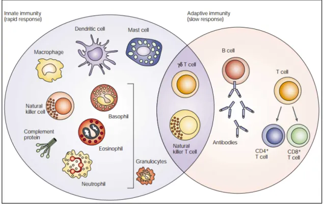
Figure 1: Cellular components of innate and adaptive immune cells. 4
Figure 2: Polarized CD4+ T cell subsets. 6
Figure 3. IL12 and IL4 driven T helper differentiation. 8
Figure 4. Stepwise induction of tTreg cells and pTreg cells. 12
Figure 5. Basic mechanisms used by Treg cells. 14
Figure 6. Mechanisms of Th17 cell induction. 16
Figure 7. Functions of Th17 cytokines and chemokines. 19
Figure 8. A common requirement for transforming growth factor-β in the induced
regulatory T cell and T helper 17 cell lineages. 21
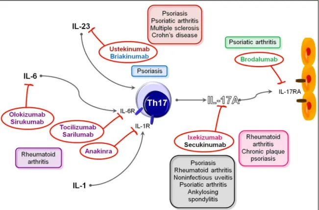
Figure 9. Drugs targeting Th17 pathways and their use in human diseases. 25
Figure 10: Types of autophagy. 31
Figure 11: Schematic model of autophagy. 37
Figure 12: Signalling regulation of mammalian autophagy. 39
Figure 13. Regulation of the mTOR pathway by nutrients
(amino acids, glucose), stress and insulin/IGF-1. 41
Figure 14. The role of autophagy in human disease. 44
Figure 15. Autophagy and its opposing role in cancer development. 45
Figure 16. ATG proteins in MHC-restricted antigen presentation. 49
Figure 17: A schematic representation of the proposed
mechanisms of action of IVIG on cellular immunity. 55
Figure 18. Immunoglobulin A. 63
List of Tables
Table 1: Mammalian homologs of yeast ATG proteins. 33
Table 2: Comparison of different IVIG preparations. 53
Abbreviations
Ab Antibody
ADCC Antibody-dependent cell mediated cytotoxicity
Ag Antigen
AHR Aryl hydrocarbon receptor
AMP Adenosine monophosphate
AMPK AMP-activated protein kinase
ANCA Anti-neutrophil cytoplasmic antibodies
APC Antigen presenting cell
ATG Autophagy related protein
BCR B cell receptor
Bcl B-cell lymphoma/leukemia
BSA Bovine serum albumin
CD Cluster of differentiation
CD Crohn disease
CIA Collagen-induced arthritis
CIDP Chronic inflammatory demyelinating polyneuropathy
CMA Chaperon-mediated autophagy
CNS Central nervous system
CTLA-4 Cytotoxic T lymphocyte antigen 4
CTL Cytotoxic T lymphocyte
CVID Common variable immunodeficiency
DAMPs Damage-associated molecular patterns
DC Dendritic cell
DCIR Dendritic cell immunoreceptor
DC-SIGN Dendritic cell-specific intercellular adhesion molecule-3-
grabbing non-integrin
DMSO Dimethyl sulphoxide
DNA Deoxyribonucleic acid
EAE Experimental autoimmune encephalitis
ELISA Enzyme link immunosorbent assay
ER Endoplasmic reticulum
F(ab’)2 Fragment antigen binding
Fc Fragment crystallizable
FcRI Fc epsilon receptor I
FcR Fc receptor
FcRn Neonatal FcR
FCS Foetal calf serum
FDA Food and Drug Administration
FIP Focal adhesion kinase family-interacting protein
Foxp3 Forkhead box 3
GALT Gut-associated lymphoid tissue
GBS Guillain Barré syndrome
GlcNAc N-acetylglucosamine
GM-CSF Granulocyte–macrophage colony-stimulating factor
HSA Human serum albumin
HSC Hematopoietic stem cell
H2O2 Hydrogen peroxide
IBD Inflammatory bowel disease
Ig Immunoglobulin
IL Interleukin
ITAM Immunoreceptor tyrosine-based activation motif
iTreg Induced regulatory T cell
ITP Immune thrombocytopenic purpura
IVIG Intravenous immunoglobulin
JNK c-Jun amino-terminal kinase
LAG-3 Lymphocyte activation gene-3
LAMP Lysosome-associated membrane protein
LPS Lipopolysaccharide
MALT Mucosal associated lymphoid tissues
MAPKs Mitogen activated protein kinases
MHC Major histocompatibility complex
mIgA Monomeric Immunoglobulin A
MNN Multifocal Motor Neuropathy
MO Monocyte
M Macrophage
MS Multiple sclerosis
mTORC Mammalian target of rapamycin complex
NAbs Natural antibodies
NK cell Natural Killer cell
NO Nitric oxide
nTreg Natural regulatory T cell
O2- Superoxide anion
PAMPs Pathogen-associated molecular patterns
PBS Phosphate buffer saline
PGE2 Prostaglandin E2
PI3K Phosphatidylinositol 3-kinase
PI3P Phosphatidylinositol 3-phosphate
RA Rheumatoid Arthritis
RORC/RORt Retinoic acid-related orphan receptor
SEM Standard error of mean
SLE Systemic lupus erythematous
SNPs Single nucleotide polymorphisms
STAT Signal transducing and activating transcription factor
T-bet T box transcription factor
TCR T cell receptor
Tc cell Cytotoxic T cell
Tfh Follicular helper T cell
TfR Transferrin receptor
TGF- Transforming growth factor
Th cell T helper cell
TIGIT T cell immunoglobulin and ITIM domain
TLR Toll like receptor
Treg Regulatory T cell
TNF Tumor necrosis factor
TSC Tuberous sclerosis complex
1. The immune system
1.1 Overview
The immune system evolved to protect multicellular organisms from pathogens and maintaining homeostasis. The fully functional immune system involves many organs, molecules, cells, and pathways in such an interconnected and sometimes circular process that it is often difficult to know where to start! The complex host immune system is capable of recognising the self and the non-self components (antigens) to mount an array of defense mechanisms referred as ‘immune response’ against foreign components without harmful effects on the host. Immune system possesses diversity; to respond against enormous variety of pathogens as well as specificity; to mediate appropriate and efficient response against the particular pathogen. However, in certain conditions, the dysregulation of immune system either in the self and non-self recognition or an excessive immune response lead to various autoimmune and inflammatory diseases.
Immune system can be functionally separated into four related activities. Immune recognition is the primary task by which immune system recognizes foreign pathogen. Upon recognition, it mounts an appropriate effector response to eliminate the foreign pathogen. The effector response needs to be regulated to avoid immunopathology by means of immune regulation. Further, immune system provides memory response for protection on later exposure to the same foreign invaders characterized by a more rapid immune reaction.
Although blood is not an immunological organ per se, it is the conduit for most immune cells circulating in the body, especially after an immunological stimulus such as vaccination. All blood cells arise from the hematopoietic stem cell (HSC) by a process haematopoiesis. Early in haematopoiesis, a multipotent stem cell differentiates along one of two pathways, giving rise to either a common lymphoid progenitor cell or a common myeloid. Lymphoid progenitor cells give rise to B, T, and NK (natural killer) cells and some DC. Myeloid progenitor cells generate progenitors of red blood cells (erythrocytes), many of the various white blood cells (neutrophils, eosinophils, basophils, monocytes, leukocytes, mast cells, DC, and platelets (1). After haematopoiesis white blood cells circulate though out the body where they encounter antigens. Development and maturation of lymphocytes takes place in the thymus and bone marrow, the primary (central) lymphoid organs. Mature lymphocytes migrate to secondary lymphoid organs
that include the lymph nodes, spleen, and various mucosal associated lymphoid tissues (MALT) such as gut-associated lymphoid tissue (GALT), where they interact with antigens.
The immune system can be simplistically viewed as having two lines of defense: innate
immunity and adaptive immunity. Innate immunity represents the first line of defense to an
intruding pathogen. It is an antigen-independent (non-specific) defense mechanism that is used by the host immediately or within hours of encountering an antigen. The innate immune response has no immunologic memory and, therefore, it is unable to recognize or memorize the same pathogen should the body be exposed to it in the future. Adaptive immunity, on the other hand, is antigen-dependent and antigen-specific and, therefore, involves a lag time between exposure to the antigen and maximal response. The hallmark of adaptive immunity is the capacity for memory which enables the host to mount a more rapid and efficient immune response upon subsequent exposure to the antigen.
1.2 Innate immunity
The primary function of innate immunity is the recruitment of immune cells to sites of infection and inflammation through the production of cytokines. The innate immune response also promotes clearance of dead cells or antibody complexes and removes foreign substances present in organs, tissues, blood and lymph. Numerous cells are involved in the innate immune response such as phagocytes (macrophages and neutrophils), DC, mast cells, basophils, eosinophils and NK cells (Figure 1). Responses from innate immune system are triggered upon pathogen recognition by a set of patter recognition receptos (PRRs) expressed on the surface of innate immune cells. PRRs identify two classes of molecules: pathogen-associated molecular patterns (PAMPs), which expressed only by microbes, but not by host and damage-associated molecular patterns (DAMPs), which are associated with cell components that are released during cell damage or death (2). In addition to their phagocytic properties, neutrophils and macrophages also produce a variety of other toxic products such as hydrogen peroxide (H2O2), the superoxide anion (O2-) and nitric oxide (NO), which assist in the elimination of pathogens. Unlike neutrophils (which are short-lived cells), macrophages are long-lived cells that not only play a role in phagocytosis, but are also involved in antigen presentation to T cells.
DCs also phagocytose and function as professional antigen-presenting cells (APCs) and act as important messengers between innate and adaptive immunity. DCs develop in the bone marrow and migrate to the tissues in an immature form. They express high levels of PRRs and exhibit high antigen uptake capacity. DCs undergo maturation when they are exposed to a number of danger signals including the PAMP as well as various cytokines and tissue factors (3). Mature DCs express co-stimulatory molecules and produce variety of cytokines that facilitate the differentiation of CD4+ T cells. They can load antigenic peptides on both MHC class I and MHC class II molecules, allowing presentation to both CD8 and CD4 T cells (4, 5). Mast cells and basophils share many salient features with each other and both are instrumental in the initiation of acute inflammatory responses, such as those seen in allergy and asthma. Unlike mast cells, which generally reside in the connective tissue surrounding blood vessels, basophils reside in the circulation. Eosinophils are granulocytes that possess phagocytic properties and play an important role in the destruction of parasites that are too large to be phagocytosed. Along with mast cells and basophils, they also control mechanisms associated with allergy and asthma. NK cells (also known as large granular lymphocytes [LGLs]) play a major role in the rejection of tumours and the destruction of cells infected by viruses. Destruction of infected cells is achieved through the release of perforins and granzymes from NK-cell granules which induce apoptosis (programmed cell death) (6).
1.3 Adaptive immunity
The hallmarks of the adaptive immune response are flexibility and memory. T and B lymphocytes are the cellular elements of the adaptive immune system. In addition, there is a minor population of dg T cells and NKT cells which share properties of T cells and NK cells (Figure 3). T cells derive from hematopoietic stem cells in the bone marrow and, following migration, mature in the thymus. They are primarily responsible for cell mediated immune response. B cells develop in bone marrow and are involved in humoral immune responses. Unlike cells in the innate system, which use a fixed repertoire of inherited receptors, T and B cells undergo a recombination of antigen receptor genes to create novel and unique antigen receptors capable of recognizing virtually any antigen. B and T cells that have encountered antigen persist over the long term within an organism and provide rapid and specific responses to reinfection, a concept known as
immunologic memory. T cells are activated when they encounter an APC that has digested an antigen and is displaying antigen fragments bound to its MHC molecules. The MHC-antigen complex activates the TCR and the T cell secretes cytokines which further control the immune response. This antigen presentation process stimulates either activation of cytotoxic T cells (CD8+ T cells) or CD4+ T helper (Th) cells subset differentiation. Cytotoxic T cells are primarily involved in the destruction of cells infected by foreign agents. They are activated by the interaction of their TCR with peptide-bound MHC class I molecules. CD4+ T cells play an important role in establishing and maximizing the immune response. These cells have no cytotoxic or phagocytic activity, and cannot kill infected cells or clear pathogens. However, they “mediate” the immune response by directing other cells to perform these tasks. They are activated through TCR recognition of antigen bound to class II MHC molecules. Once activated, CD4+ T cells release cytokines that influence the activity of many cell types, including the APCs that activate them.
Figure 1. Cellular components of innate and adaptive immune cells. The innate immune
response functions as the first line of defence against infection. It consists of soluble factors, such as complement proteins, and diverse cellular components including granulocytes (basophils, eosinophils and neutrophils), mast cells, macrophages, dendritic cells and natural killer cells. The adaptive immune response is slower to develop, but manifests as increased antigenic specificity and memory. It consists of antibodies, B cells, and CD4+ and CD8+ T lymphocytes. Natural killer
T cells and γδ T cells are cytotoxic lymphocytes that straddle the interface of innate and adaptive immunity. (Adapted from Dranoff G, Nat Rev Cancer. 2004)
1.3.1 Activation of CD4+ T cells:
Human CD4+T cells are critical regulators of immune system. CD4+ T cells are highly heterogeneous as they are generated in response to different pathogens and increasing in number of various subsets with specialized functions (7). Activation of CD4+T cells is the most critical step in developing immunity and requires 3 signals for their lineage commitment (8). The first signal is generated following the interaction between T-cell receptor (TCR) and the peptide presented in the context of MHC class II on an APC (9). However, this signal alone is insufficient and further signals in the form of accessory molecules and adhesion molecules are crucial for optimum T cell activation. T cells that do not receive costimulatory signal are likely to be rendered unresponsive or anergic. The second signal is generated following the interaction between the CD28 co-receptor on the T cell and B7 family of co-stimulatory molecules such as CD80 or CD86 on the APC. The third signal is generated by polarizing cytokines produced by the APC or other cells at the site of T cell activation. These cytokines direct differentiation of naïve CD4+T cells into a particular effector subset.
1.3.2 CD4+ T cells differentiation:
The affinity of the MHC/peptide/TCR interaction is thought to be one factors determining the differentiation of naïve CD4+ T cells. However, the local cytokine environment at the time of priming is critical, both in terms of effects on the responding T cells and on the APC (10). The type of APC is important, as are the costimulatory molecules expressed by the APC (11). TCR stimulation enhances IL-2 production from T cells, which signals in an autocrine fashion inducing various cytokine receptors.
Effector CD4+ T cells can be categorized into three major subsets based on the type of cytokine they produce and the major transcription factor (TF) they express. In the presence of interleukin (IL)-12, naïve CD4+ T cells differentiate into Th1 effectors. Th1 effectors express a transcription factor T-bet and secrete the cytokines IFN-γ and TNF-α; these cells play an essential
role in inhibiting replication of intracellular pathogens such as viruses (12, 13, 8). Under the influence of IL-4, naïve CD4+ T cells differentiate into Th2 effectors. Th2 cells express TF GATA-3, secrete cytokines IL-4, IL-5, and IL- 13 (14, 15); these cells are critical during infection by extracellular pathogens such as extracellular bacteria and helminthes. In the presence of IL-6/IL-21 and transforming growth factor (TGF)-β, naïve CD4+ T cells differentiate into Th17 cells. Th17 cells express a transcription factor ROR-γt and produce cytokines IL-17 and IL-22 (16, 17); these cells are important for control of certain bacterial and fungal infections. Th1, Th2, and Th17 cells are considered to be the major effector CD4+ T cells (18-30). Tregs express TF FoxP3; these cells secrete anti-inflammatory cytokines like TGF-β and IL-10. Tregs maintain immune homeostasis by limiting the magnitude of immune response against pathogens and control inflammatory reactions (31). T follicular helper cells (Tfh) express a TF Bcl-6 and these cells are essential for the production of high affinity IgG antibodies (32).
In addition to the aforementioned Th subsets, existence of other Th subsets has been described, such as, Th9 and Th22 subsets, which secrete IL-9 and IL-22 respectively (33-35), Th3 (TGF-b-secreting CD4 T cells) (36, 37), Tr1 (IL-10 secreting CD4 T cells) (38) and Tfr cells (39), which has been implicated in the inhibiting the functions of Tfh cells (Figure 2) (40).
Figure 2. Polarized CD4+ T cell subsets. Each CD4+ T cell subset can be defined by their distinct
abilities to sense (red), programme (orange) and function (blue) in the control of specific pathogens or immune pathologies. The inductive cytokines, polarizing transcription factors and cytokines or chemokine receptors that are characteristic of each subset are shown, along with their association with specific forms of immune defence. AHR, aryl hydrocarbon receptor; BATF, B cell-activating transcription factor; BCL-6, B cell lymphoma 6; CCR, CC-chemokine receptor; CD40L, CD40 ligand; CTLA4, cytotoxic T lymphocyte antigen 4; CXCR, CXC-chemokine receptor; EOMES, eomesodermin; FOXO, forkhead box O; FOXP3, forkhead box P3; GATA3, GATA-binding protein 3; GFI1, growth-factor independent 1; HIF1α, hypoxia-inducible factor 1α; ICOS, inducible T cell co-stimulator; IFNγ, interferon-γ; IL, interleukin; IRF4, interferonregulatory factor 4; MAF, macrophage-activating factor; NR4A, nuclear receptor 4A; PD1, programmed cell death 1; pTReg cell, peripherally derived regulatory T cell; RAR, retinoic acid receptor; ROR, retinoic acid receptor-related orphan receptor; RUNX3, runt-related transcription factor 3; SHM, somatic hypermutation; STAT, signal transducer and activator of transcription; TCF1, T cell factor 1; TCR, T cell receptor; TFH, T follicular helper; TGFβ, transforming growth factor-β; TH, T helper; TReg cell, regulatory T cell; tTReg cell, thymus-derived regulatory T cell. (Adapted from DuPage M, Bluestone JA, Nat Rev Immunol. 2016)
1.3.2.1 Th1 differentiation
Interleukin 12 (IL12) and interferon γ (IFNγ) are the critical cytokines initiating the downstream signalling cascade to develop Th1 cells (41). IL12 is secreted in large amounts by APCs after their activation through the pattern recognition receptors (42-44). The IL12, in turn, induces NK cells to produce IFNγ. Several transcription factors in coordination induce full differentiation of the Th1 cells. T-bet is the principal transcription factor, as it significantly enhances the production of IFNγ, and plays important role in suppressing the development of Th2 and Th17 (45, 46). T-bet expression was found to be strongly dependent on signal transducer and activator of transcription 1 (STAT1) (47, 48). STAT1, is in turn activated by IFNγ. T-bet suppresses development of Th2 cell by inhibiting the crucial IL4 gene and impairing the function of the Th2 master regulator GATA3 (49, 50). Th17 lineage is inhibited by the interaction of T-bet
with Rorc promoter, which encodes RORγt, the principal transcription factor of Th17 (46). IL12-induced STAT4 is another important transcription factor involved in the Th1 cell differentiation (51). STAT4 induces IFNγ production. However, STAT4 and T-bet do not function in a linear way in the differentiation of Th1 cell, with each having their unique signaling pathway. But for complete Th1 cell differentiation, these-lineage specific transcription factors need to operate in coordination with one another (52). In later stages of differentiation, IL12/STAT4 pathway upregulates IL-18Rα. IL12 along with IL18 induces IFNγ production independent of TCR activation, thus creating a pathway for enhancing Th1 response (Figure 3).
Th1 cells are involved with the elimination of intracellular pathogens and are associated with organ pecific autoimmunity (53). They mainly secrete IFNγ, lymphotoxin α (Lfα), and IL2. IFNγ is essential for the activation of mononuclear phagocytes, including macrophages, microglial cells, thereby resulting in enhanced phagocytic activity (54). Lfα is a member of the TNF super family. Lfα is associated with autoimmune diseases. The depletion of Lfα has shown to inhibit the development of experimental autoimmune encephalitis (55, 56).
Figure 3. IL12 and IL4 driven T helper differentiation. Th1 induction by IL12: Initial TCR
the IL12 receptor results in STAT4 mediated promotion of Ifng expression (2). Binding of the IFNg receptor by low initial auto/paracrine produced IFNg activates STAT1 (3), which strongly promotes expression of the Tbx21 gene (4). T-bet then enhances the transcriptional competence of the Ifng gene (5) leading to increased production of this cytokine (6). In addition, T-bet prevents Th2 differentiation by inhibiting Gata3 (7). Finally, T-bet promotes expression of the IL12 receptor b2 chain (8), resulting in greater IL12 responsiveness (9) and yet further elevated production of IFNg (10). Th2 induction by IL4: Initial TCR signaling induces low-level expression of the Il4 and Gata3 genes (11). IL4 receptor signaling strongly promotes expression of these two genes (12). Gata3 reorganizes chromatin structure in the Th2 locus, encompassing the Il4, Il5, and Il13 genes, enhancing their transcription competence (13). Increased IL4 production further enhances TH2-cell differentiation in a feed forward loop (14). Finally, Gata3 prevents the Th1 differentiation program by inhibiting expression of the IL12 receptor b2 chain (15) and of the Stat4 gene (not depicted). Primary events are indicated with black arrows, secondary events with red arrows, and tertiary events with blue arrows. (Adapted from Amsen et al., 2009; Curr Opin Immunol).
1.3.2.2 Th2 differentiation
IL4 and IL2 are critical for Th2 differentiation. The major transcription factor involved in Th2 lineage differentiation includes the IL4-induced STAT6, which upregulates the expression of the master regulator GATA3 (GATA-binding protein) (57-59). Three distinct mechanisms of GATA3 involvement in Th2 differentiation have been postulated, including enhanced Th2 cytokine production, selective proliferation of Th2 cells through recruitment of Gfi-1, and inhibition of Th1 differentiation presumably by interacting with T-bet (60). Moreover, GATA3 was found to suppress Th1 differentiation by downregulating STAT4 (61). In GATA3 deficient mice, differentiation of naïve cells was diverted towards the Th1 lineage (62). Recent studies showed that GATA3 by itself cannot regulate all the Th2-specific genes, but instead needed the collaboration of STAT6 (63). STAT5 has an important role in the Th2 lineage commitment. It is readily activated by IL2 (64, 65). STAT5 activation is independent of IL4 signaling and does not induce GATA3 expression (66). For full differentiation of Th2 cells, the coordinated activity of STAT5 and GATA3 is required, since GATA3 alone cannot induce the production of IL4 (Figure 3).
Th2 cells mount immune response to extracellular parasites, including helminthes, and play major role in induction and persistence of asthma as well as other allergic diseases (53, 67). The key effector-cytokines include IL4, IL5, IL9, IL13, IL10, IL25, and amphiregulin. IL4 is a major cytokine involved in allergic inflammation. It is involved in IgE switching and secretion by B cells. IL4 also upregulates low-affinity IgE receptor (FcεRII) on B-lymphocytes and mononuclear phagocytes, and also high-affinity IgE receptor (FcεRI) on mast cells and basophils, with subsequent degranulation of the cells and release of several active metabolites, including histamine and serotonin (68). IL5 mainly targets eosinophils (69). IL9 participates actively in the immune pathogenesis of asthma (70).
1.3.2.3 Th9 Cells
Initially characterized as a subset of Th2 cells, ongoing researches tend to classify IL9 secreting-Th9 cells as a distinct subset of CD4+ T cells. TGF-β was found to divert the differentiation of Th2 towards the development of Th9 cells. Moreover, TGF-β in combination with IL 4 directly induces the differentiation of Th9 cells (71). IRF4 also plays an important role. IRF4 was found to directly bind to the IL9 promoter (72).
1.3.2.4 Follicular Helper (Tfh) T Cells
Tfh are C-X-C motif receptor-5 (CXCR 5+)-expressing cells and are located in follicular areas of lymphoid tissue, where they participate in the development of antigen-specific B-cell immunity (73, 74). IL6 and IL21 are the main cytokines involved in the differentiation process (75, 76). STAT3, activated downstream to cytokine signaling, is an important transcription factor of Tfh. Inducible costimulator (ICOS), member of CD28 family, is also required for Tfh development (77, 78).
1.3.2.5 Regulatory T cells (Tregs) differentiation
CD4+CD25+ regulatory T (Treg) cells play a pivotal role in the maintenance of immune homeostasis, where the X-linked master transcription factor forkhead box P3 (FOXP3) determines Treg cell development and function (79, 80). Tregs are primarily generated in the thymus (tTreg), but also may be generated extra thymically at peripheral sites (pTreg), or in the presence of signal from APC and TGFβ (iTreg). tTregs also called nTregs (81). Naturally occurring thymus-derived CD4+CD25+ Tregs are a subset of T cells which have immunosuppressive properties and are ~2% of the total peripheral CD4+ T cells. Thymic induction of Foxp3 requires TCR signalling of
increased strength. Besides TCR, engagement of common g-chain (gc) cytokine receptors,
foremost IL-2R, is required for both tTregs and pTregs (82, 83). Interestingly, unlike conventional T cells, Tregs do not produce IL-2 due to chromatin inaccessibility of the IL-2 locus (84). Consequently, survival and growth of Tregs is dependent on paracrine IL-2. According to a two-step model of Treg differentiation, TCR engagement first leads to CD25 upregulation making precursor cells receptive to receiving IL-2 signals leading to Stat5 activation (85, 86). Finally, CD28 signaling induced upon binding of its ligands CD80 and CD86 has also a cell-intrinsic role in Foxp3 induction in the thymus (87, 88). Further, activation of TCR and costimulatory signalling recruitment of a number of transcription factors including NFAT, cRel, Creb, and Stat5 to the promoter and several conserved noncoding regulatory elements of the Foxp3 gene and its expression (89-91).
iTregs develop outside of the thymus in a tolerogenic environment. Induction of iTregs requires TGF-β and IL-2 (Figure 4) (92). Smad2 and Smad3, activated through TGF-β signaling pathways, are involved in the iTreg differentiation process by inducing FOXP3 (93, 94). Smad3 can differentially enhance iTreg development by upregulating FOXP3 expression and inhibit Th17 differentiation by blocking RORγt (95). STAT5-induced downstream to IL2 signaling is required for the differentiation of iTreg (96, 97). STAT5 was found to enhance FOXP3 expression and subsequently downstream to FOXP3 signaling and promote iTreg development. Additionally, programmed death-1- programmed death-1-ligand-1, ICOS-ICOSL, OX40-OX40L signaling expand Tregs.
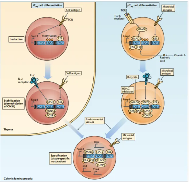
Figure 4. Stepwise induction of tTreg cells and pTreg cells. The thymus-derived regulatory T
(tTreg) cell differentiation programme is initiated by strong T cell receptor (TCR) signals induced by self-antigens in the thymus, which results in nuclear entry of REL. In mice, REL, together with several other transcription factors (not depicted) bind to conserved non-coding sequence 3 (CNS3) and the forkhead box P3 (Foxp3) promoter to induce Foxp3 gene expression. These cells become a stable Treg cell lineage when CNS2 is demethylated, and signal transducer and activator of transcription 5 (STAT5) activated by interleukin‑2 (IL‑2) receptor signalling contributes to this demethylation. Demethylated CNS2 allows binding of FOXP3 and runt‑related transcription
factor 1 (RUNX1), which results in stable Foxp3 expression. tTreg cells migrate to the intestines and further undergo functional maturation in response to environmental stimuli. Peripherally derived Treg (pTreg) cell differentiation from uncommitted naive CD4+ T cells occurs in the intestines but progresses in a stepwise manner similarly to tTreg cells. The differentiation is initiated by TCR signalling induced by microbial antigens together with transforming growth factor‑β (TGFβ) and retinoic acid‑mediated signals. These signals promote REL binding to CNS3, as well as SMAD3 and retinoic acid receptor (RAR)–retinoid X receptor (RXR) binding to CNS1 of Foxp3. Similarly, to tTreg cells, pTreg cells establish their Treg cell character and lineage commitment by demethylation of CNS2, which is induced by IL‑2 and STAT5 signalling. Microbiota‑derived butyrate contributes to chromatin remodelling and lineage commitment by its histone deacetylase (HDAC) inhibitory activity. pTreg cells further acquire functionally specialized features such as expression of RAR‑related orphan receptor γt (RORγt; encoded by Rorc), IL‑10 and cytotoxic T lymphocyte antigen 4 (CTLA4) to adapt to the intestinal environment. (Adapted from Tanoue et al., 2016; Nat Rev Immunol).
Tregs have emerged as key players in the development and maintenance of peripheral immune tolerance. In normal conditions, Tregs regulate ongoing immune responses and prevent autoimmunity. Imbalanced function or number of these cells, either enhanced or decreased, might lead to tumour development and autoimmunity, respectively. These cells thus play a major role in autoimmune diseases, transplantation tolerance, infectious diseases, allergic disease and tumour immunity. The cellular targets of Treg-mediated suppressor functions include CD4+, CD8+ T cells, DC, B cells, macrophages, monocytes, mast cells, NK cells and NKT cells (98-100). Tregs exert their suppressive functions through several mechanisms, some of which require cell-cell contact and other mediated through cytokines (Figure 5) (101). Their main effector cytokines include IL10, TGF- β, and IL35. IL10 is a potent inhibitory cytokine, with the ability to suppress proinflammatory response and thus limits tissue damage by the inflammatory process (102, 103). IL10 and TGF-β potently suppress IgE production, thereby showing their important role in attenuating allergic inflammation (104). Small molecules elaborated by Treg cells have also been implicated in restraining inflammation. Specifically, Foxp3- dependent expression of the ectonucleotidases CD39 and CD73 endows Treg cells with the ability to sequentially convert
highly inflammatory extracellular ATP, which accumulates under hypoxic conditions, into adenosine via an AMP intermediate (105).
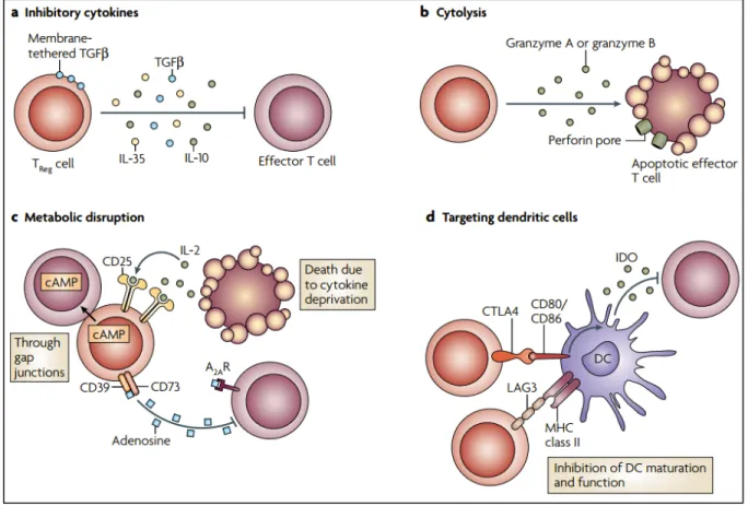
Figure 5. Basic mechanisms used by Treg cells. Depiction of the various regulatory T
(Treg)-cell mechanisms centred around four basic modes of action. (a) Inhibitory cytokines include interleukin-10 (IL-10), IL-35 and transforming growth factor-β (TGFβ). (b) Cytolysis includes granzyme-A- and granzyme-B-dependent and perforin-dependent killing mechanisms. (c) Metabolic disruption includes high-affinity CD25 (also known as IL-2 receptor α)-dependent cytokinedeprivation-mediated apoptosis, cyclic AMP (cAMP)-mediated inhibition, and CD39- and/or CD73-generated, adenosine receptor 2A (A2AR)-mediated immunosuppression. (d) Targeting dendritic cells (DCs) includes mechanisms that modulate DC maturation and/or function such as lymphocyte-activation gene 3 (LAG3; also known as CD223)–MHC-class-IImediated suppression of DC maturation, and cytotoxic T-lymphocyte antigen-4 (CTLA4)–CD80/CD86-mediated induction of indoleamine 2,3-dioxygenase (IDO), which is an immunosuppressive molecule made by DCs (Adapted from Vignali et al., 2008; Nat Rev Immunol)
In addition to cytokine-mediated modulation of inflammatory responses, Treg cells display high amounts of CTLA4 and TIGIT (T cell immunoglobulin and ITIM domain), two Ig family members that are able to inhibit the immune stimulatory potential of DCs by reducing expression of the costimulatory molecules CD80 and CD86 and by inducing IL-10 production in DCs, respectively (106-108). Besides immunomodulation or inactivation, perforin- and granzyme-mediated cytolysis of immune effector and accessory cells by Treg cells has been considered as a means of suppression (109, 110). In addition to direct killing, an indirect way of elimination of effector T cells is through induction of apoptosis due to a growth factor withdrawal. In this regard, Treg cells are able to deprive effector T cells of IL-2 due to a much higher level of CD25 expression (111).
1.3.2.6 Th17 cells differentiation
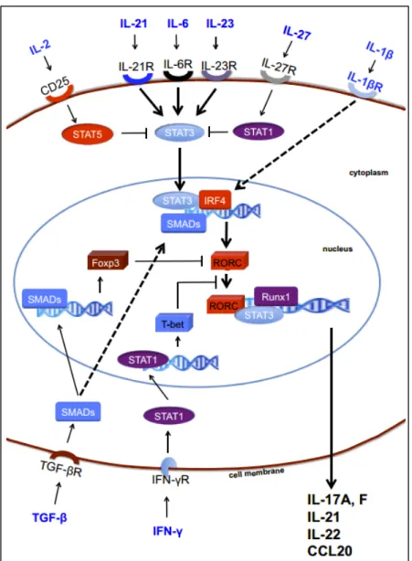
Th17 cells are a uniquely pro-inflammatory lineage of effector/memory Th cells first identified by (and named for) production of IL-17A (hereafter referred to as IL-17). Th17 differentiation, survival, and expansion rely on a variety of cytokines and transcription factors that work in concert to drive the induction of increased Th17 numbers (Figure 6).
Figure 6. Mechanisms of Th17 cell induction. TGF-b is essential for the generation of both induced regulatory T cells and Th17 cells via the induction of FoxP3 and RORC. However, in the absence of inflammation, FoxP3 represses RORC and promotes iTregs. Signaling via inflammatory cytokines, such as IL-6, IL-21, and IL-23, results in STAT3 phosphorylation, relieves RORC from the suppression of FoxP3, and initiates Th17 programming. STAT3 in combination with IFN regulatory factor 4 (IRF4) further induces RORC expression. The transcription factors STAT3, RORC, and Runx1 bind to the promoter regions of the IL17, IL21, IL22, and CCL20 genes and induce IL-17, IL-21, IL-22, and CCL20. Th17 programming can be antagonized by cytokines, such as IFN-g, IL-2, and IL-27. IL-2-mediated and IL-27-mediated activation of STAT5 and STAT1 inhibit STAT3, whereas T-bet induced by IFN-g can block RORC (Adapted from Maddur et al., 2012; Am J Pathol).
The full differentiation of Th17 cells requires three different steps: induction, amplification and stabilization/maintenance. (1) The differentiation is initiated by the combined actions of IL-6 and TGF-β; (2) the amplification of the Th17 response is driven through the production of IL-21 by Th17 cells; (3) the stabilization/maintenance of the Th17 phenotype is achieved by IL-23. Several studies found that a combination of the immunoregulatory cytokine TGF-β and the proinflammatory and pleiotropic cytokine IL-6 is required to induce IL-17 in naive T cells (112-115). However, TGF-β signaling pathways also play significant role in the development of iTreg. TGF-𝛽 promotes Th17 and iTreg development by inducing the expression of the transcription factors ROR𝛾t and FoxP3, respectively. In fact, both transcription factors are initially up-regulated after naïve CD4+ T-cells encounter TGF-β (115). Whether cells are shuttled towards a proinflammatory Th17 phenotype or a regulatory phenotype depends largely on the surrounding cytokine environment. Up-regulated Foxp3 initially binds to RORγt thereby inhibiting the development of Th17-cells and favoring Treg development (116-118). IL-6 has been identified as an important mediator driving the development of Th17 cells via activation of STAT3 (112, 114, 115). In the presence of IL-6, STAT3 activation releases FoxP3 inhibition. Eventually, this leads to the differentiation of Th17-cells and up-regulation of the receptor IL-23R.
Also, IL-21, a member of the IL-2 family of cytokines, is produced in overwhelming amounts by Th17 cells and could, in combination with TGF-β, induce Th17-differentiation (Korn et al., 2007; Ivanov et al., 2007). These findings point to a relevant function of IL-21, produced by newly generated Th17 cells, in amplifying the precursor frequency of differentiating Th17 cells (119-121).
IL-23 is another inflammatory cytokine that contributes to Th17 expansion and stability. A stronger connection between IL- 23 and Th17 cells was established when investigators showed that IL-23 promotes the production of IL-17 by activated T cells (122) and that IL-23-expanded T cells are able to transfer EAE and CIA (collagen-induced arthritis) (123, 124). IL-23R is clearly not expressed on naive T cells, and after the identification of the factors (IL-6, IL-21, and TGF-β) required for the differentiation of Th17 cells, it became clear that IL-23 was not involved in the initial differentiation of Th17 cells. IL-23p19-deficient mice have limited numbers of Th17 cells and that prolonged culture of Th17 cells in vitro requires the addition of IL- 23. IL-23 synergizes with IL-6 to enhance survival, and stabilization of Th17 cells (121, 125). IL-23 can amplify Th17
cells by inducing pro-inflammatory cytokines, such as IL-1β, TNF-a, and IL-6 in innate immune cells (126).
Besides the master regulator RORγt, several other transcription factors need to collaborate for full differentiation of Th17 cells. As such, deficiency of RORγt does not lead to complete interruption of Th17 cytokine expression (118). STAT3, activated downstream to IL6, IL21, IL23 signaling plays an important role in the differentiation process. It enhances RORγt expression. STAT3 deficiency was found to cause enhanced expression of T-bet and FOXP3, which are involved in the development of opposing cell lineages (127). STAT3 binds to IL-17A and IL-17F promoters (128). RORα, another member of the ROR family, also participates in the lineage commitment pathway. Together RORα and RORγt synergistically enhance Th17 differentiation, and their absence completely aborted the development of Th17 cells (117). Runx1 also influences Th17 differentiation. Runx1 through the induction of RORγt, promotes differentiation. However, Runx1/FOXP3 interaction negatively regulates Th17 development (129). Moreover, T-bet in collaboration with Runx1 leads to the interruption of Runx1-mediated transactivation of Rorc, thereby suppressing Th17 development (130). Aryl hydrocarbon receptor (AHR), a ligand-dependent transcription factor, was found to promote Th17 differentiation, presumably through the inhibition of STAT1 and STAT5, which negatively regulate Th17 development. However, its absence did not cause complete abortion of Th17 differentiation, but was associated with inability to produce IL22 (131, 132).
Translation of the Th17 differentiation knowledge from mice to human met with discrepancies with regard to the cytokine cocktail required for the Th17 differentiation. TGF-β was believed to be dispensable for the differentiation of human Th17 cells from naïve T cells. One of the initial studies in human T cells reported that T cell receptor (TCR) cross-linking leads to IL-17 production (133). Other studies showed that ThIL-17 differentiation can achieved by a combination of IL-1β and IL-6/IL-23 or IL-21 alone (134, 135). However, the problem with these early studies was that they did not separate effects on memory T cells versus naive T cells. Also, the endogenous source of TGF-β was not rigorously controlled in those reports. It was only more recently studies done with rigorously sorted naïve T cells and with serum free medium suggested that TGF-β is indispensable for the differentiation of human Th17 cells from naïve T cells (136-138). Importantly, it was shown that Th17 differentiation can be achieved by using TGF-β and IL-21, whereas IL-1β and IL-6 was important for amplification (138). Mechanistically, TGF-β and
IL-6/IL-21 induces the expression of IL-1R and IL-23R, making the cells receptive to IL-1β and IL-23 (139, 136, 138).
Th17 is responsible to mount immune response against extracellular bacteria and fungi. They are also involved in the generation of autoimmune diseases (140, 141, 17). The key effector cytokines include IL17A, IL17F, IL21, and IL22. IL17A and IL17F signaling occurs through a common receptor, IL17RA, thereby suggesting similar functions (Figure 7) (142). IL-17A and IL-17F are the key cytokines for recruitment and activation of neutrophils. The effect of IL17 extends beyond T cell-mediated inflammatory response. IL17 leads to the induction of proinflammatory cytokines, including IL6, IL1, TNFα, and also proinflammatory chemokines ensuring the chemotaxis of inflammatory cells to sites of inflammation (17). IL21, along being an amplifying cytokine for TH17 development, has pleiotropic functions, including activating T cells, inducing B cells to differentiate into plasmocytes and memory cells, and activating NK cells (119, 143). IL22 is known to mediate both inflammatory response and exhibits tissue protective properties. In acute liver disease, IL22 was shown to be involved in limiting liver tissue damage (144). IL22 participates actively in mucosal host defense against bacterial pathogens, by inducing antimicrobial peptides and increasing cell proliferation (145).
Figure 7. Functions of Th17 cytokines and chemokines. Th17 cells secrete several effector
molecules, including IL-21, IL-22, IL-17A/F, and CCL20. These soluble factors act on both immune and nonimmune cells and mediate several functions, such as differentiation of cells; release of antimicrobial molecules, cytokines, and chemokines; and recruitment of cells to sites of inflammation. B, B cell; CD8, cytotoxic T cell; DC, dendritic cell; EC, endothelial cell; EpC, epithelial cell; FB, fibroblast; HC, hepatic cell; MØ, macrophage; NK, natural killer cell; NØ, neutrophil; T, T cell (Adapted from Maddur et al., 2012; Am J Pathol).
1.3.3 Reciprocal relationship between Th17 cells and Tregs:
The balance between factors promoting iTreg and Th17 development is critical in determining homeostatic versus inflammatory condition. Th17 and Treg developmental programs as well as their functions are reciprocally interconnected. TGF-b is a critical factor for both lineages. TGF-b is required for the differentiation and maintenance of Tregs and is also essential for the differentiation of Th17. In steady state (in the absence of inflammatory stimuli) produces TGF-β, which induces the generation of iTregs, which together with nTregs keep activated/effector memory cells in check. IL-6/IL-21, induced during an acute-phase response, inhibits the function of nTregs and prevents the generation of iTregs, but instead induces Th17 cells. The mechanism by which IL-6 and IL-21 act as switch factors relies on the control of the Foxp3/RORγt balance (116). TGF-β is required for the expression of both Foxp3 and RORγt, although the signaling cascades downstream of the TGF-β receptor might be different for the induction of Foxp3 versus RORγt. For example, whereas Smad4 appears to be essential for the induction of Foxp3, it is probably dispensable for the induction of RORγt (146). Both RORγt and RORα physically associate with Foxp3 to antagonize each other’s functions (116, 146). Both RORα and RORγt have been identified as binding partners of FoxP3 (116, 147, 148), and binding of FoxP3 inhibits the transcriptional activity of RORα and RORγt. Importantly, the repression of RORγt-mediated effects by FoxP3 depends on the local concentration of TGFβ (146, 116). Th17 cell-promoting cytokines can act through STAT3-dependent pathways to reverse the FoxP3-mediated repression of RoRγt and RoRα. TGFβ signals initiated by IL-6 through STAT3 and sustained by IL-21 and IL-23, override FoxP3-mediated repression of RoRγt (Figure 8) (116).
Additional evidence for a reciprocal developmental relationship between Foxp3+ Tregs and Th17 cells comes from studying the effects of retinoic acid, a vitamin A metabolite, on T cell differentiation. Retinoic acid could drive the generation of Tregs while abrogating the differentiation of Th17 cells, but not of Th1 cells (149). CD103+ DCs appear to be a relevant source of retinoic acid and, together with TGF-β, are efficient in inducing Foxp3+ Tregs (150, 151). Mechanistically, retinoic acid enhances the TGF-β-driven phosphorylation of Smad3 but reduces the TGF-β-induced upregulation of the IL-6Rα subunit on T cells (152).
The reciprocal relationship between Tregs and Th17 cells is further strengthen by IL-2. IL-2 signaling is critically involved in Treg as well as Th17 cell differentiation. IL-2 is consumed preferentially by Treg since they express high affinity IL-2 receptors (101) (54). IL-2 also acts to stabilize Foxp3 induced by TGF-β (153, 154). TGF-β and IL-2, along with other proinflammatory cytokines such as IL-1β, IL-6, IL-21, or IL-23, facilitate Th17 differentiation (155).
Figure 8. A common requirement for transforming growth factor-β in the induced regulatory T cell and T helper 17 cell lineages. Dendritic cells (DCs) that are conditioned in
environments rich in transforming growth factor-β (TGF-β), such as the intestines, present microorganism-derived antigens to induce the differentiation of naive CD4+ T cells to either induced regulatory T (Treg) cells or T helper 17 (Th17) cells, depending on the dominance of retinoic acid or interleukin-6 (IL-6), respectively. During homeostasis, DCs loaded with antigens from commensal bacteria or food produce retinoic acid derived from dietary vitamin A, which favours the upregulation of forkhead box P3 (FOXP3) expression and the differentiation of induced Treg cells. Under conditions of microbial breach of the intestinal epithelial cell barrier, pro-inflammatory stimuli activate DCs, which mature and produce IL-6 and stop retinoic acid production, thereby inducing Th17 cell differentiation. IL-6 and IL-21 (induced by IL-6) suppress Foxp3 expression and upregulate expression of the retinoic acid-related orphan receptor genes RORC and RORA (which encode RORγt and RORα, respectively), leading to expression of the inducible component of the IL-23 receptor (IL-23r) and further Th17 cell development. IL-17A and IL-17F produced by Th17 cells increase production of neutrophils in the bone marrow and their recruitment to the gut through intermediary cytokines (such as granulocyte colony-stimulating factor (G-CSF) and IL-8, respectively) and can also act to induce epithelial cell production of antimicrobial peptides. IL-22 produced by Th17 cells promotes enhanced epithelial barrier function and the production of antimicrobial peptides. collectively, therefore, the Th17 cell response acts both to clear invasive microorganisms and to restore barrier function. (Adapted from Weaver and Hatton, 2009; Nat Rev Immunol).
1.3.4 Th17 cells in the pathogenesis of autoimmune and inflammatory diseases:
The role of Th17 cells in autoimmunity and inflammation was demonstrated first in mice that were deficient for the p19 chain of the IL-23, in which the IL-17-producing T cells were significantly lower than in wild-type mice, highlighting the importance of the IL-23/Th17 axis in the pathogenicity of these autoimmune and autoinflammatory diseases (156). Since then, the study of the pathogenic role of Th17 subset cells has focused on autoimmune inflammatory diseases, such as multiple sclerosis (MS), rheumatoid arthritis (RA), psoriasis and others (157, 158).
Th17 cells were found to be the central effector lineage involved in the pathogenicity of murine EAE, an important model of MS (159). L-17 expression is correlated with disease activity. Human blood-brain barrier endothelial cells were found to express receptors for IL-17 and IL-22, thus making it possible for IL-17 and IL-22 to disrupt blood brain barrier junctions (160). Further studies continued to correlate the number of Th17 cells and amount of IL-17 expression found in MS lesions with active disease (161, 162). IL-23 that expands IL-17-producing cells is critical for the induction of EAE. It has been shown that myelin antigen-specific Th17 cells directly interact with neuronal cells in demyelinating lesions (163, 164). Two independent studies have identified the critical nonredundant role of Th17-produced GM-CSF in the pathogenesis of EAE. RORgt drives the production of GM-CSF in Th17 cells under the influence of the IL-1 and IL-23 pathways. GM-CSF acts on CD103+ dermal DCs to stimulate the secretion of IL-23 and the activation of encephalitogenic T cells, creating a positive feedback loop during inflammation (165-167). In consensus, absence of GM-CSF confers resistance to EAE in mice, even in the presence of IL-17 and/or IFN-g (165).
Psoriasis is a chronic, relapsing, and immune mediated inflammatory skin disease (168). Psoriasis is defined as a Th1/Th17/Th22-based inflammatory disease (169). IL-23, overproduced by dermal DCs and keratinocytes, stimulates Th17 cells within the dermis to elevate the amounts of IL-17, TNF-a, IL-22, and CCL20. These soluble mediators of Th17 cells might further enhance inflammation by their distinct actions. For example, CCL20 can attract CCR6+ cells to lesions, and IL-17 can induce IL-6 and IL-8 in human skin keratinocytes (170). Further, an experimental model of psoriasis-like skin inflammation indicates that IL-22 plays a central role in Th17 cell– mediated skin pathology (171).
RA is a systemic autoimmune disease characterized by progressively destructive joint inflammation, destruction of articular cartilage, and bone and synovial hyperplasia. In 2003 Murphy et al. saw that IL-23p19−/− mice were protected from collagen-induced arthritis (CIA), a mouse model for RA (124). Blockage of IL-23 expression protected mice from joint and bone destruction, and an anti-IL-17 antibody has been shown to inhibit osteoclast formation in human rheumatoid arthritis samples (124, 172). PGE2, a known positive regulator of Th17 differentiation, was shown capable of enhancing CIA severity through enhanced DC-derived IL-6 production,