Technology researchers and makes it freely available over the web where possible.
This is an author-deposited version published in: https://sam.ensam.eu Handle ID: .http://hdl.handle.net/10985/15601
To cite this version :
Youngwoo KIM, Claudio VERGARI, François GIRINON, JeanYves LAZENNEC, Wafa SKALLI -Stand-to-Sit Kinematics of the Pelvis Is Not Always as Expected: Hip and Spine Pathologies Can Have an Impact - The Journal of Arthroplasty p.ePub - 2019
Any correspondence concerning this service should be sent to the repository Administrator : archiveouverte@ensam.eu
a Institut de Biomécanique Humaine Georges Charpak, Arts et Métiers Paris Tech, Paris, France, Paris, France
b Department of Orthopaedic and Trauma Surgery, Pitié-Salpêtrière Hospital, Assistance Publique-Hôpitaux de Paris, France c Department of Orthopaedic Surgery, Graduate School of Medicine, Kyoto University, Kyoto, Kyoto Prefecture, Japan d Anatomy Department Faculté Pitié-Salpêtrière, Médecine Sorbonne Université, Paris, France, Paris, France
Stand to Sit Kinematics of the Pelvis is not always as
Expected: Hip and Spine Pathologies can Have Impact.
Youngwoo Kim a, b,c, Claudio Vergari a, François Girinon a,
Jean Yves Lazennec b,d, Wafa Skalli a
Abstract
Introduction; Stand to sit pelvis kinematics is commonly considered as a rotation around the bicoxofemoral axis. However, abnormal kinematics could occur for patients with musculoskeletal disorders affecting the hip-spine complex. The aim of this study is to perform a quantitative analysis of the stand to sit pelvis kinematics using 3D reconstruction from bi-planar x-rays.
Material & Methods; Thirty volunteers as a control group (C), 30 patients with hip pathology (Hip) and 30 patients with spine pathology (Spine) were evaluated. All subjects underwent standing and sitting full-body bi-planar x-rays. 3D reconstruction was performed in each configuration and then translated such as the middle of the line joining the center of each acetabulum corresponds to the origin. Rigid registration quantified the finite helical axis (FHA) describing the transition between standing and sitting with two specific parameters. The orientation angle (OA) is the signed 3D angle between FHA and bicoxofemoral axis and the rotation angle (RA) represents the signed angle around FHA.
Results; Mean OA was -1.8° for C group, 0.3° for Hip group and -2.4° for Spine group. There was no significant difference in mean OA between groups. However, variability was higher for Spine group with a standard deviation of 16.4° compared to 10.8° in C group and 12.3° in Hip group. Mean RA in C group was 18.1° (SD 9.1°). There was significant difference in RA between Hip and Spine groups (21.1° SD 8.0°) and 16.4° (SD 10.8°), respectively) (p=0.04). Conclusion; Hip and spine pathologies affect stand to sit pelvic kinematics.
Keywords
Pelvis kinematics, stand to sit, spine pathology, hip pathology, bicoxofemoral axis, 3D reconstruction
Introduction
Stand to sit movement is an essential activity in daily living [1,2]. This movement can be problematic for patients with hip and spine pathologies [3,4]. Stand to sit pelvis kinematics is commonly considered as a rotation around the bicoxofemoral axis, leading to anterior or posterior pelvic tilt [5]. However, pelvis kinematics could be more complex than just a 2D movement in the sagittal plane, since it could have a three-dimensional component which could lead to a deviation of the rotation axis from the bicoxofemoral axis. An accurate knowledge of the axis for pelvic rotation is essential for a more precise evaluation of sagittal pelvic alignment in healthy subjects and potential kinematic disorders in patients with hip or spine impairment [3,4,6]. Previous studies reported that patients with hip osteoarthritis demonstrated abnormal hip kinematics during daily activity [7,8]. Moreover, other studies reported that spinal fusion may alter the adaptation of the spinopelvic junction [9,10]. This can result in less than optimal acetabular implant anteversion and inclination in standing and sitting positions, leading to potential dislocations or subluxations after total hip arthroplasty (THA) [3]. However, no report can be found in the literature on the precise evaluation of the axis of pelvic rotation between standing and sitting in pathological and physiological conditions.
Previous studies were based on radiographs of the lateral lumbar spine and pelvis in standing position or CT scans in supine position [11–13]. Conventional radiographs are two dimensional and provide insufficient data to evaluate the accurate 3D axis of pelvis rotation between standing and sitting. Current CT imaging is performed in supine position, which makes difficult the assessment of the pelvic rotation in standing and sitting positions. The EOS imaging system provides simultaneously anteroposterior and lateral images and offers new opportunities regarding the analysis of pelvic and lower extremities alignment in functional position [14–16]. These images can be used to reconstruct a three-dimensional model with more accurate computation of the pelvis parameters and pelvic orientation than 2D methods [15].
The purpose of this study was to perform a quantitative analysis of stand to sit pelvic kinematics in physiological and pathological condition using 3D reconstruction from bi-planar EOS imaging. These measurements were used to investigate the following questions:
1) Is the bicoxofemoral axis the true axis of pelvic rotation in physiological condition between standing and sitting positions?
2) How does the pelvis rotate around the true axis in physiological condition between standing and sitting?
3) Do hip and spine pathologies affect pelvic kinematics between standing and sitting?
Materials & Methods
Subjects
Thirty healthy volunteers were retrospectively included as a control group (C), together with 30 consecutive hip pathology (Hip) patients, and 30 consecutive preoperative spine pathology (Spine) patients between July 2011 and July 2016. The demographics of all groups is reported in Table 1. The ʺCʺ group included 14 males and 16 females, averaging 34.7 years (SD 11.6, range 18 to 68). The ʺHipʺ group included 9 males and 21 females, averaging 62.3 years (SD 10.4, range 41 to 79). The ʺSpineʺ group included 10 males and 20 females, averaging 59.8 years (SD 16.6, range 22 to 80). The C group individuals had no pathologies in either hip joint (no joint space narrowing and no osteophytes) and no clinical symptoms or previous surgery in spine, knee and ankle joint. The Hip group patients had preoperative symptomatic unilateral hip degenerative joint diseases (DJD) planned for THA but no symptomatic spinal pathology and no joint pathologies of any other lower limb joint. Within the Hip group, 14 had right hip DJD and 16 had left hip DJD. The Spine group only included patients planned for surgery with preoperative symptomatic degenerative scoliosis or spondylosis of the lumbar spine. None of these cases had hip abnormalities or joint pathologies of any other lower limb joint. Of the Spine group, 6 patients had scoliosis and 24 had spondylosis. The exclusion criteria in all groups were previous surgeries to the spine, pelvis or lower limbs. Our institutional review board approved the study, which was conducted in accordance with the 1964 Helsinki declaration and its later amendments or comparable ethical standards.
Table 1. Demographics of the volunteers (Control) who participated in the study, and those
of the patients with hip (Hip) and spine (Spine) pathologies
Control group Hip group Spine group
Number of subjects 30 30 30
Gender (M/F) 14 / 16 9 / 21 10 / 20
Imaging and data processing
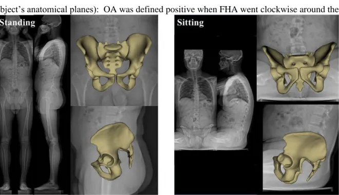
All subjects underwent standing and sitting full-body bi-planar EOS low-dose radiographs (EOS Imaging, Paris, France) [17]. Hip and Spine patients did this within their clinical routine, while C subjects were volunteers; all subject signed informed consent. Radiographs were acquired in standardized free-standing position [14], by taking care that subject’s coronal plane was aligned with the antero-posterior radiographic plane when standing. Then sitting bi-planar radiographs were obtained using a previously published protocol [3,18]. The patients sat on a backless stool with both feet resting flat on the floor and legs spread apart, and was asked to sit comfortably upright [19,20]. The simultaneous bi-planar acquisition was used to perform 3D reconstruction of pelvis and femoral head using a validated research software based on previously described method [14,15] (Fig. 1).
3D reconstruction of the pelvis in standing and sitting positions were performed in each configuration by an expert operator (Y.K.). The two models were translated so that the middle of the line joining each acetabulum corresponded to the origin. The relative reorientation of the pelvis between standing and sitting was characterized by calculating the finite helical axis (FHA) between the two configurations. The orientation angle (OA) between bicoxofemoral axis and FHA (Fig. 2a) and the rotation angle (RA) around FHA (Fig. 2b) were also computed. These axes and angles were expressed in the subject’s coordinate system (i.e, respecting the subject’s anatomical planes): OA was defined positive when FHA went clockwise around the
Fig. 1 Frontal and lateral EOS radiographs with 3D reconstructions of the pelvis in standing and
pelvis posteroanterior axis (negative if counterclockwise). RA was positive in case of posterior pelvic tilt (pelvic extension) and negative if anterior pelvic tilt (pelvic flexion). Pelvic parameters (pelvic incidence (PI), sacral slope (SS) and pelvic tilt (PT)) were also calculated from 3D reconstructions.
Statistical analysis
To interpret these parameters according to the three groups, corridors of normality were calculated for each parameter of the C group. The values of pathological patients were superimposed on the same graph to compare each group. For each parameter, the mean (M) and standard deviation (SD) of the C group were used to class as normal if within M ± SD range, subnormal high between M + SD and M + 2SD (low between M - SD and M - 2SD), or abnormal when out of the range M ± 2 SD (high or low). This representation aimed to highlight the parameters that distinguish pathological subjects from the healthy ones.
The statistical analysis was performed Matlab1 (version R2011a; The Mathworks Inc., Natick, MA, USA). Quantitative variables were described using the mean, mean difference, standard deviation, and intraclass correlation coefficient and its 95% confidence interval. Normal distribution of the values was checked with Shapiro-Wilk normality test for each series of measurements. For data with normal distribution, paired
Fig. 2 Representation of the two axis (bicoxofemoral axis and finite helical axis (FHA)) and two
radiological parameters (orientation angle (OA) and rotation angle (RA)) calculated on the 3D pelvis reconstruction between standing and sitting: a) OA between bicoxofemoral axis and FHA, b) RA around the FHA
Students-t-test (for differences stand to sit) and ANOVA were used for multiple comparison between groups. The significance level was set at less than 0.05.
Results
Table 2 shows the pelvic parameters including pelvic incidence, sacral slope and pelvic tilt. The mean PI was 52.3°, 58.0° and 56.6° for C, Hip and Spine group in standing position, respectively, with no significant difference between groups. The difference between standing and sitting was always lower than 5°, which is within the uncertainty of measurement of this parameter. In the C group, the mean SS was 40.5° in standing and 22.7° in sitting position. The mean PT was 12.2° in standing and 29.3° in sitting. There was a significant difference in SS and PT between standing and sitting in C group (p<0.05). In the Hip and Spine groups, the mean SS decreased significantly (43.3° to 23.8° and 37.6° to 21.8°, respectively) and the mean PT increased significantly (14.9° to 36.4° and 19.5° to 34.6°, respectively) between standing and sitting. In the Hip group, SS in standing was significantly larger than in Spine group, but not different from C group (p < 0.05).
Orientation angle of the finite helical axis relative to bicoxofemoral axis
Fig. 3 shows the orientation angle of Hip and Spine patients relative to the C group. Mean OA was -1.8° (SD 10.8°) for C group, 0.3° (SD 12.3°) for Hip group and -2.4° (SD 16.4°) for Spine group. There was no statistically significant difference in OA between groups. However, variability was higher for Spine group with a standard deviation of 21.5° compared to 10.8° in C group and 12.3° in Hip group. Seven patients (23.3%) with abnormal values were found for Spine group compared to 3 patients (10.0%) for Hip group. Analysis of the angle between bicoxofemoral axes in standing and sitting position showed no significant difference between groups (ANOVA test, p = 0.6). While the average difference was lower than 2° in all anatomical planes, with no difference between planes (p = 0.9), large angles could be observed in some subjects, up to 13° in the axial plane and 12° in the coronal plane. In the Hip group, 15 patients showed subnormal axial rotation of the pelvis, against only 8 and 7 in the C and Spine groups, respectively.
Rotation angle around the finite helical axis
Figure 4 shows the RA in control and pathology groups. Mean RA was 18.1° (SD 9.1°) for C group, 21.1° (SD 8.0°) for Hip group and 16.4° (SD 10.8°) for Spine group. There was significant statistical difference in RA between Hip and Spine groups (p=0.04). Fifteen patients (50.0%) with subnormal values were found for Spine group (10 low values and 5 high values) compared to 8 subnormal values (26.7%) for Hip group (1 low value and 7 high values).
Fig. 3 Orientation angle between standing and sitting in healthy, hip pathology and spine pathology
patients. Repartition of parameters values compared with intervals given by the mean (M) and standard deviation (SD) of healthy group
Fig. 4 Rotation axis between standing and sitting in healthy, hip pathology and spine pathology
patients. Repartition of parameters values compared with intervals given by the mean (M) and standard deviation (SD) of healthy group
Discussion
The concept of ʺpelvic vertebraʺ was originally introduced by Dubousset in 1984 [21]. Other investigators have shown the importance of the pelvic rotation between standing and sitting in the understanding of pelvic mobility in physiological and pathological condition [3,22]. Most of those publications considered that the pelvic movement in the sagittal plane consists in a rotation around the bicoxofemoral axis, which passes through the center of the femoral heads [10,11,23]. Literature reports that abnormal pelvic movement has substantial effect on hip joint kinematics with potential consequences on hip instability in patients after THA [24,25]. However, there was no quantitative analysis about precise assessment of the axis of pelvic rotation. This study is among the first to focus on the quantitative analysis of the true 3D axis of pelvic rotation between standing and sitting for physiological and pathological condition.
Ninety subjects were enrolled, including 30 controls and 30 for each group defined by hip or spine pathologies. Control group was not age-matched with the pathology groups; the interest of the control group was to determine a normal range for pelvic reorientation when sitting, so the implied hypothesis is that healthy aging would not alter sitting strategy. Despite the limited number in each group, significant differences among the three groups were found, and the analysis of outliers showed a large number of subnormal values in Hip and Spine patients, suggesting they might find different strategies for achieving the sitting position than
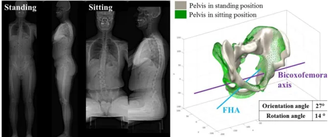
Fig. 5 Case study for a 43 years old male patient with spine pathology (L5/S1 spondylolisthesis).
Abnormal pelvis kinematics from standing to sitting: orientation angle was 27° and rotation angle was 14°. This abnormal kinematics induced an asymmetrical movement for right and left acetabulum between standing and sitting.
control subjects. This demonstrates the potential interest of an improved knowledge of the finite helical axis for pelvic rotation with regard to bicoxofemoral axis. That should be confirmed in a larger scale patient population. We excluded patients with combined spine, hip or lower limb surgeries in order to investigate well defined groups. The findings of this study should also be investigated in these more complex patients.
According to the hypothesis that bicoxofemoral axis is true axis of pelvic rotation, the expected motion would imply the FHA to be aligned with this axis (OA is null). Results of the present study indicated that the asymptomatic subject performed pelvic rotation around the bicoxofemoral axis, despite a large variability between subjects. Patients with hip and spine pathology had even larger variability and abnormality in pelvic kinematics between standing and sitting. Going more in detail, we can first investigate each group with following questions.
1) Is the bicoxofemoral axis the true axis of pelvic rotation in physiological condition between standing and sitting positions?
Previous studies have reported that hip joint center, modelled as a ball-socket joint, can be estimated using either a functional or a predictive approach [26,27]. Orthopedic surgeons consider that the pelvis moves, rotating around the hip, leading to either anterior tilt or posterior tilt [5,10]. In this study, the mean OA between bicoxofemoral axis and FHA was -1.8° in C group. However, there was a wide range in values for OA in this control group. 68% of population were between -12.6° and 9.0° and 95% of population were between -23.4° and 19.8°. Moreover, analysis of the angle between the standing and sitting bicoxofemoral axes show differences up to 12° in both the coronal and axial planes; from a methodological point of view, the angle between the FHA and bicoxofemoral axis represents the deviation of pelvic reorientation from pure ante/retro-version, while the projections of the angle between standing and sitting bicoxofemoral axes represents the direction of the reorientation. These results indicated that the asymptomatic subject without spine and hip pathologies has a large variability of 3D axis of pelvic rotation between standing and sitting. This kinematic variability of the pelvis can have significant consequences on the functional position of the acetabulum and should be considered for an optimized analysis of pelvic movement.
2) How does the pelvis rotate around the true axis (FHA) in physiological condition between standing and sitting?
Recent research revealed that pelvis moves between standing and sitting according to hip joint and spine movements and compensations [5,10]. The rotation of the pelvis between standing and sitting is considered as the main adaptation mechanism for postural change. In this study, the mean SS of control group in standing position was 40.5° and the mean RA around the FHA was 18.1°. Our findings match the previous data reported in the literature [4], with new quantitative information regarding the stand to sit pelvic kinematics using 3D reconstruction from bi-planar radiographies. Range of rotation was large: 68% of population were between 9.0° and 27.2° for RA between standing and sitting. These results indicated that the asymptomatic subject has a large variability of rotation angle around the true axis (FHA) between standing and sitting.
3) Do hip and spine pathologies affect pelvic kinematics between standing and sitting?
Because the motion of the spine, pelvis and hip is coordinated during postural changes, any pathology of this area may affect stand to sit pelvis kinematics [10]. According to the concept of the hip-spine relations, end stage hip DJD can cause flexion contractures of the hip joint, which leads to compensatory change of the pelvis and the lumbar spine [28]. Okuda et al. reported that SS and lumbar lordosis angles were significantly greater in patients with hip DJD than in healthy volunteers [29]. In this study, the patients in Hip group had a significant higher SS in standing position compared to other groups, which is consistent with the previously reported data in the literature [23,30]. The mean OA in Hip group was similar to C group (Hip: 0.3° vs C: -1.8°). However, the variability of the OA was slightly higher for Hip group with a standard deviation of 12.3° compared to 10.8° in C group. Furthermore, the mean RA in Hip group was slightly higher than in C group (Hip: 21.1° vs C: 18.1°). Previous studies reported that abnormal motion of the pelvis may be due to hip flexion contracture and pain [28,30]. This abnormal motion of the pelvis linked to the hip disease affects the functional orientation of the acetabular implants in THA patients. Previous paper reported 0.5 to 0.7° change in the functional acetabular anteversion per 1° change in posterior pelvic tilt [13,22,31]. However, these studies did not find the abnormal kinematics of the pelvis because the investigators used only the 2D lateral radiographs. We suggest that the hip pathology affect the abnormality of kinematics of pelvic rotation between standing and sitting and that these abnormal kinematics could be evaluated using 3D reconstruction from bi-planar radiographies for preoperative planning such THA.
Previous studies have reported that the patients with spinal fusion demonstrated less adaptability of the lumbosacral junction and had a risk of complication like dislocation and revision after THA because of abnormal spine-pelvis-hip biomechanics [3,4,32]. A recent study reported that a predictive factor for falling outside the functional safe zone of the prosthesis after THA was the decreased spinopelvic mobility due to spine pathology [33]. Heckmann et al reported that the patients with late dislocation after THA had spinopelvic imbalance with a decreased pelvic rotation in sitting[25]. However, the exact mechanism leading to postoperative dislocation is not clear yet.
In this study, the mean OA in the Spine group was much lower than C group (Spine: -4.7° vs C: -1.8°). Furthermore, the variability of the OA was higher for Spine group with a standard deviation of 21.5° compared to 10.8° in C group and 12.3° in Hip group. Previous studies reported that the abnormal motion of pelvis results in an unbalanced spine and pelvis [19,34]. 23.3% of the patients in Spine group who presented abnormal values of OA (OA <-23.4°, 19.8°<OA) (Fig. 5). These results demonstrate an asymmetrical movement for right and left acetabulum between standing and sitting in these outliers, directly linked to spinal pathology.
The mean RA in Spine group was lower than C group (Spine: 16.0° vs C: 18.1°). Many studies suggested that decreased mobility of the pelvis could be observed in spine pathology [18,19]. These findings suggest that the spine pathology affect the stand to sit pelvic kinematics much more than hip pathology. Surgeons should be aware of abnormal kinematics in patients with spine disease: 3D reconstruction from bi-planar radiographies in standing and sitting could help preoperative planning of THA implantation. This could allow taking potential abnormalities into account, particularly in patients with severe spine disease.
Conclusion
This study has highlighted the quantitative analysis of rotation of the pelvis between standing and sitting in healthy, hip pathology patients and spine pathology patients using 3D reconstruction from bi-planar radiographs. Hip or spine pathology affected the abnormal stand to sit pelvic kinematics. The EOS 3D reconstruction of the pelvis in standing and sitting postures provided a new insight of pelvic kinematics. Surgeons should be aware of potential abnormal stand to sit pelvis kinematics when preoperative planning a surgery such as THA. Further studies are needed to provide stronger evidence and define surgery planning in order to improve the clinical outcome in the complex cases mixing hip and spine degeneration.
References
[1] Kralj A, Jaeger RJ, Munih M. Analysis of standing up and sitting down in humans: Definitions and normative data presentation. J Biomech 1990;23:1123–38. doi:https://doi.org/10.1016/0021-9290(90)90005-N.
[2] Kerr KM, White JA, Barr DA, Mollan RAB. Analysis of the sit-stand-sit movement cycle in normal subjects. Clin Biomech 1997;12:236–45. doi:https://doi.org/10.1016/S0268-0033(96)00077-0.
[3] Lazennec JY, Clark IC, Folinais D, Tahar IN, Pour AE. What is the Impact of a Spinal Fusion on Acetabular Implant Orientation in Functional Standing and Sitting Positions? J Arthroplasty 2017;32:3184–90. doi:10.1016/j.arth.2017.04.051.
[4] Stefl M, Lundergan W, Heckmann N, McKnight B, Ike H, Murgai R, et al. Hip arthroplasty: Avoiding and managing problems spinopelvic mobility and acetabular component position for total hip arthroplasty. Bone Jt. J., vol. 99B, 2017, p. 37–45. doi:10.1302/0301-620X.99B1.BJJ-2016-0415.R1.
[5] Lazennec JY, Brusson A, Rousseau MA. Hip-spine relations and sagittal balance clinical consequences. Eur Spine J 2011;20 Suppl 5:686–98. doi:10.1007/s00586-011-1937-9.
[6] Hunt MA, Gunether JR, Gilbart MK. Kinematic and kinetic differences during walking in patients with and without symptomatic femoroacetabular impingement. Clin Biomech 2013;28:519–23. doi:10.1016/j.clinbiomech.2013.05.002.
[7] Hara D, Nakashima Y, Hamai S, Higaki H, Ikebe S, Shimoto T, et al. Dynamic hip kinematics in patients with hip osteoarthritis during weight-bearing activities. Clin Biomech 2016;32:150– 6. doi:10.1016/j.clinbiomech.2015.11.019.
[8] Constantinou M, Loureiro A, Carty C, Mills P, Barrett R. Hip joint mechanics during walking in individuals with mild-to-moderate hip osteoarthritis. Gait Posture 2017;53:162–7. doi:10.1016/j.gaitpost.2017.01.017.
[9] Ochi H, Baba T, Homma Y, Matsumoto M, Nojiri H, Kaneko K. Importance of the spinopelvic factors on the pelvic inclination from standing to sitting before total hip arthroplasty. Eur Spine J 2016;25:3699–706. doi:10.1007/s00586-015-4217-2.
[10] Ike H, Dorr LD, Trasolini N, Stefl M, Mcknight B, Heckmann N, et al. Spine-Pelvis-Hip Relationship in the Functioning of a Total Hip Replacement. J Bone Joint Surg Am 2018;0:1606–15. doi:10.2106/JBJS.17.00403.
[11] Kanawade V, Dorr LD, Wan Z. Acetabular Component Angular Change from Sitting to Standing. J Bone Jt Surg - Am Vol 2014;96–A:978–86. doi:10.2106/JBJS.M.00765.
[12] Endo K, Suzuki H, Nishimura H, Tanaka H, Shishido T, Yamamoto K. Sagittal lumbar and pelvic alignment in the standing and sitting positions. J Orthop Sci 2012;17:682–6. doi:10.1007/s00776-012-0281-1.
[13] Maratt JD, Esposito CI, McLawhorn AS, Jerabek SA, Padgett DE, Mayman DJ. Pelvic tilt in patients undergoing total hip arthroplasty: when does it matter? J Arthroplast 2015;30:387–91. doi:10.1016/j.arth.2014.10.014.
[14] Chaibi Y, Cresson T, Aubert B, Hausselle J, Neyret P, Hauger O, et al. Fast 3D reconstruction of the lower limb using a parametric model and statistical inferences and clinical measurements calculation from biplanar X-rays. Comput Methods Biomech Biomed Engin 2012;15:457–66. doi:10.1080/10255842.2010.540758.
[15] Ghostine B, Sauret C, Assi A, Bakouny Z, Khalil N, Skalli W, et al. Influence of patient axial malpositioning on the trueness and precision of pelvic parameters obtained from 3D reconstructions based on biplanar radiographs. Eur Radiol 2017;27:1295–302. doi:10.1007/s00330-016-4452-x.
[16] Lazennec JY, Folinais D, Florequin C, Pour AE. Does Patients’ Perception of Leg Length After Total Hip Arthroplasty Correlate With Anatomical Leg Length? J Arthroplasty 2018. doi:10.1016/j.arth.2017.12.004.
[17] Dubousset J, Charpak G, Skalli W, Deguise J, Kalifa G. EOS: A NEW IMAGING SYSTEM WITH LOW DOSE RADIATION IN STANDING POSITION FOR SPINE AND BONE & JOINT DISORDERS. J Musculoskelet Res 2010;13:1–12. doi:10.1142/S0218957710002430.
[18] Lazennec JY, Riwan A, Gravez F, Rousseau MA, Mora N, Gorin M, et al. Hip spine relationships: Application to total hip arthroplasty. HIP Int 2007;17:91–104.
[19] Esposito CI, Miller TT, Kim HJ, Barlow BT, Wright TM, Padgett DE, et al. Does Degenerative Lumbar Spine Disease Influence Femoroacetabular Flexion in Patients Undergoing Total Hip Arthroplasty? Clin Orthop Relat Res 2016;474:1788–97. doi:10.1007/s11999-016-4787-2. [20] Rouissi J, Arvieu R, Dubory A, Vergari C, Bachy M, Vialle R. Intra and inter-observer reliability
of determining degree of pelvic obliquity in neuromuscular scoliosis using the EOS-CHAIR® protocol. Child’s Nerv Syst 2017;33:337–41. doi:10.1007/s00381-016-3326-5.
[21] Andre C, Badoual J, Kalifa G, Dubousset J. [African histoplasmosis. A case]. Arch Fr Pediatr 1984;41:429–31.
[22] Lazennec JY, Boyer P, Gorin M, Catonné Y, Rousseau MA. Acetabular anteversion with CT in supine, simulated standing, and sitting positions in a THA patient population. Clin Orthop Relat Res 2011;469:1103–9. doi:10.1007/s11999-010-1732-7.
[23] Stefl M, Lundergan W, Heckmann N, Mcknight B, Ike H, Murgai R, et al. Spinopelvic mobility and acetabular component position for total hip arthroplasty 2017;99:37–45. doi:10.1302/0301-620X.99B1.BJJ-2016-0415.R1.
[24] Lazennec JY, Brusson A, Rousseau MA. Lumbar-pelvic-femoral balance on sitting and standing lateral radiographs. Orthop Traumatol Surg Res 2013;99:S87–103. doi:10.1016/j.otsr.2012.12.003.
[25] Heckmann N, Mcknight B, Ste M, Trasolini NA, Ike H, Stefl M, et al. Late Dislocation Following Total Hip Arthroplasty. J Bone Jt Surg - Am Vol 2018;100:1845–53. doi:10.2106/JBJS.18.00078.
[26] Della Croce U, Leardini A, Chiari L, Cappozzo A. Human movement analysis using stereophotogrammetry Part 4: Assessment of anatomical landmark misplacement and its effects on joint kinematics. Gait Posture 2005;21:226–37. doi:10.1016/j.gaitpost.2004.05.003.
[27] Begon M, Monnet T, Lacouture P. Effects of movement for estimating the hip joint centre. Gait Posture 2007;25:353–9. doi:10.1016/j.gaitpost.2006.04.010.
[28] Offierski CM, Macnab I. Hip-spine syndrome. Spine (Phila Pa 1976) 1983;8:316–21. doi:10.1097/00007632-198304000-00014.
[29] Okuda T, Fujita T, Kaneuji A, Miaki K, Yasuda Y, Matsumoto T. Stage-specific sagittal spinopelvic alignment changes in osteoarthritis of the hip secondary to developmental hip dysplasia. Spine (Phila Pa 1976) 2007;32. doi:10.1097/BRS.0b013e31815ce695.
[30] Weng WJ, Wang WJ, Wu M Da, Xu ZH, Xu LL, Qiu Y. Characteristics of sagittal spine–pelvis– leg alignment in patients with severe hip osteoarthritis. Eur Spine J 2015;24:1228–36. doi:10.1007/s00586-014-3700-5.
[31] Lazennec JY, Charlot N, Gorin M, Roger B, Arafati N, Bissery A, et al. Hip-spine relationship: A radio-anatomical study for optimization in acetabular cup positioning. Surg Radiol Anat 2004;26:136–44. doi:10.1007/s00276-003-0195-x.
[32] Sultan AA, Khlopas A, Piuzzi NS, Chughtai M, Sodhi N, Mont MA. The Impact of Spino-Pelvic Alignment on Total Hip Arthroplasty Outcomes: A Critical Analysis of Current Evidence. J Arthroplasty 2017. doi:10.1016/j.arth.2017.11.021.
[33] Tezuka T, Heckmann ND, Bodner RJ, Dorr LD. Functional Safe Zone Is Superior to the Lewinnek Safe Zone for Total Hip Arthroplasty: Why the Lewinnek Safe Zone Is Not Always Predictive of Stability. J Arthroplasty 2018:1–6. doi:10.1016/j.arth.2018.10.034.
[34] Phan D, Bederman SS, Schwarzkopf R. The influence of sagittal spinal deformity on anteversion of the acetabular component in total hip arthroplasty. Bone Jt J 2015;97–B:1017–23. doi:10.1302/0301-620X.97B8.35700.