Science Arts & Métiers (SAM)
is an open access repository that collects the work of Arts et Métiers Institute of
Technology researchers and makes it freely available over the web where possible.
This is an author-deposited version published in: https://sam.ensam.eu
Handle ID: .http://hdl.handle.net/10985/19220
To cite this version :
Luc LAVISSE, Armand KANJER, P. BERGER, V. OPTASANU, Cyril GORNY, P. PEYRE, T.
MONTESIN, M.C. MARCO DE LUCAS - High temperature oxidation resistance and
microstructure of laser-shock peened Ti-Beta-21S - Surface and Coatings Technology - Vol. 403,
p.1-8 - 2020
Any correspondence concerning this service should be sent to the repository
Administrator : archiveouverte@ensam.eu
High temperature oxidation resistance and microstructure of laser-shock
peened Ti-Beta-21S
L. Lavisse
a,⁎, A. Kanjer
a, P. Berger
b, V. Optasanu
a, C. Gorny
c, P. Peyre
c, T. Montesin
a,
M.C. Marco de Lucas
a,⁎aLaboratoire Interdisciplinaire Carnot de Bourgogne, UMR 6303 CNRS, Université Bourgogne-Franche Comté, 9 Avenue A. Savary, BP 47 870, F-21078 Dijon Cedex,
France
bUniversité Paris-Saclay, CEA, CNRS, NIMBE, 91191 Gif-sur-Yvette, France
cLaboratoire PIMM - Ensam CNRS Cnam - Arts et Métiers – Sciences et Technologies - Ensam 151 Bd de l’Hôpital, 75013 Paris, France
Keywords:
Mechanical surface treatments Metastable beta-titanium High temperature oxidation Oxidation kinetics Oxygen diffusion
A B S T R A C T
Improving the high temperature (HT) resistance of titanium alloys is currently a technological challenge for extending their use in aerospace engines. Ti-Beta-21S is a metastable β titanium alloy specifically designed for high temperature applications up to 593 °C. We report the effect of a surface treatment by laser-shock peening (LSP) on the high temperature behavior of Ti-Beta-21S in order to increase further its maximum service tem-perature. The oxidation kinetics at 700 °C for duration up to 3000 h showed that the LSP treatment increases the oxidation resistance of Ti-Beta-21S. The effects of the LSP treatment on the alloy microstructure, its evolution at high temperature and the diffusion of light atmospheric elements (oxygen and nitrogen) are also reported.
1. Introduction
Aviation is currently one of the fastest-growing sources of green-house gas emissions. This is a huge ecological challenge for aeronautics. In order to reduce pollutant emissions, aircrafts of the future require the development of components with improved lightness, efficiency and durability. The excellent combination of lightness and good physical and chemical properties makes titanium alloys very attractive in this context [1,2]. They are especially found in compressor blades and discs and engine housings, as well as in structures. However, their use is still limited to temperatures below 550 °C because of their low resistance to oxidation above this limit [3]. Implementing Ti alloy components which could resist to higher temperatures would provide a mass gain of approximately 50% compared to nickel-based superalloys currently in use above 550 °C. Extending the use of these alloys to the turboengines of the aircraft of the future would make titanium alloys even more at-tractive.
Mechanical surface treatments such as shot-peening (SP) or laser-shock peening (LSP) appear as possible technological solutions to extent the service temperature range of various metals [4,5]. Those techniques are already used in the industry to improve some particular properties of metals at ambient temperature: mechanical (fatigue resistance) [6,7], tribological (resistance to wear) [8,9] and electrochemical
(resistance to pitting) [10]. The beneficial effect of SP and LSP treat-ments on the high temperature (HT) oxidation resistance of pure tita-nium at 700 °C has been reported by Kanjer et al. [11,12]. The reduc-tion of both the mass gain and the diffusion length of oxygen in the Ti-α matrix [12], which corresponds to the so-called α-case, were remark-able in LSP treated samples. To the best of our knowledge, the possi-bility of using this kind of surface treatments to increase the maximum application temperature of metastable beta titanium alloys has not been studied. The improved high specific strength and cool formability of metastable β-titanium alloys are great assets in aeronautics applica-tions.
The aim of this study is to investigate the effect of the LSP treatment on the high temperature oxidation resistance of a metastable β-titanium alloy, as well as on the microstructure and its evolution at high tem-perature.
The material used for this work was the Ti-Beta-21S alloy from TIMET (TIMETAL 21S), which has been specifically designed for im-proved oxidation resistance, elevated temperature strength, creep re-sistance and thermal stability [13–15]. Ti-Beta-21S is reported by TIMET as suitable for high temperature applications up to 593 °C.
The study of the oxidation kinetics of LSP treated plates at 700 °C in dry air during 3000 h showed higher oxidation resistance of LSP treated samples compared to the reference material. The structural
⁎Corresponding authors.
E-mail addresses:luc.lavisse@u-bourgogne.fr(L. Lavisse),delucas@u-bourgogne.fr(M.C. Marco de Lucas).
characterization of LSP treated samples and the analysis of the spatial distribution of the main alloying elements (molybdenum and alu-minum) and light atmospheric elements (oxygen and nitrogen) after exposure at high temperature were addressed to explain the improve-ment of the oxidation resistance. Electron Backscattering Diffraction (EBSD) and scanning electron microscopy coupled to electron microp-robe (EDS) were used for this purpose. Moreover, ion beam micro-analysis was used for mapping the distribution of light elements in the cross-section of oxidized samples. The diffusion of oxygen and the in-sertion of nitrogen under the oxide scale have been investigated in this way.
2. Experimental details
2.1. The material
The material used in this study was the Ti-Beta-21S metastable β titanium alloy (TIMET supplier). This alloy is used in aeronautics for its good mechanical and chemical resistance up to 600 °C. Stabilized mainly in the metastable β-Ti phase, its chemical composition (in wt%) is: Ti 78, Mo 15.13, Nb 2.65, Fe 0.26, Si 0.2, Al 3.45, C 0.013, O 0.13, N 0.02.
For LSP treatments, pieces of 25 × 50 mm2were cut from 1.8 mm
thick cold-rolled strips produced by TIMET. High temperature oxidation experiments were made on samples of 10 × 10 × 1.8 mm3.
In order to facilitate the reading of the following results, the un-treated samples will be designated here by the abbreviation US, while those treated by laser-shock peening will be called LSP. Untreated and laser treated samples oxidized 3000 h at 700 °C will be called US-3000 h and LSP-US-3000 h, respectively.
2.2. Surface mechanical treatments and HT oxidation experiments Laser-shock peening was performed at the PIMM laboratory of ENSAM Paris, using a GAIA HP laser source [4]. After coating the surface of the sample with an aluminum foil tape, the samples were placed in a water container. The water media was used to confine the plasma plume produced by the laser beam. The aluminum coating ab-sorbs the thermal effects generated by the laser irradiation and the water media amplifies both the amplitude and the duration of the shock effect within the treated material. The wavelength of the laser beam was 532 nm and the diameter of the focal spot was 4 mm. The duration of the laser shots was 7 ns and the frequency 0.5 Hz. The deposited laser irradiance was 9.1 GW/cm2. The laser beam scanned the sample surface
on snake-shaped parallel lines with a velocity of 1.1 mm/s. The area of the overlap between two successive shots was 30%. The two faces of the sample were treated successively.
The treated and untreated samples (LSP and US) were oxidized under synthetic dry air at 700 °C during 3000 h by using a SETNAG quartz tube furnace. The samples were extracted every 160 h, weighed to evaluate their mass gain, and replaced into the furnace. Under these conditions, the material was thus cooled to room temperature and then put back to the oven after each weighing. A purge was carried out before introduction of the oxidizing gas, to avoid any presence of water vapors.
2.3. Characterization techniques
The surface topography of the samples was studied using a VEECO WYCO NT 9100 optical profilometer.
X-ray diffraction phase analysis was carried out by using a BRUKER D8-A25 DISCOVER diffractometer. A Cu anticathode was used with beam incidence fixed at 2°.
The chemical state of the samples before HT oxidation experiments was analyzed by XPS (PHI Versaprobe 5000). In order to clean the sample surface, the very first superficial layers of the material were
removed by bombardment with a 5 keV argon ion beam (Ar+) for
1 min.
After LSP treatment and oxidation for different durations of ex-posure to HT, the samples were cross-sectioned, resin-embedded and mechanically polished up to 50 nm colloidal silica suspension. Cross-sections of US and LSP samples were analyzed by SEM coupled to an electron microprobe in order to investigate the distribution of the al-loying elements (Mo, Al, …). Two microscopes coupled to energy-dis-persive spectroscopy (EDS) probes were used here, a TESCAN VEGA 3 and a field-emission microscope JEOL JSM-7600F.
The grain orientation was analyzed by Electron Backscattering Diffraction (EBSD) using a TSL EDAX OIM X4M EBSD system coupled with a JEOL-7600F microscope. The working distance was 20 mm, the tension 20 kV, the magnification ×70 and the step of scanning 1 μm. Cross-sections of US-3000 h and LSP-3000 h samples were prepared as follows for EBSD analysis. The specimens were embedded in a cold-cure resin with rapid polymerisation and small shrinkage. They were me-chanically polished with P600 and P1200 grit SiC papers. Then they were mirror-finished and slightly chemically etched using a 70:30 mixture of a colloidal silica suspension and H2O2on a polishing cloth
(Presi, Supra).
Ion beam microanalysis was mainly used for mapping the dis-tribution of light elements (oxygen and nitrogen) in the cross-section of oxidized samples. The operating principle of these techniques is sum-marized in Fig. S1 (Supplementary Material). The nuclear microprobe of CEA-Saclay was used for this study. Depending on the implemented technique, the experimental beam conditions were as follows:
•
Proton Induced X-ray Emission (PIXE) and Proton Induced Gamma-Ray Emission (PIGE) spectra were recorded with a 3 MeV proton beam (spot size 4.5 μm × 3.5 μm) for Ti (Kαand Kβlines) and27Al(1013 keV line), respectively.
•
Nuclear Reaction Analysis (NRA) and Deuteron Induced Gamma-ray Emission (DIGE) spectra were recorded with a 1.9 MeV deuteron beam (spot size: 4.5 μm × 3.5 μm) for the reactions of14N (14N(d,pi)15N and14N(d,αi)12C), and16O (871 keV line), respectively. 3. Results
3.1. Consequences of LSP treatments on the raw material
Fig. 1presents a 3D reconstruction of the surface topography of the US and LSP samples of Ti-Beta-21S before and after the LSP surface treatment. The roller marks are clearly visible on the US sample, whereas they have practically disappeared after the laser treatment. The total roughness, Rt, slightly decreased from 6.4 μm for US sample to
6.2 μm for LSP sample. The average roughness, Ra, remained
un-changed and equal to 0.4 μm for both US and LSP samples.
The hardness profiles along the thickness (Fig. S2) were measured on the cross-section of samples before and after LSP treatment. The variation of hardness along the thickness was negligible for both kinds of samples, and the mean value was around 330 HV. The absence of strain hardening leads to the conclusion that the LSP treatment does not induce plastic deformation. The same result was obtained for the LSP treatment of pure α-Ti [12].
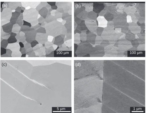
The cross-sections of US and LSP samples were observed by SEM in back-scattering electrons mode (BSE) (Fig. 2). The size distribution and the shape of the grains are similar for both US and LSP samples. However, only the images of LSP samples show in many areas a net-work of parallel straight lines (bright lines inFig. 2b) which cross the grains and which can extend into neighboring grains. High magnifica-tion views given inFig. 2c and d show these bright lines near the grain boundaries. The in-depth hardness profile has shown that the plastic deformation of the treated samples is negligible, so these lines cannot be explained as dislocations induced by the LSP treatment. Similar lines were also observed in the cross-section SEM images of LSP treated pure
α-Ti [12], and they were assigned to mainly compressive twins.
Fig. 3displays the XRD patterns obtained with the beam incidence fixed at 2° for untreated and laser treated samples. The depth of the analyzed area was estimated around 1 μm. The patterns of untreated samples show only the peaks of the β-Ti phase (ICSD: 0773482). For
laser treated samples, the same diffraction peaks of the β-Ti phase are observed, but their absolute intensity is smaller, and they are slightly broader. These effects can be mainly attributed to the disorder induced by the treatment and/or a refinement of crystallites on the surface. Nonetheless, the relative intensity between matching peaks of the β-Ti phase is quite similar in the patterns of both samples. This means that the laser treatment does not result in a different texturing than that produced after rolling.
The XRD patterns of LSP samples display also two additional small peaks at 35.1 and 40.1°, which reveal the presence of a second phase formed as a consequence of the LSP treatment. The position of these
Fig. 1. Surface optical profilometry of Ti-Beta-21S samples before (a) and after
(b) the LSP treatment.
Fig. 2. Cross-section SEM views of (a) US and (b) LSP samples. The surface of the cross-sectioned sample is at the top of both images. (c, d) High magnification views
of the grain boundaries taken in the cross section of the LSP sample.
Fig. 3. Grazing incidence X-ray diffraction patterns (incidence angle: 2°) of
untreated (US) and LSP treated samples. The peaks assigned to the β-Ti phase (ICSD: 0773482) are indicated. The intensity has been normalized to that of the β-Ti(100) reflection. The peaks tagged by stars (*) have been tentatively as-signed to the martensitic α″-Ti phase.
peaks is close to that given in Ref. [16] for the α-Ti phase formed in Ti-Beta-21S by oxidation at 650 °C during 50 h. However, the heating induced by the LSP treatment is too low to induce the β → α phase transition. These small peaks at 35.1 and 40.1° could correspond to the martensitic α″-Ti phase which in general can be formed by deformation of β-Ti at room temperature [17–20]. Here, the formation of α″-Ti would be induced by the recoiled pressure of the laser-shock wave.
In order to investigate the chemical composition at the extreme surface, XPS analyses were conducted for US and LSP samples. The results are shown inFig. 4in the spectral ranges corresponding to the XPS lines Ti2p, Al2p and O1s. The spectra showing the XPS lines Mo3d, Mo3p and Nb3d are given in Fig. S3 (Supplementary Material).
For the US sample,Fig. 4a displays the characteristic spectrum of metallic titanium which shows the Ti2p3/2and Ti2p1/2lines at 453.7
and 459.8 eV, respectively [21]. The slight broadening of both peaks towards higher binding energies observed for LSP samples can be in-terpreted as due to a slight oxidation of the surface. As a reference, Ti4+2p
3/2line is given at 458.5 eV and Ti4+2p1/2at 464.2 eV for TiO2
[21].
For the Al2p line (Fig. 4b), the main peak corresponds for both samples to metallic aluminum, Al02p
3/2 at 72.7 eV [22]. For the LSP
sample, a small component at about 75.2 eV can be attributed to Al3+2p
3/2[22]. The XPS lines of molybdenum and niobium (Fig. S3)
suggest also a slight surface oxidation of the LSP sample.
As regards oxygen, the shape of the O1s line (Fig. 4c) is slightly different for US and LSP samples. The O1s line corresponding to OeTi bonds has been reported at about 530 eV, while that corresponding to OeAl bonds is at about 532 eV and that for OeTi bonds at about 530 eV [22,23]. Compared to the US sample, the relative intensity of the contribution associated to TieO bonds is higher in the spectrum of LSP samples. This shows that the surface oxidation of LSP samples is slightly higher compared to US samples.
3.2. Kinetics of the high temperature oxidation
Fig. 5shows the variation of the mass gain reported to the surface of the sample, Δm/S, in US and LSP samples for non-isothermal oxidation at 700 °C. The mass gain of LSP treated samples after exposure to high temperature for 3000 h is about 2.5 times smaller for laser-shock treated samples compared to untreated ones. So, the mass gain is about 60% smaller for laser-shock treated Ti-Beta-21S. This clearly demon-strates the benefits of the LSP treatment to increase the oxidation re-sistance of Ti-Beta-21S at 700 °C.
The analysis of the oxidation kinetics was done on the basis of the most-general expression of parabolic kinetics as proposed by Monceau and Pieraggi [24]:
= + +
t a0 a1( m S/ ) a2( m S/ )2 (1) where t is the time, Δm the mass intake, S the sample surface, and ai
(i=0,1,2) the fitting coefficients. The reciprocal of a2is equal to the
parabolic rate constant, kp, which describes the effect of the diffusion
processes on the mass intake. A parabolic law is associated to an ef-fective protection of the metal against the oxidation provided by the oxidation scale, which grows with a continuing decreasing rate [25]. The reciprocal of a1is the linear rate constant, kl, which together with
the coefficient a0depends on the initial thickness of the oxide and the
oxidation mechanisms involved in a transient period.
The experimental data were fitted to Eq.(1)in the 0–3000 h range. The fitting parameters are given inTable 1, together with the corre-sponding linear and the parabolic rate constants. The linear rate con-stant, kl, is almost 2 times higher for the untreated Ti-Beta-21S
com-pared to the laser treated alloy. The parabolic rate constant, kp, is about
7 times higher for the US sample than for the LSP one.
3.3. Effects of high temperature exposure on the microstructure and the diffusion of alloying and light atmospheric elements
Fig. 6shows the XRD patterns of US and LSP samples oxidized for 3000 h at 700 °C in dry air. The main phase observed for both samples is TiO2-Rutile. A small contribution of Al2O3-corundum can also be
identified, mainly for the US-3000 h sample. This is in agreement with the results reported by Wallace et al. [26] and by Behera et al. [16] showing the formation of an alumina layer on the top of the rutile scale. Stringer et al. [27] also showed the formation of a small amount of
Fig. 4. XPS spectra of untreated (US) and laser treated samples (LSP) in the spectral ranges corresponding to the following XPS lines: (a) Ti2p, (b) Al2p and (c) O1s.
Fig. 5. Mass gain curves of US and LSP samples during oxidation at 700 °C
under synthetic dry air. Dotted lines correspond to the fitting of the experi-mental data to Eq.(1)with the parameters given inTable 1.
molybdenum oxide in the scale in the high temperature oxidation of a Ti-15% Mo alloy. However, the volatilization process of molybdenum oxide, MoO3, at high temperature is well known [28]. Here, no
dif-fraction peaks of MoO3or mixed oxides could be clearly identified.
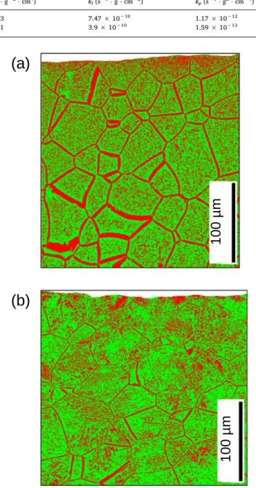
Fig. 7displays cross-section maps of oxidized US-3000 h and LSP-3000 h samples showing the spatial distribution of β-Ti (in green) and α-Ti (in red) phases obtained by EBSD study of both samples.
For the US-3000 h sample, the β-Ti phase is found in about 60% of the cross-section image, while 40% corresponds to α-Ti phase. For the LSP-3000 h sample, the percentage of β-Ti phase is higher, about 68%, while 32% corresponds to α-Ti phase. The ratio β-Ti/α-Ti increases from 1.5 for US-3000 to 2.1 for LSP-3000 h, which shows that the LSP treatment acts to inhibit the β → α phase transition.
Moreover, the distribution of the α-Ti phase is different in both samples. For the untreated sample, the α-Ti phase is mainly found in the grain boundaries which are broadened. For the LSP-3000 h sample, the α-Ti is not mainly formed in the grain boundaries, but inside the grains. For both samples, the percentage of α-Ti phase is higher near the surface (on top of both images in Fig. 7). In a range going from the surface to 30 μm in-depth, the percentage of α-Ti phase increases to about 53% for the SP-3000 h sample, while it is about 39% for the LSP-3000 h sample.
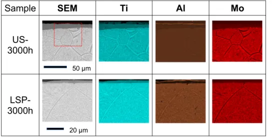
In order to investigate the segregation of the alloying elements due to high temperature exposure, SEM observations were coupled to EDS mapping of different elements. Cross-section SEM views and EDS maps showing the spatial distribution of Ti, Al and Mo are given inFig. 8for US and LSP samples.
After 3000 h at 700 °C, the thickness of the oxide scale has increased in both samples, but it is clearly thinner in the LSP sample than in the US one. For the US sample the thickness of the oxide scale is not homogeneous, it can vary from about 10 to 35 μm thick. In these latter zones the scale is stratified and then provides little protection against
oxidation. For the LSP sample, the oxide scale is quite homogeneous and only about 6 μm thick. It can provide an effective protection to the metal. Aluminum is clearly observed in a layer at the extreme outer surface of US and LSP samples. This agrees with XRD results, and supports the existence of a thin layer of alumina on top of the scale [16,26].
Underneath the oxide scale,Fig. 8shows also a clear difference in the grain boundaries, which are quite broader for the US sample (about 2–5 μm thick) than for the LSP sample (about 1–2 μm thick). The spatial resolution of EDS is barely sufficient for the analysis of the grain
Table 1
Results of fitting the experimental oxidation curves (Fig. 5) to a complete parabola (Eq.(1)). The linear rate constant, klis the reciprocal of a1, and the parabolic rate
constant, kpis the reciprocal of a2.
Sample a0(h−1) a1(h ⋅ g−1⋅cm2) a2(h ⋅ g−2⋅cm4) kl(s−1⋅g ⋅ cm−2) kp(s−1⋅g2⋅cm−4)
US −64.3 371.7 238.3 7.47 × 10⁻¹⁰ 1.17 × 10⁻¹²
LSP −46.5 703.0 1751.1 3.9 × 10⁻¹⁰ 1.59 × 10⁻¹³
Fig. 6. Grazing incidence XRD patterns of untreated (US) and laser treated
samples (LSP) after oxidation at 700 °C during 3000 h. ICDS number of the compounds and phases identified in the pattern: TiO2-rutile: 070-7347, Al2O3
(corundum): 070-5679.
Fig. 7. EBSD results showing the spatial distribution of β-Ti (in green) and α-Ti
(in red) phases in the cross section of (a) US-3000 h and (b) LSP-3000 h sam-ples. The surface of the cross-sectioned sample is at the top of both images. (For interpretation of the references to color in this figure legend, the reader is re-ferred to the web version of this article.)
boundaries in LSP-3000 h, but it is suitable for their analysis in US-3000 h. The segregation between α- and β-stabilizing elements in the metal is clearly observed. At the grain boundaries, the concentration of aluminum is higher, whereas that of molybdenum is lower. This seg-regation of α- and β-stabilizing elements is also observed inside the grains.
The spatial distribution of light elements (oxygen and nitrogen) in the cross-section of these samples was also studied by EDS, but the overlap between the Kαlines of nitrogen and oxygen with the Lαand K1
lines titanium penalizes the accuracy of the results. Fig. S4 (Supplementary Material) displays the concentration profiles obtained for both metallic and non-metallic elements in this way.
To overcome this problem, ion-beam analysis techniques were used here to study the distribution of light elements in oxidized samples.
Fig. 9 displays cross-section maps showing the spatial distribution of light elements (O and N) over 200 × 200 μm, as well as the distribution of metallic elements (Ti, Al and Mo) over 300 × 300 μm.
The spatial distribution of oxygen clearly shows the oxidation scale at the top of the images for both US-3000 h and LSP-3000 h samples, but its thickness is quite bigger for the untreated sample. Moreover, the spatial distribution of titanium shows the stratification of the oxidation layer in some areas (on the left inFig. 9). Between two sublayers of the oxidation scale the Al-maps shows a higher concentration of aluminum, which reveals the formation of alumine Al2O3on the top of the
sub-layers. A similar effect is not observed for molybdenum.
Under the oxide scale, the spatial distribution of Ti shows inFig. 9
the grain boundaries in the metal, as it was previously observed in EDS maps (Fig. 8). Unfortunately, the low signal-to-noise ratio in aluminum and molybdenum maps prevents observing the segregation of these elements in the grain boundaries.
As regards the distribution of light elements under the oxide scale,
Fig. 9shows the diffusion of oxygen in the metal, which seems to be smaller in LSP treated samples than in untreated samples. To better study this effect, the variation in depth of the DIGE signal of oxygen has been plotted inFig. 10by using the experimental data ofFig. 9for oxygen. For both US and LSP samples, the highest signal corresponds to the oxide scale. However, the low thickness of the scale (similar to the step in IBA maps) for the LSP-3000 h prevents obtaining the high in-tensity measured for the US-3000 h sample. Under the oxide scale, the signal of oxygen drastically falls down for both samples, but there are significant differences between both samples. For the treated sample, the signal of oxygen is very low and nearly constant in the analyzed range. For the untreated sample, the oxygen signal far deep from the oxide is slightly higher than for the treated sample, moreover, it is higher in a 20 μm thick strip under the oxide scale. It is worth to note the large thickness and the stratification of the oxide above this area.
The atomic concentration of oxygen as a function of the oxygen signal has been calculated by using the signal of the oxide scale as a reference corresponding to TiO2The difference in the slowing of the
incident beam of deuterons as a function of the TiOxstoichiometry was
Fig. 8. Cross-section SEM views of US-3000 h
and LSP-3000 h samples recorded in back-scat-tering electrons (BSE) configuration, and ele-mental EDS maps corresponding to the spatial distribution of titanium, aluminum and mo-lybdenum. The surface of the cross-sectioned sample is at the top of the images. A higher magnification was used for the observations of the LSP-3000 h sample. For easier comparison, a dotted red rectangle of 60 × 50 μm2displays in
the SEM image of US-3000 h sample the size of the area analyzed for the LSP-3000 h sample. (For interpretation of the references to color in this figure legend, the reader is referred to the web version of this article.)
Fig. 9. Spatial distribution of different
elements (Ti, Al, Mo, O and N) in the cross-section of US-3000 h and LSP-3000 h samples obtained by different ion-beam microanalysis techniques (see Section 2.3). The surface of the cross-sectioned sample is at the top of the images. The characteristics of the ion beam and the IBA technique used for detecting each element are also in-dicated.
addressed by using the Pyrole software [29]. Further details are given as Supplementary Material (Fig. S5). For the US-3000 h sample, the stoichiometry is about TiO0.092(8.4 at.% O) in a 20 μm thick strip under
the oxide scale, but it decreases to TiO0.012(1.2 at.% O) 150 μm far in
depth from the oxide scale. For the LSP-3000 h sample, the con-centration of oxygen in the metal is smaller compared to US-300. It is negligible far in-depth in the metal, and it slightly increases up to about 0.5 at.% just under the oxide scale.
IBA experiments revealed the presence of nitrogen in the oxidized samples (Fig. 9). Nitrogen was clearly detected in both untreated and treated samples under the oxide scale, but its distribution is different. For the laser treated sample, nitrogen is located in a thin layer just under the oxide scale, while it spreads more in the metal for the un-treated sample without forming a continuous layer under the oxide scale. The narrow in-depth spatial distribution of nitrogen and the low intensity of the NRA signal do not allow extracting an in-depth profile as done for the oxygen.
4. Discussion
The study of the HT oxidation resistance of untreated and laser treated Ti-Beta-21S at 700 °C during 3000 h has shown the beneficial effects of the laser treatment. They can be summarized as follows:
•
The mass gain is reduced about 60% for non-isothermal oxidation during 3000 h.•
The oxidation scale is homogeneous and unstratified.•
The in-depth diffusion of oxygen in the alloy under the oxide scale is also reduced.•
The insertion of nitrogen in a thin and continuous layer at the in-terface between the oxide scale and the metal.•
The β → α phase transition at high temperature is partially in-hibited, even 200 μm under the sample surface.The reasons explaining these changes in the HT oxidation behavior of laser treated Ti-Beta-21S should be sought in the changes induced by the laser treatment in the sample surface and also in-depth in the alloy. The changes at the sample surface concern mainly the topography of the sample. The LSP treatment flattens the roller marks due to the
lamination process of Ti-Beta-21S and slightly reduces the total roughness. It induces also a slight oxidation of the surface. The induced hardening is negligible. The surface modified in this way can promote the growth at high temperature of a more compact oxide scale com-pared to untreated samples. However, the changes induced in the sur-face cannot explain all the effects summarized above.
The in-depth effects induced by the LSP treatment must be related to the propagation of the shock-wave in the sample. The analysis of laser treated samples has shown a partial transformation of the β-Ti phase into the martensitic α″-Ti phase. We have related this effect with the observation in SEM cross-section observations of laser treated samples of a network of lines crossing the grains of β-Ti. Taking into account that the hardening induced by the treatment is negligible, these lines cannot be assigned to dislocations. The experimental conditions used here for the laser treatment lead to the formation of a plasma over the surface sample which extends over about 10 μs for each shot [10]. This plasma induces a pressure smaller than the yield strength of Ti-Beta-21S. The shock-wave propagates in-depth in the sample at around 5000 m/s and it can be reflected on the bottom sample surface 1.8 mm below. The interference between incident and reflected waves can in-duce shearing strains which can promote twinning. The lines observed in the cross section of LSP samples can be due to this effect. This ex-planation is supported by the presence of peaks assigned to the α″-Ti phase in the XRD patter recorded for LSP samples before the oxidation experiments.
Different works [20,30–32] have studied the stress-induced mar-tensitic transition (SIMT) of β-Ti alloys during deformation at room temperature. They have shown that in metastable β-Ti alloys with a low molybdenum equivalency (about 8.1 wt%), SIMT is the primary de-formation mode. The start of yielding can correspond to the triggering of SIMT eventually followed by dislocation slip at a higher stress, but both effects can also operate simultaneously [30]. α″-Ti plates have been observed by SEM and TEM [30]. α″-Ti is mechanically twinned, and its substructure can influence the twinning in the β-Ti phase.
For Ti-Beta-21S, the molybdenum equivalency about 13 wt% is quite higher than the stability limit α-Ti+β-Ti/ β-Ti about 10 wt%. The aluminum equivalency is low, about 5 wt%. The shock-laser treatment induces fast mechanical stress with very low heating. In these condi-tions, SIMT can appear without dislocation slip. The presence of α″-Ti could modify the β-Ti → α-Ti phase transition at high temperature by reducing the diffusion of alpha-stabilizing aluminum towards the grain boundaries where the β-Ti → α-Ti transition is mainly observed in untreated Ti-Beta-21S.
Oxygen is also an alpha-stabilizing element. As regards its in-depth diffusion from the sample surface and its spatial distribution in the cross-section maps, the results do not show a significant increase of the concentration in oxygen at the grain boundaries of oxidized untreated (US) samples, where the α-Ti phase is formed at high temperature. This suggests that the diffusion of oxygen does not occur preferentially fol-lowing the grain boundaries, but it takes place in the grains. The in-depth diffusion of oxygen in the alloy is also reduced by the laser treatment. This could be explained by the insertion of nitrogen in a thin and continuous layer at the interface between the oxide scale and the metal shown by IBA experiments. This layer of nitrogen would reduce the diffusion of oxygen towards the metal. This effect has been pre-viously reported for α-Ti [12].
5. Conclusions
This work shows the beneficial effect of a laser-shock peening (LSP) treatment on the high temperature oxidation resistance of the me-tastable β titanium alloy Ti-Beta-21S which has been studied for a long exposure time at 700 °C in dry air. After 3000 h, the mass gain in LSP treated samples is reduced by about 60% compared to untreated Ti-Beta-21S. The oxidation scale is thinner and more homogeneous for LSP treated samples compared to untreated specimens that present stratified
Fig. 10. Oxygen depth profile in the US-3000 h and LSP-3000 h samples. The
intensity of the oxygen signal in the DIGE maps ofFig. 9was summed at each depth X in the 40 μm large rectangles (red delimited domains in the image) to increase the S/N ratio, and plotted as a function of depth (X). (For interpreta-tion of the references to color in this figure legend, the reader is referred to the web version of this article.)
oxide scales. In both cases, the oxidation layer is mainly composed of rutile with a thin layer of alumina on top of the scale. Below the oxide, we have detected the insertion of nitrogen in a narrow strip. The con-centration of nitrogen is higher in laser treated samples.
The LSP treatment modifies the topography of the surface of the samples by flattening the roller marks. It also induces a slight oxidation of the surface while the induced hardening is negligible. Beyond the surface, the impact of the LSP treatment on the microstructure was revealed in SEM cross-section views by the apparition of a network of lines crossing the grains of laser treated samples. This effect was as-signed to a partial transformation of the β-phase into the martensitic α″-phase detected by XRD.
The analysis of oxidized untreated and laser treated samples under the oxidation scale has shown that the LSP treatment partially inhibits the β → α phase transition at the grain boundaries. It is mainly found for untreated samples together with the segregation of molybdenum and aluminum. For laser treated samples, the α-phase is formed mainly inside the grains. The following explanation has been proposed based on the martensitic transformation induced by the laser treatment: at high temperature the α″ phase transforms into the α which reduces the diffusion of aluminum towards the grain boundaries and of mo-lybdenum towards the grains core.
The diffusion of oxygen in the metal is also smaller in laser treated samples. This is noteworthy to preserve the properties of Ti-Beta-21S under high temperature exposure. The compactness of the oxide scale and the insertion of nitrogen in a thin layer under the oxide scale, acting as a break to oxygen diffusion, could explain this behavior.
In summary, the surface treatment of Ti-Beta-21S alloy by laser-shock peening is a suitable method for extending its maximum appli-cation temperature in dry air.
CRediT authorship contribution statement
L. Lavisse: Conceptualization, Methodology, Investigation,
Validation, Writing - review & editing, Project administration. A.
Kanjer: Investigation, Formal analysis, Visualization. P. Berger:
Investigation, Methodology, Formal analysis, Writing - review & editing. V. Optasanu: Conceptualization, Methodology, Validation, Writing - review & editing. C. Gorny: Resources, Investigation. P.
Peyre: Resources, Validation, Supervision. T. Montesin: Validation,
Supervision. M.C. Marco de Lucas: Conceptualization, Methodology, Investigation, Validation, Writing - review & editing, Project adminis-tration.
Declaration of competing interest
The authors declare that they have no known competing financial interests or personal relationships that could have appeared to influ-ence the work reported in this paper.
Acknowledgements
The authors thank O. Heintz, N. Geoffroy and F. Herbst from the ICB laboratory and M. Saint-Jean from the IUT Chalon sur Saône for their contribution to the experimental observations and analyses, and Prof. B. Domenichini for his expertise in the analysis of XPS results. This work has been supported by the EIPHI Graduate School (contract ANR-17-EURE-0002).
Appendix A. Supplementary data
Supplementary data to this article can be found online athttps:// doi.org/10.1016/j.surfcoat.2020.126368.
References
[1] J.C. Williams, E.A. Starke, Progress in structural materials for aerospace systems, Acta Mater. 51 (2003) 5775–5799.
[2] K. Inagaki, T. Takechi, Y. Shirai, N. Ariyasu, Application and features of titanium for the aerospace industry, technical report, Nippon Steel Sumitomo Metal 106 (2014) 22–27.
[3] J. Stringer, The oxidation of titanium in oxygen at high temperatures, Acta Metall. 8 (1960) 758–766.
[4] R. Fabbro, P. Peyre, L. Berthe, X. Scherpereel, Physics and applications of laser-shock processing, J. Laser Appl. 10 (1998) 265–279.
[5] L. Raceanu, V. Optasanu, T. Montesin, G. Montay, M. François, Shot-peening of pre-oxidized plates of zirconium: influence of residual stress on oxidation, Oxid. Met. 79 (2012) 135–145.
[6] X.C. Zhang, Y.K. Zhang, J.Z. Lu, F.Z. Xuan, Z.D. Wang, S. Tu, Improvement of fa-tigue life of Ti–6Al–4V alloy by laser shock peening, Mater. Sci. Eng. A 527 (2010) 3411–3415.
[7] E. Maawad, Y. Sano, L. Wagner, H.-G. Brokmeier, C. Genzel, Investigation of laser shock peening effects on residual stress state and fatigue performance of titanium alloys, Mater. Sci. Eng. A 536 (2012) 82–91.
[8] S.A. Chamgordani, R. Miresmaeili, M. Aliofkhazraei, Improvement in tribological behavior of commercial pure titanium (CP-Ti) by surface mechanical attrition treatment (SMAT), Tribol. Int. 119 (2018) 744–752.
[9] M. Rajabi, R. Miresmaeili, M. Aliofkhazraei, Hardness and wear behavior of surface mechanical attrition treated titanium, Mater. Res. Exp. 6 (2019) 065003. [10] P. Peyre, X. Scherpereel, L. Berthe, C. Carboni, R. Fabbro, G. Béranger, C. Lemaitre,
Surface modifications induced in 316L steel by laser peening and shot-peening. Influence on pitting corrosion resistance, Mater. Sci. Eng. A 280 (2000) 294–302. [11] A. Kanjer, V. Optasanu, L. Lavisse, M.C. Marco de Lucas, S. Dejardin, M. François,
P. Berger, P. Peyre, C. Gorny, T. Montesin, Influence of mechanical surface treat-ment on high-temperature oxidation of pure titanium, Oxid. Met. 88 (2017) 383–395.
[12] A. Kanjer, L. Lavisse, V. Optasanu, P. Berger, C. Gorny, P. Peyre, F. Herbst, O. Heintz, N. Geoffroy, T. Montesin, M.C. Marco de Lucas, Effect of laser shock peening on the high temperature oxidation resistance of titanium, Surf. Coat. Technol. 326 (2017) 146–155.
[13] K. Chaudhuri, J. Perepezko, Microstructural study of the titanium alloy Ti-15Mo-2.7Nb-3Al-0.2Si (TIMETAL 21S), Metall. Mater. Trans. A 25 (1994) 1109–1118. [14] I. Polmear, D. StJohn, J.-F. Nie, M. Qian, 7— Titanium alloys, in: I. Polmear,
D. StJohn, J.-F. Nie, M. Qian (Eds.), Light Alloys, Fifth ed., Butterworth-Heinemann, Boston, 2017, pp. 369–460.
[15] K.K. Sankaran, R.S. Mishra, Chapter 5 — Titanium alloys, in: K.K. Sankaran, R.S. Mishra (Eds.), Metallurgy and Design of Alloys With Hierarchical Microstructures, Elsevier, 2017, pp. 177–288.
[16] A. Behera, S. Nag, K. Mahdak, H. Mohseni, J. Tiley, R. Banerjee, Influence of oxygen ingress on fine scale precipitation of α-Ti during oxidation of beta 21S β-Ti alloy, J. Mater. Sci. 48 (2013) 6700–6706.
[17] C. Li, G. Li, Y. Yang, M. Varlioglu, K. Yang, Martensitic twinning in alpha + beta Ti-3.5Al-4.5Mo titanium alloy, J. Metallur. 2011 (2011) 1–5.
[18] Y. Mantani, Y. Takemoto, M. Hida, A. Sakakibara, M. Tajima, Phase transformation of α′ martensite structure by aging in Ti-8 mass%Mo alloy, Mater. Trans. 45 (2004) 1629–1634.
[19] S. Guo, Q. Meng, X. Zhao, Q. Wei, H. Xu, Design and fabrication of a metastable β-type titanium alloy with ultralow elastic modulus and high strength, Sci. Rep. 5 (2015) 14688.
[20] D. Pionnier, M. Humbert, M. Philippe, Y. Combres, Study of the α″ phase texture obtained by martensitic β–α″ phase transformation induced by tensile test in a sheet of Ti5Al2Sn4Zr4Mo2Cr1Fe, Acta Mater. 46 (1998) 5891–5898.
[21] M.C. Biesinger, L.W.M. Lau, A.R. Gerson, R.S.C. Smart, Resolving surface chemical states in XPS analysis of first row transition metals, oxides and hydroxides: Sc, Ti, V, Cu and Zn, Appl. Surf. Sci. 257 (2010) 887–898.
[22] M.R. Alexander, G.E. Thompson, G. Beamson, Resolving surface chemical states in XPS analysis of first row transition metals, oxides and hydroxides: Sc, Ti, V, Cu and Zn, Surf. Interface Anal. 257 (2000) 468–477.
[23] J.F. Moulder, W.F. Stickle, P.E. Sobol, K.D. Bomben, J. Chastein, R.K. Jr, Handbook of X-ray Photoelectron Spectroscopy, Physical Electronics, Eden Prairie, 1995. [24] D. Monceau, B. Pieraggi, Determination of parabolic rate constants from a local
analysis of mass-gain curves, Oxid. Met. 50 (1998) 477–493.
[25] P. Kofstad, High temperature corrosion, Physical Electronics, Applied Science Publishers Ltd., London–New York, 1988, p. 1988.
[26] T.A. Wallace, R.K. Clark, K.E. Wiedemann, Oxidation Characteristics of Beta21S in Air in the Temperature Range 600–800 °C, NASA Technical Memorandum 104217 (1992), Langley Res. Center, Hampton, VA (United States), 1992.
[27] J. Stringer, G. Metcalf, M. Nicholson, The high-temperature oxidation of a com-mercial titanium-molybdenum alloy, J. Less Common Metals 4 (1962) 69–77. [28] P.E. Blackburn, M. Hoch, H.L. Johnston, The vaporization of molybdenum and
tungsten oxides, J. Phys. Chem. 62 (1958) 769–773.
[29] P. Trouslard, Pyrole: un logiciel au service des analyses par faisceau d’ions, Rapport CEA-R5703, (1995).
[30] A. Zafari, K. Xia, Stress induced martensitic transformation in metastable βTi-5Al-5Mo-5V-3Cr alloy: triggering stress and interaction with deformation bands, Mater. Sci. Eng. A 724 (2018) 75–79.
[31] T. Grosdidier, C. Roubaud, M.-J. Philippe, Y. Combres, The deformation mechan-isms in the β-metastable β-Cez titanium alloy, Scr. Mater. 36 (1997) 21–28. [32] X. Jiang, H. Zhao, R. Han, X. Zhang, M. Ma, R. Liu, Grain refinement and tensile
properties of a metastable TiZrAl alloy fabricated by stress-induced martensite and its reverse transformation, Mater. Sci. Eng. A 722 (2018) 8–13.