Structural models of the different trimers
present in the core of phycobilisomes from
Gracilaria chilensis based on crystal structures
and sequences
Jorge Dagnino-Leone1☯, Maximiliano Figueroa1☯, Claudia Mella1, Marı´a Alejandra Vorphal1, Fre´de´ric Kerff2, Aleikar Jose´ Va´squez1, Marta Bunster1
*, Jose´ Martı´nez-Oyanedel1*
1 Departamento de Bioquı´mica y Biologı´a Molecular, Facultad de Ciencias Biolo´gicas, Universidad de
Concepcio´n, Concepcio´n, Chile, 2 Centre d’Inge´nie´rie des Prote´ines, Universite´ de Liège, Liège, Belgium ☯These authors contributed equally to this work.
*mbunster@udec.cl(MB);jmartine@udec.cl(JMO)
Abstract
Phycobilisomes (PBS) are accessory light harvesting protein complexes that directionally transfer energy towards photosystems. Phycobilisomes are organized in a central core and rods radiating from it. Components of phycobilisomes in Gracilaria chilensis (Gch) are Phy-cobiliproteins (PBPs), Phycoerythrin (PE), and Phycocyanin (PC) in the rods, while Allophy-cocyanin (APC) is found in the core, and linker proteins (L). The function of such complexes depends on the structure of each component and their interaction. The core of PBS from cyanobacteria is mainly composed by cylinders of trimers ofαandβsubunits forming het-erodimers of Allophycocyanin, and other components of the core including subunitsαIIand
β18. As for the linkers, Linker core (L
C) and Linker core membrane (LCM) are essential for the
final emission towards photoreaction centers. Since we have previously focused our studies on the rods of the PBS, in the present article we investigated the components of the core in the phycobilisome from the eukaryotic algae, Gracilaria chilensis and their organization into trimers. Transmission electron microscopy provided the information for a three cylinders core, while the three dimensional structure of Allophycocyanin purified from Gch was deter-mined by X-ray diffraction method and the biological unit was deterdeter-mined as a trimer by size exclusion chromatography. The protein sequences of all the components of the core were obtained by sequencing the corresponding genes and their expression confirmed by tran-scriptomic analysis. These subunits have seldom been reported in red algae, but not in Gra-cilaria chilensis. The subunits not present in the crystallographic structure were modeled to build the different composition of trimers. This article proposes structural models for the dif-ferent types of trimers present in the core of phycobilisomes of Gch as a first step towards the final model for energy transfer in this system.
a1111111111 a1111111111 a1111111111 a1111111111 a1111111111 OPEN ACCESS
Citation: Dagnino-Leone J, Figueroa M, Mella C,
Vorphal MA, Kerff F, Va´squez AJ, et al. (2017) Structural models of the different trimers present in the core of phycobilisomes from Gracilaria chilensis based on crystal structures and sequences. PLoS ONE 12(5): e0177540.https:// doi.org/10.1371/journal.pone.0177540
Editor: Jiajie Diao, University of Illinois at
Urbana-Champaign, UNITED STATES
Received: February 2, 2017 Accepted: April 28, 2017 Published: May 18, 2017
Copyright:© 2017 Dagnino-Leone et al. This is an open access article distributed under the terms of theCreative Commons Attribution License, which permits unrestricted use, distribution, and reproduction in any medium, provided the original author and source are credited.
Data Availability Statement: All protein structure
files are available from the PDB database (accession code 5TJF).
Funding: This work was supported by FONDECYT
113.0256 grant to MB, FONDECYT Doctoral grant 21120260 to JDL, CONICYT scholarship to JDL and CR_Lab Biofı´sica Molecular 2015-2016. The funders had no role in study design, data collection and analysis, decision to publish, or preparation of the manuscript.
Introduction
Aquatic organisms, such as cyanobacteria, eukaryotic red alga and cryptophyta, present acces-sory light harvesting protein complexes called phycobilisomes (PBS) [1]. The structure of PBS allows the energy to be transferred directionally to photosystem II (PSII), thus improving the use of the 400-650nm range of wavelengths. Phycobilisomes are organized in a central core and rods radiating from it. Components of phycobilisomes inGracilaria chilensis are
Phycobi-liproteins (PBPs): Phycoerythrin (PE), and Phycocyanin (PC) in the rods, Allophycocyanin (APC) is found in the core, and linker proteins (L) present along the whole system. PBPs are chromophorylated proteins that share a general architecture. They are organized as heterodi-mers (αβ), which assembly themselves into triheterodi-mers or hexaheterodi-mers. The subunits of PBPs are mono or multi chromophorylated with linear tetrapyrrols covalently bound to cysteine resi-dues. Efficient energy transfer is achieved through a combination of position and geometry of the chromophores and the protein environment [1–3]. In the present article, we focused on the principal component of the phycobilisomes´ core, the phycobiliprotein Allophycocyanyn or APC. The Allophycocyanin is organized as cylinders, and different numbers of these cylin-ders have been reported for the core of PBS including bicylindrics, tricylindrics and pentacy-lindrics, being the tricylindric the most common in cyanobacteria and red alga [4–6]. The tricylindrical core has been proposed to be formed by a foundation of two cylinders associated to membrane and a third cylinder is located at the top of the base forming a pyramid [7]. Four subunits have been identified for APC in the core:α, β, αIIandβ18, which are codified by apcA, apcB, apcD and apcF genes, respectively [8]. Studies reported onSynechococcus show
that subunitsαIIandβ18are expressed in minor quantities, which make difficult to study the expressed proteins in native conditions [9,10]. Indeed, studies of the same nature in eukaryotic red alga are still missing.
Besides PBPs, the two linkers Core (LC) and core-membrane (LCM) have been identified in
the core [1]. The LCof 7.8 kDa is codified by the apcC gene. This linker is associated to APC
trimers in cylinders, suggesting a specific role on stability and energy transfer [9,11–13]. The structure of LChas been determined by X-ray diffraction as a complex with APC trimer in
Mastigocladus laminosus (PDB ID: IB33) [14]. The LCM, codified by the apcE gene, in a
tricy-lindrical core is a 90kDa chromophorylated linker protein that has been proposed as the final acceptor of the light harvested by the PBS, besides its role in the anchoring of the PBS to the photosynthetic membrane [9,12,13,15,16]. Different domains have been identified in the linker core-membrane; for example, the N-terminal region, called PB domain, is homologous to PBP alpha subunits, and shows 30% similarity withα subunit of APC, and also contains a phycocyanobilin chromophore. This PB domain presents an insertion of 50 residues (PB loop). The PB loop in cyanobacteria has been associated to fluorescence quenching by its inter-action with the orange carotenoid protein (OCP) [17], which has not been detected in eukary-otic alga. Zhao and his group [15] produced a recombinant chromophorylated PB domain in absence of lyases, a fact that induced the authors to propose an autocatalytic binding of phyco-cyanobilin to LCM. Due to their similarity, the PB domain can replace theα subunit and form a
PBβ heterodimer, which can associate itself to other αβ heterodimers to form a trimer. Different trimers composition present in a tricylindrical core have been reported for Syne-chocystis [18] APC (α3β3), APC_1 (αα3β3Lc), APC_2 (α2αIIβ3Lc) and APC_3 (α2PBβ2β18).
Watanabe and Ikiushi [19], in an interesting review proposed also a model for the position of each type of trimer in the three cylinders core, which inGracilaria chilensis has not been
assessed yet.
Gracilaria chilensis (Rhodophyta, Gigartinalis) (G.ch) [20] is one of the economically important red macroalga in the South Pacific. Our research on the structure and function of
Competing interests: The authors have declared
the PBS in this algae has been focused on understanding how the organization,i.e.
composi-tion, structure or component interactions of the PBS provides the frame for the high efficiency of the energy transfer towards PSII. Until now, the structure of PE and PC fromGch has been
reported previously by our group [21,22], as well as information about interaction between hexamers in a rod and its role in the function of energy transfer [23]. In this article, we report i) the three cylinders nature of the core of the PBS inGch, ii) the sequences of the subunits
components of the core, iii) the sequence analysis and iv) the X-ray structure for Allophyco-cyanin fromGch (αβ)3. This structure was used to model the rest of the subunits, which may
also form a functional Allophycocyanin, and thereby to build the trimers proposed for the core in cyanobacteria, as our first approach to the analysis of the contribution of both the subunits in the core and LCto the energy transfer pathways in an eukaryotic red alga.
Material and methods
Purification of phycobilisomes
PBS were purified using the condition presented inS1 File, from 200g of intertidalG.ch
col-lected at Coliumo Bay (36˚ 32’S, 072˚ 56’ W). The fraction containing complete PBS was iden-tified by its spectroscopic properties (emission at 660nm after excitation at 566nm) and was used for Electron Microscopy.
Electron microscopy: The micrographs were obtained in a Philips Tecnai 12 Bio Twin EM
(120kv) at the Pontificia Universidad Cato´lica de Chile facility, using the methodology described [23] for complete phycobilisomes.
The sequences of the subunits
Primers design. The primers were designed by sequence alignment of the genes reported
in literature, using the sequences displayed onS2 File. The primers were designed manually considering the conserved sequences, the %GC, Tm and the size of the oligonucleotides. The design was verified with Oligonucleotide PrimerCheck (http://depts.washington.edu/bakerpg/ primertemp/primermelttemp.html) and synthesized by IDT. The primers used were:
α subunit Sense: 5’atgagtattattactaaatcaatcgttaatgctgatgcagaagctcgg 3` Antisense: 3’ttactgcattgcacctaatgtataatcaaaataaaatcct 5’ βsubunit Sense: 5’atgcaagatgctattacttctgtaattaatgcagctgatgtacaagg 3’ Antisense: 3’ctagcttaaaccagaacaaatataatcaaaatatactccc5’ αII subunit Sense: 5’atgagcttagttagccaaattattttaaatgcagataatgaattaagat 3’ Antisense: 3’ ttatgacataccttgtattataaaatcaaagtagggttct 5’ β18 Sense: 5’atgcaagacgctattactacaattttaaatcgatatgatttaacaggaaa 3’ Antisense: 3’ ttagatatcttcttcgcttaagtttttaattatatattgaaatggttcaa 5’
Sequencing of the genes codifying for the subunits of Allophycocyanin. RNA was
extracted from 0.1g of fresh washed and frozen alga, ground in a mortar with TRI-Reagent (Molecular Research Center) using the protocol recommended by the supplier. cDNA was obtained using Reverse Transcriptase RevertAidTMkit (Fermentas) according to recommen-dations of suppliers. The cDNA was used for the amplification assays by PCR according toS3 File. The sequences translated to proteins were used for the resolution of the 3D structure of Allophycocyanin and for the molecular modeling of the subunitsαIIandβ18.
The sequence of the linkers associated to the CORE. The PB domain of the LCM:Primers
were designed according to the sequences reported for LCMof the red algaeChondrus crispus
[24] andGracilaria tenuistipitata [25]. The sequencing and the amplification methodology were similar to the methodology described above forα and β subunits of APC.
The LC: The sequence of the Linker core ofGracilaria chilensis was obtained from a direct
analysis of the transcriptome (AN: SRX1507975) [26].
Purification of Allophycocyanin
APC was obtained as reported inS4 File. The pure protein was characterized by its spectro-scopic properties of absorption (λA
max= 651nm) and emission (λEmax= 660nm) and the
rela-tionship A651/A280as purity index. This sample was used for the determination of the
oligomerization state of Allophycocyanin, the spectroscopic studies and for crystallization.
Determination of the oligomerization state
50μL of 1 mg/mL of purified APC was separated in a Superdex200 HR/30 column (Amershan Biosciences), equilibrated with 5mM phosphate buffer pH7, and associated to a Merck-Hitachi FPLC with a flux of 0.5ml/min provided by a L6210 Intelligent Pump with a L-4250 UV-Vis Detector. For the calibration, BioRad standards were used. The elution of the proteins was fol-lowed at 651 and 280nm.
Protein crystallization
A general screening was performed in the robotic unit at Universidad de Chile. The final crys-tallization conditions were 2mg/mL protein concentration, 300mM NaCl, 20% v/v PEG 6000, and 100mM MES pH7.
Determination of the three dimensional structure of APC
Crystals were diffracted at Soleil Synchrotron up to 2.3Å resolution. The phases were obtained by molecular replacement using the data of 1KN1 [27] and the software Phenix [28]. The final model was built in Coot [29] and refined by Phenix Refine [30].
Molecular models for the different trimers present in the core
The trimer for APC was obtained by crystallographic symmetry (P321). The different subunits were modeled with Modeller v 9.1 [31]. The templates were theα subunit of APC from the structure reported here (PDB ID: 5TJF), the PB domain in 4XXI [32], and theβ subunit of 5TJF forαII, PB domain andβ18, respectively. Trimers APC_1, APC_2, APC_3 were built by substitution of the corresponding subunits followed by a molecular dynamics protocol for ste-ric rearrangement of residues. LCwas modelled also with Modeller using the chain O of the
reported structure forMastigoclaudus laminosus (PDB ID: 1B33) [14] as template, and a dock-ing procedure was performed between the LCand (αβ)3or (αIIα2β3) using ClusPro [33]
with-out restrictions. Each final model was evaluated with PROSA [34] and RAMPAGE [35].
Results and discussion
The phycobilisome’s core of G. chilensis is tricylindrical
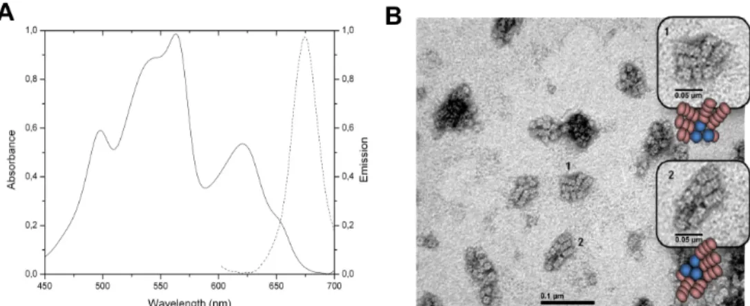
The PBS enriched fractions were analyzed by electron microscopy. The absorption spectrum (Fig 1A) showed the assembly of the PBS. Upon excitation at 566nm corresponding to the absorption maximum of PE (Phycoerythrin). The only emission corresponded to the emission
of APC, which means that the assembly was intact. Moreover, the energy has been transferred through the complex to emit at 660nm. The micrograph (TEM) for the PBS is shown inFig 1B. The image clearly showed three cylinders core of phycobilisomes, which are compatible with the presence of 90kDa LCM, and 5–6 rods formed by the piling of 3–4 hexamers. This is
the first time that a photomicrograph of a phycobilisome ofGracilaria chilensis is reported,
and at least in shape and organization resembles the PBS from prokaryotes [2].
Allophycocyanin is mainly present as trimer (
αβ
)
3, despite the
transcription of other subunits
The absorption and emission spectra of the purified samples of Allophycocyanin are shown in
Fig 2A. This sample was used to determine the oligomerization state using size exclusion chro-matography (Fig 2B).
Fig 1. Characterization of phycobilisomes of Gracilaria chilensis. A) Absorption(-) and emission(..)
spectra. B) Transmission electron micrograph of purified phycobilisomes.The inserts show amplified images. Schematic drawings of PBS are also shown.
https://doi.org/10.1371/journal.pone.0177540.g001
Fig 2. Spectroscopic characterization and oligomerization state of Allophycocyanin. A) Absorption (-) and emission (–) spectra
of purified Allophycocyanin. B) Molecular sieve chromatogram; the standards and the sample are indicated.
The result showed that Allophycocyanin fromGracilaria chilensis was purified in an
oligo-meric state (αβ)3. The purified APC was shown to contain onlyα and β subunits by mass
spectrometry experiments. Studies reported inSynechocystis [19] and our results from the sequencing of the genes of the phycobiliproteins in the core ofGracilaria chilensis, revealed
that they also containαIIandβ18, which are not present in the Allophycocyanin purified. Therefore, in this article the sequences of these two additional subunits are also reported (Fig 3). The sequences for apcA, apcB, apcD and apcF genes (corresponding toα, β, αII
andβ18
subunits, respectively) are shown inS5 File.Fig 3shows the amino acid sequence of theα and β subunits corresponding to the apcA and apcB genes, respectively, and comparison with sequences of their homologous subunitsαIIandβ18(genes apcD and apcF, respectively). The sequence of the PB domain and the LCused for this work are also displayed. These sequences
were confirmed by transcriptomics analysis [26]. The analysis of the sequences showed thatα and αII
subunits have 161 residues. They shared 41% identity and the position of the cysteines is conserved (Cys81) in both molecules. β and β18
subunits have 161 and 169 residues, respectively. They share 43% identity and the position of cysteines are also conserved (81 and 82, respectively). The sequence ofα subunit and the fragment of the linker core-membrane used for modeling (PB domain) shared 22% identity (Fig 3), with an insertion of 55 amino acids in the PB domain. Cys81 inα subunit is replaced by Ser153 in the sequence alignment, but the chromophore is bound to Cys191 in the sequence of PB domain according to the reported recombinant structure reported (PDB ID: 4XXI) [32].
The X-ray structure of Allophycocyanin form G. chilensis showed a high
structural conservation
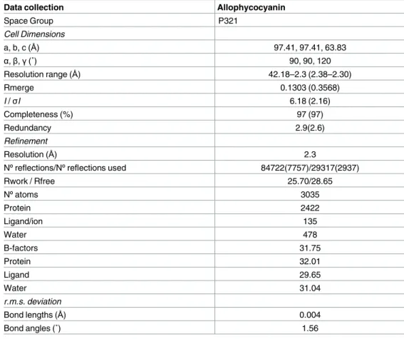
The purified APC was used for crystallization experiments. The statistics and the refinement data are shown inTable 1. The coordinates have been deposited at the Protein Data Bank under the accession code 5TJF.
The crystal structure of Allophycocyanin fromG. chilensis was determined in the space
group P321 with one heterodimerαβ per asymmetric unit using molecular replacement. The final structure was refined to 2.3Å resolution with a final R-work and R-free of 25.7 and 28.69 respectively (Table 1). The resolution allowed the chromophores to be clearly defined in the electron density map (|2Fo-Fc|) (Fig 4B).
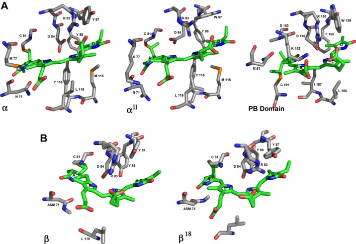
The structure of the (αβ heterodimer is shown inFig 4A. Indeed,α and β subunits con-tained one phycocyanobilin bound to cysteine 81 and are stabilized by Asn71, Arg83 and Asp84 inα subunit, and by methyl-Asn71 (Asm71) and Asp84 in β subunit. The biological unit is a trimer of heterodimers as it was determined by size exclusion chromatography. The structure reported in this article is very similar to the structures of APC previously reported, 1B33 that contains the Lc [14] and 1KN1 from the rhodophytaPorphyra yezoensis [27].
The environment of the chromophore phycocyanobilin is highly
conserved among different subunits
SubunitsαII,β18and PB domain were modeled and analyzed. A comparison among subunits in APC ofM. laminosus [14],P. yezoensis [27],G. chilensis (PDB IDs 1B33, 1KN1, and 5TJF
(this paper), respectively) and theαIImodel showed that the residues interacting with the phy-cocyanobilin chromophore are highly conserved (S1 Table). There is also high degree of con-servation for the conformation of the chromophore (ASA) [36,37], which is confirmed by the distances between rings A and D in the phycocyanobilin (Fig 5AandS1 Table). Instead the PB domain presents a SSA conformation that could account for the different spectroscopic
characteristics of the LCM. A comparison between theβ and β18subunit is also shown inFig
5B. In general, there are not important differences betweenβ and β18
subunits and betweenα andαIIsubunits as it was expected by the sequence identity.
The same comparison was performed betweenα subunit of 5TJF, the structure reported for the PB domain fromNostoc sp. (PDB ID: 4XXI) [32], and the model for the PB domain in Fig 3. Sequence comparison betweenαandαII(ApcA and ApcD),βandβ18(Apcb and ApcF) andαand the PB_domain of the L
CM
(ApcA and ApcE). Chromophore binding region are enclosed in green rectangles, and the conserved residues are highlighted in green. The
cysteine residues that bind the chromophores are shown in red background. The PB-loop sequence is enclosed in blue lines.
Gracilaria chilensis. The PB domain can replace an α subunit in a trimer of heterodimers.
Nev-ertheless, there are obvious differences, such as the insertion of 55 residues between residues 81–136 in the PB domain; 58% of this sequence was recognized as intrinsically disordered by Disprot [38] and also contains 12 Ser, 4 Thr and 1 Tyr, all hydroxylated residues. Four of these residues (Ser98, Ser99, Thr101 and Thr102) were recognized as phosphorylation sites by PRO-SITE and one of them (Thr101) in a motif recognized by protein kinase C. In prokaryote, this peptide segment has been described as an interaction region for the Orange Carotenoid Pro-tein (OCP), which has not been reported in eukaryotes yet, and also a site for regulation by phosphorylation [39], fact that has not been tested in eukaryotic red algae for potential significance.
Attention is drown to the fact that the LCMand its chromophore have been proposed to be
responsible for the final transfer of energy towards chlorophylls in Photosystems. In this regard, it was important to analyze the differences in the binding site of phycocyanobilins inα subunit, the modeled PB domain and the structure reported at the PDB ID: 4XXI [32]. The position of Cys81 in the alignment (Fig 3), which binds the PCB in all the otherα subunits, is occupied by Ser153 in the PB domain. The Cys191 is the residue that binds to the phycocyano-bilin. In our model, the chromophore was stabilized by Asp156, Arg155, Trp159, and Phe160 (S1 Table). Besides the difference in the cysteine residue, it is possible to see minor differences in the stabilization of the ligand (Fig 5AandS1 Table). Considering the binding site and the Table 1. Information on the data collection and refinement for the determination of the three dimen-sional structure of Allophycocyanin from Gracilaria chilensis.
Data collection Allophycocyanin
Space Group P321 Cell Dimensions a, b, c (Å) 97.41, 97.41, 63.83 α,β,γ(˚) 90, 90, 120 Resolution range (Å) 42.18–2.3 (2.38–2.30) Rmerge 0.1303 (0.3568) I /σI 6.18 (2.16) Completeness (%) 97 (97) Redundancy 2.9(2.6) Refinement Resolution (Å) 2.3
Nºreflections/Nºreflections used 84722(7757)/29317(2937)
Rwork / Rfree 25.70/28.65 Nºatoms 3035 Protein 2422 Ligand/ion 135 Water 478 B-factors 31.75 Protein 32.01 Ligand 29.65 Water 31.04 r.m.s. deviation Bond lengths (Å) 0.004 Bond angles (˚) 1.56
The values in parenthesis correspond to the last resolution shell.
conformation of the chromophore, the phycocyanobilin in the PB domain has a SSA confor-mation instead of ASA for the chromophore in theα subunit [36,37]. This conformation is the same in the crystal structure of PB domain fromNostoc sp. (PDB ID: 4XXI) [32]. The absorp-tion spectrum, calculated for phycocyanobilin in the model of the PB domain ofGch, using
Gaussian 09[40] with a ZINDO-MN semiempirical approach [40], gave theλmaxAbs= 661nm,
which agreed with the maximum absorption of the PB domain reported forNostoc sp. Other
relevant difference is that Cys 81 ofα subunit binds from the top of PCB, while Cys 165 binds from the bottom in the PB domain (SeeFig 5). This difference may be related to the fact that PCB needs a specific cysteine lyase to be bound to Cys 81 inα subunit. However, it has been reported that for LCM, the process of binding the chromophore would be autocatalytic [15]. It
has been also reported a mechanism for the addition of PCB to Cys 155 in Phycocyanin in cya-nobacteria. It involves the presence of an aspartic acid participating in the formation of an acy-limmonium cation on ring A of the phycocyanobilin. After that, the addition reaction to cysteine is produced with the assistance of a tyrosine residue. The authors also report that after the addition, a conformational change is produced in the chromophore. The PB domain Fig 4. Crystallographic structure of Allophycocyanin from Gracilaria chilensis. A) Ribbon representation
of the asymmetric unit, the heterodimer. B) A section of the |2F-Fo| omit electron density map is shown for phycocyanobilin inαsubunit. The residues interacting with the chromophore are also shown.
model fromGch locates also a tyrosine and an aspartic acid in the vicinity of the
phycocyanobi-lin; however the position of the functional groups seems to be located too far in the binding state. It is possible that before the reaction to occur and the positioning of the chromophores, these residues could be part of the catalysis, and after the binding their location changes to the final position in the molecule.
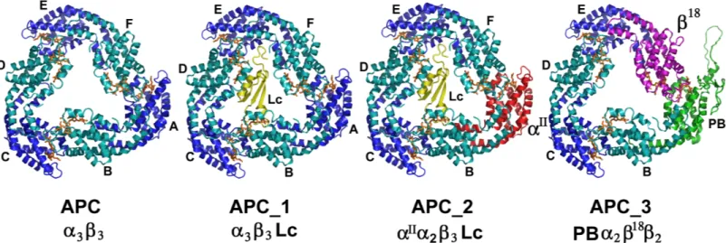
The crystal structure of Allophycocyanin allowed the building of the
different type of trimers present in the core
Fig 6shows the models created for APC, APC_1, APC_2 and APC_3 using the structure of APC reported here, with the corresponding substitution of subunits as is shown.
Each trimer was analyzed considering the number and type of interactions among subunits, the interaction surfaces on each trimer as a whole and considering the substitutions. (The data is shown inS2 Table). A comparison between trimers as follows:
1. APC/APC_1: The presence of the Lc (Chain G) did not affect the interaction surface
(aprox.1450Å2) between subunits, yet the number of hydrogen bonds between heterodi-mers decreases. The Lc interacted mainly with theβ (chains B, D and F) subunits by hydro-gen bonding.
Fig 5. Binding sites of phycocynobilin. A) Sticks representation of binding site of phycocyanobilin inαsubunit of 5TJF (this paper),αII and the PB domain in APC_3. The chromophores are represented in green. B) Sticks representation ofβsubunit in 5TJF andβ18in APC_3. The chromophores are also in green.
2. APC_1/APC_2: Both trimers contained the Lc, but APC_2 has the substitution of α by αII
(Chain A). The interactions of Lc in both oligomers were very similar withβ chains, espe-cially with chain D.
3. APC/APC_3: The substitution of chain A (α) by PB domain and chain F (β) by β18produced a trimer that contained these two special subunits in neighbor heterodimers. Theαβ he-terodimers conserved the interactions (H-bonds/salt bridges), but less salt bridges were observed in heterodimers with the substitutions. The interaction surfaces in both trimers are similar (aprox 1450Å2), except for the lower value measured for the interaction between subunits F and E (1154Å2)
The structural analysis of the models proposed in this article allowed to arrange them face to face to form hexamers, which interact back to back with other hexamers to organize the cyl-inders present in the core as it was proposed forSynechococcus [41].
The experimental data available so far, for the subunit composition of trimers proposed in this article, indicated that LCM(PB_domain) andβ18were not in the same heterotrimer
withαII[41]. According to spectroscopic data obtained by Kuzminovet al [42],β18should transfer energy to the LCM, a fact that could be accomplished only if they are in the same
trimer but neighboring heterodimers. Finally, they propose thatβ18is a mediator in the energy transfer towards the final emitter, with the chromophore present in the LCM
(PB_domain).
The modeling of the 55 extra residues (PB_loop) and the analysis of the sequence (Fig 3) indicated a very disordered region that is probably interacting with other proteins. Cross-link-ing experiments involvCross-link-ing cyanobacterial phycobilisomes [43] do not provide information on the possible interactions of this segment of PB domain.
The model for the Linker core fits very well the space in the center of APC; clearly, this small molecule contributes to give asymmetry to allophycocyanin. It has been reported that the LCmodifies the spectroscopic properties of the PCB in APC_1 and APC_2. According to
our results and those from Reuteret al [14], in the trimeric state, one explanation is the differ-ent environmdiffer-ent of the chromophore upon binding of LC.
Fig 6. Structural models proposed for each trimer of Allophycocyanin. A) Schematic representation of the different composition trimers. B) Structure of
APC, APC_1, APC_2, and APC_3.
Future perspectives
Our work on the structure of the phycobilisome components of an eukaryotic algae such as
Gracilaria chilensis, including the study of the components of its core in this article, pretends
to fill a gap produced by the enormous amount of information published in prokaryotes, espe-ciallySynechocystis and the small amount of information produced about this system in
eukaryotic organisms. From the beginning, the purpose of our study on Phycobilisomes was to understand how the structure of this super complex supported the high efficiency of the energy transfer towards the photosystems. In this article, we are reporting new pieces of the big puzzle.
Supporting information
S1 File. Methodology for phycobilisomes purification.
(DOCX)
S2 File. Species and sequences used to design the primers for PCR.
(DOCX)
S3 File. Methodology for amplification assays.
(DOCX)
S4 File. Methodology for Allophycocyanin purification.
(DOCX)
S5 File. Sequence of the genes codifying for the proteins present in the core of a phycobili-some ofGracilaria chilensis.
(DOCX)
S1 Table. Comparison of the PCB binding sites forα subunits.
(DOCX)
S2 Table. Interface characteristics of different trimers of Allophycocyanin. Interface area,
numbers of hydrogen bonds and salt bridges between subunits in different trimers. (DOCX)
Acknowledgments
We would like to thanks to the Soleil Synchrotron and its beamline Proxima 1 for the x-ray data collection. Also, we would like to thanks to the Robotic Unit at Universidad de Chile for the initial screening of crystallization conditions.
Author Contributions
Conceptualization: MB JDL MF JMO. Data curation: JDL.
Formal analysis: JDL MF. Funding acquisition: MB.
Investigation: MAV AJV CM JDL MF FK. Methodology: MF JDL MB JMO.
Resources: MB JMO. Software: JMO MF JDL. Supervision: JMO MB. Validation: JDL MF. Visualization: JMO JDL MF.
Writing – original draft: JMO JDL MF MB. Writing – review & editing: JMO JDL MF MB.
References
1. Glazer AN. Light guides. Directional energy transfer in a photosynthetic antenna. J Biol Chem. 1989; 264: 1–4. PMID:2491842
2. MacColl R. Cyanobacterial Phycobilisomes. J Struct Biol. 1998; 124: 311–334.https://doi.org/10.1006/ jsbi.1998.4062PMID:10049814
3. Adir N. Elucidation of the molecular structures of components of the phycobilisome: reconstructing a giant. Photosynth Res. 2005; 85: 15–32.https://doi.org/10.1007/s11120-004-2143-yPMID:15977057 4. Sun L, Wang S, Zhao M, Fu X, Gong X, Chen M, et al. Phycobilisomes from Cyanobacteria. Handbook
on Cyanobacteria: Biochemistry, Biotechnology and Applications. Nova Science Publishers; 2009. pp. 105–160.
5. Sidler WA. Phycobilisome and Phycobiliprotein Structures. In: Bryant DA, editor. The Molecular Biology of Cyanobacteria. Springer Netherlands; 1994. pp. 139–216.
6. MacColl R. Allophycocyanin and energy transfer. Biochim Biophys Acta BBA—Bioenerg. 2004; 1657: 73–81.
7. Arteni AA, Ajlani G, Boekema EJ. Structural organisation of phycobilisomes from Synechocystis sp. strain PCC6803 and their interaction with the membrane. Biochim Biophys Acta BBA—Bioenerg. 2009; 1787: 272–279.
8. Hagopian JC, Reis M, Kitajima JP, Bhattacharya D, Oliveira MC de. Comparative Analysis of the Com-plete Plastid Genome Sequence of the Red Alga Gracilaria tenuistipitata var. liui Provides Insights into the Evolution of Rhodoplasts and Their Relationship to Other Plastids. J Mol Evol. 2004; 59: 464–477.
https://doi.org/10.1007/s00239-004-2638-3PMID:15638458
9. Lundell DJ, Glazer AN. Molecular architecture of a light-harvesting antenna. Core substructure in Syne-chococcus 6301 phycobilisomes: two new allophycocyanin and allophycocyanin B complexes. J Biol Chem. 1983; 258: 902–908. PMID:6401721
10. Maxson P, Sauer K, Zhou J, Bryant DA, Glazer AN. Spectroscopic studies of cyanobacterial phycobili-somes lacking core polypeptides. Biochim Biophys Acta BBA—Bioenerg. 1989; 977: 40–51.
11. Houmard J, Capuano V, Colombano MV, Coursin T, Marsac NT de. Molecular characterization of the terminal energy acceptor of cyanobacterial phycobilisomes. Proc Natl Acad Sci. 1990; 87: 2152–2156. PMID:2107546
12. Gao X, Wei T-D, Zhang N, Xie B-B, Su H-N, Zhang X-Y, et al. Molecular insights into the terminal energy acceptor in cyanobacterial phycobilisome. Mol Microbiol. 2012; 85: 907–915.https://doi.org/10. 1111/j.1365-2958.2012.08152.xPMID:22758351
13. Mullineaux CW. Phycobilisome-reaction centre interaction in cyanobacteria. Photosynth Res. 2008; 95: 175.https://doi.org/10.1007/s11120-007-9249-yPMID:17922214
14. Reuter W, Wiegand G, Huber R, Than ME. Structural analysis at 2.2Åof orthorhombic crystals pres-ents the asymmetry of the allophycocyanin–linker complex, AP LC7. 8, from phycobilisomes of Mastigo-cladus laminosus. Proc Natl Acad Sci. 1999; 96: 1363–1368. PMID:9990029
15. Zhao K-H, Su P, Bo¨hm S, Song B, Zhou M, Bubenzer C, et al. Reconstitution of phycobilisome core– membrane linker, LCM, by autocatalytic chromophore binding to ApcE. Biochim Biophys Acta BBA— Bioenerg. 2005; 1706: 81–87.
16. Adir N, Dines M, Klartag M, McGregor A, Melamed-Frank M. Assembly and Disassembly of Phycobili-somes. In: Shively JM, editor. Complex Intracellular Structures in Prokaryotes. Springer Berlin Heidel-berg; 2006. pp. 47–77.
17. Jallet D, Gwizdala M, Kirilovsky D. ApcD, ApcF and ApcE are not required for the Orange Carotenoid Protein related phycobilisome fluorescence quenching in the cyanobacterium Synechocystis PCC 6803. Biochim Biophys Acta BBA—Bioenerg. 2012; 1817: 1418–1427.
18. Capuano V, Braux AS, Marsac NT de, Houmard J. The “anchor polypeptide” of cyanobacterial phycobi-lisomes. Molecular characterization of the Synechococcus sp. PCC 6301 apce gene. J Biol Chem. 1991; 266: 7239–7247. PMID:1901865
19. Watanabe M, Ikeuchi M. Phycobilisome: architecture of a light-harvesting supercomplex. Photosynth Res. 2013; 116: 265–276.https://doi.org/10.1007/s11120-013-9905-3PMID:24081814
20. Bird CJ, McLachlan J, Oliveira EC de. Gracilaria chilensis sp.nov. (Rhodophyta, Gigartinales), from Pacific South America. Can J Bot. 1986; 64: 2928–2934.
21. Contreras-Martel C, Matamala A, Bruna C, Poo-Caamaño G, Almonacid D, Figueroa M, et al. The structure at 2Åresolution of Phycocyanin from Gracilaria chilensis and the energy transfer network in a PC–PC complex. Biophys Chem. 2007; 125: 388–396.https://doi.org/10.1016/j.bpc.2006.09.014
PMID:17118524
22. Contreras-Martel C, Martinez-Oyanedel J, Bunster M, Legrand P, Piras C, Vernede X, et al. Crystalliza-tion and 2.2Åresolution structure of R-phycoerythrin from Gracilaria chilensis: a case of perfect hemi-hedral twinning. Acta Crystallogr D Biol Crystallogr. 2001; 57: 52–60. PMID:11134927
23. Figueroa M, Martı´nez-Oyanedel J, Matamala AR, Dagnino-Leone J, Mella C, Fritz R, et al. In silico model of an antenna of a phycobilisome and energy transfer rates determination by theoretical Fo¨rster approach. Protein Sci. 2012; 21: 1921–1928.https://doi.org/10.1002/pro.2176PMID:23047609 24. Colle´n J, Porcel B, Carre´ W, Ball SG, Chaparro C, Tonon T, et al. Genome structure and metabolic
fea-tures in the red seaweed Chondrus crispus shed light on evolution of the Archaeplastida. Proc Natl Acad Sci. 2013; 110: 5247–5252.https://doi.org/10.1073/pnas.1221259110PMID:23503846 25. Guan X, Qin S, Zhao F, Zhang X, Tang X, others. Phycobilisomes linker family in cyanobacterial
genomes: divergence and evolution. Int J Biol Sci. 2007; 3: 434–445. PMID:18026567
26. Vorphal MA, Gallardo-Esca´rate C, Valenzuela-Muñoz V, Dagnino-Leone J, Va´squez JA, Martı´nez-Oya-nedel J, et al. De novo transcriptome analysis of the red seaweed Gracilaria chilensis and identification of linkers associated with phycobilisomes. Mar Genomics. 2017; 31: 17–19.https://doi.org/10.1016/j. margen.2016.11.001PMID:27843115
27. Liu J-Y, Jiang T, Zhang J-P, Liang D-C. Crystal Structure of Allophycocyanin from Red AlgaePorphyra yezoensis at 2.2-ÅResolution. J Biol Chem. 1999; 274: 16945–16952. PMID:10358042
28. Adams PD, Afonine PV, Bunko´czi G, Chen VB, Davis IW, Echols N, et al. PHENIX: a comprehensive Python-based system for macromolecular structure solution. Acta Crystallogr D Biol Crystallogr. 2010; 66: 213–221.https://doi.org/10.1107/S0907444909052925PMID:20124702
29. Emsley P, Cowtan K. Coot: model-building tools for molecular graphics. Acta Crystallogr D Biol Crystal-logr. 2004; 60: 2126–2132.https://doi.org/10.1107/S0907444904019158PMID:15572765
30. Afonine PV, Grosse-Kunstleve RW, Echols N, Headd JJ, Moriarty NW, Mustyakimov M, et al. Towards automated crystallographic structure refinement with phenix.refine. Acta Crystallogr D Biol Crystallogr. 2012; 68: 352–367.https://doi.org/10.1107/S0907444912001308PMID:22505256
31. Webb B, Sali A. Comparative Protein Structure Modeling Using MODELLER. Current Protocols in Bio-informatics. John Wiley & Sons, Inc.; 2002.
32. Tang K, Ding W-L, Ho¨ppner A, Zhao C, Zhang L, Hontani Y, et al. The terminal phycobilisome emitter, LCM: A light-harvesting pigment with a phytochrome chromophore. Proc Natl Acad Sci. 2015; 112: 15880–15885.https://doi.org/10.1073/pnas.1519177113PMID:26669441
33. Comeau SR, Gatchell DW, Vajda S, Camacho CJ. ClusPro: a fully automated algorithm for protein–pro-tein docking. Nucleic Acids Res. 2004; 32: W96–W99.https://doi.org/10.1093/nar/gkh354PMID:
15215358
34. Wiederstein M, Sippl MJ. ProSA-web: interactive web service for the recognition of errors in three-dimensional structures of proteins. Nucleic Acids Res. 2007; 35: W407–W410.https://doi.org/10.1093/ nar/gkm290PMID:17517781
35. Lovell SC, Davis IW, Arendall WB, de Bakker PIW, Word JM, Prisant MG, et al. Structure validation by Cαgeometry:ϕ,ψand Cβdeviation. Proteins Struct Funct Bioinforma. 2003; 50: 437–450.
36. Tu P, Yao Y, Li Y, Liu B. Conformational flexibility of phycocyanobilin: Monte-Carlo and DFT study. J Mol Struct THEOCHEM. 2009; 894: 9–13.
37. Go¨ller AH, Strehlow D, Hermann G. Conformational Flexibility of Phycocyanobilin: An AM1 Semiempiri-cal Study. ChemPhysChem. 2001; 2: 665–671.https://doi.org/10.1002/1439-7641(20011119) 2:11<665::AID-CPHC665>3.0.CO;2-OPMID:23686901
38. Piovesan D, Tabaro F, MičetićI, Necci M, Quaglia F, Oldfield CJ, et al. DisProt 7.0: a major update of the database of disordered proteins. Nucleic Acids Res. 2017; 45: D1123–D1124.https://doi.org/10. 1093/nar/gkw1279PMID:27965415
39. Harris D, Tal O, Jallet D, Wilson A, Kirilovsky D, Adir N. Orange carotenoid protein burrows into the phy-cobilisome to provide photoprotection. Proc Natl Acad Sci. 2016; 113: E1655–E1662.https://doi.org/10. 1073/pnas.1523680113PMID:26957606
40. Frisch MJ, Trucks GW, Schlegel HB, Scuseria GE, Robb MA, Cheeseman JR, et al. Gaussian 09 Revi-sion A.1.
41. Tal O, Trabelcy B, Gerchman Y, Adir N. Investigation of Phycobilisome Subunit Interaction Interfaces by Coupled Cross-linking and Mass Spectrometry. J Biol Chem. 2014; jbc.M114.595942.
42. Kuzminov FI, Bolychevtseva YV, Elanskaya IV, Karapetyan NV. Effect of APCD and APCF subunits depletion on phycobilisome fluorescence of the cyanobacterium Synechocystis PCC 6803. J Photo-chem Photobiol B. 2014; 133: 153–160.https://doi.org/10.1016/j.jphotobiol.2014.03.012PMID:
24727864
43. Chang L, Liu X, Li Y, Liu C-C, Yang F, Zhao J, et al. Structural organization of an intact phycobilisome and its association with photosystem II. Cell Res. 2015; 25: 726–737.https://doi.org/10.1038/cr.2015. 59PMID:25998682