AN APPROACH FOR DETECTION OF GLOMERULI IN MULTISITE DIGITAL PATHOLOGY
R. Mar´ee
a,b, S. Dallongeville
a, J.-C. Olivo-Marin
a, V. Meas-Yedid
a ∗ aBioImage Analysis Unit, CNRS UMR 3691,
Institut Pasteur, 25 rue du docteur Roux, 75015, Paris, France
b
University of Li`ege, Systems and Modeling
Montefiore Institute & GIGA-Research, Grande Traverse 10, 4000 Li`ege Sart-Tilman, Belgique
ABSTRACT
We present a novel bioimage informatics workflow that com-bines Icy and Cytomine software and their algorithms to en-able large-scale analysis of digital slides from multiple sites. In particular, we apply this workflow on renal biopsies and evaluate empirically our approach for the automatic detection of glomeruli in hundreds of tissue sections.
Index Terms— whole slide imaging, image processing, color normalization, machine learning, glomeruli detection
1. INTRODUCTION
Digital pathology is an active field of research which raises many image analysis challenges [14]. One of the key chal-lenge for pattern recognition algorithm is the wide variety of preparation and imaging protocols that imply highly variable image appearances of tissue structures, within a given labora-tory and across research centers. Indeed, tissue sections are prepared using coloured histochemical stains that bind selec-tively to cellular components. Colour variation is a problem in histopathology based on transmitted microscopy due to the several factors such as variable chemical colouring/reactivity from different manufacturers/batches of stains, colouring be-ing dependent on stainbe-ing procedures (timbe-ing, concentrations, etc.), and light transmission being a function of section thick-ness and the use of different scanners.
Because of the lack of efficient tools to share datasets be-tween histology labs, image analysis algorithms are often de-signed and applied on lab-specific datasets with locally stan-dardized protocols that reduce image variabilities. It is there-fore currently unknown if more generic pattern recognition approaches could recognize tissue structures accross several histology labs, which would be of great practical value. Cur-rent practice is indeed to train and apply recognition models using specific images for each new lab setting, e.g. [6] recog-nizes glomerular structures using a lab-specific IHC-stained protocol, [5] compares algorithms to detect mitotic figures from 5 breast cancer H&E biopsy slides from one hospital,
∗e-mail: [email protected]
and [13] recognizes lung tumors in hundreds of experimental mice H&E whole-slide images but from one research labora-tory only. Recently, normalization of H&E images was pro-posed [1] to reduce staining variations.
Ideally, pattern recognition algorithms should be able to recognize tissue structures regardless of preparation and imaging conditions, as human experts are capable of doing after histology training. Evaluating automatic algorithms in such realistic settings is challenging. It requires softwares and algorithms able to organize persistently in databases large sets of digital slides from multiple centers, allow semantic annota-tion of digital slides by experts, as well as protocols and user interfaces to visualize and evaluate algorithm results. In this work we propose to combine and extend softwares (namely Cytomine1 [12] and Icy2 [2]), and their algorithms (based
on machine learning and image processing, respectively) to enable multisite digital pathology studies.
1.1. Image analysis in renal pathology
In particular, we will focus here on image analysis in renal pathology. Biopsy is a key diagnostic tool for many renal pathologies because it allows early detection of lesions in the kidney before the clinical symptoms appear. Examination of the biopsy is currently done by several pathologists from a visual examination of biopsy slides stained by several stain-ing, through a microscope. This screening is very tedious and subject to inter and intra-observer variabilities. One of the first visual task consists in counting glomeruli, a critical ele-ment of the kidney, in 2D tissue sections to determine if the tissue sampling is representative of the 3D organ. Due to the slicing, the glomeruli on an image section show varying size. Furthermore, the slide images are subject to high variations in staining. As far as we know, there are few works on automatic detection of glomeruli. [9] used an edge detector and genetic algorithms but the validation has been done on 13 glomeruli only. More recently, [6] and [7] proposed a glomeruli detec-tor by using a linear Support Vecdetec-tor Machine (linear-SVM)
1http://www.cytomine.be/ 2http://icy.bioimageanalysis.org/
on respectively the Histogram of Oriented Gradients (HOG) and Segmental HOG (S-HOG) features. The HOG reaches the recognition rate of 85% of glomeruli detection and the S-HOG, improves the result of HOG. However both works are mono-centric studies on mouse renal samples that show less variations than our dataset which comes from a human, multisite study. Indeed, our approach, described in Section 2, enabled an empirical study on hundreds of tissue sections which results are presented in Section 3.
2. METHODS
In this Section, we describe the proposed workflow for ef-fective analysis of large sets of digital slides from multiple centers. Our workflow combines algorithms and protocols to exchange data and results between Icy and Cytomine, two open-source software for bioimage analysis.
2.1. Image and data storage using Cytomine
Cytomine is an open-source rich internet application for col-laborative analysis of large images [12]. Through HTTP transfers, we centralize images on a Cytomine server and or-ganize them into a project with a user-defined vocabulary of terms describing renal tissue structures. Cytomine supports various slide scanner formats and provide web services to retrieve image thumbnails and tiles corresponding to specific image area at any resolution level. Cytomine data models also allow to store annotation geometries drawn manually or generated by image analysis softwares. Each user or soft-ware has its own layer of annotations and these data can be visualized and edited through a web interface and they can also be retrieved or updated by third party softwares through a RESTful API.
2.2. Icytomine plugin for image and data exchange Icy is a collaborative platform that combines a free and open-source Java software designed for bioimage analysis and a community website for contributing and sharing algorithms and protocols. It already implements various image process-ing algorithms but is does not handle very large images.
Icytomine is a bridge that we developed, schematically represented in Figure 1 to exchange image data and anno-tations between Icy and Cytomine. Our implementation al-lows batch processing of hundreds of whole slide images by relying on Icy API and Cytomine RESTful API and JSON lightweight data-interchange format. As explained before, the slide images are first stored in Cytomine database at high magnification. Then Icytomine downloads whole slide im-age at lower resolution from Cytomine (at 1.5x magnifica-tion) to performs section detection using an automatic thresh-olding method. The detected region of interest (ROI) within
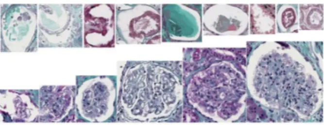
Fig. 2. Example of candidate glomeruli detected by step 1. Top: False positives, Bottom: True positives.
Icy are converted into corresponding whole slide polygon co-ordinates in WKT format and sent back to Cytomine using its annotation web services. For glomeruli detection, image tiles of each section are downloaded from Cytomine at higher magnification (20X magnification) and converted to Icy inter-nal data structures for tile processing, as described in the next section.
2.3. Detection of glomeruli candidates using Icy
This step (Step 1) aims at detecting glomeruli candidates by using image processing algorithms in Icy. As we want an ap-proach that can detect glomeruli whatever the color staining is, our detection step is based on geometric invariance, more precisely it relies on the ellipse-shape of glomeruli. First, the luminance component is thresholded in order to obtain the lumen regions, from which we fit the ellipses by the Fitzgib-bon’s method [3]. It minimizes the algebraic distance subject to the constraint 4ac − b2 = 1, by incorporating the
ellip-ticity constraint into the normalization factor. This algorithm requires only five points and has the advantages to be very ro-bust, efficient and simple to implement. However, it detects too many candidates (many of them are non-glomerular struc-tures), as illustrated by Figure 2. We therefore propose to send them back to Cytomine for further processing based on clas-sification algorithms. As these detected objects are stored as ROI objects in Icy, we again used Icytomine to convert them and send their whole-slide coordinates to Cytomine.
2.4. Automatic Classification of candidate images The detection step generates many non-glomerular structures that would require manual intervention to filter them out. In Step 2, we want to evaluate the use of classification algo-rithms to filter out non-glomerular objects and reduce manual workload. In this work, we propose to evaluate two generic classifiers readily available, namely WND-CHARM [15] and the ET-FL (Extremely Randomized Trees for Feature Learn-ing) tree-based method of [11]. These methods have been shown effective on various classification tasks. However, as our large empirical study reveals that none of these methods
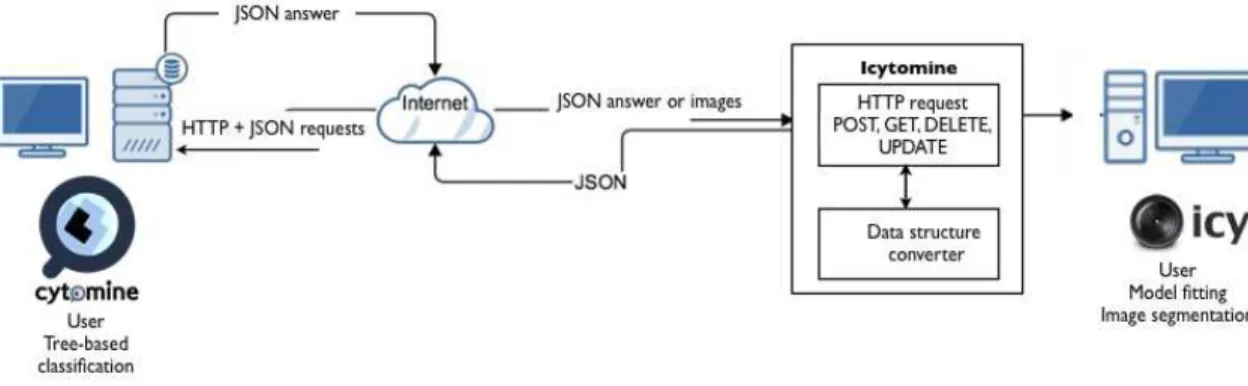
Fig. 1. An overview of our workflow and its communication mechanisms between Icy and Cytomine.
is robust enough on our realistic dataset, we propose to also consider color normalization algorithms to improve recogni-tion performances. More specifically, we combined color nor-malization with ET-FL method only as this method performed better on non-normalized images.
2.4.1. Color normalization
We have tested 4 color normalization methods in order to im-prove the classification performance: i) the simple color his-togram equalization i.e. hishis-togram equalization on each color channel, red, green, blue. ii) Automatic Color Enhancement [4] is an effective color correction and enhancement method based on a simple model of the human visual system and which has a connection with histogram equalization. iii) The color transfer method of Reinhard et al. [16] that maps the im-ages to the color distribution of a target image on a per-pixel basis by equalizing the mean and standard deviations sepa-rately for each dimension of a perceptual colorspace. iv) Ma-cenko et al. [10] uses a singular value decomposition (SVD)-based approach to directly estimate the color deconvolution matrices.
2.4.2. Supervised classification 2.4.2.1. WND-CHARM
We first used [15] that proposed a generic approach for image classification based on the extraction of a large set of features. For each candidate image, it computes polynomial decompositions (Chebyshev-Fourier statistics, Chebyshev statistics, Zernike polynomials), high contrast features (edge and object statistics), pixel and texture statistics (Gabor tex-tures, first four moments, Haralick textex-tures, Multiscale his-tograms, Tamura textures, Radon). These features are com-puted on the raw image, transforms of the image (Wavelet, Chebyshev and Fast Fourier transforms), and transforms of transforms of the image (Chebyshev transform of the Fourier
transform). In total, this approach generates 2919 features. As in [15], these are then used by a weighted nearest neighbor classifier.
2.4.2.2. ET-FL
We also evaluate systematically the ET-FL classification method of [11] based on random subwindows and extremely randomized tree learning method. This method first performs random extraction of a large number of square subwindows in candidate images then it uses trees to build a novel image description (a global, sparse, feature vector that encodes sub-window frequencies in tree leaves) subsequently classified by a linear SVM classifier. For each of the normalization variant presented previously, we trained models using different con-figurations of subwindow size intervals (as in [11]), and we report the best obtained result. Default tests were made using 100 training subwindows per candidate image. For each con-figuration, subwindows are subsequently resized by nearest neighbor interpolation to patches of fixed size (16 × 16) and their pixel values are encoded in HSV colorspace unless oth-erwise stated (a few tests were performed using graylevels). For the tree parameters, we used the same set of parameter values: k = 28 (square root of the total number of attributes), T = 10, and nmin= 1000.
3. EXPERIMENTAL DATA AND RESULTS 3.1. Data
Our dataset contains tissue sections from 200 slides (one slide contains several tissue sections from the same patient) prepared using trichrome Masson protocol within 13 re-search centers and acquired using a Nanozoomer scanner with 0.452µm pixel resolution. We used a random set of 100 slides (containing a total of 2927 glomeruli and 13648 non-glomerular structures) to train the recognition algorithms, while the remaining sections from 100 slides (containing
Method Avg. acc. NonGlom acc. Glomeruli acc. WND-CHARM GRAY 73.71 81.38 47.39 ET-FL GRAY 87.17 96.87 53.91 ET-FL HS 89.01 98.85 55.29 ET-FL COLOR 91.67 98.61 67.89 ET-FL ACE 91.04 98.71 64.73 ET-FL RH 92.62 98.95 70.93 ET-FL MM 93.78 98.97 75.98
Table 1. Summary of recognition performances.
a total of 2853 glomeruli and 9785 non-glomerular struc-tures) were used to evaluated recognition performances. To build the ground truth data of glomeruli and non-glomerular structures in both training and test sets, we used Cytomine web proofreading tools to re-classify manually candidates detected by Step 1.
3.2. Results
Table 1 shows classification accuracies for all method variants on the test set. Our experiments show that using graylevels only yields lower performances for both WND-CHARM and ET-FL classifiers. ET-FL is best combined with the color normalization method of [10]. If we increase the num-ber of training subwindows up to 500, we further increase recognition rate up to 94.74 (98.71 nonglom accuracy, 81.10 glomeruli accuracy) which also corresponds to 0.95 preci-sion, 0.81 recall, and 0.87 F 1 score. However, although the method succeeds in rejecting most of the non-glomeruli objects, it also incorrectly rejects many glomeruli. Future work should therefore concentrate on balancing true/false positive/negative rates to better meet practical expectations.
4. CONCLUSION
In this work we proposed a novel workflow combining Cy-tomine and Icy softwares and their algorithms for glomeruli detection in human renal biopsies from multiple centers. Al-though results are not yet satisfactory for practical use, our open-source developments3 open the door for large-scale, multisite, digital pathology studies. In the future we will evaluate other color normalization techniques as well as data augmentation and classification approaches inspired by works in natural image classification [8].
5. ACKNOWLEDGMENTS
We thank Institut Carnot Maladies Infectieuses for R.M. post-doctoral grant as well as Wallonia (DGO6) for research grant 1017072. We also thank Florian Aubin for preliminary imple-mentation, and Eric Thervet for providing renal slide images.
3Icytomine plugin will be made available together with the paper
publi-cation.
6. REFERENCES
[1] B.E. Bejnordi, N. Timofeeva, I. Otte-Hller, N. Karssemeijer, and J. van der Laak. Quantitative analysis of stain variability in histology slides and an algorithm for standardization. In SPIE, editor, Proc.
Med-ical Imaging: Digital Pathology, volume 9041, March 2014. [2] F. de Chaumont, S. Dallongeville, N. Chenouard, N. Herve, S. Pop,
T. Provoost, V. Meas-Yedid, P. Pankajakshan, T. Lecomte, Y. Le Mon-tagner, T. Lagache, A. Dufour, and J. C. Olivo-Marin. Icy: an open bioimage informatics platform for extended reproducible research. Nat
Methods, 9(7):690–6, 2012.
[3] A. W. Fitzgibbon, M. Pilu, and R. B. Fisher. Direct least-squares fitting of ellipses. IEEE Transactions on PAMI, 21(5):476–480, May 1999. [4] Pascal Getreuer. Automatic Color Enhancement (ACE) and its Fast
Implementation. Image Processing On Line, 2:266–277, 2012. [5] Alessandro Giusti, Dan Claudiu Ciresan, Claudio Caccia, J ¨urgen
Schmidhuber, and Luca Maria Gambardella. A comparison of algo-rithms and humans for mitosis detection. In International Symposium
on Biomedical Imaging (ISBI), 2014.
[6] Y. Hirohashi, R. Relator, T. Kakimoto, R. Saito, Y. Horai, A. Fukunari, H. Utsumi, K. Okada, and T. Kato. Automated quantitative image anal-ysis of glomerular desmin immunostaining as a sensitive injury marker in spontaneously diabetic torii rats. Journal of Biomedical Image
Pro-cessing, 1(1):20–28, May 2014.
[7] T. Kato, R. Relator, H. Ngouv, Y. Hirohashi, O. Takaki, T. Kakimoto, and K. Okada. Segmental hog: new descriptor for glomerulus detection in kidney microscopy image. BMC Bioinformatics, 16:316, 2015. [8] Alex Krizhevsky, Ilya Sutskever, and Geoffrey E. Hinton. Imagenet
classification with deep convolutional neural networks. In F. Pereira, C.J.C. Burges, L. Bottou, and K.Q. Weinberger, editors, Advances in
Neural Information Processing Systems 25, pages 1097–1105. Curran Associates, Inc., 2012.
[9] J. Ma and Zhang J. an Hu J. Glomerulus extraction by using genetic al-gorithm for edge patching. In Proc. of IEEE Congress on Evolutionary
Computation, pages 2474–2479, 2009.
[10] M. Macenko, M. Niethammer, J.S. Marron, D. Borland, J.T. Woosley, Xiaojun Guan, C. Schmitt, and N.E. Thomas. A method for normal-izing histology slides for quantitative analysis. In Proc. ISBI, pages 1107–1110, June 2009.
[11] R. Mar´ee, P. Geurts, and L. Wehenkel. Towards generic im-age classification: a extensive empirical study. Technical re-port, University of Lige, December 2014. Available online at http://orbi.ulg.ac.be/handle/2268/175525.
[12] R. Mar´ee, L. Rollus, B. Stevens, R. Hoyoux, G. Louppe, R. Van-daele, J.M. Begon, P. Kainz, P. Geurts, and L. Wehenkel. Col-laborative analysis of multi-gigapixel imaging data using cytomine.
Accepted for publication (minor revision) in Bioinformatics, 2015. http://www.cytomine.be/.
[13] R. Mar´ee, L. Rollus, B. Stevens, G. Louppe, O. Caubo, N. Rocks, S. Bekaert, D. Cataldo, and L. Wehenkel. A hybrid human-computer approach for large-scale image-based measurements using web ser-vices and machine learning. Proc. ISBI, pages 902–906, 2014. [14] MT. McCann, C. Castro, JA. Ozolek, B. Parvin, and J. Kovacevic.
Au-tomated histology analysis: opportunities for signal processing. IEEE
Signal Processing, 32(1):78–87, 2014.
[15] N. Orlov, L. Shamir, T. Macura, J. Johnston, D. M. Eckley, and I. Gold-berg. Wnd-charm: Multi-purpose image classification using compound transforms. Pattern Recognition Letters, 29(11):1684–1693, 2008. [16] E. Reinhard, M. Adhikhmin, B. Gooch, and P. Shirley. Color
trans-fer between images. Computer Graphics and Applications, IEEE, 21(5):34–41, Sep 2001.

![[PDF] Cours de PL SQL en pdf | Formation informatique](data:image/gif;base64,R0lGODlhAQABAIAAAP///wAAACH5BAEAAAAALAAAAAABAAEAAAICRAEAOw==)