Published in: Laureys, Steven (ed), Gosseries, Olivia (ed), Tononi, Giulio (ed), The Neurology of Consciousness – 2nd edition (2016), pp.155-166
DOI: 10.1016/B978-0-12-800948-2.00010-8 Status : Postprint (Author’s version)
The Assessment of Conscious Awareness in the Vegetative
State
Adrian M. Owen1, Nicholas D. Schiff2, and Steven Laureys3
1The Brain and Mind Institute, The University of Western Ontario, London, ON, Canada
2Department of Neurology and Neuroscience, Weill Medical College of Cornell University, New York, NY, USA 3Coma Science Group, Neurology Department and GIGA, University of Liege, Liege, Belgium
INTRODUCTION
In recent years, improvements in intensive care have increased the number of patients who survive severe acute brain injuries. Although the majority of these patients recover from coma within the first days of the insult, others evolve to a state of "wakeful unawareness" or vegetative state. Clinically, recognizing unambiguous signs of conscious perception of the environment and of the self in such patients can be extremely challenging. This difficulty is reflected in frequent misdiagnoses of the condition and confusion between the vegetative state and related conditions such as minimally conscious state and locked-in syndrome (Andrews et al., 1996; Childs et al., 1993). Like all severely brain-injured patients, bedside evaluation of residual brain function in vegetative state is difficult because motor responses may be very limited or inconsistent. In addition, the clinical assessment of cognitive function relies on inferences drawn from present or absent responses to external stimuli at the time of the examination (Wade and Johnston, 1999). Recent advances in functional neuroimaging suggest a novel solution to this problem; in several cases, the so-called "activation" studies have been used to identify residual cognitive function and even conscious awareness in patients who are assumed to be vegetative, yet retain cognitive abilities that have evaded detection using standard clinical methods. Indeed, in some patients, communication with the outside world via simple "yes" and "no" questions has been achieved, even in cases where no possibility for behavioral interaction exists. In this chapter, we first describe the major clinical characteristics of vegetative state following severe brain injury. We then discuss the contribution of neuroimaging studies to the assessment of conscious awareness in the vegetative state, including the recent use of reproducible and task-dependent fMRI responses as a form of "communication" in patients who are assumed to be vegetative. Finally, we review the major methodological impediments to conducting studies in disorders of consciousness.
CLINICAL DESCRIPTION
Patients in the vegetative state are awake, but are assumed to be entirely unaware of self and environment (Jennett, 2002; Jennett and Plum, 1972). Jennett and Plum cited the Oxford English Dictionary to clarify their choice of the term "vegetative": to be vegetative is to "live a merely physical life devoid of intellectual activity or social intercourse" and vegetative describes "an organic body capable of growth and development but devoid of sensation and thought." "Persistent vegetative
Published in: Laureys, Steven (ed), Gosseries, Olivia (ed), Tononi, Giulio (ed), The Neurology of Consciousness – 2nd edition (2016), pp.155-166
DOI: 10.1016/B978-0-12-800948-2.00010-8 Status : Postprint (Author’s version)
state" is arbitrarily coined as a vegetative state present 1 month after acute traumatic or non-traumatic brain injury but does not imply irreversibility ("Medical aspects of the persistent vegetative state (1). The Multi-Society Task Force on PVS," 1994). "Permanent vegetative state" denotes irreversibility. The Multi-Society Task Force on vegetative state concluded that 3 months following a non-traumatic brain injury and 12 months after traumatic injury, the condition of vegetative patients may be regarded as "permanent." These guidelines are best applied to patients who have suffered diffuse traumatic brain injuries and post anoxic events; other non-traumatic etiologies may be less well predicted (see e.g., Menon et al., 1998; Wilson et al., 2001) and require further considerations of etiology and mechanism in evaluating prognosis. Even after long and arbitrary delays, some exceptional patients may show limited recovery. This is more likely in patients with non-traumatic coma without cardiac arrest who survive in the vegetative state for more than 3 months. The diagnosis of vegetative state should be questioned when there is any degree of sustained visual pursuit, consistent and reproducible visual fixation, or response to threatening gestures ("Medical aspects of the persistent vegetative state (1). The Multi-Society Task Force on PVS," 1994). It is essential to establish the formal absence of any sign of conscious perception or deliberate action before making the diagnosis.
RESTING BRAIN FUNCTION
In the vegetative state, the brainstem is relatively spared whereas the gray or white matter of both cerebral hemispheres is widely and severely injured. Overall cortical metabolism of vegetative patients is 40—50% of normal values (Beuthien-Baumann et al., 2003; Boly et al., 2004; DeVolder et al., 1990; Edgren et al., 2003; Laureys et al., 1999a,b; Levy et al., 1987; Momose et al., 1989; Rudolf et al., 1999, 2002; Schiff et al., 2002; Tommasino et al., 1995). Some studies, however, have found normal cerebral metabolism (Schiff et al., 2002) or blood flow (Agardh et al., 1983) in patients in a persistent vegetative state. In permanent vegetative state (i.e., 12 months after a trauma or 3 months following a non-traumatic brain injury), brain metabolism values drop to 30—40% of normal values (Figure 10.1) (Tommasino et al., 1995). This progressive loss of metabolic functioning over time is the result of progressive Wallerian and transsynaptic neuronal degeneration. Characteristic of vegetative patients is a relative sparing of metabolism in the brainstem (encompassing the pedunculopontine reticular formation, the hypothalamus, and the basal forebrain) (Laureys et al., 2000b). The functional pres-ervation of these structures allows for the preserved arousal and autonomic functions in these patients.
Published in: Laureys, Steven (ed), Gosseries, Olivia (ed), Tononi, Giulio (ed), The Neurology of Consciousness – 2nd edition (2016), pp.155-166
DOI: 10.1016/B978-0-12-800948-2.00010-8 Status : Postprint (Author’s version)
BOX 10.1
VEGETATIVE PATIENTS WITH ATYPICAL BEHAVIORAL FRAGMENTS
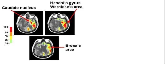
Stereotyped responses to external stimuli, such as grimacing, crying, or occasional vocalization are frequently observed on examination of vegetative state patients. These behaviors are assumed to arise primarily from brainstem circuits and limbic cortical regions that are preserved in the vegetative state. Rarely, however, patients meeting the diagnostic criteria for the vegetative state exhibit behavioral features that prima facie appear to contravene the diagnosis. A series of studies of chronic vegetative patients examined with multimodal imaging techniques identified three such patients with unusual behavioral fragments. Using FDG-PET, structural MRI and magnetoencephalography (MEG) preserved islands of higher resting brain metabolism measured by PET imaging and incompletely preserved evoked MEG gamma-band responses were correlated with structural imaging and behavioral fragments (Schiff et al., 2002). Among those studied was a patient who had been in a vegetative state for 20 years who infrequently expressed single words (typically epithets) in isolation of environmental stimulation (Schiff et al., 1999). MRI images demonstrated severe subcortical injuries. Resting FDG-PET measurements of the patient’s brain revealed a global cerebral metabolic rate of <50% of normal across most brain regions with small regions in the left hemisphere expressing higher levels of metabolism (Figure 10.2). MEG responses to bilateral auditory stimulation were confined to the left hemisphere and localized to primary auditory areas. Taken together, the imaging and neurophysiological data appeared to identify isolated sparing of left sided thalamo-cortical- basal ganglia loops that normally support language function in Heschl’s gyrus, Broca’s area, and Wernicke’s area. Similar observations in two other vegetative state patients provide novel evidence that isolated cerebral networks may remain active in rare vegetative state patients. Importantly, the preservation of these isolated behaviors does not herald further recovery in patients in chronic vegetative states who have been repeatedly examined and carefully studied with imaging tools. Reliable observations of such unusual features should
Published in: Laureys, Steven (ed), Gosseries, Olivia (ed), Tononi, Giulio (ed), The Neurology of Consciousness – 2nd edition (2016), pp.155-166
DOI: 10.1016/B978-0-12-800948-2.00010-8 Status : Postprint (Author’s version)
prompt further investigation in an individual patient.
FIGURE 10.2 Preservation of regional cerebral metabolic activity in a vegetative state patient. FDG-PET data for vegetative state patient with occasional expression of isolated words is displayed co-registered with structural MRI (Source: Data from Schiff et al., 2002). PET voxels are normalized by region and expressed on a color scale ranging from 55% to 100% of normal.
Another hallmark of the vegetative state is a systematic impairment of metabolism in the polymodal associative cortices (bilateral prefrontal regions, Broca’s area, parieto-temporal and posterior parietal areas and precuneus) (Laureys et al., 1999a). These regions are known to be important in various functions that are necessary for consciousness, such as attention, memory, and language (Baars et al., 2003). It is still unknown whether the observed metabolic impairment in this large cortical network reflects an irreversible structural neuronal loss (Rudolf et al., 2000) or functional and potentially reversible damage. However, in those rare and fortunate cases where vegetative patients recover awareness of self and environment, positron emission tomography (PET) shows a functional recovery of metabolism in these same cortical regions (Laureys et al., 1999b). Moreover, the resumption of long-range functional connectivity between these associative cortices and the intralaminar thalamic nuclei parallels the restoration of their functional integrity (Laureys et al., 2000c). The cellular mechanisms which underlie this functional normalization remain unclear: axonal sprouting, neurite outgrowth, cell division (known to occur predominantly in associative cortices in normal primates) (Gould et al., 1999) have been proposed as candidate processes (Laureys et al., 2000d) (Box 10.1).
PASSIVE NEUROIMAGING STUDIES
While metabolic studies are useful, they can only identify functionality at the most general level; that is, mapping cortical and subcortical regions that are potentially recruitable, rather than relating neural activity within such regions to specific cognitive processes (Momose et al., 1989). On the other hand, methods such as H215O PET and functional magnetic resonance imaging (fMRI) can be used to
link residual neural activity to the presence of covert cognitive function. In short, passive functional neuroimaging studies have the potential to demonstrate distinct and specific physiological responses
Published in: Laureys, Steven (ed), Gosseries, Olivia (ed), Tononi, Giulio (ed), The Neurology of Consciousness – 2nd edition (2016), pp.155-166
DOI: 10.1016/B978-0-12-800948-2.00010-8 Status : Postprint (Author’s version)
(changes in regional cerebral blood flow (rCBF) or changes in regional cerebral hemodynamics) to controlled external stimulation in the absence of any overt response (e.g., a motor action) on the part of the patient. In the first of such studies, H215O PET was used to measure rCBF in a post-traumatic
vegetative patient during an auditorily presented story told by his mother (de Jong et al., 1997). Compared to non-word sounds, activation was observed in the anterior cingulate and temporal cortices, possibly reflecting emotional processing of the contents, or tone, of the mother's speech. In another patient diagnosed as vegetative, Menon et al. (1998) also used PET, but to study covert visual processing in response to familiar faces. During "experimental" scans, the patient was presented with pictures of the faces of family and close friends, while during "control" scans scrambled versions of the same images were presented which contained no meaningful visual information whatsoever. Previous imaging studies in healthy volunteers have shown that such tasks produce robust activity in the right fusiform gyrus, the so-called human "face area" (e.g., Haxby et al., 1991, 1994). The same visual association region was activated in the vegetative patient when the familiar face stimuli were compared to the meaningless visual images (Menon et al., 1998).
In cohort studies of patients unequivocally meeting the clinical diagnosis of the vegetative state, simple noxious somatosensory (Laureys et al., 2002) and auditory (Boly et al., 2004; Laureys et al., 2000a) stimuli have shown systematic activation of primary sensory cortices and lack of activation in higher-order associative cortices from which they were functionally disconnected. High intensity noxious electrical stimulation activated midbrain, contralateral thalamus, and primary somatosensory cortex in each and every one of the 15 vegetative patients studied, even in the absence of detectable cortical evoked potentials (Laureys et al., 2002). However, secondary somatosensory, insular, posterior parietal, and anterior cingulate cortices, which were activated in all control subjects, failed to show significant activation in a single vegetative patient.
Moreover, in the vegetative state patients, the activated primary somatosensory cortex was shown to exist as an island, functionally disconnected from higher-order associative cortices of the pain-matrix. Similarly, although simple auditory click stimuli activated bilateral primary auditory cortices in vegetative patients, hierarchically higher-order multimodal association cortices were not activated. Moreover, a cascade of functional disconnections were observed along the auditory cortical pathways, from primary auditory areas to multimodal and limbic areas (Laureys et al., 2000a), suggesting that the observed residual cortical processing in the vegetative state does not lead to integrative processes which are thought to be necessary for awareness (Box 10.2).
A question that is often asked of such studies, however, is whether the presence of "normal" brain activation in patients who are diagnosed as vegetative indicates a level of conscious awareness. Many types of stimuli, including faces, speech and pain will elicit relatively "automatic" responses from the brain; that is to say, they will occur without the need for willful intervention on the part of the participant (e.g., you cannot choose to not recognize a face, or to not understand speech that is presented clearly in your native language). By the same argument, "normal" neural responses in patients who are diagnosed as vegetative do not necessarily indicate that these patients have any conscious experience associated with processing those same types of stimuli. Thus, such patients
Published in: Laureys, Steven (ed), Gosseries, Olivia (ed), Tononi, Giulio (ed), The Neurology of Consciousness – 2nd edition (2016), pp.155-166
DOI: 10.1016/B978-0-12-800948-2.00010-8 Status : Postprint (Author’s version)
awareness.
The logic described above exposes a central conundrum in the study of conscious awareness and in particular, how it relates to the vegetative state. Deeper philosophical considerations notwithstanding, the only reliable method that we have for determining if another being is consciously aware is to ask him/her. The answer may take the form of a spoken response or a non-verbal signal (which may be as simple as the blink of an eye, as documented cases of the locked-in syndrome have demonstrated), but it is this answer that allows us to infer conscious awareness. In short, our ability to know unequivocally that another being is consciously aware is ultimately determined, not by whether they are aware or not, but by their ability to communicate that fact through a recognized behavioral response. But what if the ability to blink an eye or move a hand is lost, yet conscious awareness remains? By definition, patients who are diagnosed as vegetative are not able to elicit such behavioral responses. Following the logic of this argument then, even if such a patient were consciously aware, he/she would, by definition, have no means for conveying that information to the outside world.
BOX 10.2
METHODOLOGICAL ISSUES
The acquisition, analysis, and interpretation of neuroimaging data in severe brain injury is methodologically extremely complex. In quantitative PET studies, the absolute value of cerebral metabolic rates depends on many assumptions for which a consensus has not been established in cases of cerebral pathology. For example, the estimation of the cerebral metabolic rate of glucose using 18F-FDG-PET requires a correction factor, known as the lumped constant. It is generally accepted that this lumped constant is stable in normal brains. However, in traumatic brain injury, a significant global decrease in lumped constant has recently been reported (Wu et al., 2004) and in severe ischemia, regional lumped constant values are known to increase significantly as a result of glucose transport limitation (Hamlin et al., 2001). Second, cerebral glucose use as measured by
18F-FDG may not always be tightly coupled with oxygen use in patients because altered metabolic
states, including anaerobic glycolysis, may occur acutely after brain injury (Bergsneider et al., 2001; Goodman et al., 1999; Hovda et al., 1992). Third, because PET provides measurements per unit volume of intracranial contents, they may be affected by the inclusion of metabolically inactive spaces such as cerebrospinal fluid or by brain atrophy which may artificially lower the calculated cerebral metabolism (Herscovitch et al., 1986; Videen et al., 1988).
As described in the main text, the so-called “activation studies” using H215O PET or fMRI together
with established sensory paradigms provide a direct method for assessing cognitive processing and even conscious awareness in severely brain-injured patients. However, like metabolic studies, these investigations are methodologically complex and the results are rarely equivocal. For example, in brain-injured patients, the coupling between neuronal activity and local hemodynamics, essential for all H215O PET and fMRI activation measurements, is likely to be
different from healthy controls (Gsell et al., 2000; Hamzei et al., 2003; Rossini et al., 2004; Sakatani et al., 2003), making interpretation of such data sets extremely difficult. Notwithstanding this basic
Published in: Laureys, Steven (ed), Gosseries, Olivia (ed), Tononi, Giulio (ed), The Neurology of Consciousness – 2nd edition (2016), pp.155-166
DOI: 10.1016/B978-0-12-800948-2.00010-8 Status : Postprint (Author’s version)
methodological concern, the choice of experimental paradigm is also critical. For example, abnormal brainstem auditory evoked responses may make the use of auditory stimuli inappropriate and alternative stimuli (i.e., visual) should be considered. The paradigm should also be sufficiently complex to exercise the cognitive processes of interest, preferably beyond those that are simply involved in stimulus perception, yet not so complex that they might easily overload residual cognitive capacities in a tired or inattentive patient. In addition, it is essential that the experimental paradigm chosen produces well-documented, anatomically specific, robust, and reproducible activation patterns in healthy volunteers in order to facilitate interpretation of imaging data in patients. In vegetative state, episodes of low arousal and sleep are also frequently observed and close patient monitoring (preferably by means of simultaneous electroencephalographic recording) during activation scans is essential to avoid such periods. Spontaneous movements during the scan itself may also compromise the interpretation of functional neuroimaging data, particularly scans acquired using fMRI. Data processing of functional neuroimaging data may also present challenging problems in patients with acute brain injury. For example, the presence of gross hydrocephalus or focal pathology may complicate co-registration of functional data (e.g., acquired with PET or fMRI) to anatomical data (e.g., acquired using structural MRI), and the normalization of images to a healthy reference brain. Under these circumstances statistical assessment of activation patterns is complex and interpretation of activation foci in terms of standard stereotaxic coordinates may be impossible. Finally, where PET methodology is employed, issues of radiation burden must also be considered and may preclude longitudinal or follow-up studies in many patients.
ACTIVE NEUROIMAGING STUDIES
A novel approach to this conundrum has been the development of the so-called “active” fMRI paradigms that render awareness reportable in patients who are either entirely behaviorally non-responsive and therefore diagnosed as vegetative (Owen et al., 2006; Boly et al., 2007) or partially responsive and diagnosed as minimally conscious state (Bardin et al., 2011, 2012). In contrast to passive functional neuroimaging studies, active studies require participants to respond to task-specific demands by willfully modulating their own neural activity. In other words, the neural responses required are not produced automatically by the eliciting stimulus, but rather depend on time-dependent and sustained responses generated by the participants themselves. Such behavior (albeit neural “behavior”) provides a proxy for a motor action and is, therefore, an appropriate vehicle for reportable awareness (Zeman, 2009).
BOX 10.3
OTHER FMRI APPROACHES TO DETECTING CONSCIOUSNESS IN NON-RESPONSIVE PATIENTS
In addition to the use of mental imagery, another approach to detecting covert awareness after brain injury is to target processes that require the willful adoption of “mind-sets” in carefully matched (perceptually identical) experimental and control conditions. For example, Monti et al.
Published in: Laureys, Steven (ed), Gosseries, Olivia (ed), Tononi, Giulio (ed), The Neurology of Consciousness – 2nd edition (2016), pp.155-166
DOI: 10.1016/B978-0-12-800948-2.00010-8 Status : Postprint (Author’s version)
(2009) presented healthy volunteers with a series of neutral words, and alternatively instructed them to just listen, or to count, the number of times a given word was repeated. As predicted, the counting task revealed the frontoparietal network that has been previously associated with target detection and working memory. When tested on this same procedure, a severely brain-injured patient produced a very similar pattern of activity, confirming that he could willfully adopt differential sets as a function of the task conditions and could actively maintain these mind-sets across time; covert abilities that were entirely absent from his documented behavioral repertoire. As in the tennis/spatial navigation examples described in the main text, because the external stimuli (a series of words) were identical in the two conditions any difference in brain activity observed cannot reflect an “automatic” brain response (i.e., one that can occur in the absence of consciousness). Rather, the activity must reflect the fact that the patient has performed a particular action (albeit a “brain action”) in response to the stimuli on one (but not the other) presentation; in this sense, the brain response is entirely analogous to a (motor) response to command and should carry the same weight as evidence of awareness.
Following similar logic, Monti et al. (2013) used an entirely different type of approach to demonstrate that a patient who was entirely unable to exhibit any signs of command following during standard behavioral testing, could nevertheless demonstrate reliable and robust responses in predefined brain regions by willfully modulating his brain activity. The stimuli used were super-imposed pictures of faces and houses. When healthy volunteers are requested, following a cue tone, to shift their attentional focus from a face to a house (or vice versa), a distinct shift in fMRI activity from the fusiform gyrus (the FFA), to the parahippocampal gyrus (the “parahippocampal place area”) is observed (or vice versa) (Monti et al., 2013). With continuous, repeated cues, this effect manifests as a time-locked alternation of activity between these two functionally distinct brain regions, despite the fact that the stimulus remains unchanged throughout. Thus, this change is driven, not by the external stimulus per se, but by the will, or the intention, of the participant to focus on one, or the other, aspect of the stimulus and is therefore a reliable indicator of conscious intent. When asked to perform the same task, the activity observed in the patient closely resembled the activity observed in the healthy volunteers and, as such, provided the only conclusive evidence that he could indeed follow commands (Monti et al., 2013).
The most successful techniques in active paradigms make use of the general principle observed in studies of healthy participants that imagining performing a particular task generates a robust and reliable pattern of brain activity in the fMRI scanner that is similar to actually performing the activity itself. For example, imagining moving or squeezing the hands will generate activity in the motor and premotor cortices (Jeannerod and Frak, 1999), while imagining navigating from one location to another will activate the same regions of the parahippocampal gyrus and the posterior parietal cortex that have been widely implicated in map-reading and other so-called spatial navigation tasks (Aguirre et al., 1996) (Box 10.3).
In one study (Boly et al., 2007), 34 healthy volunteers were asked to imagine hitting a tennis ball back and forth to an imaginary coach when they heard the word “tennis” (thereby eliciting vigorous
Published in: Laureys, Steven (ed), Gosseries, Olivia (ed), Tononi, Giulio (ed), The Neurology of Consciousness – 2nd edition (2016), pp.155-166
DOI: 10.1016/B978-0-12-800948-2.00010-8 Status : Postprint (Author’s version)
imaginary arm movements) and to imagine walking from room to room in their house when they heard the word “house” (thereby eliciting imaginary spatial navigation). Imagining playing tennis was associated with robust activity in the supplementary motor area in each and every one (100%) of the participants scanned. In contrast, imagining moving from room to room in a house activated the parahippocampal cortices, the posterior parietal lobe and the lateral premotor cortices; all regions that have been shown to contribute to imaginary, or real, spatial navigation (Aguirre et al., 1996; Boly et al., 2007). A recent follow-up study has demonstrated that such responses can be reliably produced in single-participants (classified correctly in at least 80% of cases) using a hospital-grade 1.5T scanner, lending the technique to widespread clinical use (Fernandez-Espejo et al., 2014).
The robustness and reliability of these fMRI responses across individuals means that activity in these regions can be used as a neural proxy for behavior, confirming that the participant retains the ability to understand instructions, to carry out different mental tasks in response to those instructions and, therefore, is able to exhibit willed, voluntary behavior in the absence of any overt action. Thus, like any other form of action that requires response selection, these brain responses require awareness of the various contingencies that govern the relationship between any given stimulus (in this case, the cue word for one of two possible imagery tasks) and a response (in this case, imagining the task). Put simply, fMRI responses of this sort can be used to measure awareness because awareness is necessary for them to occur.
Owen et al. (2006, 2007) used this same logic to demonstrate that a young woman who fulfilled all internationally agreed criteria for the vegetative state was, in fact, consciously aware and able to make responses of this sort using her brain activity (Figure 10.3). The patient, who was involved in a complex road traffic accident and had sustained very severe traumatic brain injuries, had remained entirely unresponsive for a period of 6 months prior to the fMRI scan. During the scanning session, the patient was instructed to perform the two mental imagery tasks described above. When she was asked to imagine playing tennis, statistically significant activity was observed repeatedly in the supplementary motor area (Owen et al., 2006) that was indistinguishable from that observed in the healthy volunteers (Boly et al., 2007). Moreover, when she was asked to imagine walking through her home, significant activity was observed in the parahippocampal gyrus, the posterior parietal cortex and the lateral premotor cortex which was again, indistinguishable from those observed in healthy volunteers (Owen et al., 2006, 2007). On this basis, it was concluded that, despite fulfilling all of the clinical criteria for a diagnosis of vegetative state, this patient retained the ability to understand spoken commands and to respond to them through her brain activity, rather than through speech or movement, confirming beyond any doubt that she was consciously aware of herself and her surroundings. In a follow-up study of 23 patients who were behaviorally diagnosed as vegetative,
Monti et al. (2010) showed that four (17%) were able to generate reliable responses of this sort in the fMRI scanner.
FIGURE 10.3 (A) Supplementary motor area (SMA) activity during tennis imagery and parahippocampal gyrus (PPA), posterior parietal lobe (PPC) and lateral premotor cortex (PMC) activity while imagining moving around a house in the patient described by Owen et al. (2006). (B) Statistically indistinguishable activity in all four brain regions in a group of 12 healthy volunteers asked to perform the same imagery tasks. (C) The result when a healthy volunteer underwent exactly the same fMRI procedure as the patient described by Owen et al. (2006), with the
Published in: Laureys, Steven (ed), Gosseries, Olivia (ed), Tononi, Giulio (ed), The Neurology of Consciousness – 2nd edition (2016), pp.155-166
DOI: 10.1016/B978-0-12-800948-2.00010-8 Status : Postprint (Author’s version)
exception that non-instructive sentences (e.g., "The man played tennis," "The man walked around his house") were used. Using an identical statistical model to that used with the patient, no significant sustained activity was observed in the SMA, PPA, the PPC, the PMC, nor any other brain region. All results are similarly thresholded at FDR p < 0.05, corrected for multiple comparisons.
After a severe brain injury, when the request to move a hand or a finger is followed by an appropriate motor response, the diagnosis can change from vegetative state (no evidence of awareness) to minimally conscious state (some evidence of awareness). By analogy then, if the request to activate the supplementary motor area of the brain by imagining moving the hand is followed by an appropriate brain response, shouldn't we give that response the very same weight? Skeptics may argue that brain responses are somehow less physical, reliable or immediate than motor responses but, as is the case with motor responses, all of these arguments can be dispelled with careful measurement, replication, and objective verification (Boly et al., 2007; Fernandez-Espejo and Owen, 2013; Hampshire et al., 2012; Monti et al., 2010; Naci et al., 2013; Naci and Owen, 2013; Owen et al., 2006, 2007). For example, if a patient who was assumed to be unaware raised his/her hand to command on just one occasion, there would remain some doubt about the presence of awareness given the possibility that this movement was a chance occurrence, coincident with the instruction. However, if that same patient were able to repeat this response to command on ten occasions, there would remain little doubt that the patient was aware. By the same token, if that patient was able to activate his/her supplementary motor area in response to command (e.g., by being told to imagine
Published in: Laureys, Steven (ed), Gosseries, Olivia (ed), Tononi, Giulio (ed), The Neurology of Consciousness – 2nd edition (2016), pp.155-166
DOI: 10.1016/B978-0-12-800948-2.00010-8 Status : Postprint (Author’s version)
hand movements), and was able to do this on every one of ten trials, would we not have to accept that this patient was consciously aware? Like most neuroimaging investigations, replication of this sort was inherent in both of the studies described above (Monti et al., 2010; Owen et al., 2006), because correct classification of the characteristic neural signatures required statistically similar significant results across repeated trials.
It has been suggested that fMRI responses of this sort could reflect an "implicit preconscious neural response" to the key words that were used in those studies (Greenberg, 2007; Nachev and Husain, 2007). While no empirical evidence exists to support this possibility, it is nevertheless important to consider its theoretical plausibility. In the volunteers studied by (Boly et al., 2007), and in the patients reported by Owen et al. (2006) and Monti et al. (2010), the observed activity was not transient, but persisted for the full 30 s of each imagery task that is, far longer than would be expected, even given the hemodynamics of the fMRI response. In fact, these task-specific changes persisted until the volunteers and the patients were cued with another stimulus indicating that they should switch tasks. No evidence exists to show that single-word stimuli (such as "tennis," "house," or "rest") can unconsciously elicit sustained (i.e., 30 s) hemodynamic responses in the supplementary motor area, the parahippocampal gyrus, the posterior parietal cortex or the lateral premotor cortex premotor cortex, yet considerable data exists to suggest that they cannot. For example, although it is well documented that some words can, under certain circumstances, elicit wholly automatic neural responses, such responses are typically transient and last for just a few seconds. In addition, the activation patterns observed in the studies by Boly et al. (2007), Owen et al. (2006), and Monti et al. (2010), were entirely predicted and were not in brain regions that are known to be involved in word processing, but rather, in regions that are known to be involved in the two imagery tasks (also see Weiskopf et al., 2004). In short, temporally sustained fMRI responses in these regions of the brain are impossible to explain in terms of automatic responses to either single "key" words or to short sentences containing those words. In fact, non-instructive sentences containing the same key words (e.g., "The man enjoyed playing tennis") have been shown to produce no sustained activity in any of these brain regions in healthy volunteers, nor is activity seen when the words "tennis" and "house" are presented to naïve participants who have not been previously instructed to perform the imagery tasks (Owen et al., 2007) (Figure 10.3). The most parsimonious explanation is, therefore, that patients who display sustained and reproducible brain activation during visual mental imagery tasks are consciously aware and following instructions, even in the complete or partial absence of behavioral responsiveness.
COMMUNICATION
Owen and Coleman (2008) extended the general principles discussed above, by which active mental rehearsal is used to signify awareness, to show that communication of "yes" and "no" responses was possible using the same approach. Thus, a healthy volunteer was able to reliably convey a "yes" response by imagining playing tennis and a "no" response by imaging moving around a house, thereby providing the answers to simple questions posed by the experimenters using only their brain activity. This technique was further refined by Monti et al. (2010) who successfully decoded three "yes" and "no" responses from each of 16 healthy participants with 100% classification accuracy using
Published in: Laureys, Steven (ed), Gosseries, Olivia (ed), Tononi, Giulio (ed), The Neurology of Consciousness – 2nd edition (2016), pp.155-166
DOI: 10.1016/B978-0-12-800948-2.00010-8 Status : Postprint (Author’s version)
only their real time changes in the supplementary motor area (during tennis imagery) and the parahippocampal place area (during spatial navigation). Moreover, in one traumatic brain injury patient, who had been repeatedly diagnosed as vegetative over a 5-year period, similar questions were posed and successfully decoded using the same approach (Monti et al. 2010). Thus, this patient was able to convey biographical information that was not known to the experimenters at the time (but was verified as factually correct) such as his father's name and the last place that he had visited on vacation before his accident 5 years earlier. In contrast, and despite a re-classification to minimally conscious state following the fMRI scan, it remained impossible to establish any form of communication with this patient at the bedside.
An obvious application for approaches of this sort is to begin to involve some of these patients in the decision-making processes involved in their own therapeutic care and management. To date, this has only been achieved successfully in one patient, who had been repeatedly diagnosed as vegetative for 12 years following a severe closed head injury (Fernandez- Espejo and Owen, 2013). Indeed, over one 14-month period from 2011 to 2013, the patient underwent 20 standardized behavioral assessments by a multidisciplinary team, at different times of the day and in different postural positions, using the Coma Recovery Scale-Revised (Giacino et al., 2004). In February 2012, 12 years and 2 months after his accident, the patient was first scanned using the fMRI mental imagery approach described above (Monti et al., 2010; Owen et al., 2006). The patient was able to provide correct answers to multiple externally verifiable questions, including autobiographical and other basic factual information. Two non-verifiable questions were then posed, including one pertaining to his care preferences (e.g., whether he liked watching hockey games on TV), and another to details about his current clinical con-dition (e.g., whether he was in any physical pain). Within the time-constrains of the scanning visits, the majority of responses to these questions were verified in independent sessions that posed the reverse questions (e.g., "Is your name Mike?" vs "Is your name Scott?"). At the time of the patient's death in 2013, answers to 12 different questions had been obtained across several sessions, despite the fact that the patient remained entirely physically non-responsive at the bedside (Fernandez-Espejo and Owen, 2013).
In the technique described above, the patient's ability to turn his or her attention to a specific scenario is required and serves as a "neural proxy" for a physical "response to command." By linking scenarios to "yes" responses and "no" responses, respectively, communication is possible. Similarly, Naci and colleagues (Naci et al., 2013; Naci and Owen, 2013) developed a novel tool for communicating with non- responsive patients based on how they selectively directed their attention to sounds while in the fMRI scanner. It is well established that selective attention can significantly enhance the neural representation of attended sounds (Bidet-Caulet et al., 2007). In their first study (Naci et al., 2013), 15 healthy volunteers answered questions (e.g., "Do you have brothers or sisters?") in the fMRI scanner by selectively attending to the appropriate word ("yes" or "no"), which was played to them auditorily, interspersed with "distractor" stimuli (digits 1—9). Ninety percent of the answers were decoded correctly based on activity changes within the attention network of the brain (i.e., 90% classification accuracy). Moreover, majority of the volunteers conveyed their answers with less than 3 min of scanning, which represents a significant time saving over the mental imagery methods described above (Boly et al., 2007; Owen et al., 2006, 2007). Indeed, a formal comparison between the two
Published in: Laureys, Steven (ed), Gosseries, Olivia (ed), Tononi, Giulio (ed), The Neurology of Consciousness – 2nd edition (2016), pp.155-166
DOI: 10.1016/B978-0-12-800948-2.00010-8 Status : Postprint (Author’s version)
approaches revealed improved individual success rates and an overall reduction in the scanning times required to correctly detect responses; 100% of volunteers showed significant task-appropriate activity to the selective attention task, compared to 87% to the motor imagery. This result is consistent with previous studies showing that a proportion of healthy volunteers do not produce reliably classifiable brain activation during mental imagery tasks (Boly et al., 2007).
In their follow-up study (Naci and Owen, 2013), the same approach was used to test for residual conscious awareness and communication abilities in three behaviorally non-responsive, brain-injured patients. As in the previous study of healthy participants, the patients had to either "count" or "relax" as they heard a sequence of sounds. The word count at the beginning of the sequence instructed the patient to count the occurrences of a target word (yes or no), while the word relax instructed them to relax and ignore the sequence of words. Reliable activity increases in the attention network of the brain after the word count relative to the word relax was taken as evidence of command following. All three patients (two of whom were diagnosed as being in a minimally conscious state and one as being in a vegetative state) were able to convey their ability to follow commands inside the fMRI scanner by following the instructions in this way. In stark contrast, extremely limited or a complete lack of behavioral responsivity was observed in repeated bedside assessments of all three patients. These results confirm that selective attention is an appropriate vehicle for detecting covert awareness in some behaviorally non-responsive patients who are presumed to mostly or entirely lack any cognitive abilities whatsoever.
In a following series of scans, communication was also attempted in two of the patients. The communication scans were similar to those in the command-following scan, with one exception. Instead of an instruction (count or relax), a binary question (e.g., "Is your name Steven?") preceded each sound sequence. Thus, each patient then had to willfully choose which word to attend to (count) and which to ignore, depending on which answer he wished to convey to the specific question that had been asked. Using this method, the two patients (one diagnosed as minimally conscious state and one diagnosed as vegetative state) were able to use selective attention to repeatedly communicate correct answers to questions that were posed to them by the experimenters (Naci and Owen, 2013). In the absence of external cues as to which word the patient was attending to, the functional brain activation served as the only indicator of the patient's intentions—and in both cases, led to the correct answers being decoded. For example, when asked, "Are you in a supermarket?" one patient showed significantly more activation for "no" than "yes" sequences in a network of brain areas that had been previously activated when that patient was focusing attention on external cues. Conversely, when asked, "Are you in a hospital?" the patient showed significantly more activation for "yes" than "no" sequences in the same regions. Despite his diagnosis (vegetative state for 12 years), the fMRI approach allowed this patient to establish interactive communication with the research team in four different fMRI sessions. The patient's brain responses within specific regions were remarkably consistent and reliable across two different scanning visits, 5 months apart, during which the patient maintained the long-standing vegetative state diagnosis. For all four questions, the patient produced a robust neural response and was able to provide the correct answer with 100% classification accuracy. The patient's brain activity in the communication scans not only further corroborated that he was, indeed, consciously aware but also revealed that he had far richer cognitive
Published in: Laureys, Steven (ed), Gosseries, Olivia (ed), Tononi, Giulio (ed), The Neurology of Consciousness – 2nd edition (2016), pp.155-166
DOI: 10.1016/B978-0-12-800948-2.00010-8 Status : Postprint (Author’s version)
reserves than could be assumed based on his clinical diagnosis. In particular, beyond the ability to pay attention, these included autobiographical knowledge and awareness of his location in time and space.
CONCLUSIONS
Vegetative state presents unique problems for diagnosis, prognosis, treatment, and everyday management. At the patient's bedside, the evaluation of possible cognitive function in these patients is difficult because voluntary movements may be very small, inconsistent, and easily exhausted. Functional neuro-imaging offers a complimentary approach to the clinical assessment of patients with vegetative state and other altered states of consciousness and can objectively describe (using population norms) the regional distribution of cerebral activity at rest and under various conditions of stimulation. In addition, functional neuroimaging has demonstrated preserved cognitive function, and even covert awareness, in patients who are assumed to be vegetative, yet retain cognitive abili-ties that have evaded detection using standard clinical methods. Most recently, the use of task-dependent fMRI responses as a form of "communication" in these patients represents an important milestone. In our opinion, the future use of functional neuroimaging will not only substantially increase our understanding of severely brain-injured patients, but will allow us to make high-level assessments of residual cognitive function and answer clinically relevant questions.
Acknowledgments
SL is Research Associate at the Fonds National de la Recherche Scientifique de Belgique (FNRS). NS is supported by NS02172, NS43451, and the Charles A. Dana Foundation. AMO is supported by the Canada Excellence Research Chairs (CERC) program, the Canadian Institutes for Health Research (CIHR) and National Sciences and Engineering Research Council of Canada (NSERC). All three authors are also supported by the James S. McDonnell Foundation.
References
Agardh, C.D., Rosen, I., Ryding, E., 1983. Persistent vegetative state with high cerebral blood flow following profound hypoglycemia. Ann. Neurol. 14 (4), 482-486.
Aguirre, G.K., Detre, J.A., Alsop, D.C., D'Esposito, M., 1996. The parahippocampus subserves topographical learning in man. Cereb. Cortex. 6 (6), 823 —829.
Andrews, K., Murphy, L., Munday, R., Littlewood, C., 1996. Misdiagnosis of the vegetative state: retrospective study in a rehabilitation unit. BMJ. 313 (7048), 13-16.
Baars, B.J., Ramsoy, T.Z., Laureys, S., 2003. Brain, conscious experience and the observing self. Trends Neurosci. 26 (12), 671—675.
Bardin, J.C., Fins, J.J., Katz, D.I., Hersh, J., Heier, L.A., Tabelow, K., et al., 2011. Dissociations between behavioural and functional magnetic resonance imaging-based evaluations of cognitive function after brain injury. Brain. 134 (Pt 3), 769—782.
Bardin, J.C., Schiff, N.D., Voss, H.U., 2012. Pattern classification of volitional functional magnetic resonance imaging responses in patients with severe brain injury. Arch. Neurol. 69 (2), 176—181.
Bergsneider, M., Hovda, D.A., McArthur, D.L., Etchepare, M., Huang, S.C., Sehati, N., et al., 2001. Metabolic recovery following human traumatic brain injury based on FDG-PET: time course and relationship to neurological
Published in: Laureys, Steven (ed), Gosseries, Olivia (ed), Tononi, Giulio (ed), The Neurology of Consciousness – 2nd edition (2016), pp.155-166
DOI: 10.1016/B978-0-12-800948-2.00010-8 Status : Postprint (Author’s version)
disability. J. Head Trauma Rehabil. 16 (2), 135-148.
Beuthien-Baumann, B., Handrick, W., Schmidt, T., Burchert, W., Oehme, L., Kropp, J., et al., 2003. Persistent vegetative state: evaluation of brain metabolism and brain perfusion with PET and SPECT. Nucl. Med. Commun. 24 (6), 643 -649.
Bidet-Caulet, A., Fischer, C., Besle, J., Aguera, P.E., Giard, M.H., Bertrand, O., 2007. Effects of selective attention on the electrophysiological representation of concurrent sounds in the human auditory cortex. J. Neurosci. 27 (35), 9252—9261.
Boly, M., Faymonville, M.E., Peigneux, P., Lambermont, B., Damas, P., Del Fiore, G., et al., 2004. Auditory processing in severely brain injured patients: differences between the minimally conscious state and the persistent vegetative state. Arch. Neurol. 61 (2), 233-238.
Boly, M., Coleman, M.R., Davis, M.H., Hampshire, A., Bor, D., Moonen, G., et al., 2007. When thoughts become action: an fMRI paradigm to study volitional brain activity in non-communicative brain injured patients. Neuroimage. 36 (3), 979—992.
Childs, N.L., Mercer, W.N., Childs, H.W., 1993. Accuracy of diagnosis of persistent vegetative state. Neurology. 43 (8), 1465—1467.
de Jong, B.M., Willemsen, A.T., Paans, A.M., 1997. Regional cerebral blood flow changes related to affective speech presentation in persistent vegetative state. Clin. Neurol. Neurosurg. 99 (3), 213—216.
DeVolder, A.G., Goffinet, A.M., Bol, A., Michel, C., de Barsy, T., Laterre, C., 1990. Brain glucose metabolism in postanoxic syndrome. Positron emission tomographic study. Arch. Neurol. 47 (2), 197-204.
Edgren, E., Enblad, P., Grenvik, A., Lilja, A., Valind, S., Wiklund, L., et al., 2003. Cerebral blood flow and metabolism after cardiopulmonary resuscitation. A pathophysiologic and prognostic positron emission tomography pilot study. Resuscitation. 57 (2), 161—170.
Fernandez-Espejo, D., Owen, A.M., 2013. Detecting awareness after severe brain injury. Nat. Rev. Neurosci. 14 (11), 801—809.
Fernandez-Espejo, D., Norton, L., Owen, A.M., 2014. The clinical utility of fMRI for identifying covert awareness in the vegetative state: a comparison of sensitivity between 3T and 1.5T. PLoS One. 9 (4), e95082.
Giacino, J.T., Kalmar, K., Whyte, J., 2004. The JFK Coma Recovery Scale-Revised: measurement characteristics and diagnostic utility. Arch. Phys. Med. Rehabil. 85 (12), 2020-2029.
Goodman, J.C., Valadka, A.B., Gopinath, S.P., Uzura, M., Robertson, C.S., 1999. Extracellular lactate and glucose alterations in the brain after head injury measured by microdialysis. Crit. Care Med. 27 (9), 1965-1973.
Gould, E., Reeves, A.J., Graziano, M.S., Gross, C.G., 1999. Neurogenesis in the neocortex of adult primates. Science. 286 (5439), 548—552.
Greenberg, D.L., 2007. Comment on "Detecting awareness in the vegetative state". Science. 315 (5816), 1221. Gsell, W., De Sadeleer, C., Marchalant, Y., MacKenzie, E.T., Schumann, P., Dauphin, F., 2000. The use of cerebral blood flow as an index of neuronal activity in functional neuroimaging: experimental and pathophysiological considerations. J. Chem. Neuroanat. 20 (3-4), 215-224.
Hamlin, G.P., Cernak, I., Wixey, J.A., Vink, R., 2001. Increased expression of neuronal glucose transporter 3 but not glial glucose transporter 1 following severe diffuse traumatic brain injury in rats. J. Neurotrauma. 18 (10), 1011—1018.
Published in: Laureys, Steven (ed), Gosseries, Olivia (ed), Tononi, Giulio (ed), The Neurology of Consciousness – 2nd edition (2016), pp.155-166
DOI: 10.1016/B978-0-12-800948-2.00010-8 Status : Postprint (Author’s version)
Hampshire, A., Parkin, B.L., Cusack, R., Espejo, D.F., Allanson, J., Kamau, E., et al., 2012. Assessing residual reasoning ability in overtly non-communicative patients using fMRI. Neuroimage Clin. 2, 174-183.
Hamzei, F., Knab, R., Weiller, C., Rother, J., 2003. The influence of extra- and intracranial artery disease on the BOLD signal in FMRI. Neuroimage. 20 (2), 1393-1399.
Haxby, J.V., Grady, C.L., Horwitz, B., Ungerleider, L.G., Mishkin, M., Carson, R.E., et al., 1991. Dissociation of object and spatial visual processing pathways in human extrastriate cortex. Proc. Natl. Acad. Sci. USA. 88 (5), 1621-1625.
Haxby, J.V., Horwitz, B., Ungerleider, L.G., Maisog, J.M., Pietrini, P., Grady, C.L., 1994. The functional organization of human extrastriate cortex: a PET-rCBF study of selective attention to faces and locations. J. Neurosci. 14 (11 Pt 1), 6336—6353.
Herscovitch, P., Auchus, A.P., Gado, M., Chi, D., Raichle, M.E., 1986. Correction of positron emission tomography data for cerebral atrophy. J. Cereb. Blood Flow Metab. 6 (1), 120—124.
Hovda, D.A., Becker, D.P., Katayama, Y., 1992. Secondary injury and acidosis. J. Neurotrauma. 9 (Suppl. 1), S47— S60.
Jeannerod, M., Frak, V., 1999. Mental imaging of motor activity in humans. Curr. Opin. Neurobiol. 9 (6), 735— 739.
Jennett, B., 2002. The vegetative state. J. Neurol. Neurosurg. Psychiatry. 73 (4), 355-357.
Jennett, B., Plum, F., 1972. Persistent vegetative state after brain damage. A syndrome in search of a name. Lancet. 1 (7753), 734—737.
Laureys, S., Goldman, S., Phillips, C., Van Bogaert, P., Aerts, J., Luxen, A., et al., 1999a. Impaired effective cortical connectivity in vegetative state: preliminary investigation using PET. Neuroimage. 9 (4), 377-382.
Laureys, S., Lemaire, C., Maquet, P., Phillips, C., Franck, G., 1999b. Cerebral metabolism during vegetative state and after recovery to consciousness. J. Neurol. Neurosurg. Psychiatry. 67 (1), 121.
Laureys, S., Faymonville, M.E., Degueldre, C., Fiore, G.D., Damas, P., Lambermont, B., et al., 2000a. Auditory processing in the vegetative state. Brain. 123 (Pt 8), 1589—1601.
Laureys, S., Faymonville, M.E., Goldman, S., 2000b. Impaired cerebral connectivity in vegetative state. In: Gjedde, A., Hansen, S.B., Knudsen, G.M., Paulson, O.B. (Eds.), Physiological Imaging of the Brain with PET. Academic Press, San Diego, CA, pp. 329—334.
Laureys, S., Faymonville, M.E., Luxen, A., Lamy, M., Franck, G., Maquet, P., 2000c. Restoration of thalamocortical connectivity after recovery from persistent vegetative state. Lancet. 355 (9217), 1790-1791.
Laureys, S., Faymonville, M.E., Moonen, G., Luxen, A., Maquet, P., 2000d. PET scanning and neuronal loss in acute vegetative state. Lancet. 355 (9217), 1825-1827.
Laureys, S., Faymonville, M.E., Peigneux, P., Damas, P., Lambermont, B., Del Fiore, G., et al., 2002. Cortical processing of noxious somatosensory stimuli in the persistent vegetative state. Neuroimage. 17 (2), 732-741.
Levy, D.E., Sidtis, J.J., Rottenberg, D.A., Jarden, J.O., Strother, S.C., Dhawan, V., et al., 1987. Differences in cerebral blood flow and glucose utilization in vegetative versus locked-in patients. Ann. Neurol. 22 (6), 673-682.
Medical aspects of the persistent vegetative state (1). The Multi-Society Task Force on PVS. 1994. N. Engl. J. Med. 330 (21), 1499-1508.
Published in: Laureys, Steven (ed), Gosseries, Olivia (ed), Tononi, Giulio (ed), The Neurology of Consciousness – 2nd edition (2016), pp.155-166
DOI: 10.1016/B978-0-12-800948-2.00010-8 Status : Postprint (Author’s version)
in persistent vegetative state. Wolfson Brain Imaging Centre Team. Lancet. 352 (9123), 200.
Momose, T., Matsui, T., Kosaka, N., Ohtake, T., Watanabe, T., Nishikawa, J., et al., 1989. Effect of cervical spinal cord stimulation (cSCS) on cerebral glucose metabolism and blood flow in a vegetative patient assessed by positron emission tomography (PET) and single photon emission computed tomography (SPECT). Radiat. Med. 7 (5), 243 -246.
Monti, M.M., Coleman, M.R., Owen, A.M., 2009. Executive functions in the absence of behavior: functional imaging of the minimally conscious state. Prog. Brain Res. 177, 249—260.
Monti, M.M., Vanhaudenhuyse, A., Coleman, M.R., Boly, M., Pickard, J.D., Tshibanda, L., et al., 2010. Willful modulation of brain activity in disorders of consciousness. N. Engl. J. Med. 362 (7), 579—589.
Monti, M.M., Pickard, J.D., Owen, A.M., 2013. Visual cognition in disorders of consciousness: from V1 to top-down attention. Hum. Brain Mapp. 34 (6), 1245-1253.
Nachev, P., Husain, M., 2007. Comment on "Detecting awareness in the vegetative state". Science. 315 (5816), 1221.
Naci, L., Owen, A.M., 2013. Making every word count for nonresponsive patients. JAMA Neurol. 70 (10), 1235— 1241.
Naci, L., Cusack, R., Jia, V.Z., Owen, A.M., 2013. The brain's silent messenger: using selective attention to decode human thought for brainbased communication. J. Neurosci. 33 (22), 9385—9393.
Owen, A.M., Coleman, M.R., 2008. Detecting awareness in the vegetative state. Ann. N.Y. Acad. Sci. 1129,130— 138.
Owen, A.M., Coleman, M.R., Boly, M., Davis, M.H., Laureys, S., Pickard, J.D., 2006. Detecting awareness in the vegetative state. Science. 313 (5792), 1402.
Owen, A.M., Coleman, M.R., Boly, M., Davis, M.H., Laureys, S., Jolles, D., et al., 2007. Response to comments on "Detecting awareness in the vegetative state". Science. 315 (5816), 1221.
Rossini, P.M., Altamura, C., Ferretti, A., Vernieri, F., Zappasodi, F., Caulo, M., et al., 2004. Does cerebrovascular disease affect the coupling between neuronal activity and local haemodynamics? Brain. 127 (Pt 1), 99-110.
Rudolf, J., Ghaemi, M., Ghaemi, M., Haupt, W.F., Szelies, B., Heiss, W.D., 1999. Cerebral glucose metabolism in acute and persistent vegetative state. J. Neurosurg. Anesthesiol. 11 (1), 17—24.
Rudolf, J., Sobesky, J., Grond, M., Heiss, W.D., 2000. Identification by positron emission tomography of neuronal loss in acute vegetative state. Lancet. 355 (9198), 115—116.
Rudolf, J., Sobesky, J., Ghaemi, M., Heiss, W.D., 2002. The correlation between cerebral glucose metabolism and benzodiazepine receptor density in the acute vegetative state. Eur. J. Neurol. 9 (6), 671 —677.
Sakatani, K., Murata, Y., Fukaya, C., Yamamoto, T., Katayama, Y., 2003. BOLD functional MRI may overlook activation areas in the damaged brain. Acta Neurochir. Suppl. 87, 59—62.
Schiff, N., Ribary, U., Plum, F., Llinas, R., 1999. Words without mind. J. Cogn. Neurosci. 11 (6), 650 -656.
Schiff, N., Ribary, U., Moreno, D.R., Beattie, B., Kronberg, E., Blasberg, R., et al., 2002. Residual cerebral activity and behavioural fragments can remain in the persistently vegetative brain. Brain. 125 (Pt 6), 1210-1234.
Tommasino, C., Grana, C., Lucignani, G., Torri, G., Fazio, F., 1995. Regional cerebral metabolism of glucose in comatose and vegetative state patients. J. Neurosurg. Anesthesiol. 7 (2), 109—116.
Published in: Laureys, Steven (ed), Gosseries, Olivia (ed), Tononi, Giulio (ed), The Neurology of Consciousness – 2nd edition (2016), pp.155-166
DOI: 10.1016/B978-0-12-800948-2.00010-8 Status : Postprint (Author’s version)
Videen, T.O., Perlmutter, J.S., Mintun, M.A., Raichle, M.E., 1988. Regional correction of positron emission tomography data for the effects of cerebral atrophy. J. Cereb. Blood Flow Metab. 8 (5), 662-670.
Wade, D.T., Johnston, C., 1999. The permanent vegetative state: practical guidance on diagnosis and management. BMJ. 319 (7213), 841-844.
Weiskopf, N., Mathiak, K., Bock, S.W., Scharnowski, F., Veit, R., Grodd, W., et al., 2004. Principles of a brain-computer interface (BCI) based on real-time functional magnetic resonance imaging (fMRI). IEEE Trans. Biomed. Eng. 51 (6), 966-970.
Wilson, B.A., Gracey, F., Bainbridge, K., 2001. Cognitive recovery from "persistent vegetative state": psychological and personal perspectives. Brain Inj. 15 (12), 1083—1092.
Wu, H.M., Huang, S.C., Hattori, N., Glenn, T.C., Vespa, P.M., Yu, C. L., et al., 2004. Selective metabolic reduction in gray matter acutely following human traumatic brain injury. J. Neurotrauma. 21 (2), 149-161.