UNIVERSITÉ DE LILLE
FACULTÉ DE MÉDECINE HENRI WAREMBOURG
Année : 2019
T H È S E P O U R L E D I P L Ô M E D ' É T A T D E D O C T E U R E N M É D E C I N E
Augmentation de l'incidence des maladies inflammatoires
chroniques de l'intestin chez la femme jeune : résultats en
population générale du Registre EPIMAD sur une période de 27 ans
(1988-2014).
The changing pattern of Inflammatory Bowel Disease Incidence in
Northern France: a continuing increase in the young women
(1988-2014).
Présentée et soutenue publiquement le 16 octobre 2019 à 18h
au Pôle Formation
Par Thibaut CRETIN
_______________
JURY
Président :
Monsieur le Professeur Benjamin PARIENTE
Assesseurs :
Monsieur le Professeur Pierre DESREUMAUX
Monsieur le Professeur Damien LUCIDARME
Monsieur le Docteur Luc DAUCHET
Directrice de Thèse :
2
AVERTISSEMENT
La Faculté n'entend donner aucune approbation aux opinions émises
dans les thèses : celles-ci sont propres à leurs auteurs.
3
LISTE DES ABREVIATIONS
MICI : Maladies inflammatoires chroniques de l’intestin MC : Maladie de Crohn
RCH : Rectocolite hémorragique IBD: inflammatory bowel disease CD: Crohn’s disease
UC: ulcerative colitis
IBDU: inflammatory bowel disease unclassified IP: interviewer practitioner
IQR: interquartile range
APC: average percentage change EIM: extra-intestinal manifestation CI: confidence interval
4
TABLE DES MATIERES
REMERCIEMENTS ... ERREUR ! SIGNET NON DEFINI.
LISTE DES ABREVIATIONS ...3
RESUME ...5
ABSTRACT ...7
INTRODUCTION ...9
PATIENTS AND METHODS ... 11
I. PATIENTS AND METHODOLOGY ... 11
II. ADDITIONAL DATA COLLECTED FOR THE PRESENT STUDY... 12
A. Age groups ... 12
B. Definitions of disease locations and behaviours ... 12
1. Crohn’s disease ... 12
2. Ulcerative colitis ... 13
III. STATISTICAL ANALYSIS ... 13
RESULTS... 15
I. INCIDENCE OF DISEASE ... 15
II. CLINICAL PRESENTATION AT DIAGNOSIS... 15
A. Adult-onset IBD ... 15
B. Sex differences ... 16
C. Age group differences ... 18
III. CHANGES OVER TIME IN CD AND UC ... 20
A. Incidence ... 20
1. Inflammatory bowel disease in general ... 20
2. Sex and age group differences ... 21
B. Disease locations ... 25
DISCUSSION ... 28
REFERENCES ... 35
5
RESUME
Introduction : L’incidence des Maladies Inflammatoires Chroniques de l’Intestin (MICI) semble s’être stabilisée chez l’adulte dans les pays développés, contrairement à chez l’enfant. Le but de ce travail était de décrire l’évolution de l’incidence et de la présentation phénotypique des MICI de l’adulte au sein du registre EPIMAD sur une période de 27 ans.
Patients et méthodes : Nous avons inclus tous les patients adultes (17 ans ou plus au diagnostic) enregistrés au sein du registre EPIMAD dans quatre départements du nord-ouest de la France, entre 1988 et 2014. La période était divisée en 9 périodes de 3 ans. Les taux d’incidence standardisée étaient calculés pour la maladie de Crohn (MC) et la rectocolite hémorragique (RCH) dans la population globale, ainsi que selon la tranche d’âge (17-39 ans, 40-59 ans et plus de 60 ans) et le sexe. La localisation de la maladie était définie selon la classification de Montréal.
Résultats : 17686 cas incidents de MICI adultes ont été enregistrés, incluant 10206 MC (58%), 6839 RCH (38%) et 641 colites indéterminées (CI) (4%). Le taux d’incidence global sur la période était de 13,5/105 (IC95% : 13,3-13,7) pour les MICI,
7,5/105 (7,4-7,7) pour la MC, 5,4/105 (5,3-5,6) pour la RCH et 0,51/105 (0,47-0,55)
pour les CI. L’âge médian au diagnostic était plus bas pour la MC que pour la RCH (28 ans [22-40] vs 36 [27-49] ; p<0,0001). La proportion de femmes était plus élevée dans la MC (ratio femmes/hommes = 1,36) que dans la RCH (ratio = 0,84) (p<0,0001). Au diagnostic, les femmes étaient significativement plus jeunes que les hommes, surtout dans la RCH : 33 ans (26-45) vs 39 (28-51) (p<0,0001), et 28 ans (21-39) vs 29 (22-40) dans la MC (p<0,0001). Entre 1988 et 2014, l’incidence globale des MICI a augmenté de 11,5/105 en 1988-1990 à 12,9/105 en 2012-2014 (p=0,053). On
6 constatait une augmentation significative continue de l’incidence de la MC de 5,5 à 7,9/105 avec une augmentation annuelle moyenne de +1% (0,5-1,4) (p<0,0001) alors
que l’incidence de la RCH était stable à 5,0/105. L’augmentation la plus importante
était observée chez la femme jeune (17-39 ans) avec une augmentation annuelle moyenne de +1,9% dans la MC (p<0,0001 ) et de +1,7% dans la RCH (p<0,0001). Le sex-ratio femmes/hommes était stable dans le temps pour la MC (p=0,28) alors que dans la RCH il s’inversait, passant de 0,70 en 1988-1990 à 1,10 en 2012-2014 (p=0,0001). Parmi les patients ayant eu une exploration intestinale complète (colon et grêle) dans la MC, la proportion d’atteinte du grêle augmentait de 19% en 1988-1990 à 26% en 2012-2014 pour L1 et de 50 à 58% pour L3, tandis que le taux d’atteinte colique diminuait de 31% à 16% (p<0,0001). Le phénotype était stable dans le temps. Pour la RCH, les formes E1 diminuaient de 42 à 34% alors que les formes E3 augmentaient de 19 à 28% (p<0,0001).
Conclusion : Dans cette étude en population générale dans le Nord-Ouest de la France sur une période de 27 ans chez 17686 patients, l’incidence de la MC et de la RCH continue d’augmenter significativement chez la femme jeune. Dans la MC, on constate une augmentation des formes iléales tandis que la proportion des formes coliques diminue. Ces résultats suggèrent qu’un ou plusieurs facteurs environnementaux prédisposent les femmes aux MICI dans notre région, et que ces facteurs impliquent plus particulièrement l’intestin grêle.
7
ABSTRACT
Background: In industrialized countries, inflammatory bowel disease (IBD) incidence rates appear to have stabilized in adults but not in children.
Objective: To describe changes in the incidence and phenotype of adult-onset IBD in northern France over a 27-year period.
Patients & Methods: We included all adult-onset IBD patients (aged 17 or over at IBD diagnosis) recorded the EPIMAD population-based IBD registry between 1988 and 2014. Standardized incidence rates for Crohn’s disease (CD) and for ulcerative colitis (UC) were calculated in the population as a whole and by age and sex. Patients were divided into three age groups: 17-39, 40-59, and 60 and over. Disease locations and behaviours were defined according to the Montreal classification.
Results: A total of 17,686 incident adult-onset IBD cases were recorded, including 10,206 cases of CD (58%), 6,839 cases of UC (38%) and 641 cases of IBD unclassified (IBDU; 4%). The mean [95% confidence interval (CI)] annual incidences of adult-onset IBD, CD, UC, and IBDU were 13.5 per 105 [13.3-13.7], 7.5 per 105 [7.4-7.7], 5.4 per
105 [5.3-5.6] and 0.51 per 105 [0.47-0.55], respectively. The median [interquartile range
(IQR)] age at diagnosis was lower in the CD group (28 [22-40]) than in the UC group (36 [27-49]) (p<0.0001). The female/male ratio was significantly higher in the CD group (1.36) than in the UC group (0.84) (p<0.0001). The median [IQR] age at diagnosis was lower in women than in men, especially in the UC group; the values were respectively 28 [21-39] vs. 29 [22-40] in the CD group (p<0.0001), and 33 [26-45] vs. 39 [28-51] (p<0.0001) in the UC group. The incidence of IBD rose from 11.5 per 105 in 1988-1990
to 12.9 per 105 in 2012-2014 (p=0.053). Over the same period, the incidence of CD
steadily increased from 5.5 to 7.9 per 105 (average percent change per year [APC]
8 increase in the IBD incidence rate was seen in young women (the 17-39 age group), with an APC of +1.9% per year for CD (p<0.0001) and +1.7% per year for UC (p<0.0001). The sex ratio did not vary over time for CD (p=0.28) but changed from 0.70 in 1988-1990 to 1.10 in 2012-2014 for UC (p=0.001). Among CD patients having undergone a full assessment of the ileum and colon, the proportion with L1 lesions increased over time from 19% to 26% for L1 (p<0.0001), the proportion with L3 lesions increased from 50% to 58% (p<0.0001), and the proportion with L2 lesions decreased from 31% to 16% (p<0.0001). The disease behaviour was stable over time. In the UC group, the proportion of patients with E1 involvement decreased from 42% to 34%, whereas the proportion with E3 involvement increased from 19% to 28% (p<0.0001). Conclusions: In a large, population-based study over a period of 27 years, we found that the incidences of CD and UC in the young women in northern France are still rising quickly. Among patients with CD, we observed an increase in ileal involvement and a concomitant decrease in colonic involvement. Our present results suggest that one or more environmental factors may significantly predispose women in northern France to IBD in general and disease affecting the ileum in particular.
9
INTRODUCTION
Les Maladies Inflammatoires Chroniques de l’Intestin (MICI) comprennent la maladie de Crohn (MC) et la rectocolite hémorragique (RCH). Ce sont des maladies qui évoluent par poussées, entrecoupées de périodes de rémission. Dans la MC, l’atteinte inflammatoire peut toucher tout le tube digestif, de la bouche à l’anus, avec une atteinte préférentielle de la région iléo-caecale (1). Concernant la RCH, l’inflammation se limite au rectum et au colon (2). Ces maladies débutent le plus souvent chez le jeune adulte et entrainent, compte-tenu de leurs symptômes et de leur chronicité, un important handicap fonctionnel (3) et une altération de la qualité de vie sociale et professionnelle (4). Leur étiopathogénie est mal connue. Elle ferait intervenir une activation inappropriée du système immunitaire vis-à-vis du microbiote intestinal, chez des individus génétiquement prédisposés, et sous l’influence de facteurs environnementaux.
L'épidémiologie descriptive étudie la fréquence et la répartition des problèmes de santé dans la population. L’évolution d’un évènement de santé dans le temps et dans l’espace permet d’envisager des hypothèses diagnostiques mais également des plans politiques de prise en charge. Concernant les MICI, l’épidémiologie permet de mesurer l’importance des maladies et leur impact pour la société mais aussi de soulever des hypothèses étiologiques, étant donnée leur cause inconnue.
Depuis la seconde moitié du XXe siècle, l’incidence (nombre de nouveaux cas dans une population donnée sur une période définie) des MICI a fortement augmenté dans les pays développés, notamment en Amérique du Nord, Europe de l’Ouest, Australie et Nouvelle-Zélande (5,6). Au tournant du XXIe siècle, on constate dans les pays occidentaux une stabilisation de l’incidence des MICI chez l’adulte (7), alors
10 qu’elle augmente rapidement dans les régions récemment industrialisées telles que l’Amérique du Sud, l’Asie et l’Afrique. Cette augmentation mondiale de l’incidence des MICI souligne le rôle étiopathogénique de facteurs de risques environnementaux liés à l’industrialisation et au mode de vie occidental (8,9).
Le registre EPIMAD (Étude Prospective d’Incidence des MAladies Digestives) recense depuis 1988, en population générale, les cas incidents de MICI dans la région Nord-Ouest de la France. Il surveille près de 6 millions d’habitants (9,3% de la population française totale) et constitue le plus grand registre des MICI au monde. Le recueil des données en population générale permet la réalisation d’études de grande échelle, rendu possible par un réseau ville-hôpital unique. La principale source des données est l’ensemble des gastroentérologues adultes (n=250) et pédiatres (n=15) des 4 départements concernés (Nord, Pas-de-Calais, Somme et Seine-Maritime), quel que soit leur mode d’exercice (public, libéral ou mixte). EPIMAD a précédemment montré que, l’incidence des MICI, MC comme RCH, explosait chez l’adolescent et adulte jeune, avec une augmentation moyenne annuelle de près de 7% (10).
Les objectifs de notre travail étaient : 1) Évaluer en population générale l’incidence et la présentation phénotypique de tous les cas de MICI adultes (âgés de plus de 17 ans) au cours d’une période de 27 ans (de 1988 à 2014) ; et 2) Évaluer les tendances temporelles de ces paramètres.
11
PATIENTS AND METHODS
I. Patients and methodology
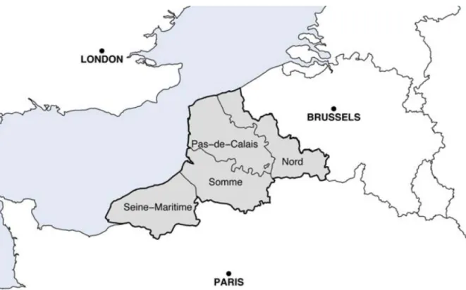
We included all patients with adult-onset IBD recorded in the EPIMAD registry between January 1988 and December 2014. The EPIMAD registry covers northern France (comprising 5,841,156 inhabitants, according to the 2014 national census performed by the French National Institute for Statistics and Economic Studies (Institut national de la statistique et des études économiques, INSEE)). The study area is divided into four administrative areas: Nord (2,603,472 inhabitants), Pas-de-Calais (1,472,589 inhabitants), Somme (571,632 inhabitants) and Seine-Maritime (1,257,920 inhabitants) (Figure 1).
Figure 1: The area covered by the EPIMAD registry.
The EPIMAD registry’s methodology and procedures have been described in detail elsewhere (11–14). Briefly, eight interviewer practitioners (IPs) collected data on
12 all incident IBD patients diagnosed by gastroenterologists (n=265), regardless of the latter’s type of practice (private and/or public). Inclusion in the study was limited to patients who were resident in the area at the time of diagnosis. Each gastroenterologist reported on the patients at the time of the first consultation with clinical symptoms suggestive of a diagnosis of IBD. The IP subsequently contacted gastroenterologist by phone at least three times each year. For each new case, the IP visited the gastroenterologist’s office, and collected medical chart data on a standardized questionnaire. The main items of data recorded were age, sex, year of diagnosis, the time interval between symptom onset and diagnosis, the clinical presentation (including extra-intestinal manifestations [EIMs], defined as joint, skin, eye and hepatobiliary manifestations), and the radiological, endoscopic and histological findings at the time of diagnosis. Only investigations completed within six weeks of diagnosis were taken into account. On this basis, a diagnosis of IBD was established by two expert gastroenterologists and recorded as definite, probable or possible, according to published criteria (11). The present study only considered patients with definite or probable CD, UC or IBDU diagnosed between 1988 and 2014, and who were aged 17 or over at the time of diagnosis. Data on the CD phenotype were recorded from 2006 onwards only.
II. Additional data collected for the present study
A. Age groupsPatients were divided into three age groups: 17-39, 40-59, and 60 and over.
B. Definitions of disease locations and behaviours 1. Crohn’s disease
The sites affected by CD were defined according to the Montreal Classification (15) as ileal involvement (L1); colonic involvement (L2), ileocolonic involvement (L3),
13 and upper gastrointestinal disease (L4, which could be combined with L1, L2 or L3). Patients with ileocaecal involvement were classified as L3. The presence or absence of perianal lesions (‘p’, including abscesses and/or fistulae) was noted. The type of CD behaviour was classified as B1 (inflammatory / nonstricturing nonpenetrating), B2 (stricturing) or B3 (penetrating). The ‘p’ index was added to B1, B2, or B3 when concomitant perianal disease was present. To adequately assess changes over time in the CD behaviour, only patients having undergone a full assessment of the ileum and colon were included in these analyses.
2. Ulcerative colitis
The UC site was defined according to the Montreal Classification (15), as follows: proctitis was defined as involvement limited to the rectum (E1); left-sided colitis was defined as involvement limited to the colorectum below the splenic flexure (E2); and extensive colitis was defined as involvement of the colorectum above the splenic flexure (E3).
III. Statistical analysis
Incidence rates were computed as the number of incident cases (i.e. new diagnoses) divided by the population at risk, in nine equal 3-year periods (1988-1990, 1991-1993, 1994-1996, 1997-1999, 2000-2002, 2003-2005, 2006-2008, 2009-2011, and 2012-2014). The mean annual incidence rates were calculated for the entire study period and for each 3-year period. Incidence rates were standardized by age (European population, with weightings provided by Eurostat, 2013) and were determined for the whole population and for each age group (17-39, 40-59, and 60 and over). Standardized incidence rates were presented with their 95% confidence interval (CI), based on a gamma distribution. For each of the four administrative areas, yearly
14 population data by age group were obtained from the INSEE. The data were based on a procedure that mixes the exhaustive census prior to 2004 and random sampling after 2004. Continuous variables were expressed as the median (interquartile range [IQR]), and categorical variables were expressed as the frequency (percentage). Intergroup comparisons of continuous variables were performed using the Mann-Whitney test. Intergroup comparisons of qualitative variables were performed using a chi-squared tests or (depending on the number of expected events) Fisher’s exact test.
Time trends in incidence were tested using a log-linear Poisson regression analysis that took account of the number of person-years at risk (introduced as an offset variable) and over-dispersion, when necessary. The trends were presented as the average percentage change (APC) per year calculated from the Poisson regression estimates and using the equation exp(β)-1, where β is the estimated time trend. The statistical analysis was performed using SAS software (version 9.4, SAS Institute Inc., Cary, NC, USA). The threshold for statistical significance was set to p ≤ 0.05.
15
RESULTS
I. Incidence of disease
For the period between January 1st, 1988, and December 31st, 2014, we identified
19,389 incident cases of IBD, including 11,439 cases of CD (59%), 7269 cases of UC (37%), and 681 cases of IBDU) (4%). There were 17,686 cases (91%) in adults (patients aged 17 and over at diagnosis, according to the EPIMAD registry), with 10,206 cases of CD (58%), 6,839 cases of UC (38%), and 641 cases of IBDU (4%). Adult-onset cases of CD and UC accounted respectively for 88% and 94% of all patients with CD and UC.
During the study period (1988-2014), the mean [95%CI] annual incidence of adult-onset IBD in the general population was 13.5 per 105 [13.3-13.7], and the
incidences of CD, UC, and IBDU were respectively 7.5 per 105 [7.4-7.7], 5.4 per 105
[5.3-5.6], and 0.51 per 105 [0.47-0.55].
II. Clinical presentation at diagnosis
A. Adult-onset IBDThere were significantly more women in the CD group (female/male ratio: 1.36) than in the UC group (0.84; p<0.0001). The median [IQR] age at diagnosis was 28 [22-40] for CD and 36 [27-49] for UC. A total of 1,626 patients (9%) had a family history of IBD in a first-degree relative. The median [IQR] time interval between symptom onset and diagnosis was 3 months [1-9] in the CD group and 2 months [1-6] in the UC group. Extra-intestinal manifestations (EIMs) were present in 7% (n=1,289) of the patients.
During the 1988-2014 period, 8,034 (79%) patients with CD had a complete bowel investigation (i.e. the ileum and the colon): 4,732 patients (59%) had ileocolonic (L3) involvement, 1,656 (21%) had colonic (L2) disease, and 1,646 (20%) had ileal
16 (L1) involvement. Near of a quarter of the patients (n=2,275, 22%) displayed upper gastrointestinal track involvement (L4). We noted that 477 (5%) of the patients had perianal lesions at diagnosis. With regard to the disease behaviour, 72% (n=2,122) of the patients had an inflammatory phenotype, 19% (n=560) had a structuring phenotype, and 9% (n=264) had a penetrating phenotype.
Over the same study period, proctitis (E1) was observed in 2,566 patients with UC (38%), left sided colitis (E2) was observed in 2,522 (37%), and extensive colitis (E3) was observed in 1,670 (25%).
B. Sex differences
The IBD patients’ sociodemographic and clinical data are summarized by sex in Table 1.
In the CD group, the mean [95%CI] incidence rate was significantly higher in women (7.5 [7.3–7.7]) than in men (6.0 [5.8–6.2]) (p<0.0001). The median [IQR] age at diagnosis was lower for women than for men (28 years [21-39] vs. 29 [22-40], respectively; p<0.0001). Women and men did not differ significantly with regard to the disease behaviour and locations, with the exception of L4 involvement (in 24% of men and 21% of women, p<0.0001) and the presence of perianal lesions (6% of men vs. 4% of women; p<0.0001). Extra-intestinal manifestations were more frequent in women than in men (11% vs. 9%, respectively; p<0.0001).
In the UC group, the mean incidence rate was significantly lower in women (4.1; 4.0-4.3) than in men (5.3; 5.1-5.5) (p<0.0001) and the median age at diagnosis was significantly lower for women than for men (33 [26-45] vs. 39 [28-51] respectively; p<0.0001).
17 A family history of IBD was more frequent among women than among men in both the CD group (12% vs. 11%, respectively; p=0.015) and the UC group (7% vs. 5%, respectively; p<0.0001).
Table 1. Sociodemographic and clinical data for patients with IBD, by sex. Variables at diagnosis Male
(n=8,364) (n=9,322) Female P-value Type of IBD CD n (%) 4,319 (51.6%) 5,887 (63.1%) <0.0001 UC n (%) 3,718 (44.5%) 3,121 (33.5%) IBDU n (%) 327 (3.9%) 314 (3.4%) CD
Median [IQR] age, years 29 [22-42] 28 [21-39] <0.0001
IBD family history n (%) 467 (10.8%) 729 (12.4%) 0.015
Median (IQR) time between symptom onset and diagnosis, months
3 [1; 9] 3 [1; 9] 0.001 Location* µ L1, n (%) 725 (21.6%) 921 (19.7%) 0.059 L2, n (%) 660 (19.6%) 996 (21.3%) L3, n (%) 1,977 (47.3%) 2,755 (48.1%) L4, n (%) 1,046 (24.2%) 1,229 (20.9%) <0.0001 Behaviour* £ B1, n (%) 870 (70.1%) 1,252 (73.4%) 0.090 B2, n (%) 246 (19.8%) 314 (18.4%) B3, n (%) 125 (10.1%) 139 (8.1%) Perianal disease, n (%) 247 (5.7%) 230 (3.9%) <0.0001 Extra-intestinal manifestations, n (%) 383 (8.9%) 674 (11.4%) <0.0001 UC
Median [IQR] age, years 39 [28-51] 33 [26-45] <0.0001
IBD family history (%) 179 (4.8%) 221 (7.1%) <0.0001
Median [IQR] time between symptom onset and diagnosis, months
2 [1; 6] 2 [1; 6] 0.039 Location* E1, n (%) 1,268 (34.5%) 1,298 (42.1%) <0.0001 E2, n (%) 1,436 (39.0%) 1,086 (35.3%) E3, n (%) 974 (26.5%) 696 (22.6%) Extra-intestinal manifestations, n (%) 96 (2.6%) 101 (3.1%) 0.107
* According to the Montreal classification (15)
µ Only recorded in patients with a full assessment (ileum and colon)
18 C. Age group differences
The IBD patients’ sociodemographic and clinical data are summarized by age group in Table 2.
In the CD group, the mean [95%CI] incidence rate was significantly higher in the 17-39 age group (14.6; [14.2-14.9]) than in the 40-59 age group (5.1; [4.8–5.3]) and the 60 and over age group (2.3; [2.2-2.5]; p<0.0001). Likewise, in the UC group, the mean incidence rate was significantly higher in the 17-39 age group (8.0; [7.7-8.2]) than in the 40-59 age group (5.2; [4.9–5.4]) and the 60 and over age group (2.9; [2.7-3.1]; p<0.0001).
In the CD group, ileal involvement (L1 + L3) was more common in the 17-39 age group (73%) than in the 60 and over age group (38%; p<0.0001). In contrast, isolated colonic involvement (L2) at diagnosis was more frequent in the 60 and over age group (62%) than in the 17-39 age group (27%; p<0.0001).
In the UC group, proctitis (E1) was the most frequent disease location (in 41% of patients) in the 17-39 group, while left-sided colitis (E2) was the most frequent in the 60 and over age group (54%).
19 Table 2. Sociodemographic and clinical data for patients with IBD, by age group.
Variables at diagnosis 17-39 40-59 60 and over+ P-value Type of IBD CD n (%) 7,557 (63.5%) 1,965 (47.5%) 684 (41.5%) <0.0001 UC n (%) 3,984 (33.5%) 1,999 (48.4%) 856 (52.0%) IBDU n ‘%) 364 (3.0%) 170 (4.1%) 107 (6.5%) CD
Family history of IBD, n (%) 986 (13.0%) 174 (8.8%) 36 (5.3%) <0.0001
Women, n (%) 4,452 (58.9%) 1,022 (52.0%) 413 (60.4%) <0.0001
Median [IQR] time between symptom onset and diagnosis, months
3 [1; 9] 3 [1; 9] 3 [1; 7] 0.033 Location* µ L1, n (%) 1,231 (19.8%) 355 (23.9%) 60 (17.0%) <0.0001 L2, n (%) 1,101 (17.8%) 420 (28.3%) 135 (38.2%) L3, n (%) 3,865 (62.4%) 709 (47.8%) 158 (44.8%) L4, n (%) 1,881 (24.9%) 306 (15.6%) 88 (12.9%) <0.0001 Behaviour* £ B1, n (%) 1,615 (73.0%) 382 (68.8%) 125 (70.2%) 0.190 B2, n (%) 400 (18.1%) 125 (22.6%) 35 (19.7%) B3, n (%) 198 (8.9%) 48 (8.6%) 18 (10.1%) Perianal disease n (%) 337 (4.5%) 99 (5.0%) 41 (6.0%) 0,133 Extra-intestinal manifestations, n (%) 800 (10.6%) 213 (10.8%) 44 (6.4%) 0.002 UC
Family history of IBD , n (%) 285 (7.1%) 87 (4.3%) 28 (3.3%) <0.0001
Women, n (%) 2,052 (51.5%) 723 (36.2%) 346 (40.4%) <0.0001
Median [IQR] time between symptom onset and diagnosis, months
2 [1; 6] 2 [1; 6] 2 [1; 5] 0.369 Location* E1 n (%) 1,623 (41.2%) 770 (39.1%) 173 (20.4%) <0.0001 E2 n (%) 1,293 (32.8%) 769 (39.1%) 460 (54.2%) E3 n (%) 1,025 (26.0%) 430 (21.8%) 215 (25.4%) Extra-intestinal manifestations n (%) 118 (3.0%) 54 (2.7%) 25 (2.9%) 0.848
* According to the Montreal classification (15)
µ Only recorded in patients with a full assessment (ileum and colon)
20
III. Changes over time in CD and UC
A. Incidence
1. Inflammatory bowel disease in general
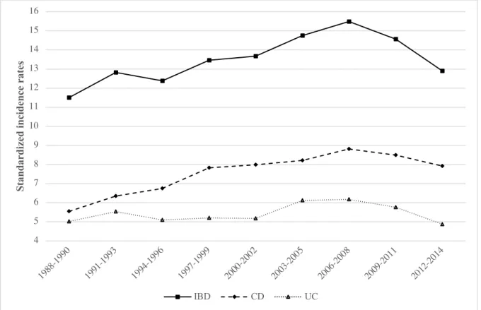
During the study period, the overall incidence of IBD increased from 11.5 per 105 in 1988-1990 to 12.9 per 105 in 2012-2014 (p=0.053). The incidence of CD
increased from 5.5 in 1988–1990 to 7.9 in 2012–2014 (APC: +1%; [0.5; 1.4]; p<0.0001), whereas the incidence of UC remain relatively stable; it was 5.0 in 1988– 1990 and 4.9 in 2012–2014 (APC +0.1%; [-0.4; 0.6]; p=0.67) (Figure 2).
Figure 2. Changes in standardized incidence rates for IBD, CD and UC over a 27-year period.
Each data point corresponds to the mean value for a 3-year period. 4 5 6 7 8 9 10 11 12 13 14 15 16 198 8-1990 199 1-1993 199 4-1996 199 7-1999 200 0-2002 200 3-2005 200 6-2008 200 9-2011 201 2-2014 St andar di ze d inc ide nc e rat es IBD CD UC
21 2. Sex and age group differences
The yearly APCs in the incidence of IBD by age group and by sex are shown in Table 3.
Overall, IBD increased in women (with an APC of +0.8% [0.3; 1.2]; p<0.001) but was stable in men (0.0% [-0.5; 0.4]; p=0.82).
Among patients with CD, the incidence rate increased by 1.9% a year in the 17-39 group for both men and women (p<0.0001). In the 40-59 group, the incidence rate increased by 1.1% a year in women only (p<0.0001). In the 60 and over group, the incidence rate was stable over time (p=0.423) (Figure 3). Among patients with CD, the sex ratio was stable over time (p=0.28).
22
Figure 3. Changes in the standardized incidence rates for CD over a 27-year period, by sex
and by age group. Each data point corresponds to the mean value for a 3-year period. 0 5 10 15 198 8-1990 199 1-1993 199 4-1996 199 7-1999 200 0-2002 200 3-2005 200 6-2008 200 9-2011 201 2-2014 St andar di ze d inc ide nc e rat es
Men
17-39 years 40-59 years > 60 years
0 5 10 15 20 25 198 8-1990 199 1-1993 199 4-1996 199 7-1999 200 0-2002 200 3-2005 200 6-2008 200 9-2011 201 2-2014 St andar di ze d inc ide nc e rat es
Women
23 Among the patients with UC, the incidence rates increased significantly in the 17-39 and 40-59 groups in women only (by 1.7% and 1.1% a year, respectively; p<0.0001 and p=0.024, respectively). The overall female/male ratio changed over time, with an inversion from 0.70 in 1988-1990 to 1.10 in 2012-2014 (p=0.001). We observed a significant decrease in the incidence of UC in the 60 and over age group (APC: -2% [-3.3; -0.8]; p=0.002) (Figure 4).
24
Figure 4. Changes in standardized incidence rates of UC by sex and by age group over a
27-year period. Each data point corresponds to the mean value for a 3-27-year period. 0 2 4 6 8 10 198 8-1990 199 1-1993 199 4-1996 199 7-1999 200 0-2002 200 3-2005 200 6-2008 200 9-2011 201 2-2014 St andar di ze d inc ide nc e rat es
Men
17-39 years 40-59 years >60 years
0 2 4 6 8 10 12 198 8-1990 199 1-1993 199 4-1996 199 7-1999 200 0-2002 200 3-2005 200 6-2008 200 9-2011 201 2-2014 St andar di ze d inc ide nc e rat es
Women
25 B. Disease locations
Between 1988 and 2014, the proportion of patients with CD having undergone a complete bowel assessment increased from 68% to 83% (p<0.0001). The proportion of UC patients having undergone an assessment of the colon was stable (approximately 80%) over the study period.
In the CD group, the proportion of patients with L1 lesions increased from 19% to 26% (p<0.0001), the proportion with L3 lesions rose from 50% to 58% (p<0.0001), and the proportion with L2 lesions decreased from 31% to 16% (p<0.0001) (Figure 5). The proportion of patients with upper digestive tract lesions (L4) increased from 12% to 25% (p<0.0001). The incidence of perianal lesions decreased from 6% to 3% (p=0.0024). The proportions of disease behaviours were stable over time.
In the UC group, the proportion of patients with E1 lesions decreased from 42% to 34%, whereas the proportion of patients with E3 lesions increased from 19% to 28% (p<0.0001) (Figure 5). The incidence of EIMs decreased significantly in the CD group (from 14% to 9%; p<0.0001) but not in the UC group (from 4% to 3%; p=0.084). There were no changes over time with regard to the sex ratio, the time interval between symptom onset and diagnosis, or age at diagnosis in either the CD group or the UC group.
26
Figure 5. Changes in disease locations in patients with CD and in patients with UC over a
27-year period. Each data point corresponds to the mean value for a 3-27-year period. 0 10 20 30 40 50 60 70 198 8-1990 199 1-1993 199 4-1996 199 7-1999 200 0-2002 200 3-2005 200 6-2008 200 9-2011 201 2-2014 Di se as e lo ca ti on ( % )
Crohn's disease
L1 L2 L3 0 10 20 30 40 50 198 8-1990 199 1-1993 199 4-1996 199 7-1999 200 0-2002 200 3-2005 200 6-2008 200 9-2011 201 2-2014 Di se as e lo ca ti on ( % )Ulcerative colitis
E1 E2 E327 Table 3. The yearly APC in the incidence of IBD, by age group and by sex (%).
IBD CD UC
APC p-value APC p-value APC p-value
All ages All 0.4 [0; 0.8] 0.053 1.0 [0.5; 1.4] <0.0001 0.1 [-0.4; 0.6] 0.67 Females 0.8 [0.3; 1.2] <0.001 1.0 [0.4; 1.6] <0.001 0.9 [0.3; 1.4] 0.002 Males 0.0 [-0.5; 0.4] 0.82 0.9 [0.3; 1.4] 0.001 -0.5 [-1.1; 0.0] 0.071 17-39 All 1.4 [1.1; 1.7] <0.0001 1.9 [1.5; 2.3] <0.0001 1.0 [0.5; 1.4] <0.0001 Females 1.8 [1.4; 2.1] <0.0001 2.0 [1.5; 2.4] <0.0001 1.7 [1.1; 2.3] <0.0001 Males 1.0 [0.5; 1.4] <0.0001 1.8 [1.2; 2.4] <0.0001 0.2 [-0.5; 0.8] 0.59 40-59 All 0.1 [-0.4; 0.6] 0.733 0.8 [0.1; 2.6] 0.028 0.0 [-0.6; 0.7] 0.95 Females 0.9 [0.2; 1.6] <0.001 1.1 [0.2; 2.1] 0.019 1.1 [0.1; 2.2] 0.024 Males -0.5 [-1.2; 0.1] 0.118 0.5 [0.0; 1.5] 0.339 -0.6 [-1.4; 0.2] 0.168 >60 All -1.4 [-2.2; -0.5] 0.001 -0.5 [-1.7; 0.7] 0.423 -1.4 [-2.4; -0.4] 0.007 Females -1.2 [-2.2; -0.2] 0.018 -1.0 [-2.5; 0.4] 0.152 -0.6 [-1.9; 0.8] 0.411 Males -1.6 [-2.6; -0.5] 0.005 0. 3 [-1.5; 2.2] 0.709 -2.0 [-3.3; -0.8] 0.002
28
DISCUSSION
Over a 27-year period (1988-2014), this study of a large population-based registry included 17,686 incident cases of IBD in patients aged 17 and over. The mean [95%CI] annual incidence was 13.5 per 105 [13.3-13.7] for all types of IBD, 7.5 per 105
[7.4-7.7] for CD and 5.4 per 105 [5.3-5.6] for UC. In the literature, the annual incidence
rates in Europe range from 0.3 to 12.7 per 105 for CD, and from 0.6 to 24.3 per 105 for
UC (5,6). In the present study, the CD/UC ratio was greater than 1. This finding contrast with most of the other population-based studies performed in Western Europe, where UC was more frequent than CD (16–18). A similar ratio was found in and around the city of Liège in Belgium (19), which is close to northern France - suggesting that one or several risk factors for CD might be present in this geographic area. Since the 1990s, epidemiological studies of Western countries have shown that incidence rates have changed: 73% of the studies of CD and 83% of the studies of UC reported that the incidence was stable or was falling (7). In a recent Canadian study, the incidence of both CD and UC decreased significantly between 1990 and 2012 (20). This result contrasts with our present observation of an increase in the incidence of IBD in general and CD in particular in northern France. Although the incidence of IBD seems to have stabilized in most Westernized countries, the overall estimated prevalence of IBD (~0.5% of the general population) is still increasing because of a low mortality rate (5). A predictive model developed in Canada was used to estimate that the prevalence of IBD in that country was 0.6% in 2015, and predicted a rise to 0.9% by 2025 (7). In a Scottish study based on a capture-recapture design, it was suggested that the prevalence of IBD in Scotland would rise to 1% within 10 years (21).
29 Our study’s major finding was that the incidences of adult-onset CD and UC are still increasing among the young and middle-aged women. Indeed, we found an APC of +2% among women aged 17-39 and +1.1% among women aged 40-59. The corresponding APCs for UC were +1.7% and +1.1%. In contrast, the incidence of CD increased only in the 17-39 aged group in men (APC: +1.8%). To the best of our knowledge, the present population-based study is the first to have highlighted this type of finding. Shah et al. (22) recently described sex differences in IBD incidence as a function of the age at diagnosis. The risk of CD was lower in women than in males up until the 10-14 age range; thereafter, the sex ratio inverted, with a significantly higher risk among women in the 25-29 group and the 35 and over group. With regard to UC, female predominance was observed in the 5-9 group. The incidences in men and women were similar up until the age of 45, after which male predominance was observed. These findings suggest that female hormones have role; we observed a shift in the sex ratio around puberty in CD and around the menopause in UC. The results of an animal study suggested that oestrogens may modulate epithelial barrier function via the oestrogen receptor-β (23) - a mechanism thought to be significantly involved in CD (24). Similarly, Khalili et al.’s analysis of the Nurses Health Study cohort (25) suggested that current use of oral contraceptives was associated with a three-fold elevation in the risk of CD. For UC, this risk was limited to women with a history of active smoking. In a meta-analysis published in 2008, the use of oral contraceptives was significantly associated with an elevated risk of CD (hazard ratio (95%CI) 1.46; 1.26–1.70; p<0.001) and UC (1.28 (1.06–1.54); p=0.011) after adjustment for smoking (26). More recently, endocrine disruptors have been linked to a large number of chronic diseases (such as asthma (27) and type II diabetes (28)) and sexual dysfunction (29,30). Like ethinyl estradiol, bisphenol A (an oestrogenic compound widely used in
30 the food-packaging industry) can alter the gut’s flora and has systemic effects (31); however, the results of animal studies are contradictory. A study in rats demonstrated that reference doses of bisphenol A influenced the intestine’s barrier function and gut nociception, and that perinatal exposure promoted the development of severe inflammation in adult offspring - but only in women (32). However, a study in a mouse model of inflammatory colitis found that neither developmental exposure nor direct exposure to bisphenol A had a significant effect on the course of colonic disease (33). Even though these results were contradictory, exposure to pollutants is still a solid lead that is underpinned by the geospatial heterogeneity in the incidence of IBD.
Exposure to medications is another hypothesis for the pathogenesis of IBD. Antibiotic treatment has been shown to alter the gut flora, and exposure to antibiotics is a known risk factor for developing IBD (34,35). Antibiotic use in France is amongst the highest in Europe, and use in northern France is above the French national average. Moreover, women accounted for 58% of antibiotic consumers in France in 2015 (36). It is well known that the chronic inflammatory skin disease acne vulgaris affects between 20% and 40% of adolescents, and is more frequent among females (37). Since the 2000s, tetracycline antibiotics have been frequently prescribed for the treatment of moderate acne. One can hypothesize that by modifying the microbiota, tetracyclines are involved in the increase in the incidence of IBD in general and CD in particular (38,39). The results for isotretinoid (a drug used to treat severe acne) are not consistent: an association with UC was found in one case-control study (40) but not in another (41).
There are other possible explanation for the dramatic increase in the incidence of IBD in young women. The association between smoking and IBD is well known, with opposite effects on CD and UC. In Mahid et al.’s (42) meta-analysis, 12 out of 13 studies found that current smoking protected against UC. In contrast, former smoker
31 status was associated with an increased risk of UC - mostly in the first five years (42) after smoking cessation. Data from the INSEE evidence a rise in regular tobacco consumption among women since the early 1980s (43). This rise could explain the increased incidence in CD but not that in UC. Women quit smoking earlier in life than men because of pregnancy; this might play a role in the increased incidence of UC in young women. In the elderly population, UC is more prominent than CD. Interesting, the influence of smoking cessation in the elderly has been mentioned as an explanation for this disease predominance (44). In the present study, the incidence of UC decreased over time in both men and women aged ≥60 - suggesting that changed in tobacco consumption in this particular population are also present. Unfortunately, data on active smoking is missing for ~30% of cases in the EPIMAD registry; this prevented us from being taken into account as an explanatory variable.
Appendectomy for inflammatory conditions (appendicitis or lymphadenitis) is reportedly associated with a low risk of UC (45). A meta-analysis of 13 case-control studies showed that appendectomy reduced the risk of developing UC by 69% (46). The link between appendectomy and CD is less clear. A meta-analysis published in 2008 showed that the risk of developing CD after an appendectomy was significantly higher than expected by chance (risk ratio [95%CI] = 1.61; [1.28-2.02]). However, after 5 years, the risk of developing CD no longer depend on whether or not the subject had undergone appendectomy (47). The results of a Swedish study performed in 2003 suggested that non-perforated appendicitis increased the risk of developing CD in women but not in men (48). Nevertheless, the same research group suggested that a false-positive diagnosis of non-perforated appendicitis is probably more frequent among women because of the high incidence of surgery for non-inflamed appendices in women (24). This would increase the frequency of missed patients with CD, which
32 could explain the sex difference in the association with non-perforated appendicitis (49). In 1997 in France, appendectomy was more frequent among women than among men (54% vs. 46%, respectively; M/F gender ratio = 0.85). However, the incidence of appendectomy has declined sharply over the two last decades - especially in women, with an inversion of the M/F sex ratio (1.05 in 2012) (50). These factors might have prompted an increased risk of developing UC in women.
Dietary composition has also been studied - but mostly in retrospective case-control studies - as a possible risk factor for IBD (51,52). In the prospective E3N cohort of women (the French counterpart of the EPIC study), high protein intake (only meat and fish) was significantly associated with an increased risk of IBD (hazard ratio [95%CI]: 3.31 [1.41–7.77]) among women aged 40 to 65 (53).
Another possible explanation for the increase in the incidence of IDB in both sexes is related indirectly to medical care. The development of new diagnostic tools might have led to the earlier, more widespread diagnosis of IBD. If this were true, however, we would have notice a transient peak in the incidence, a decrease, and then stabilization at a lower level. In fact, our present results evidenced a continuous increase in incidence. Moreover, the time between symptom onset and IBD diagnosis did not change over a period of almost three decades – suggesting strongly that diagnostic capacities remained more or less stable over the study period.
Another major finding of our study concerned changes over time in the IBD disease location. Between 1988 and 2014, the incidence of CD with ileal involvement (L1 and L3) rose significantly from 69% to 84%, whereas the incidence of CD with isolated colonic location (L2) decreased from 31% to 16%. Given that we only considered patients having undergone a full bowel assessment at diagnosis, these
33 variations in disease location over a 27-year period are likely to be real and not related to changes in diagnostic methods. In CD, ileal involvement was more frequent in the youngest patients. It is well known that CD location is age-related. Indeed, ileal CD is linked to the involvement of Payer’s patches (PP) located in lymphoid follicles that have a major role in intestinal immunity (54). In an autopsy study, PPs were mostly found in the terminal ileum, where they formed a lymphoid ring (55). The PPs increase in number and in size throughout childhood but then start to involute after puberty: this might explain the low frequency of ileal involvement in children and elderly (56). In UC, the proportion of patients with ulcerative proctitis (E1) accounted for about one third of the incident cases (38%). This proportion is considered to be a good marker of case ascertainment in the literature, since (i) E1 corresponds to the onset of UC (57), and (ii) a low frequency of E1 disease in a prospective, population-based study of UC incidence suggests that recording was not exhaustive (58).
The present study’s major strengths were its population-based design, the large sample size (17,686 incident cases of IBD in patients aged 17 or over), exhaustive recording over a 27-year period, and the use of validated, published diagnostic criteria (11–14). All the charts were analyzed by two gastroenterologists, using a diagnostic algorithm. The study’s major limitation was related to the EPIMAD registry’s methodology, in which radiological, endoscopic and histological investigations of the incident case have to be completed within six weeks of diagnosis. This probably leads to underestimation of the extent of the disease at the time of diagnosis. However, we sought to limit this limitation by reporting on disease locations only in patients having undergone a full assessment of the ileum and colon. A second limitation relates to the absence of data on smoking status or oral contraceptive use.
34 In conclusion, the results of a large, population-based study over a 27-year period showed that the incidence of CD and UC are still rising dramatically among young women in northern France. This finding suggests that one or more major environmental factors may predispose the women in this area to IBD. Over the 27-year study period, we observed an increase in ileal involvement and a concomitant decrease in colonic involvement, whereas the age at diagnosis and the time interval between symptom onset and diagnosis did not change significantly. We hypothesize that the environmental factors associated with CD in our region may be particularly active in the ileum of young women. Specific epidemiological and fundamental studies would be required to assess this hypothesis.
35
REFERENCES
1. Schofield PF. The natural history and treatment of Crohn's disease. Ann R Coll Surg Engl. 1965 May;36:258–79.
2. Cullinan ER, Macdougall IP. The natural history of ulcerative colitis. Lancet Lond Engl. 1957 Mar 9;272(6967):487–9.
3. Burisch J, Kiudelis G, Kupcinskas L, Kievit HAL, Andersen KW, Andersen V, et al. Natural disease course of Crohn’s disease during the first 5 years after diagnosis in a
European population-based inception cohort: an Epi-IBD study. Gut. 2019 Mar;68(3):423–33. 4. Peyrin-Biroulet L, Loftus EV, Colombel J-F, Sandborn WJ. The Natural History of Adult Crohnʼs Disease in Population-Based Cohorts: Am J Gastroenterol. 2010
Feb;105(2):289–97.
5. Molodecky NA, Soon IS, Rabi DM, Ghali WA, Ferris M, Chernoff G, et al. Increasing Incidence and Prevalence of the Inflammatory Bowel Diseases With Time, Based on
Systematic Review. Gastroenterology. 2012 Jan;142(1):46-54.e42.
6. Cosnes J, Gower–Rousseau C, Seksik P, Cortot A. Epidemiology and Natural History of Inflammatory Bowel Diseases. Gastroenterology. 2011 May;140(6):1785-1794.e4.
7. Ng SC, Shi HY, Hamidi N, Underwood FE, Tang W, Benchimol EI, et al. Worldwide incidence and prevalence of inflammatory bowel disease in the 21st century: a systematic review of population-based studies. The Lancet. 2017 Dec;390(10114):2769–78.
8. Frolkis A, Dieleman LA, Barkema HW, Panaccione R, Ghosh S, Fedorak RN, et al. Environment and the Inflammatory Bowel Diseases. Can J Gastroenterol. 2013;27(3):e18–24. 9. Piovani D, Danese S, Peyrin-Biroulet L, Nikolopoulos GK, Lytras T, Bonovas S. Environmental Risk Factors for Inflammatory Bowel Diseases: An Umbrella Review of Meta-analyses. Gastroenterology. 2019;157(3):647-659.e4.
10. Ghione S, Sarter H, Fumery M, Armengol-Debeir L, Savoye G, Ley D, et al. Dramatic Increase in Incidence of Ulcerative Colitis and Crohnʼs Disease (1988–2011): A Population-Based Study of French Adolescents: Am J Gastroenterol. 2018 Feb;113(2):265–72.
11. Gower-Rousseau C, Salomez JL, Dupas JL, Marti R, Nuttens MC, Votte A, et al. Incidence of inflammatory bowel disease in northern France (1988-1990). Gut. 1994 Oct 1;35(10):1433–8.
12. Molinie F. Opposite evolution in incidence of Crohn’s disease and ulcerative colitis in Northern France (1988-1999). Gut. 2004 Jun 1;53(6):843–8.
13. Chouraki V, Savoye G, Dauchet L, Vernier-Massouille G, Dupas J-L, Merle V, et al. The changing pattern of Crohn’s disease incidence in northern France: a continuing increase in the 10- to 19-year-old age bracket (1988-2007): Epidemiology of inflammatory bowel disease. Aliment Pharmacol Ther. 2011 May;33(10):1133–42.
14. Gower-Rousseau C, Vasseur F, Fumery M, Savoye G, Salleron J, Dauchet L, et al. Epidemiology of inflammatory bowel diseases: New insights from a French population-based registry (EPIMAD). Dig Liver Dis. 2013 Feb;45(2):89–94.
15. Silverberg MS, Satsangi J, Ahmad T, Arnott ID, Bernstein CN, Brant SR, et al. Toward an Integrated Clinical, Molecular and Serological Classification of Inflammatory Bowel Disease: Report of a Working Party of the 2005 Montreal World Congress of Gastroenterology. Can J Gastroenterol. 2005;19(suppl a):5A-36A.
16. Vind I, Riis L, Jess T, Knudsen E, Pedersen N, Elkjær M, et al. Increasing Incidences of Inflammatory Bowel Disease and Decreasing Surgery Rates in Copenhagen City and County, 2003–2005: A Population-Based Study from the Danish Crohn Colitis Database. Am J Gastroenterol. 2006 Jun;101(6):1274–82.
36 Limonard CB, Kester ADM, et al. Inflammatory Bowel Disease in South Limburg (the
Netherlands) 1991–2002: Incidence, diagnostic delay, and seasonal variations in onset of symptoms. J Crohns Colitis. 2009 Jun;3(2):115–24.
18. Tragnone A, Corrao G, Miglio F, Caprilli R, Lanfranchi GA. Incidence of Inflammatory Bowel Disease in Italy: A Nationwide Population-Based Study. Int J Epidemiol. 1996;25(5):1044–52.
19. Latour P, Louis E, Belaiche J. Incidence of inflammatory bowel disease in the area of Liège: a 3 years prospective study (1993-1996). Acta Gastro-Enterol Belg. 1998
Dec;61(4):410–3.
20. Torabi M, Bernstein CN, Yu BN, Wickramasinghe L, Blanchard JF, Singh H. Geographical Variation and Factors Associated With Inflammatory Bowel Disease in a Central Canadian Province. Inflamm Bowel Dis. 2019 Aug 23;
21. Jones G-R, Lyons M, Plevris N, Jenkinson PW, Bisset C, Burgess C, et al. IBD prevalence in Lothian, Scotland, derived by capture-recapture methodology. Gut. 2019 Jul 11;10.1136/gutjnl-2019-318936.
22. Shah SC, Khalili H, Gower-Rousseau C, Olen O, Benchimol EI, Lynge E, et al. Sex-Based Differences in Incidence of Inflammatory Bowel Diseases-Pooled Analysis of Population-Based Studies From Western Countries. Gastroenterology. 2018;155(4):1079-1089.e3.
23. Looijer-van Langen M, Hotte N, Dieleman LA, Albert E, Mulder C, Madsen KL. Estrogen receptor-β signaling modulates epithelial barrier function. Am J Physiol Gastrointest Liver Physiol. 2011 Apr;300(4):G621-626.
24. Barreau F, Madre C, Meinzer U, Berrebi D, Dussaillant M, Merlin F, et al. Nod2 regulates the host response towards microflora by modulating T cell function and epithelial permeability in mouse Peyer’s patches. Gut. 2010 Feb;59(2):207–17.
25. Khalili H, Higuchi LM, Ananthakrishnan AN, Richter JM, Feskanich D, Fuchs CS, et al. Oral contraceptives, reproductive factors and risk of inflammatory bowel disease. Gut. 2013 Aug;62(8):1153–9.
26. Cornish JA, Tan E, Simillis C, Clark SK, Teare J, Tekkis PP. The Risk of Oral Contraceptives in the Etiology of Inflammatory Bowel Disease: A Meta-Analysis. Am J Gastroenterol. 2008 Sep;103(9):2394–400.
27. Spanier AJ, Kahn RS, Kunselman AR, Hornung R, Xu Y, Calafat AM, et al. Prenatal exposure to bisphenol A and child wheeze from birth to 3 years of age. Environ Health Perspect. 2012 Jun;120(6):916–20.
28. Silver MK, O’Neill MS, Sowers MR, Park SK. Urinary Bisphenol A and Type-2 Diabetes in U.S. Adults: Data from NHANES 2003-2008. PLOS ONE. 2011 Oct
26;6(10):e26868.
29. Takeuchi T, Tsutsumi O, Ikezuki Y, Takai Y, Taketani Y. Positive relationship between androgen and the endocrine disruptor, bisphenol A, in normal women and women with ovarian dysfunction. Endocr J. 2004;51(2):165–9.
30. Galloway Tamara, Cipelli Riccardo, Guralnik Jack, Ferrucci Luigi, Bandinelli Stefania, Corsi Anna Maria, et al. Daily Bisphenol A Excretion and Associations with Sex Hormone Concentrations: Results from the InCHIANTI Adult Population Study. Environ Health Perspect. 2010 Nov 1;118(11):1603–8.
31. Javurek AB, Spollen WG, Johnson SA, Bivens NJ, Bromert KH, Givan SA, et al. Effects of exposure to bisphenol A and ethinyl estradiol on the gut microbiota of parents and their offspring in a rodent model. Gut Microbes. 2016 Nov;7(6):471–85.
32. Braniste V, Jouault A, Gaultier E, Polizzi A, Buisson-Brenac C, Leveque M, et al. Impact of oral bisphenol A at reference doses on intestinal barrier function and sex
37 33. Roy A, Gaylo A, Cao W, Saubermann LJ, Lawrence BP. Neither direct nor
developmental exposure to bisphenol A alters the severity of experimental inflammatory colitis in mice. J Immunotoxicol. 2013 Dec;10(4):334–40.
34. Shaw SY, Blanchard JF, Bernstein CN. Association Between the Use of Antibiotics and New Diagnoses of Crohnʼs Disease and Ulcerative Colitis: Am J Gastroenterol. 2011 Dec;106(12):2133–42.
35. Aniwan S, Tremaine WJ, Raffals LE, Kane SV, Loftus EV. Antibiotic Use and New-Onset Inflammatory Bowel Disease in Olmsted County, Minnesota: A Population-Based Case-Control Study. J Crohns Colitis. 2018 Jan 24;12(2):137–44.
32. Evolution des consommations d’antibiotiques en France entre 2000 et 2015 - Point d’Information - ANSM 2017.
37. Collier CN, Harper JC, Cafardi JA, Cantrell WC, Wang W, Foster KW, et al. The prevalence of acne in adults 20 years and older. J Am Acad Dermatol. 2008 Jan;58(1):56–9. 38. Margolis D, Fanelli M, Hoffstad O, Lewis J. Potential Association Between the Oral Tetracycline Class of Antimicrobials Used to Treat Acne and Inflammatory Bowel Disease. Am J Gastroenterol. 2010 Dec;105(12):2610–6.
39. Li W-Q, Cho E, Khalili H, Wu S, Chan AT, Qureshi AA. Rosacea, Use of Tetracycline, and Risk of Incident Inflammatory Bowel Disease in Women. Clin
Gastroenterol Hepatol Off Clin Pract J Am Gastroenterol Assoc. 2016 Feb;14(2):220-225.e3. 40. Crockett SD, Porter CQ, Martin CF, Sandler RS, Kappelman MD. Isotretinoin Use and the Risk of Inflammatory Bowel Disease: A Case Control Study. Am J Gastroenterol. 2010 Sep;105(9):1986–93.
41. Bernstein CN, Nugent Z, Longobardi T, Blanchard JF. Isotretinoin is not associated with inflammatory bowel disease: a population-based case-control study. Am J Gastroenterol. 2009 Nov;104(11):2774–8.
42. Mahid SS, Minor KS, Soto RE, Hornung CA, Galandiuk S. Smoking and
Inflammatory Bowel Disease: A Meta-analysis. Mayo Clin Proc. 2006 Nov;81(11):1462–71. 22. Tabac : évolution de l’usage occasionnel ou régulier parmi les 18-75 ans - OFDT 2019.
44. Charpentier C, Salleron J, Savoye G, Fumery M, Merle V, Laberenne J-E, et al.
Natural history of elderly-onset inflammatory bowel disease: a population-based cohort study. Gut. 2014 Mar;63(3):423–32.
45. Andersson RE, Olaison G, Tysk C, Ekbom A. Appendectomy and protection against ulcerative colitis. N Engl J Med. 2001 Mar 15;344(11):808–14.
46. Koutroubakis IE. Appendectomy and the Development of Ulcerative Colitis: Results of a Metaanalysis of Published Case-Control Studies. 2000;95(1):6.
47. Kaplan GG, Jackson T, Sands BE, Frisch M, Andersson RE, Korzenik J. The risk of developing Crohn’s disease after an appendectomy: a meta-analysis. Am J Gastroenterol. 2008 Nov;103(11):2925–31.
48. Andersson RE, Olaison G, Tysk C, Ekbom A. Appendectomy is followed by increased risk of Crohn’s disease. Gastroenterology. 2003 Jan;124(1):40–6.
49. Andersson R, Hugander A, Thulin A, Nyström PO, Olaison G. Indications for operation in suspected appendicitis and incidence of perforation. BMJ. 1994 Jan 8;308(6921):107–10.
29. Philippe Oberlin, Marie-Claude Mouquet, 2014, « La longue diminution des appendicectomies en France », Études et résultats, n°868, Drees, février 2014.
51. Järnerot G, Järnmark I, Nilsson K. Consumption of refined sugar by patients with Crohn’s disease, ulcerative colitis, or irritable bowel syndrome. Scand J Gastroenterol. 1983 Nov;18(8):999–1002.
38 Modern life’ in the epidemiology of inflammatory bowel disease: a case-control study with special emphasis on nutritional factors. Eur J Gastroenterol Hepatol. 1998 Mar;10(3):243–9. 53. Jantchou P, Morois S, Clavel-Chapelon F, Boutron-Ruault M-C, Carbonnel F. Animal protein intake and risk of inflammatory bowel disease: The E3N prospective study. Am J Gastroenterol. 2010 Oct;105(10):2195–201.
54. Caprilli R. Why does Crohn’s disease usually occur in terminal ileum? J Crohns Colitis. 2008 Dec;2(4):352–6.
55. Van Kruiningen HJ, West AB, Freda BJ, Holmes KA. Distribution of Peyer’s Patches in the Distal Ileum: Inflamm Bowel Dis. 2002 May;8(3):180–5.
56. Meinzer U, Ideström M, Alberti C, Peuchmaur M, Belarbi N, Bellaïche M, et al. Ileal involvement is age dependent in pediatric Crohn’s disease. Inflamm Bowel Dis. 2005
Jul;11(7):639–44.
56. Calkins’ BM, Mendeloff AI. Epidemiology of inflammatory bowel disease. Inflamm Bowel Dis. :32.
58. Ritchie JK, Powell-Tuck J, Lennard-Jones JE. Clinical outcome of the first ten years of ulcerative colitis and proctitis. Lancet Lond Engl. 1978 May 27;1(8074):1140–3.
39
ANNEXE
Remerciements :
Les auteurs souhaitent remercier tous les enquêteurs qui ont participé au Registre EPIMAD depuis 1988: B Lemaire, M Lemahieu, N. Wauquier, N. Guillon, I. Rousseau, A. Pétillon, B. Turck, L. Damageux, H. Pennel, L. Yzet, P. Fosse, S. Auzou, M. Leconte, C. Le Gallo, D. Rime, B. David ainsi que tous les gastro-entérologues participant au Registre :
Andre JM, Antonietti M, Aouakli A, Armand A, Aroichane I, Assi F, Aubet JP, Auxenfants E, Ayafi-Ramelot F, Azzouzi K, Bankovski D, Barbry B, Bardoux N, Baron P, Baudet A, Bazin B, Bebahani A, Becqwort JP, Benet V, Benali H, Benguigui C, Ben Soussan E, Bental A, Berkelmans I, Bernet J, Bernou K, Bernou-Dron C, Bertot P, Bertiaux-Vandaële N, Bertrand V, Billoud E, Biron N, Bismuth B, Bleuet M, Blondel F, Blondin V, Bohon P, Boniface E, Bonnière P, Bonvarlet E, Bonvarlet P, Boruchowicz A, Bostvironnois R, Boualit M, Bouche B, Boudaillez C, Bourgeaux C, Bourgeois M, Bourguet A, Bourienne A, Branche J, Bray G, Brazier F, Breban P, Bridenne M, Brihier H, Brung-Lefebvre V, Bulois P, Burgiere P, Butel J, Canva JY, Canva-Delcambre V, Capron JP, Cardot F, Carpentier P, Cartier E, Cassar JF, Cassagnou M, Castex JF, Catala P, Cattan S, Catteau S, Caujolle B, Cayron G, Chandelier C, Chantre M, Charles J, Charneau T, Chavance-Thelu M, Chirita D, Choteau A, Claerbout JF, Clergue PY, Coevoet H, Cohen G, Collet R, Colombel JF, Coopman S, Corvisart J, Cortot A, Couttenier F, Crinquette JF, Crombe V, Dadamessi I, Dapvril V, Davion T, Dautreme S, Debas J, Degrave N, Dehont F, Delatre C, Delcenserie R, Delette O, Delgrange T, Delhoustal L, Delmotte JS, Demmane S, Deregnaucourt G, Descombes P, Desechalliers JP, Desmet P, Desreumaux P, Desseaux G, Desurmont P, Devienne A, Devouge E, Devred M, Devroux A, Dewailly A, Dharancy S, Di Fiore A, Djeddi D, Djedir R, Dreher-Duwat ML, Dubois R, Dubuque C, Ducatillon P, Duclay J, Ducrocq B, Ducrot F, Ducrotte P, Dufilho A, Duhamel C, Dujardin D, Dumant-Forest C, Dupas JL, Dupont F, Duranton Y, Duriez A, El Achkar K, El Farisi M, Elie C, Elie-Legrand MC, Elkhaki A, Eoche M, Evrard D, Evrard JP, Fatome A, Filoche B, Finet L, Flahaut M, Flamme C, Foissey D, Fournier P, Foutrein- Comes MC, Foutrein P, Fremond D, Frere T, Fumery M, Gallet P, Gamblin C, Ganga S, Gerard R, Geslin G, Gheyssens Y, Ghossini N, Ghrib S, Gilbert T, Gillet B, Godard D, Godard P, Godchaux JM, Godchaux R, Goegebeur G, Goria O, Gottrand F, Gower P, Grandmaison B, Groux M, Guedon C, Guillard JF, Guillem L, Guillemot F, Guimberd D, Haddouche B, Hakim S, Hanon D, Hautefeuille V, Heckestweiller P, Hecquet G, Hedde JP, Hellal H, Henneresse PE, Heyman B, Heraud M, Herve S, Hochain P, Houssin-Bailly L, Houcke P, Huguenin B, Iobagiu S, Ivanovic A, Iwanicki-Caron I, Janicki E, Jarry M, Jeu J, Joly JP, Jonas C, Katherin F, Kerleveo A, Khachfe A, Kiriakos A, Kiriakos J, Klein O, Kohut M, Kornhauser R, Koutsomanis D, Laberenne JE, Laffineur G, Lagarde M, Lalanne A, Lannoy P, Lapchin J, Laprand M, Laude D, Leblanc R, Lecieux P, Leclerc N, Le Couteulx C, Ledent J, Lefebvre J, Lefiliatre P, Legrand C, Le Grix A, Lelong P, Leluyer B, Lenaerts C, Lepileur L, Leplat A,
Lepoutre-40
Dujardin E, Leroi H, Leroy MY, Lesage JP, Lesage X, Lesage J, Lescanne-Darchis I, Lescut J, Lescut D, Leurent B, Levy P, Lhermie M, Lion A, Lisambert B, Loire F, Louf S, Louvet A, Luciani M, Lucidarme D, Lugand J, Macaigne O, Maetz D, Maillard D, Mancheron H, Manolache O, Marks-Brunel AB, Marti R, Martin F, Martin G, Marzloff E, Mathurin P, Mauillon J, Maunoury V, Maupas JL, Mesnard B, Metayer P, Methari L, Meurisse B, Meurisse F, Michaud L, Mirmaran X, Modaine P, Monthe A, Morel L, Mortier PE, Moulin E, Mouterde O, Mudry J, Nachury M, N’Guyen Khac E, Notteghem B, Ollevier V, Ostyn A, Ouraghi A, Ouvry D, Paillot B, Panien-Claudot N, Paoletti C, Papazian A, Parent B, Pariente B, Paris JC, Patrier P, Paupart L, Pauwels B, Pauwels M, Petit R, Piat M, Piotte S, Plane C, Plouvier B, Pollet E, Pommelet P, Pop D, Pordes C, Pouchain G, Prades P, Prevost A, Prevost JC, Quesnel B, Queuniet AM, Quinton JF, Rabache A, Rabelle P, Raclot G, Ratajczyk S, Rault D, Razemon V, Reix N, Revillon M, Richez C, Robinson P, Rodriguez J, Roger J, Roux JM, Rudelli A, Saber A, Savoye G, Schlosseberg P, Segrestin M, Seguy D, Serin M, Seryer A, Sevenet F, Shekh N, Silvie J, Simon V, Spyckerelle C, Talbodec N, Techy A, Thelu JL, Thevenin A, Thiebault H, Thomas J, Thorel JM, Tielman G, Tode M, Toisin J, Tonnel J, Touchais JY, Touze Y, Tranvouez JL, Triplet C, Turck D, Uhlen S, Vaillant E, Valmage C, Vanco D, Vandamme H, Vanderbecq E, Vander Eecken E, Vandermolen P, Vandevenne P, Vandeville L, Vandewalle A, Vandewalle C, Vaneslander P, Vanhoove JP, Vanrenterghem A, Varlet P, Vasies I, Verbiese G, Vernier-Massouille G, Vermelle P, Verne C, Vezilier-Cocq P, Vigneron B, Vincendet M, Viot J, Voiment YM, Wacrenier A, Waeghemaecker L, Wallez JY, Wantiez M, Wartel
41
Date de Soutenance : 16 octobre 2019
Titre de la Thèse : Augmentation de l'incidence des maladies inflammatoires chroniques de l'intestin
chez la femme jeune : résultats en population générale du Registre EPIMAD sur une période de 27 ans (1988-2014).
Thèse - Médecine - Lille 2019
Cadre de classement : Hépato-gastro-entérologie DES + spécialité : Hépato-gastro-entérologie
Mots-clés : Maladies inflammatoires chroniques de l’intestin, Incidence, Epimad
Introduction : L’incidence des Maladies Inflammatoires Chroniques de l’Intestin (MICI) semble s’être
stabilisée chez l’adulte dans les pays développés, contrairement à chez l’enfant. Le but de ce travail était de décrire l’évolution de l’incidence et de la présentation phénotypique des MICI de l’adulte au sein du registre EPIMAD sur une période de 27 ans.
Patients et méthodes : Nous avons inclus tous les patients adultes (17 ans ou plus au diagnostic)
enregistrés au sein du registre EPIMAD dans quatre départements du nord-ouest de la France, entre 1988 et 2014. La période était divisée en 9 périodes de 3 ans. Les taux d’incidence standardisée étaient calculés pour la maladie de Crohn (MC) et la rectocolite hémorragique (RCH) dans la population globale, ainsi que selon la tranche d’âge (17-39 ans, 40-59 ans et plus de 60 ans) et le sexe. La localisation de la maladie était définie selon la classification de Montréal.
Résultats : 17686 cas incidents de MICI adultes ont été enregistrés, incluant 10206 MC (58%), 6839
RCH (38%) et 641 colites indéterminées (CI) (4%). Le taux d’incidence global sur la période était de 13,5/105 (IC95% : 13,3-13,7) pour les MICI, 7,5/105 (7,4-7,7) pour la MC, 5,4/105 (5,3-5,6) pour la RCH et 0,51/105 (0,47-0,55) pour les CI. L’âge médian au diagnostic était plus bas pour la MC que pour la RCH (28 ans [22-40] vs 36 [27-49] ; p<0,0001). La proportion de femmes était plus élevée dans la MC (ratio femmes/hommes = 1,36) que dans la RCH (ratio = 0,84) (p<0,0001). Au diagnostic, les femmes étaient significativement plus jeunes que les hommes, surtout dans la RCH : 33 ans (26-45) vs 39 (28-51) (p<0,0001), et 28 ans (21-39) vs 29 (22-40) dans la MC (p<0,0001). Entre 1988 et 2014, l’incidence globale des MICI a augmenté de 11,5/105 en 1988-1990 à 12,9/105 en 2012-2014 (p=0,053). On constatait une augmentation significative continue de l’incidence de la MC de 5,5 à 7,9/105 avec une augmentation annuelle moyenne de +1% (0,5-1,4) (p<0,0001) alors que l’incidence de la RCH était stable à 5,0/105. L’augmentation la plus importante était observée chez la femme jeune (17-39 ans) avec une augmentation annuelle moyenne de +1,9% dans la MC (p<0,0001 ) et de +1,7% dans la RCH (p<0,0001). Le sex-ratio femmes/hommes était stable dans le temps pour la MC (p=0,28) alors que dans la RCH il s’inversait, passant de 0,70 en 1988-1990 à 1,10 en 2012-2014 (p=0,0001). Parmi les patients ayant eu une exploration intestinale complète (colon et grêle) dans la MC, la proportion d’atteinte du grêle augmentait de 19% en 1988-1990 à 26% en 2012-2014 pour L1 et de 50 à 58% pour L3, tandis que le taux d’atteinte colique diminuait de 31% à 16% (p<0,0001). Le phénotype était stable dans le temps. Pour la RCH, les formes E1 diminuaient de 42 à 34% alors que les formes E3 augmentaient de 19 à 28% (p<0,0001).
Conclusion : Dans cette étude en population générale dans le Nord-Ouest de la France sur une période
de 27 ans chez 17686 patients, l’incidence de la MC et de la RCH continue d’augmenter significativement chez la femme jeune. Dans la MC, on constate une augmentation des formes iléales tandis que la proportion des formes coliques diminue. Ces résultats suggèrent qu’un ou plusieurs facteurs environnementaux prédisposent les femmes aux MICI dans notre région, et que ces facteurs impliquent plus particulièrement l’intestin grêle.
Composition du Jury :
Président : Monsieur le Professeur Benjamin PARIENTE
Directrice de thèse : Madame le Docteur Corinne GOWER-ROUSSEAU
Assesseurs : Monsieur le Professeur Pierre DESREUMAUX, Monsieur le Professeur Damien LUCIDARME, Monsieur le Docteur Luc DAUCHET