HAL Id: tel-02120786
https://tel.archives-ouvertes.fr/tel-02120786
Submitted on 6 May 2019HAL is a multi-disciplinary open access archive for the deposit and dissemination of sci-entific research documents, whether they are pub-lished or not. The documents may come from teaching and research institutions in France or abroad, or from public or private research centers.
L’archive ouverte pluridisciplinaire HAL, est destinée au dépôt et à la diffusion de documents scientifiques de niveau recherche, publiés ou non, émanant des établissements d’enseignement et de recherche français ou étrangers, des laboratoires publics ou privés.
immunosurveillance by dendritic cells
Violaine Randrian
To cite this version:
Violaine Randrian. Role of myosin IIA in the small intestine immunosurveillance by dendritic cells. Immunology. Université Sorbonne Paris Cité, 2017. English. �NNT : 2017USPCB038�. �tel-02120786�
UNIVERSITE PARIS DESCARTES
Spécialité BiologieEcole doctorale Frontières du Vivant
THESE DE DOCTORAT
Soutenue le 13 Octobre 2017 parViolaine RANDRIAN
Pour obtenir le titre deDOCTEUR EN SCIENCES
DE L’UNIVERSITE PARIS DESCARTES
Thèse dirigée par Ana-Maria LENNON DUMENIL
Jury :
Dr. Danijela Matic VIGNJEVIC Rapporteur Dr. Pierre GUERMONPREZ Rapporteur Dr. Eduardo VILLABLANCA Examinateur Dr. Emmanuel DONNADIEU Examinateur Dr. Ana-Maria LENNON DUMENIL Directeur de thèse
Role of Myosin IIA in the Small Intestine
Immunosurveillance by Dendritic Cells
To the Jury members:
I thank all the jury members to have kindly accepted to read and assess my thesis manuscript. I particularly thank Danijela Vignjevic and Pierre Guermonprez for their advice and constructive remarks as reviewers. I also thank Emmanuel Donnadieu and Eduardo Vilablanca to have accepted to be examiners. I especially thank Pierre Guermonprez and Eduardo Villablanca to have traveled to Paris to attend my defense.
A ma Directrice de Thèse:
Chère Ana, cette soutenance parachève les dix ans de notre rencontre. Il est difficile d’évaluer à quel point je me suis identifiée à toi, personnalité solaire, trilingue hypermnésique à l’enthousiasme inépuisable qui a assumé de miser sur moi dès mon premier stage de Master 1 dans ton laboratoire. Un immense merci pour cette confiance que tu m’as accordée, qui m’a permis de m’accomplir en tant que scientifique.
A mes collègues :
Martina, un grand merci pour ton accueil à Stockholm, ton énergie. Paulo et Martina, merci pour votre compétence et vos capacités à la transmettre qui ont été un tournant dans ma thèse.
Denis, je sais combien ton expérience en imagerie, ta grande capacité de mise au point, ton calme olympien et ta bonne volonté ont été un accélérateur indispensable de ma thèse : un immense merci pour cela.
Sasha, Alexandra Serguevnia, merci pour tout, ta rigueur et ta maturité scientifique, cette très impressionnante capacité de travail, ta gentillesse, nos échanges sur la science, les chefs, les hommes, la vie. Confiance, ton avenir est radieux.
Christel, un immense merci pour ta rigueur scientifique, ta compétence, ta fiabilité, qui ont été d’un grand secours.
Mathieu, ta dégaine brute de décoffrage masque ton perfectionnisme et ton exigence scientifique qui ont été très bénéfiques pour mon travail. Tout ça en me faisant rire et j’en avais bien besoin, merci.
Un grand merci à l’équipe Lennon :
Paolo, Judith, Graciela, Doriane, Zarhaa, Odile, Hélène, Mathieu, Anita pour cette atmosphère attentive et positive qui a été très bénéfique pour mon travail.
Merci aux anciens : Maria-Isabel, Anne, Pablo V., Pablo S., Dorian, Marine, Camille qui ont également beaucoup contribué à l’évolution de ma démarche scientifique.
Marine, voisine de bureau, confidente bienveillante, un grand merci pour ton soutien d’aînée.
Dorian, merci d’avoir dépassé ton aversion pour les médecins pour m’aider à me construire en tant que doctorante, à former mon esprit scientifique et mon approche technique (la mise au point, toujours la mise au point !!).
Pablo S., merci beaucoup de ton indulgence pour mon niveau d’espagnol, nos échanges enrichissants et ton aide à la construction de mon manuscrit.
Anita, chère co-doctorante, je te remercie sincèrement de ta bienveillance, ton indétrônable sourire rayonnant qui m’a permis de traverser quelques moments de panique.
Hélène, tu es maintenant le pilier de ce laboratoire sur lequel nous nous appuyons tous. Merci pour ton implication dans le laboratoire, tes talents de médiatrice/négociatrice, ton aide très précieuse et spontanée pendant la rédaction de mon manuscrit et l’élaboration de la présentation.
A l’U932 et à l’Institut Curie :
Je tiens à remercier ici toutes les personnes qui ont facilité l’accomplissement de ce travail par leurs conseils, leur esprit collaboratif : une pensée pour Florence et son soutien constructif ; Aymeric pour ta confiance en la vie, une vraie leçon ; Silvia pour m’avoir évité l’angoisse de la page blanche ; François-Xavier, que je croise mine de rien depuis mes débuts ici, pour ta bienveillance et ton esprit Géo-trouve-tout (du L2 à la stérilisation d’huile d’olive !), Ahmed pour ta bonne humeur, Nilushi pour ton aide souriante et sans accroc, Ruby pour le temps et l’énergie que tu as pris pour m’aider à débuter en cytométrie, Elodie et Cassandra pour m’aider à démêler les fils de l’administration et des financements y compris pendant les vacances de Noël.
Merci Marie pour ton énergie, ton enthousiasme et ton efficacité qui m’ont permis d’améliorer la partie imagerie de ce travail.
Je remercie également Virginie, Isabelle, Céline, Aude, Océane, Cédrick, et avant lui Pierrick, qui m’ont été d’une grande aide pendant ces trois ans à travailler avec des souris.
A mes camarades et amis:
Ralitza, merci pour ton sourire et ton aide en toute simplicité tout au long de cette thèse, jusqu’à organiser mon séjour en Bulgarie !
Pierre-Antoine, merci d’avoir partagé ton érudition de la faune, la flore, l’astronomie, de m’avoir fait découvrir le tango. Vous avez en commun avec Joël, autre cavalier, une impressionnante capacité d’organisation multi-tâches, merci beaucoup à vous !
Paul-Gydéon brillant jeune docteur de biologie, merci pour ces discussions entre co-moniteurs.
Mikaël, ton flegme britannique cache une admirable énergie, merci pour nos échanges autour de la santé de demain et un grand bravo pour tes réalisations.
autres: l’associatif constructif et sans douleur ça existe !!!
Jev, merci pour tes ondes positives et tes talents diplomatiques que j’ai découvert à YRLS et qui m’impressionnent toujours autant, je comprends maintenant ce qu’est l’intelligence sociale.
Aux amis de toujours :
Marie-Tiphaine, notre amitié traverse les années malgré tous les obstacles. Merci pour ta force intérieure, tes conseils pragmatiques et avisés.
Xavier, à travers nos déjeuners hebdomadaires entre voisins tu sais tout du double-cursus médecine-science : le Master 2, l’externat, le stress du concours, le stress de la thèse… Merci pour nos échanges de point de vue, ton soutien permanent et félicitations pour ton beau parcours.
Clémence, la palme de l’ancienneté à qui je peux demander de relire un manuscrit entre deux astreintes… euh…pour tout de suite ? Avec un retour détaillé. Avec toi je partage la vision du service public, d’exigence scientifique et ce malgré les centaines de kilomètres qui nous séparent. Ton parcours admirable et ton dévouement sont un exemples pour moi.
A ma famille :
Merci à ceux, oncles, tantes, cousins, qui remettent leurs fiches à jours en permanence et ne se lassent pas de suivre les péripéties de mon parcours estudiantin, prochaine étape Clermont-Ferrand !
Merci à ma marraine et à mon parrain qui ont jalonné ma vie intellectuelle des œuvres de Stefan Zweig, Michel Pastoureau et Marcel Proust. Vos parcours universitaires dans vos Humanités respectives ont sans nul doute contribué à mon « envie d’aller plus loin ».
Merci à mes parents qui me soutiennent avec une énergie sans faille depuis toujours. Je tiens à vous exprimer ici toute ma gratitude et mon affection qui ne sont pas des mots assez forts pour refléter mes sentiments. J’espère être capable de transmettre ces valeurs fortes et tout cet amour.
I
Several routes for antigen capture have been described in the small intestine, mainly upon pathogenic infection: direct sampling by Dendritic Cells (DCs), sampling by macrophages that deliver antigens to DCs in the stroma, antigenic passage through goblet cells. Previous in vitro work in the lab showed that myosin IIA is essential to coordinate antigen uptake and processing with DC migration. The objective of my thesis was to combine several imaging methods including intravital microscopy, ex vivo confocal microscopy and immunofluorescence on gut tissue to flow cytometry in order to unravel the impact of myosin IIA on DC physiology in vivo. My work shows that CD103+CD11b+ DCs, which are unique to the gut, constantly patrol the epithelium of the small intestine at steady state: they are recruited from the lamina propria (LP) and penetrate into the epithelium by transmigrating through the basal membrane that separates these two compartments. DC transmigration requires myosin IIA in vivo. Remarkably, we found that DC transmigration into the epithelium occurs mainly in the upper parts of the small intestine, the duodenum and the jejunum, but is not observed in the ileum. DC transmigration does not require the gut microbiota but relies on retinal, a vitamin A metabolite of that they convert into its active form all-trans retinoic acid (AtRA). Strikingly, single cell RNA-seq showed that intra-epithelial CD103+CD11b+ DCs constitute a homogenous cell population with a distinct transcriptomic signature from their LP counterpart. They are enriched with RNA related to antigen presentation, autophagy and lysosome pathways. Our results further suggest that these cells have a different function from LP CD103+CD11b+ DCs, as they do not significantly impact proliferation or differentiation of T helper lymphocytes but control the CD8+αβ intraepithelial lymphocytes (IELs) pool.
These findings highlight the importance of the epithelial tissue as a first line of defense against pathogens in the upper parts of the small intestine. They also raise new questions about the regulation of the immune response in the epithelium and the mutual influences between lumen, epithelium and intestinal lamina propria.
II
Plusieurs méthodes de capture antigénique ont été décrites dans l’intestin grêle, surtout en cas d’infection: échantillonnage direct par les cellules dendritiques (DC), capture par les macrophages qui délivrent ensuite l’antigène aux DC du stroma, passage des antigènes à travers les cellules caliciformes. Des travaux antérieurs in vitro dans le laboratoire ont montré l’importance de la myosine IIA dans la coordination de la migration des DC avec la capture et de l’apprêtement antigénique. L’objectif de ma thèse était de combiner plusieurs méthodes d’imagerie telle que la microscopie intravitale, la microscopie confocale ex vivo et l’immunofluorescence sur tissus à la cytométrie en flux pour déterminer l’impact de la myosine IIA sur la capture antigénique in vivo.
Cette étude montre que les DC patrouillent en permanence dans l’épithélium de l’intestin grêle, y compris hors conditions infectieuses. Elles sont recrutées dans la lamina propria (LP) et pénètrent dans l’épithélium par transmigration à travers la membrane basale qui sépare ces deux compartiments. La myosine IIA est indispensable à la transmigration de CD103+CD11b+DC. Ces événements de transmigration surviennent plus fréquemment dans les parties proximales de l’intestin grêle, duodénum and jéjunum, que dans l’iléon. Chez les souris adultes, ces DC ne sont pas recrutées sous l’influence du microbiote mais sont sensibles au rétinal, un métabolite de la vitamine A qu’elles transforment en une molécule active l’acide trans-rétinoïque (AtRA). D’après notre analyse transcriptomique, les DC intra-épithéliales constituent une population homogène dont le profil est distinct de celui de leurs homologues de la LP. Elles sont enrichies en ARN des voies liées à l’apprêtement antigénique, l’autophagie et les lysosomes. Ces résultats suggèrent qu’elles ont une fonction différente des CD103+CD11b+DC de la LP: elles n’agissent pas sur la prolifération ni la différenciation des lymphocytes T mais contrôlent spécifiquement l’effectif des lymphocytes intra-épithéliaux CD8+αβ.
Ces découvertes reflètent l’importance de l’épithélium comme première ligne de défense contre les pathogènes. Elles soulèvent également de nouvelles questions concernant la régulation de la réponse immune dans l’épithélium et les interactions mutuelles entre la lumière intestinale, l’épithélium et le stroma des villosités.
V
2/3D two/three dimensions
ALDH aldehyde dehydrogenase
AMP antimicrobial protein
APC antigen presenting cell
AtRA all-trans retinoic acid
BATF3 basic leucine zipper transcriptional factor ATF-like 3
BCZ B cell zone
CCL19/21/25 chemokine (C-C motif) ligand 19/21/25
CCR2/7/9 chemokine (C-C motif) receptor 2/7/9
CD4/8/11/64/68/103 cluster of differentiation 4/8/11/64/68/103
c/pDC classical/plasmacytoid dendritic cell
CDP common dendritic cell precursor
CiAN confinement induced actin network
CLIP classII-associated Invariant chain peptide
cMOP common monocyte progenitor
CRABP1/2 cellular retinoic acid binding proteins 1/2
CSF1/M-CSF colony-stimulating factor 1
CSF2/GM-CSF colony-stimulating factor 2
CX3CR1 chemokine (C-X3-C motif) receptor 1
CXCL1/8 chemokine (C-X-C motif) ligand 1/8
CYP26 cytochrome P 26
DSS dextran sulfate salt
ECM extra-cellular matrix
ELC essential light chains
ER endoplasmic reticulum
F-actin fibrillar-actin
FAE follicle-associated epithelium
GALT gut associated lymphoid tissue
GAP Goblet cell Associated Antigen Passage
HMM Heavy meromyosin
HSC haematopoietic stem cell
id2 inhibitor of DNA protein 2
IEC intestinal epithelial cell
IEL Intra-epithelial lymphocytes
IFNγ Interferon γ
IgA immunoglobulin A
Ii invariant chain
IL-1/2/4/5/10/12/13/17/34 interleukin 1/2/4/5/10/12/13/17/34
ILC1/2/3 innate lymphoid cell 1/2/3
IRF4 interferon-regulatory factor 4
IRF8 interferon regulatory protein 8
IV
LC3 1A/1B-light chain 3
LMPP lymphoid-primed multipotent progenitor
LP lamina propria
LPS lipopolysaccharide
MadCam1 mucosal addressing cell adhesion molecule-1
MHC major histocompatibility complex
MHCI MHC class I
MHCII MHC class II
MLC myosin light chain
MLCK myosin light chain kinase
MLN mesenteric lymph nodes
MMP matrix metallo-proteases
NF-κb nuclear factor kappa-b
NM II non-muscle myosin II
PAMP pathogens associated microbial peptide
PC1/2/3 principal component 1/2/3
PCA principal component analysis
PRR pattern recognition receptors
RALDH retinaldehyde dehydrogenases
RAR retinoic acid receptor
RARE retinoic acid response elements
RELMα resistin-like molecule-α
RLC regulatory light chains
RXR retinoid X receptors
SED subepithelial-dome
SFB Segmented Filamentous Bacteria
SI small intestine
SIgA secretory immunoglobulin A
SILT Solitary Isolated lymphoid tissue
SPF Specific Pathogen Free
STRA 6 stimulated by retinoic acid 6 receptor
TCZ T cell zone
TED trans-epithelial dendrite
TGFβ Tumor Growth Factor beta
Th1/2/17 helper T cell 1/2/17
TIMP tissue inhibitor of matrix proteases
TLR toll-like receptor
TNFα tumor necrosis factor α
Treg regulatore T cell
TSLP thymic stromal lymphoprotein
t-SNE t-distributed stochastic neighbor embedding
V
Introduction
Figure 1. The different players of the immune system. 1
Figure 2. Initiating a response to a new antigen. 2
Figure 3. Intracellular trafficking of antigens during MHC II presentation. 5
Figure 4. Structure of the small intestine. 7
Figure 5. Regional specialization in the gut. 9
Figure 6. Proposed classification of mononuclear phagocytes. 11 Table 1. Mononuclear phagocytes and their respective subsets in the lamina propria of the mouse intestine. 13 Figure 7. Antigen capture in the small intestine. 15 Figure 8. Several lines of defense to prevent bacterial entrance. 21 Figure 9. Antigen capture in Peyer’s patches. 23
Figure 10. AtRA metabolism in the gut. 29
Figure 11. Non.muscle Myosin IIA structure. 38
Figure 12. Cytoskeleton and Cell polarization in Dendritic Cells. 42
Figure 13. Coupling antigen capture to DC migration. 44
Results
Figure 14. Intra.epithelial cDCs come from the CD103+CD11b+ cDC pool of the lamina
propria. 52
Figure 15. Myosin IIA deficiency in DC affects only the CD103+CD11b+ subset. 55
Figure 16. Myosin IIA is required for DC transmigration into the epithelium. 58 Figure 17. Intra.epithelial CD11b+CD103+ cDCs display a gradient along the intestine. 60 Figure 18. Transmigration of CD11b+CD103+ cDCs in the epithelium depends on AtRA
but not on microbiota. 62
Figure 19. Intra.epithelial CD11b+CD103+ cDCs have an homogenous transcriptomic
profile. 64
Figure 20. CD103+CD11b+ DCs display different transcriptomic signature in the LP and in
the epithelium. 65
Figure 21. Myosin IIA deficiency in DC does not impact T lymphocytes hoeostasis. 66 Figure 22. Myosin IIA deficiency in DCs impact IEL homeostasis. 68
Discussion
Table 2. Characteristics of cell populations identified in this study. 73
Figure 23. DC transmigration in the small intestine. 78
Figure 24. Principal component Analysis (PCA) of CD103+CD11b+ DCs from the epithelium
and the lamina propria. 82
Figure 25. IgA secretion is not affected in mice with MyoIIAKO DCs. 84
Figure 26. Lack of intra.epithelial CD103+CD11b+ DCs in MyoIIAKO mice does not affect oral
tolerance or commensalism. 86
Figure 27. Different immune networks identified in small intestines of MyoIIAWT mice and
MyoIIAKO mice.
88 Figure 28. Proposed model of antigen sampling in the different regions of the small intestine 93
Remerciements
Abstract I
List of Abbreviations III
Figures and Tables Index V
Table of Content VI
Introduction
A-Linking innate and adaptative immunity 1
A-1- DCs play a crucial role connecting innate and adaptative system 3
A-2- DCs present antigens to T cells 5
B- Mucosal immunology of the small intestine 8
B-1- In the lumen of the small intestine 8
B-2- The immune response in the small intestine B-2-a Antigen presenting cells of the small intestine DCs and macrophages come from different lineages Defining DCs of the small intestine B-2-b Antigen capture in the small intestine B-2-c T cell differentiation 12 12 12 14 16 18 B-3- The epithelium maintains the boundary B-3-a The dialog between Intestinal Epithelial cells (IECs) and immune cells B-3-b The Gut Associated Lymphoid Tissue (GALT) 20 20 24 C- The Modulation of mucosal immunity in the small intestine by the lumen content 25 C-1- Interactions with microbiota 25
C-2- Impact of nutrition C-2-a From oral tolerance to tolerance to food C-2-b Focusing on AtRA, a vitamin A metabolite 27 27 30 AtRA metabolism AtRA impacts on the immune system AtRA and the microbiota AtRA and cell migration: Matrix Metalloproteinases (MMPs) regulation 30 34 35 37 D- Cytoskeleton impact on antigen capture and DC migration 37
D-1- Cytoskeleton in migration 37 D-2- Myosin is a cell conductor D-2-a Myosin II in antigen capture D-2-b Myosin II couples antigen processing and migration 43 43 45 Thesis rational and thesis goals 46
Results
49Intraepithelial DCs originate from CD11b+CD103+ DCs that migrate from the
LP in a Myosin IIA-dependent manner
53
Myosin IIA regulates transmigration of CD11b+CD103+ DCs in vivo and ex vivo 56
Myosin IIA-dependent transmigration of CD11b+CD103+ DCs preferentially
occurs in the upper region of the small intestine 59 Transmigration of CD11b+CD103+ DCs from the LP to the epithelium requires
retinoic acid production 61
Intraepithelial CD11b+CD103+ DCs exhibit a distinct and unique
transcriptional profile 63
Lack of intraepithelial CD11b+CD103+ DCs in the absence of Myosin IIA is
Discussion
72A-cDCs and intestinal epithelium: more than neighbors
74
B- cDC transmigration is a key event for intestinal cDC homeostasis 76
B-1- Myosin IIA is crucial for cDC transmigration 76 B-2- Which molecules cooperate with myosin IIA to promote cDC
transmigration? 79 B-3- Transmigration : a differentiation step?
81
C- Intra-epithelial CD103+CD11b+ cDCs regulate the immune response
in the small intestine epithelium
85 C-1 Intra-epithelial CD103+CD11b+ cDCs do not affect stromal immune
effectors 85
C-2 Intra-epithelial CD103+CD11b+ cDCs affect epithelial immune effectors
89
D- Intra-epithelial CD103+CD11b+ cDCs take part in the immune
compartmentalization of the small intestine
91 D-1-The intestinal content may drive the gradient of CD103+CD11b+ cDCs 91
D-2- The regionalization of intra-epithelial CD103+CD11b+ cDCs underlies
regionalized strategies for antigen sampling
94
Concluding remarks and Perspectives
95
Materials and Methods
98
References
109Appendices
Appendix 1 List of Genes whose RNA is enriched in…
123 Cluster 0 Cluster 1 Cluster 2 Cluster 3 124 127 129 139~ 1 ~
A-Linking innate and adaptative immunity
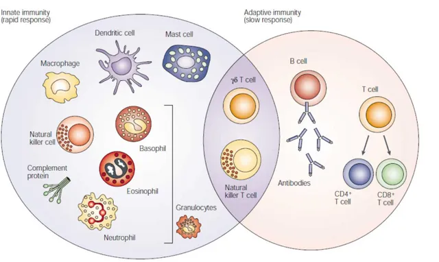
Defenses against pathogens occur through two types of responses innate and adaptative immunity as illustrated in Figure 1. Innate immunity gathers all cell types able to recognize a broad class of pathogenic micro-organisms through Pattern Recognition Receptors (PRR) and ready to react against an invader without further stimulation. In contrast, the adaptative immune system identifies accurate response to a given peptide extracted from the pathogen, called an antigen, by B and T lymphocytes.
Figure 1. The different players of the immune system. The innate immune response functions as the first line of defense against infection. It consists of soluble factors, such as complement proteins, and diverse cellular components including granulocytes (basophils, eosinophils and neutrophils), mast cells, macrophages, dendritic cells and natural killer cells. The adaptive immune response is slower to develop, but manifests as increased antigenic specificity and memory. It consists of antibodies, B cells, and CD4+ and CD8+ T lymphocytes. Natural killer T cells and T cells are cytotoxic lymphocytes that straddle the interface of innate and adaptive immunity. Dranoff G, Nat. Rev. Cancer, 2004
~ 2 ~
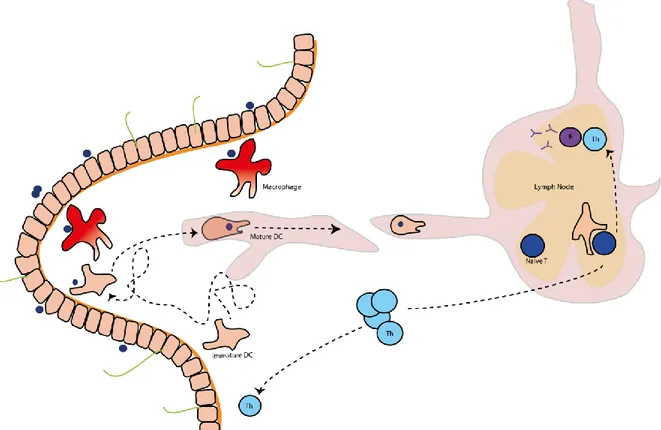
Figure 2. Initiating a response to a new antigen. Antigen encountered in peripheral tissue, here the lamina propria of the small intestine, is either cleared by macrophages or efficiently taken up by immature DCs that turn into a mature state and migrate fast to the draining lymph node. Then, antigen presentation induces T cell proliferation and differentiation into helper T cells (Th). Th are homed to the tissue where the antigen has to be cleared. Th are also involved in B cell maturation that leads to antibody production.
~ 3 ~
A-1- DCs play a crucial role connecting innate and adaptative system
A specific stimulation is required to trigger the adaptative response. Dendritic cells (DCs) ensure the key connection between the innate immune system and proper stimulation of the lymphocyte response. Peripheral tissue surveillance is under the control of two types of tissue resident mononuclear phagocytes that surveil tissues differently: DCs and Macrophages. Both can detect danger signals and internalize antigens but while tissue-resident macrophages remain sessile, DCs are rather migratory cells. Both macrophages and DCs internalize antigens through different processes called phagocytosis and macropinocytosis, respectively. Phagocytosis was first described in 1880 by Elie Metchnikoff who was awarded with the Nobel prize for this key contribution. Macrophages get attached to their target through recognition receptors to perform phagocytosis. Macrophages specifically endocyte the particles following their contours, so that there is almost no non-specific fluid uptake. Once linkage is strengthen by a set of integrins getting attached to their ligand on the particle surface, macrophages engulf the particle through pseudopod extensions. This step relies on local activation of the cytoskeleton, especially actin polymerization for extension and actomyosin ring contraction for the phagocytic cup. The newly formed vesicle called phagosome is fused with endolysosomes where the particle gets processed and degraded. Ralph M. Steinman (J Exp Med., 1973) was awarded with the Nobel Prize in 2011 for the first description of DCs he provided. He reported their various shapes: elongated or stellate due to their numerous pseudopods.
~ 4 ~
RM. Steinman did not observe as efficient phagocytosis by DCs as compared to macrophages even in conditions of cell culture that were highly favoring phagocytosis. DCs mainly perform macropinocytosis which, in contrast with phagocytosis, does not rely on the necessarily limited availability of specific receptors. Macropinocytosis is a non saturable phenomenon that relies on surface membrane recycling and involves the whole actin cytoskeleton. As a consequence DCs constantly engulf a large volume of fluid. Afterwards, they use receptors to retain and accumulate molecules in macropinosomes that are characteristic of foreigners referred to as antigens. Of note, DCs can also be infected and cytosolic antigens are processed through autophagy into autophagosomes that fuse with lysosomes. Misinterpretation of the signal brought by a self-antigen or an exogenous non-pathogenic antigen can lead to a non-adapted inflammatory response to the signal and pathologic situations of auto-immunity with extensive tissue damage.
The active suppression of pro-inflammatory signals and effectors in case of non-pathogenic situation is called tolerance. On the contrary, tolerating a pathogenic antigen leads to invasive infection. Upon antigen capture, Toll-like receptors (TLRs), a sub-family of PRRs activates a cascade of reactions leading to the activation of the nuclear factor NF-κb. On the one hand it promotes an innate immune response by anti-microbial peptides production that will have an immediate toxic action towards the pathogen. On the other hand, activated DCs secrete cytokines. Moreover, they migrate to draining lymph nodes where they present antigens to T cells. Both DCs and T lymphocytes interact with B cells resulting in their proliferation leading to their activation and the formation of germinal centers and in their maturation to high-affinity antibody producers.
~ 5 ~
A-2- DCs present exogenous antigens to T cells
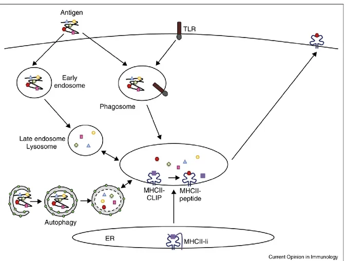
Figure 3. Intracellular trafficking of antigens during MHC II presentation. MHC II presentation involves trafficking of antigens into early endosomes and egress to late endosomes where antigen degradation elicits MHC II epitopes. Phagocytosed antigens are degraded in phagolysosomes, with this process enhanced by recruitment of TLR to phagosomes. MHC II-Ii complexes are cleaved into MHC II-CLIP before the fusion of ER vesicles with lysosomes. Green dots represent 1A/1B-light chain 3 (LC3), the main autophagy associated protein. Adapted from Mintern JD, Curr Op Immunol., 2015
~ 6 ~
To prime naïve T cells, DCs present them antigenic peptides from exogenous antigens loaded onto Major Histocompatibility Complexes (MHC) molecules. Antigen processing takes place in endolysosomes, which are endocytic compartments to which macropinosomes deliver their content. These vesicles also acquire selectively proteins needed for antigen processing such as cathepsin S and are enriched in MHC molecules. After controlled proteolysis, antigenic peptides are loaded onto MHC molecules. DCs can perform cross-presentation i.e. present antigenic peptides from endogenous antigens. They generated in the cytosol by the proteasome and delivered to lysosomes where they are loaded on a MHC I molecule. When loaded on MHC class I (MHC I), the presented antigen can prime CD8+ T cells. Thanks to cross-presentation, DCs can trigger a cytotoxic response to a pathogen without being infected. When loaded on MHC class II (MHC II) the presented antigen, usually exogenous antigen endocytosed by the presenting cell, can prime CD4+ T cells. MHC II proteins are produced in the endoplasmic reticulum (ER) as complexes of nine molecules with a ratio of two molecules of MHC class II for one molecule of Invariant chain (Ii). Ii helps MHC II folding and occupies the peptide binding site, preventing early binding of endogenous peptides. This complex of molecules is transferred to endo-lysosomes. Thanks to a dileucine motif in the cytosolic tail of Ii in this acidic environment enriched in proteases such as cathepsin S, Ii is degraded into a Class II-associated Invariant chain peptide (CLIP). Ultimately, CLIP is exchanged with an antigenic peptide onto MHC II molecules and complexes are exposed to the DC surface membrane for CD4+ T lymphocyte activation. Depending on the associated signals secreted by DCs and by the microenvironment, CD4+ T cells, also called helper T cells (Th), proliferate and differentiate into different types of subsets, the most known being Th1, Th2, Th17 that are mostly pro-inflammatory, or Treg that are essential for tolerance induction. Our group has shown that, in B cells, the cytosolic tail of Ii binds to the actin motor-protein Myosin II. These complexes are required for macropinocytosis and proper antigen processing, thereby coordinating these processes with DC migration. the MHC II (Vascotto F, JCB., 2007). A key role of Myosin II has also been shown in coordinating antigen processing with cell migration in DCs (Chabaud M, Nat Comm., 2015). As it is central in my thesis project, I will introduce in further details Myosin II role in DCs later in this introduction (p.37).
~ 7 ~
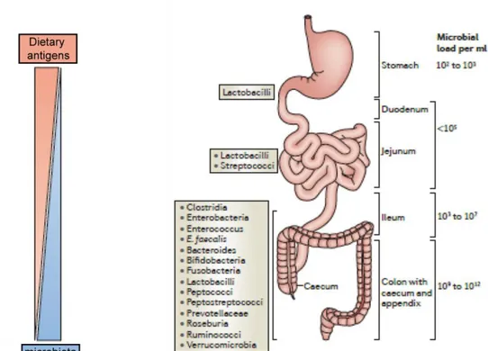
Figure 4. Structure of the small intestine. Dietary antigens and micro-organisms form reciprocal gradients along the small intestine. Adapted from Mowat AM & Agace WW, Nat Rev Immunol., 2014.
~ 8 ~
B- Mucosal immunology of the small intestine
The classical description of how immune responses are triggered is now enriched by the intensive research focusing on immune cells residing in mucosa. This new field is referred to as mucosal immunology. In the lung for instance, alveolar macrophages are constantly scanning the respiratory track lumen (Lloyd CM & Marsland BJ, Immunity, 2017). In addition, immunological memory exists at mucosal sites in mice and humans (Sathaliwayala T, Immunity, 2013). It can generate local inflammation without activation from secondary lymphoid organs. The precise cell composition and their function depend on the nature of the tissue studied. This section describes the immune specificities of the small intestine, linking them with the specific structure of this organ.
~ 9 ~
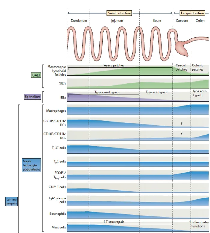
Figure 5. Regional specialization in the gut. Leukocytes populations display heterogeneous repartitions along the small intestine. Gut associated lymphoid tissue (GALT). Intra-epithelial lymphocytes (IELs). Solitary Isolated lymphoid tissue (SILT). Helper T cell (Th). Adapted from Mowat AM & Agace WW, Nat Rev Immunol., 2014.
~ 10 ~
B-1- In the lumen of the small intestine
The small intestine is the long proximal part of the gut, linking the stomach to the caecum. It has multiple histological specificities. The exchange surface with the lumen is substantially increased thanks to the folding of the organ into villi. From outside to inside, one finds: 1) smooth muscle, 2) submucosa with crypts and 3) lamina propria. Crypts are the circular structures made of stem epithelial cells that give rise to epithelium. The lamina propria is a tissue full of immune cells sitting in between blood and lymph vessels. This structure constitutes the stroma of the villi and is covered by a single epithelial layer. The enterocytes i.e. the main cells in the epithelial monolayer, are oriented in the baso-apical direction. On the luminal side, the membrane of enterocytes presents a supplemental folding called the brush border. The duodenum is the most proximal part of the gut and receives the acidic content of the stomach. Therefore, it is full of partly digested food antigens and its microbiotal content is low. Bile acids resulting of heme degradation and cholesterol metabolism are brought by bile ducts and mixed with this food content. The pancreatic duct carries into the duodenum the enzymes required for digestion and produced in the pancreas, such as lipase. The jejunum is the second part of the small intestine where nutrient absorption is very effective. The ileum makes the junction with the caecum. Duodenum, jejunum and ileum form a unique organ since they are not histologically separated but rather assembled with smooth transition between them. The lymph circulating in the small intestine is drained in mesenteric lymph nodes (MLNs) but some gut associated lymphoid tissues (GALTs) are also involved in the gut immune system.
All along the small intestine, some non-encapsulated lymphoid follicles sit right under the epithelium, the main ones are called Peyer Patches and constitute dedicated sites for B cell maturation. This continuum between duodenum, jejunum and ileum promotes gradients formation along the small intestine. In particular, a gradient of pH with a low pH in the duodenum, ranges from 7 to 9 in the jejunum and reaches alkaline values of pH in the ileum. Gradients of metabolites and cells, especially immune cells, are also established as reported in Figure 4. However, how such gradients of immune cells correspond to regions of functional immune specialization remains unclear. I will address this question in my thesis.
~ 11 ~
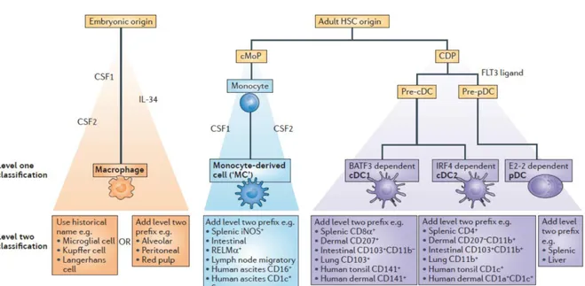
Figure 6. Proposed classification of mononuclear phagocytes. Three main groups of cells are described — namely, common dendritic cell (DC) precursor (CDP)-derived DCs, embryonic-derived macrophages and monocyte-derived cells. DCs are further subdivided into ‘classical type 1 DCs (cDC1s)’, ‘cDC2s’ and plasmacytoid DCs (pDCs). BATF3, basic leucine zipper transcriptional factor ATF-like 3; cMoP, common monocyte progenitor; CSF1, colony-stimulating factor 1 (also known as M-CSF); CSF2, colony-stimulating factor 2 (also known as GM-M-CSF); FLT3, FMS-like tyrosine kinase 3; HSC, haematopoietic stem cell; IL-34, interleukin-34; iNOS, inducible nitric oxide synthase; IRF4, interferon-regulatory factor 4; RELMα, resistin-like molecule-α. Adapted from Guilliams, Nat. Rev. Immunol 2014
~ 12 ~
B-2- The immune response in the small intestine
B-2-a Antigen presenting cells of the small intestine
DCs and macrophages come from different lineages
Several studies have shown that macrophages and DCs have different origins. First, based on growth factors requirements and second on transcription factors (Murphy TL, Annu Rev Immunol., 2016), the field is now benefiting from transcriptomics approach (Schraml BU & Reis e Sousa C, Curr Op Immunol., 2015). In mouse, tissue macrophages mainly arise from embryonic development and are maintained independently of the postnatal monocytic lineage (Yona S, Immunity, 2013). Haematopoietic stem cells differentiate in granulocytes and lymphoid-primed multipotent progenitor (LMPP). LMPP give rise to two distinct progenitors (Figure 6): a common monocyte progenitor (cMoP) and a common DC precursor (CDP). Different cell lineages develop from these two types of cells develop different cell lineage: monocyte-derived cells from cMoP and DCs from CDP. The main types of DCs residing in tissues are defined as conventional DCs (cDCs). This ontologic model that early separates the monocytic lineage from the DC lineage is validated both in lymphoid organs (Liu K, Science, 2009) and in several peripheral tissues like the skin (Tamoutounour, Immunity, 2013) and the gut (Bain CC, Mucosal Immunol., 2013; Scott CL, Mucosal Immunol., 2015). A lot of efforts have been made to determine the best markers among several candidates (CX3CR1, F4/80, CD64, CD11c) for the different cellular types. Among them, CD64 has been identified to easily distinguish DCs from macrophages and monocyte derived-cells (Tamoutounour S., Eur J Immunol., 2012; Langlet C, J Immunol., 2012). Within CD64negative cells, cDCs are identified as CD11c positive, MHC II positive cells (Joeris T, Mucosal Immunol., 2017). Additional markers are required to further dissect of the DC landscape in the small intestine (Table 1).
~ 13 ~
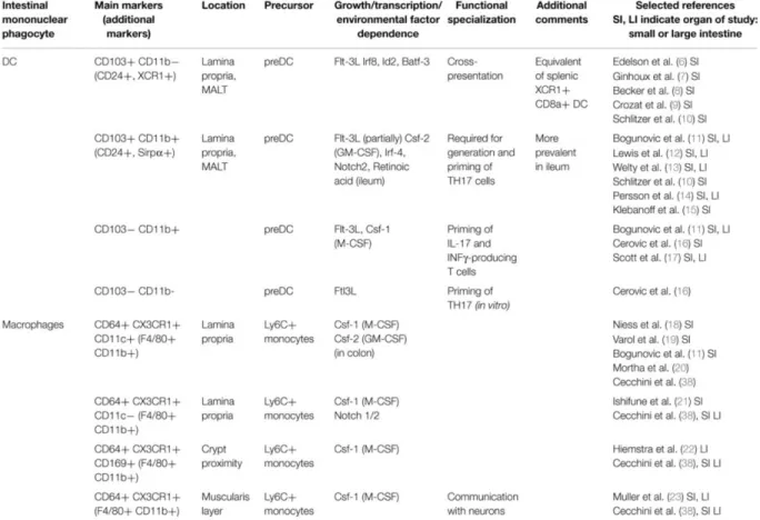
Table 1. Mononuclear phagocytes and their respective subsets in the lamina propria of the mouse intestine. From Gross M, Front Immunol., 2015
~ 14 ~
Defining DCs of the small intestine
The lamina propria (LP) of the small intestine contains a lot of DCs and macrophages whose functions partially overlap. Thanks to the work on the ontogeny of DCs and macrophages from the gut, and the identification of distinct lineages, the immune gut landscape became clearer (Bogunovic M, Immunity, 2009). In this section, I will focus on the classification of cDCs in the small intestine. Once identified as CD64 negative, CD11c positive and MHC II positive, DCs are split into groups following two additional surface markers: CD103 and CD11b, identifying CD103+CD11b+, CD103+CD11b-, CD103-CD11b+ and CD103-CD11b- (Table 1). Thanks to chimeric mice, the contribution of monocytes and CDPs to the different DC subsets was elucidated (Bogunovic M, Immunity, 2009). Monocytes, which produce some CD103-CD11b+ DCs, do not contribute at all to the CD103+CD11b+ cDC subset. CDPs and pre-DCs give rise preferentially to CD103+CD11b+ DCs in the LP. CD103-CD11b-cDCs form a small and understudied population. Monocyte-derived CD103-CD11b+ are rare in the small intestine but frequent in the colon where they prime pro-inflammatory responses. CD103+CD11b- are also derived from pre-DCs under the control of fms-like tyrosine kinase 3 (Flt3) ligand, inhibitor of DNA protein 2 (Id2), IFN regulatory protein 8 (IRF8) (Ginhoux F, J Exp Med., 2009), and Batf3 (Edelson BT, J Exp Med., 2010). They behave similarly to CD8+ DCs of the lymphoid organs and to XCR1+ splenic cells. Intestinal CD103+CD11b- cDCs perform cross-presentation of oral antigens to CD8+Tcells in MLNs, which implies their ability to migrate (Becker M, Front Immunol., 2014).
The CD103+CD11b+ cDC subset is specific to the small intestine LP. It derives from pre-DCs. The development of this subset depends on Irf4 but not on Id2 or IRF8. It relies on environmental cues such as all-trans retinoic acid (AtRA) (Klebanoff CA, J Exp Med., 2013). Parabiontic mice experiments showed that CD103+CD11b+ DCs had a great turnover rate. This result supports the idea that CD103+CD11b+ DCs constantly migrate to the MLNs to report information there and stimulate T cells with the latest antigens they have captured. CD103+CD11b+ cDCs are particularly responsive to fungi and extracellular bacteria through the Notch2 pathway (Lewis KL, Immunity, 2011). They drive Th17 pro-inflammatory response (Persson EK, Immunity, 2013). DC markers now provide us with indications about their function, which is useful for further dissection of the immune system in the small intestine.
~ 15 ~
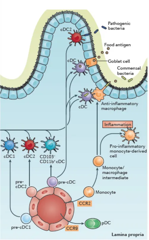
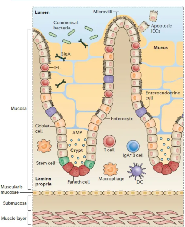
Figure 7. Antigen capture in the small intestine. The small-intestinal LP contains at least three distinct populations of pre-classical dendritic cell (cDC)-derived cDCs (cDC1s, cDC2s and CD103− CD11b+ cDCs. LP cDCs acquire antigen by handover, either from epithelial goblet cells or CX3
C-chemokine receptor 1 (CX3CR1)hi macrophages. LP cDC2s also have the propensity to migrate into
the epithelium and extend dendrites into the gut lumen for the capture of pathogenic bacteria. In Peyer's patches, cDCs acquire antigen in the subepithelial-dome (SED) through M cells BCZ, B cell zone; SCS, subcapsular sinus; TCZ, T cell zone. Adapted from Worbs T, Nat. Rev. Immunol. 2017
~ 16 ~
B-2-b Antigen capture in the small intestineThe intestinal epithelium is rather non permeable to antigens. However, in homeostatic conditions, DCs can sense and capture antigens present in the gut lumen. How do antigens get from the lumen into the LP without requiring any epithelial damage? In this section, I will detail the routes described in the literature as antigen gateways. The relative importance and potential redundancy of these different routes in homeostasis and upon infection remain unknown.
The first proposed ways resemble a passive passage with no particular energy invested by the organism to take up antigens. A possible paracellular leakage of soluble antigens through the epithelium then entering lymphatic vessels is a valid route towards MLNs. In the same line, antigens can enter blood vessels in the LP of the intestinal villi and get direct access to the liver and, if not eliminated at that step, to the general circulation (Pabst O. & Mowat AM., Mucosal Immunol., 2012). Goblet cells can form Goblet cell Associated Antigen Passages (GAPs) and are seen as holes when staining the intestinal epithelial layer as their cytosol is pushed aside by the mucin vesicles. As a result, they deliver soluble antigens to the LP below (Figure 7), in particular to CD103+ DCs. Only small molecules, up to 10kDa
according to experiments of dextran internalization, can pass through GAPs (McDole JR, Nature, 2012).
A second proposed mechanism for antigen uptake in the LP is active and direct sampling of the gut lumen by resident immune cells of the LP. Among them, CD11c+ CX3CR1+ cells have
been identified to emit trans-epithelial dendrites (TEDs) and sample gut lumen through the epithelium. They emit TEDs as a response to the epithelial secretion by goblet cells of CX3CR1 ligand the chemokine so-called fractalkine (Kim KW, Blood, 2011). CD11c+CD103+
DCs can also send TEDs through the epithelium. Indeed, they sit at the periphery of the LP where they interact with epithelial cells by opening adherens junctions between them (Rescigno M, Nat Immunol., 2001). This mechanism is very likely as CD103 is an integrin whose ligand E-cadherin is present on epithelial surface membranes and might facilitate interactions between CD103+ cells and epithelial cells. It is supported by observations showing that CD103+ cells send dendrites through the epithelium towards the gut lumen
~ 17 ~
TEDs from CD103+ CD11b+ cDCs form especially in the upper gut. Their formation is supposed to depend on the microbiota since they are reduced in germ-free mice and in Specific Pathogen Free (SPF) mice after antibiotic treatment (Chieppa M, J Exp Med., 2006). A third and last approach is cooperation between cell types. CD103+CD11b- DCs from the
Peyer patches can exit the sub-epithelial dome and migrate towards the LP to present antigen to DCs sitting there. Although CD11c+CX3CR1+cells have been proposed based on
undirect observations to migrate towards MLNs (Diehl GE, Nature, 2013), there is no formal demonstration for this. CD11c+ CX3CR1 cells are now thought of as macrophages with no migratory potential, working as intermediaries between the gut lumen and DCs to which they transfer antigens (Mazzini E, Immunity, 2014).
It is very likely that all proposed routes co-exist and the quantitative contribution of each mechanism is under debate. An immune compartmentalization between colon and small intestine is admitted, due to the differences in both lumen content and the LP DC composition (Mowat AM & Agace WW, Nat Rev Immunol., 2014). Given the metabolic, microbial and immune gradients described into the small intestine, a study comparing the different floors of the small intestine may bring helpful insights in the field.
~ 18 ~
B-2-c T cell differentiationAfter efficient antigen capture, DCs turn into a mature state and migrate to the MLNs to present antigens to T cells. Associated signals, costimulatory molecules, and cytokine secretion are necessary to reach full CD4+ and CD8+ T cell activation. The T cell subsets of the LP may be affected by a modification in DC subsets in the small intestine.
In response to intracellular bacterial infection, antigen presenting cells (APCs) secrete interleukin (IL-)12, enhancing expression of the transcription factor Tbet in CD4+T cells which then differentiate into Th1 cells. Th1 cells promote an immunogenic response through secretion of Interferon γ (IFNγ), IL-2 and tumor necrosis factor α (TNFα), which facilitates the activation of cytotoxic CD8+ T cells and phagocytosis by macrophages. In response to parasite infection, and presence of IL-4 during antigen presentation, CD4+ T cells differentiate into Th2 cells, which depend on the GATA3 transcription factor. In response to extracellular bacteria, APCs secrete low concentrations of TGFβ associated with 6 and 21. This triggers the expression of the IL-23 receptor and the RORγt transcription factor promoting differentiation towards Th17 cells (Mangan PR, Nature 2006; Aujla SJ, Nat Med., 2008). Th17 cells secrete IL-17 and IL-22, which favor neutrophil recruitment and stimulate anti-bacterial immunity (Flannigan KL, Mucosal Immunol., 2017). In the presence of high concentrations of Tumor Growth Factor beta (TGFβ) associated with interleukin 6 (IL-6), T cells express the transcription factor FOXP3, which is characteristic of differentiation towards the Treg cell fate. In contrast to the Th subsets previously described, T regs promote tolerance and prevent pro-inflammatory responses. Tregs also suppress T cell pro-inflammatory effects through IL-10 secretion. This reduction of T helper effectors also impacts on antibody production by B cells. As opposed to Th1 and Th2 cell fates that are pretty stable, Th17 and Treg cell fates present some plasticity (Bhaumik S, Front Immunol., 2017).
Besides the induction of T cell proliferation and differentiation, a key function of DCs is to confer T cells the ability to migrate towards the tissue where DCs have detected the antigen that T cells need to eliminate, i.e. the small intestine in this case. This phenomenon is referred to as “imprinting of gut homing properties” as DCs will make T cells express surface markers that will trigger their trafficking to the gut, namely the CC chemokine receptor CCR9 and the α4β7
~ 19 ~
integrin. Upon stimulation, Th cells express CCR9 whose ligand is secreted by epithelial cells only in the small intestine (Kunkel EJ, J Exp Med., 2000). DCs also enhance α4β7 integrin expression on the membrane surface of T cells (Johansson-Lindbom B, J Exp Med., 2005). This integrin binds to the mucosal addressing cell adhesion molecule-1 (MadCam1), which is present on the cellular surface of high endothelial veinules in the small intestine and in the colonic LP. As for differentiation, this phenomenon is facilitated by molecular signals from the microenvironment. Imprinting of gut homing properties to Th cells is more efficient in the presence of TGFβ and all-trans Retinoic Acid (AtRA) (Eksteen B, Gastroenterology, 2009). Once arrived into the tissue they are targeted to, Th cells promote pathogen elimination. In the small intestine their action is further tuned by the DCs that reside in the tissue and by local cues secreted by the epithelium.
~ 20 ~
B-3- The epithelium maintains the boundary
B-3-a The crosstalk between Intestinal Epithelial cells (IECs) and immune cells
Intestinal epithelium directly and undirectly modifies the local immune system. First, it is itself a barrier against pathogens. Second, it secretes cytokines that affect DC and T cell fates.
IECs constitute a monolayer strongly polarized in an apico-basal manner, the apex being the lumen and the basis corresponding to the basal membrane. IECs form an heterogenous population made of a majority of absorptive cells called enterocytes and a minority of secretory cells: goblet cells, Paneth cells, Tuft cells and enteroendocrine cells. Goblet cells mainly secrete mucus: a network of glycoproteins which form a complex viscous gel on top of the epithelial layer, limiting contacts between the content of the gut lumen and IECs. Mucus is mainly composed of mucins, the main one being MUC2, which is stored in acidic granulae within goblet cells. Release of MUC2 requires local increase of pH and local decrease of calcium to allow its correct unfolding (Ambort D., PNAS, 2012). It is more elaborated in the colon where it consists of two layers: one loose superficial layer that contains some bacteria and a lower thick layer, which sticks to the epithelium and is almost sterile. (Johansson ME, PNAS, 2008). In the small intestine, the mucus structure is drastically different, as it consists of a single thin and loose layer. It is not attached to the epithelium and easy to aspirate during an endoscopic procedure (Johansson ME, Cell Mol Life Sci., 2011). This difference in structure explains that mucus in the small intestine is permeable for nutrients intake and also partly permeable to bacteria. Some adherent bacteria can implant in mucus, such as Segmented Filamentous Bacteria (SFB) in the ileum. Mucus detachment from the epithelium by N-terminal digestion of MUC2 is crucial: it is a way to wash out micro-organisms, preventing them to grow and reach the epithelium (Johansson ME & Hansson GC, Nat Rev Immunol., 2016). Of note, Th2 cells stimulated by parasite infections secrete cytokines, among which IL-13, which enhance mucus secretion and goblet cell hyperplasia, preventing the interaction between parasites and enterocytes (Webb RA, Parasite Immunol., 2007).
The mucus contains molecules secreted by the epithelium that diffuse within the gel. All IECs secrete antimicrobial proteins (AMPs) able to kill bacteria by attacking their wall.
~ 21 ~
Figure 8. Several lines of defense to prevent bacterial entrance. Intestinal epithelial cells (IECs) are produced from stem cells near the bottom of the crypts. After a few days, IECs are lost from the tip of the villus and are replaced by new cells that are migrating upwards from the crypts. As well as the absorptive epithelial cells, stem cells in the crypts give rise to the mucus-secreting goblet cells found on the villus (indicated by the arrow), and to Paneth cells that migrate downwards to the bottom of the crypt. These Paneth cells are characterized by the presence of dense granules that contain antimicrobial peptides (AMPs). The central part of the villus comprises the LP, where the majority of intestinal immune cells are found, whereas intraepithelial lymphocytes (IELs) are found lying
between epithelial cells. DC, dendritic cell; SIgA, secretory immunoglobulin A. Adapted from Mowat AM & Agace WW, Nat. Rev. Immunol. 2014
~ 22 ~
Paneth cells are the major producers of AMPs, which act as a supplementary protection against bacterial invasion. Paneth cells are produced in the crypts of the small intestine, especially in the ileum, and migrate to the bottom of the crypt. Genetic deletions affecting the pool of Paneth cells, for instance in Nod -/- mice, lead to unadapted reactions to infections as well as to mucosal lesions compatible with Crohn’s disease, an inflammatory bowel disease that affects both the small and the large intestine (Clevers, Annu. Rev. Physiol. 2013).
TLR are expressed at low levels at the IECs surface. Thanks to these receptors, epithelial cells are able to sense Pathogens Associated Microbial Peptides (PAMPs). Upon activation, they trigger the NF-κB or Interferon Regulatory Factor (IRF) pro-inflammatory cascade downstream. Some consider epithelial cells as non-professional antigen-presenting cells since they express MHC II. Importantly, IECs also create a microenvironment which is not neutral for immune cells. For instance, it favors differentiation towards one cell fate thanks to their ability to secrete Vitamin A metabolites and cytokines such as TGFβ and Thymic Stromal LymphoProtein (TSLP). DCs cultured in the presence of human IEC supernatants turn into tolerogenic DCs (Iliev ID, Gut, 2009). IECs also shape cells of the adaptative immune system such as the pool of T cells that resides in the epithelium, the intra-epithelial lymphocytes (IELs). These cells form a gradient along the small intestine, being much more numerous in the duodenum. Recently, innate lymphoid cells (ILCs), which have a lymphoid origin and morphology but lack antigen specificity were described. ILCs can be classified into ILC1, which resemble CD4+Th1; ILC2, which resemble CD4+Th2 and ILC3, which resemble CD4+Th17 (Spits H, Nat Rev Immunol., 2013). As for IELs, IECs secrete cytokines that promote ILC2 activation: IL-33, IL-25 and TSLP. In turn, ILC2 act on crypt progenitors to influence IEC differentiation. Furthermore, in vitro, CD4+T cells from patients with Crohn disease, which behave like Th17 cells, made intestinal epithelium secrete chemokine (C-X-C motif) ligand 1 (CXCL1), and CXCL8 that attract neutrophils and macrophages (Calderon-Gomez E, Gastroenterology, 2016).
In brief, the epithelium can locally modify immune cell fate and in turn be used as an amplifier of the immune response by effector cells. This close interaction between immune cells and the epithelium raises the hypothesis that a local system refines the immune response programmed in MLNs.
~ 23 ~
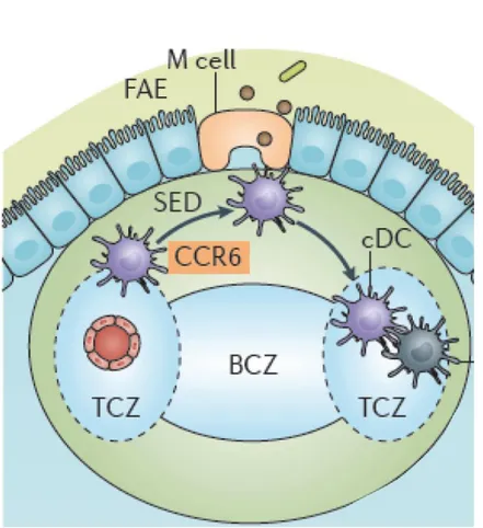
Figure 9. Antigen capture in Peyer’s patches. cDCs are recruited in a CCR6-dependent manner towards the follicle-associated epithelium (FAE) where M cells deliver them soluble antigens. They subsequently migrate from the subepithelial dome (SED) into parafollicular T cell areas of Peyer’s patches. B cell zone (BCZ); T cell zone (TCZ). Adapted from Worbs T, Nat. Rev. Immunol. 2017
~ 24 ~
B-2-b The Gut Associated Lymphoid Tissue (GALT)In addition to the immune cells present in the LP, some non-encapsulated lymphoid organs where T cell activation and B cell maturation take place B cells, are dispersed along the small intestine, in the submucosa. The biggest ones are the Peyer patches. The epithelial layer that covers Peyer patches is specific to these structures: it is enriched with microfold cells called M cells (Figure 9). These M cells transport antigens from the lumen to the stroma, easing antigen presentation to immune cells lying beneath (Owen RL, Sem Immunol., 1999). Therefore, DCs sitting in the sub-epithelial domes in Peyer patches can easily sense the lumen and its microbial content. They present antigens they sample in situ to immature B cells enhancing their maturation and class switch to IgA secreting plasma cells (Fagarasan A., Nature, 2001; Mora JR, Science, 2006). IgA plasma cells as they represent 75% of plasma cells in the duodenum and about 90% of the plasma cells in the duodenum. Eosinophils are abundant in Peyer patches and are thought to maintain IgA+ plasma cells. Plasma cells migrate out of Peyer patches in the LP where they secrete IgA (Reboldi A, Science, 2016). IgA are trans-cytosed through the epithelium thanks to a polymeric Ig-receptor, and diffuse into the mucus layer where they bind to the bacterial surface they are targeting. Receptors to IgA have been identified at the membrane surface of eosinophils, DCs and IECs (Wines BD, Tissue Antigens, 2006). Thus, IgA binding to bacteria can result in their endocytosis by IECs or by Peyer patches DCs. The submucosa of the small intestine contains some anatomically well-defined sites dedicated to B cell maturation and high-affinity IgA production. These structures complete the mucosal immune system of the small intestine.
~ 25 ~
C- The Modulation of mucosal immunity in the small intestine
by the lumen content
C-1- Interactions with microbiota
A new paradigm emerged in the 2000s considering that we constantly host trillions of bacteria that do not act as pathogens but as commensals in our organism. The mainstream idea is now that our immune system co-evolved with this flora, implying that it learned to tolerate it and that the flora in turn constantly influences the behavior of immune cells. As the quantity and composition of the flora change along the small intestine, this microbial content of the lumen may contribute to the regionalization of immune cell composition of the LP.
There is a gradient of microbiota from the duodenum to the ileum. Duodenum contains a low bacterial concentration while the ileum is perceived as a reservoir of bacteria containing 1012 bacteria per gram of luminal content. It is associated with a parallel gradient of Paneth cells, which are more numerous in the ileum. The diversity of bacteria hosted in our small intestine is huge. Most of them belong to two phylotypes: Firmicutes and Bacteroides. Very early, studies in germ-free mice have highlighted the importance of the gut microbiota to induce a normal immune response especially in germinal centers formation (Bauer H, Am. J. Pathol., 1963). Upon normal conditions, the microbiota triggers secretory IgA production and regulates the balance between different T cell fates. Bacteroides fragilis is able to orientate the differentiation of T cells towards Treg by secreting a specific capsular polysaccharide A (Mazmanian SK, Nature, 2008). Segmented filamentous bacteria (SFB) can colonize the ileum and interact directly with epithelial cells. There, they promote Th17 expansion (Ivanov IL, Cell, 2009; Gaboriau-Routhiau V, Immunity, 2009). SFB deficient mice produce less IgA and respond less to Citrobacter rodentium infections. This diverse bacterial population is under environment influence. It can be reversibly manipulated by changes in the diet (Turnbaugh PJ, Cell Host Microbe, 2008) or oral antibiotic treatments. For instance, Streptomycin induces a shift in the inflammation state, allowing
Salmonella Typhimurium to compete with the original microbiota, thus favoring infection
~ 26 ~
Clostridium difficile infection, whose major risk factor is treatment with penicillins, illustrates
how antibiotics can deeply modify the microbiota. As for prevention, commensal yeast such as
Saccharomyces-boulardii are associated to antibiotics of the Ampicillin family when they are
prescribed to patients with high risk of infection. The latest efficient treatment, fecal transplantation is now performed in routine in case of severe recurrence with excellent results (Van Nood E, NEJM., 2013). It is the best evidence for the reversibility of these antibiotics effects. Besides, it also shows the ability of a healthy human flora to restore diversity in an ill microbiota. These studies revealed that as the microbiota is in close relationship with its host, any alteration in the host microenvironment such as excessive inflammation or injury changes to the microbial composition. However, the reciprocal assumption, i.e. changes in the microbiota influence the inflammatory state, is still under debate. Administration of total fecal extracts to mice triggers the production of IL-17 by T cells, TNFα and IL1-β by monocytes, macrophages and DCs. These pro-inflammatory effects are reversed by antibiotic treatments (Bhattacharya N, Immunity, 2016). It is accepted that some bacteria, for instance invasive strains of Escherichia coli in patients with Crohn’s disease (Darfeuille-Michaud A, Gastroenterology, 2004) maintain unappropriate inflammation. However there are some difficulties to determine whether this is a consequence of inflammation or the cause. Besides, in Crohn’s disease, fecal transplantation efficiency is far more controversial than in C. difficile infection (Choi HH, Clin. Endosc, 2016). In the cancer field, manipulation of the microbiota can enhance anti-tumoral immune responses (Daillière R, Immunity, 2016) at a systemic level, leading to death of subcutaneous tumors. Consistently, some studies are in favor of a manipulation of the immune response through microbiota. Chemotherapy, for example with cyclophosphamide also alters microbiota composition thus changing adaptive response to cancer (Viaud S, Science, 2013). These apparent contradictions between cancer, infection and inflammatory bowel disease studies may find a unifying explanation when focusing on epithelial integrity. Indeed, under inappropriate inflammatory conditions such as in Crohn’s disease, epithelial barrier is disrupted and loses its power to prevent microbial entrance in the organism.
~ 27 ~
C-2- Impact of nutrition
C-2-a From oral tolerance to tolerance to food
As previously described, DCs have access to antigens present in the gut lumen. Consequently, they constantly sample dietary antigens, but this permanent intake should not trigger any pro-inflammatory response. Therefore, the small intestine, and especially its upper part is identified as a site where tolerance is particularly efficient. Oral tolerance induction is of particular interest for the immunology community since it would give a simple route for immune manipulation.
Besides anergy i.e. absence of immune response by the lack of costimulatory signals, the main mechanism identified is active T cell mediated suppression through Treg expansion (Pabst O. and Mowat A.M., Mucosal Immunol, 2012). DCs are thought to play a crucial role in this process not only by inducing Treg development in MLN but also by recruiting effector T cells to the gut, through imprinting of gut homing receptors (Païdassi H, Gastroenterology, 2011; Mora JR, Nature, 2003; Cassani B, Gastroenterology, 2011). Due to the physiological ingestion of 130-190g of proteins each day (Weiner HL, Immunol Rev 2011), obvious candidates in the small intestine lumen for immune modulation are food proteins.
Food contribution to peripheral Treg expansion has been studied in the gut (Kim KS, Science, 2016) by raising mice under strict control in bacterial content (germ free vs specific pathogen free) and food antigen content (milk vs antigen-free diet vs chow diet after weaning). Antigen-free mice are offsprings from germ Antigen-free mice raised with an antigen-Antigen-free diet. This diet impacts on peripheral Treg development in the small intestine but not in the colon where there is a loss in T cell memory, as in the colon of germ free mice. These data suggest regional specificities in the gut, T cell development relying on diet in the small intestine and more on microbiota in the colon. In addition, peripheral Treg depletion leads to stronger immune responses to dietary antigens with more symptoms of intestinal allergy. This work further suggests that both microbial and dietary antigens are required to induce a complete tolerance.
~ 28 ~
Indeed, by weaning SPF mice onto antigen-free diet or by treating them with antibiotics, the authors show that dietary antigens drive RORγt- peripheral Treg expansion and microbiota drive RORγt+ peripheral Treg expansion.
As far as DCs are concerned, the number CD103+ CD11b+ were reduced in the small intestine of antigen-free mice but not in their MLNs, suggesting a local impact of dietary antigens onto CD103+ CD11b+ cDC expansion.
In brief, the intestine is a route of antigen administration that shapes the immune system through modulation of DC capacity to regulate T cell development and differentiation both in secondary lymphoid organs and in mucosal sites. One well studied nutrient that has multiple effect on DCs and T cell is AtRA,as I will discuss in the next part of the introduction.
~ 29 ~
Figure 10. AtRA metabolism in the gut. High retinol concentrations in the small intestine (SI) likely underlie the enhanced Aldh1a2 expression and ALDH activity of SI CD103+ DCs. (a) High concentrations of retinol in the SI and draining MLNs are maintained by the diet and probably also through bile. (b). Which SI cells generate the AtRA that imprints CD103+ DCs in vivo are currently unknown but SI epithelial cells likely make an important contribution. Bile directly induces RAR signalling in BM-DCs in vitro, suggesting that bile-derived RA may directly contribute to SI CD103+ DCs imprinting. Nevertheless, the in vivo role of bile-derived retinol and RA remains to be addressed. RA directly induces Aldh1a2 expression in SI CD103+ DCs and imprints these cells with vitamin A metabolising activity. RA-induced RAR signalling in DCs appears to require ERK signalling, and RA induction of Aldh1a2 requires de novo protein synthesis and thus functions through additional intermediates. (c) Imprinted CD103+ DCs constitutively migrate to draining MLNs and present lumen-derived foreign and intestinal self-antigen to T cells.During T cell priming, vitamin A metabolising CD103+ DCs provide AtRA signals to responding T cells and probably promote the unique nature of the SI immune responses. From Agace WW & Persson EK, Trends Immunol. 2012