HAL Id: hal-01319907
https://hal.archives-ouvertes.fr/hal-01319907
Submitted on 23 May 2016
HAL is a multi-disciplinary open access
archive for the deposit and dissemination of
sci-entific research documents, whether they are
pub-lished or not. The documents may come from
teaching and research institutions in France or
abroad, or from public or private research centers.
L’archive ouverte pluridisciplinaire HAL, est
destinée au dépôt et à la diffusion de documents
scientifiques de niveau recherche, publiés ou non,
émanant des établissements d’enseignement et de
recherche français ou étrangers, des laboratoires
publics ou privés.
Estimating the EMG response exclusively to fatigue
during sustained static maximum voluntary contraction
Jing Chang, Damien Chablat, Fouad Bennis, Liang Ma
To cite this version:
Jing Chang, Damien Chablat, Fouad Bennis, Liang Ma. Estimating the EMG response exclusively
to fatigue during sustained static maximum voluntary contraction. 7th International Conference on
Applied Human Factors and Ergonomics, Jul 2016, Orlando, United States. �hal-01319907�
Estimating the EMG response exclusively to fatigue
during sustained static maximum voluntary contraction
Jing Chang1,2, Damien Chablat1, Fouad Bennis1, Liang Ma2
1 IRCCyN, Ecole Centrale de Nantes, 44321 CEDEX 3, Nantes, France
2. Department of Industrial Engineering, Tsinghua University,
Beijing, 100084,P.R.China
{Jing.chang, Damien.Chablat, Fouad.Bennis} @irccyn.ec-nantes.fr; [email protected]
Abstract.The increase of surface electromyography (sEMG) root-mean-square
(RMS) is very frequently used to determine fatigue. However, as RMS is also influenced by muscle force,its effective usage as indicator of fatigue is mainly limited to isometric, constant force tasks.This research develops a simple me-thodto preclude the effect of muscle force, hereby estimates the EMG amplitude response exclusively to fatigue with RMS. Experiment was carried out on the biceps brachiis of 15 subjects (7males, 8 females) during sustained static maxi-mum voluntary contractions (sMVC).Result shows that the sEMG RMS re-sponse to fatigue increasesto 21.27% while muscle force decreasing to 50%MVC, which implies that more and more extra effort is needed as muscle fatigue intensifies. It would be promising to use the RMS response exclusively to fatigue as an indicator of muscle fatigue.
Keywords: surface EMG· root-mean-square· muscle fatigue· sustained static max-imum voluntary contraction
1
Introduction
Muscle fatigue is a complicate phenomenon believed to be closely related to muscu-loskeletal disorder[1]. In ergonomics, fatigue is usually measured from three
as-pects:the reduction of force output capability, the self-rating discomfort, as well as the myoelectric activity change.The reduction of force output capabilityhas been well modeled[2], and been used to describe fatigueand calculate fatigue resistance[3,4]; the
self-rating discomfort was reported to be a valid estimator[5], but its usage might be
limited due to the subjectivity. The myoelectric activitymonitored by surface EMG (sEMG) shows the in situ and real-time changes of the muscles[6]. Therefore, ithas
been frequently used to determine fatigue[7].
Overall, sEMG signal is the synthesis of myoelectric and anatomic properties: the shape of motor unit action potential (MUAP) waveform[8], the firing rate of MUAP trains, the recruitment of new motor units[9]as well as thesubcutaneous tissues which
act as a volume conductor and cause a spatial low-pass filtering effect when conduct-ing myoelectric signals[10].In the process of fatigue, metabolites such as lactic acid
concentrate, for which the muscle fibre conduction velocity (CV) decreases. The de-crease of CV directly changes the shape of the MUAP waveform and leads to the
compression of EMGspectrumtowards its lower part[11]. As a result, more energy passes through the tissues which act as a low pass filter, and is detected by the surface electrodes, leading to an increase of sEMG amplitude.
The changes ofsEMG characteristic values,such as decrease of the median power frequency (MDF) and increase of the root-mean-square (RMS), are very often used to estimate fatigue[12,13,14]. But it should be noted that thesesEMGindicators are not
al-ways applicable. Firstly,sEMG spectral compressions are sometimes slight and insig-nificant statistically, especially during low force level tasks[5,15].As De Luca[7] puts it,
the usage of spectral variables should be limited toisometric, constant-load tasks greater than 30% of subjects’ maximal force. In addition, thesEMG amplitude is sig-nificantly affected bymuscle force: greater force leads to larger amplitude[16,17]. It
seemsthe RMS should only be used in constant-force fatigue process.
However, the tasks that are cared most aredynamic, inconstant-force ones. These tasks are characterized by time-varying changes in forces exerted variations as well as in working postures[12]. Both of the two factors lead to EMG changes despite of
fati-gue. To indicate fatigue with sEMG in real operations, it is logistic to start from tasks with fixed posture and inconstant force, such as sustained static maximal exer-tions.Voluntary sustained static maximal exertions, or sustained maximum voluntary contractions (sMVC), are characterized by rapid muscle fatigue and continuous force decline[18]. In this process, the impact of intensifying fatigue on sEMG amplitude is
overwhelmed by the impact of decreasing muscle force. As a result, the RMS turns to decline along with fatiguing progress[19].
In this research, a simple methodto preclude the impact of muscle force on RMS is tested during sMVC and the sEMG RMS response exclusively to fatigue is identified. The possibility of using the identified value as an indicator of fatigue is discussed.
2
Methods
2.1 Subjects
A number of 15 subjects (7 males, 8 females) took part in the experiment. They were healthy, aged between 20 and 35 and free from any upper limb pain during the pre-vious 12 months. All of them are right-handed. Other criteria include moderate (non-extreme) level of self-reported daily physical activity. After being fully informed of the nature of the experiment, they signed an informed consent. Anthropometry data were measured upon their arrival at the laboratory (see Table 1).
Table 1.Subject physical characteristics.
Characteristic Mean SD
Age (year) 28.3 4.7
Height (cm) 167.1 9.1 Weight (kg) 67.1 14.0
BMI 23.8 3.5
2.2 Experiment setup
The experiment platform consisted of a dynamometer (BET®Primus RS), a
custo-mized chair without armrest, an electromyography recorder (TeleMyo 2400T V2®)
and an interactive monitor. The muscle force and the surface EMG were recorded by the dynamometer and EMG recorder at frequency of 20 Hz and 1500 Hz respectively. Real-time muscle force was recorded by and displayed on the monitor.
In the experiment, each subject was told to be seated on the chair with torso and upper arms perpendicular to the ground. The elbow joint angle of the dominant arm was kept at 90 degree with forearm supine and horizontal. Subject's output strength was transduced to the dynamometer by a lever that is hold in hand (see Figure 1). Body movement was mechanically restrained by belts restraining legs, trunk and shoulder to the chair. During the whole experiment, subjects were closely monitored to maintain the posture as still as possible. In all the experiments, bar arms were used and the room temperature was maintained around 22°C by air-conditioning if neces-sary.
Fig. 1.Experiment posture.
2.3 Experiment protocol
Before experiment, subjects were trained to sustain submaximal muscle force without joint movements.
The experiment includes three sessions. First there was the initial Maximum Volunta-ry Contraction (iMVC) session. Subject's iMVC was obtained in the position de-scribed above. At least three short-time MVC trials (each lasting for 3s) were per-formed, with 10 min rest in between. The iMVC trials were continued until there were three measures whose Coefficient of Variation (CV) was less than 5%. The average value of the three measures was taken as iMVC.
After a recovery break of 10 minutes, there came the simple contraction sessions, where subjects perform a series of five short-time submaximal exertions, from 50% MVC to 90%MVC, in steps of 10% MVC. The sequence of the five exertions was generated by computer randomly for every subject. Each exertion lasted for only 3s to
avoid fatigue. After each exertion, at least 5 minutes rest was taken until a complete recovery was reported by the subject. In this session, the interactive monitor that dis-played the real-time force was set in front of the subjects and the target force level was clearly labelled on the screen.
Finally it was the sMVC fatigue session. After a total recovery of at least 15min, the subject was asked to exert his maximum strength to lift the transducing lever handle of the dynamometer and to sustain the maximum effort for 60 seconds. The force decline was recorded by the dynamometer automatically. Subjects received non-threatening verbal encouragement throughout the procedure.
2.4 Data collection
Surface EMG data was collected from the biceps brachii muscles by a pair of dispos-able Ag-AgCl electrodes. The electrodes were 1cm in diameter each, placed on the belly of the biceps brachii, with a 2.0 cm space in between. The skin was carefully shaved, cleared by alcohol and slightly abraded. Intra-electrodes resistance was kept below 10KΩ. A ground electrode was placed over the end of the humerusin the elbow flexor joint.
EMG signals were recorded continuously throughout the three test sessions. Raw signals were sampled at 1500 Hz. RMS was calculated using MATLAB.
2.5 Data analysis
For each 3s exertion of the simple contraction session, the RMS was calculated from the 1s-long EMG signals fragment in the central of the 3s-long signals (from 1s to 2s), noted asRMSs.
During the sMVC, the muscle force declines from almost 100% MVC all the way along. For each subject, the time points when the force reaches 90%MVC, 80% MVC, 70% MVC, 60% MVC and 50% MVC were determined and the nearby EMG signals (1s-long) corresponding to these time points were selected. RMSs were calcu-lated from these EMG signals fragments, noted as RMSf.
By comparing the RMSfwith RMSs, one may figure out the EMG amplitude changes
caused by fatigue, precluding the effect of muscle force. For each subject, all RMS values were normalized by the RMS of EMG signals when he or she reached the peak force during the sMVC fatigue session.
fe f s
RMS RMS RMS (1)
3
Results
3.1 Simple contraction session
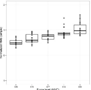
Monotonous and significantly linear relationship (Pearson’s test:r = 0.74; p< 0.001) was found between muscle force and RMS values in the simple contraction session
(shown in Figure 2). Larger muscle force corresponds stronger EMG signals.
Fig. 2. Subjects’ normalized RMS values during simple contraction session.
3.2 Fatigue sessions
Under the influence of both muscle force declines and the fatigue increases, the am-plitude of EMG decreases along the fatigue process. As shown in Figure 3, from 90% MVC to 50% MVC, the RMSfgoes from 95.4%to 70.2% (normalized by RMS
corres-ponding to maximal force).
Table 2.Normalized RMS values corresponding to five force levels during sMVC session
90% MVC 80% MVC 70% MVC 60% MVC 50% MVC
RMSf *(%) 95.38(13.55) 96.37(19.58) 89.69(18.30) 80.06(14.37) 70.19(16.90)
RMSfe *(%) 0.54(11.78) 2.24 (19.18) 14.70 (16.42) 14.67 (18.88) 21.27(18.66)
* Normalized by the RMS values corresponding to maximal force.
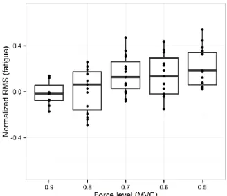
3.3 RMS response exclusively to fatigue
After precluding the influence of muscle force by equation (1), sEMG RMS response exclu-sively to fatigue was determined.Result shows thatit increaseswith fatigue strengthening:
RMSfe goes from 0.54% up to 21.27%while muscle force reduces from 90% MVC to 50%
MVC.Boxplotis shown in Figure 4.
Fig. 4.Subjects’ RMS response to fatigue during sMVC. Impact of muscle forceis precluded.
4
Discussion
4.1 Force - RMS relationship in simple contraction
In this experiment, linear relationship was found between muscle forces and RMS values during the simple contraction session. This clear relationship between mechan-ical and the electrmechan-ical responses of human muscle is well documented in previous researches[20,21,22] under voluntary isometric contractions.
As mentioned above, the RMS value of the sEMG signals is decided by: (1). the percentage of MUAP trains wave that filtered by the tissue and reach the electrodes,
0
designated as p; (2). the average length of the MUAP trains, designated asl;(3). the
firing rate of the MUAP trains, designated as f; (4). the meanamplitude of filtered MUAP trains, designated asA. Then RMS could be indicated by equation (2) ( is
the Constant coefficient).
0
RMS l p f A (2)
During the simple contractions when the muscles are fresh, the shape of MUAP waveform and the filtering threshold of the tissue remain unchanged. Therefore p is constant. With the increase of muscle force, MUs with higher firing rate of their am-plitude potential trains are recruited and the firing rates of initial MUAP trains in-crease. These factors lead to the increases of and f, and furthermore an increase of total RMS.
4.2 RMS during Fatigue Process
During the sMVC fatigue progress, the RMS reduces toabout 70% when muscle force reaches 50% MVC. Similar results have been reported in previous researches, as listed in Table 3. Generally, EMG amplitude reduced by30% to 70% during a max-imal voluntary contraction sustained for 60 s, depending on different muscles and protocols.
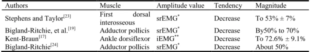
Table 3.EMG amplitude changes during sustainedmaximal voluntary contraction.
Authors Muscle Amplitude value Tendency Magnitude Stephens and Taylor[23] First dorsal
interosseous srEMG
* Decrease To 53% ± 7%
Bigland-Ritchie, et al.[19]
Adductor pollicis srEMG* Decrease By50% to 70% Kent-Braun[17] Ankle dorsiflexor iEMG** Decrease To 72.6% ± 9.1% Bigland-Ritchie[24] Adductor pollicis srEMG* Decrease About 50% *srEMG - Smoothed rectified EMG;
**
iEMG –Integreted EMG
As mentioned above, theRMSof sEMG has been found to increase in the process of constant-force fatiguing tasks[25,12,13]. When the body tries to maintain the target forces,
a progressive increase of MUAP trains firing rate take place and MUs with larger amplitude are recruited[27], which increase the f and in Eq.2. At the same time, the percentage of filtered MUAP trains pgrows due to fatigue. As a result, the total RMS increases.
Whereas in the procedure of sustained maximal exertions, both acute muscle fati-gue and rapid force decline are identified. The MUAP trains are pushed to change in two directions: on the one hand, in every moment of sMVC, the muscle is trying hard to maintain its original force, which leads to increases off and ; on the other hand, as the muscle fails to maintain its original force, muscle force continue to decline, which brings about the decreases of f and . As a consequence, the impact of force decline prevails over that of fatigue and decreases of f and are observed[27]. Despite of the
increase of the percentage of filtered MUAP trains p, the total RMS declines with time. A A A A A
4.3 EMG response exclusively to fatigue
After precludingthe impact of muscle force changes,the sEMG RMS response to fati-gue is found to increase from 0.54%to 21.27%, along with muscle force decreasing from 90%MVC to 50%MVC.The underlying implication is that in this process, al-though the firing rate of and average amplitude of MUAP trains decline significantly, increasingly extra effort is made compared with that when the fresh muscle exerts same forces. The more severe the muscle fatigue is, the more extra effort it is needed.
It is notable from Table 2 that no extra RMS increase is detected when force de-clines from 70%MVC to 60%MVC. This should be explained by the different percen-tage held by MU deactivation and firing rate slowing in force reduction at different force output level. As the muscle force increases, MUs are recruited in the order of their firing rate and twitch tension from low to high[9,28]along with the average firing
rate speeding up[29]. The recruitment of new MU was reported to terminate by
60%MVC[30] or 75%MVC[28] in biceps brachii. Conversely, during sMVC session,
before the muscle force reaches about 70%MVC, the slowing of MU firing rate con-tributes the largest partto force loss. From 70%MVC to 60%MVC, the deactivation of MU takes part in.MUs with the largest twitch tension are deactivate, which makes a great contribution to force loss. Hereby, the firing rate slowing and RMS changes are not significant.
5
Conclusions
In this research, a simple way to identify the sEMG RMS response to fatigue was tested on biceps brachii muscles during sMVC process. The impact of muscle force changes on RMS is precluded by simply subtracting the RMS of the fresh muscle when exerting corresponding force from the total RMS.Result shows that the sEMG RMS response to fatigue increasesalong with the fatigue process to as much as 20%, which implies that more and more extra effort is needed as muscle fatigueintensifies. It would be promising to use the RMS response exclusively to fatigue as an indicator of muscle fatigue.
Acknowledgments. This work was supported by the National Natural Science Foun-dation of China under Grant numbers 71101079 and 71471095, by the Tsinghua Uni-versity Initiative Scientific Research Program under Grant number 20131089234, by Chinese State Scholarship Fund, and by INTERWEAVE Project (Erasmus Mundus Partnership Asia-Europe) under Grants number IW14AC0456 and IW14AC0148.
References
1. Chaffin, D. B., Andersson, G., & Martin, B. J.: Occupational biomechanics. pp. 91-130. Wiley,New York (1999)
2. Ma, L., Chablat, D., Bennis, F., & Zhang, W.: A new simple dynamic muscle fatigue model and its validation. International Journal of Industrial Ergonomics. 39, 211-220. (2009)
3. Ma, L., Chablat, D., Bennis, F., Zhang, W., Hu, B., & Guillaume, F.: A novel approach for determining fatigue resistances of different muscle groups in static cases. International Journal of Industrial Ergonomics. 41, 10-18. (2011)
4. Ma, L., Zhang, W., Hu, B., Chablat, D., Bennis, F., & Guillaume, F.: Determination of subject-specific muscle fatigue rates under static fatiguing operations. Ergonomics. 56, 1889-1900. (2013)
5. Öberg, T. O. M. M. Y., SANDSJö, L. E. I. F., & KADEFORS, R.: Subjective and objec-tive evaluation of shoulder muscle fatigue. Ergonomics. 37, 1323-1333. (1994)
6. Cifrek, M., Medved, V., Tonković, S., & Ostojić, S. Surface EMG based muscle fatigue evaluation in biomechanics. Clinical Biomechanics.24, 327-340.(2009)
7. De Luca, C. J.: The use of surface electromyography in biomechanics. Journal of applied biomechanics. 13, 135-163. (1997)
8. De Luca, C. J.: Spectral compression of the EMG signal as an index of muscle fatigue. Neuromuscular fatigue. Royal Netherlands Academy of Arts and Sciences, Amsterdam, The Netherlands, 44-51.(1992)
9. De Luca, C. J.: Physiology and mathematics of myoelectric signals. Biomedical Engineer-ing, IEEE Transactions on. 313-325. (1979)
10. Basmajian, J. V., & De Luca, C. J.: Muscles alive. Muscles alive: their functions revealed by electromyography. 278, 126. (1985)
11. Brody, L. R., Pollock, M. T., Roy, S. H., De Luca, C. J., & Celli, B.: pH-induced effects on median frequency and conduction velocity of the myoelectric signal. Journal of Ap-plied Physiology. 71, 1878-1885. (1991)
12. Iridiastadi, H., & Nussbaum, M. A.: Muscle fatigue and endurance during repetitive in-termittent static efforts: development of prediction models. Ergonomics. 49, 344-360. (1991)
13. Bigland-Ritchie B, Furbush F, Woods J.: Fatigue of intermittent submaximal voluntary contractions: central and peripheral factors. Journal of Applied Physiology. 61,421-429.(1986)
14. Dedering, Åsa, Gunnar Németh, and Karin Harms-Ringdahl: Correlation between elec-tromyographic spectral changes and subjective assessment of lumbar muscle fatigue in subjects without pain from the lower back. Clinical Biomechanics. 14, 103-111.(1999) 15. Nussbaum, M. A.: Static and dynamic myoelectric measures of shoulder muscle fatigue
during intermittent dynamic exertions of low to moderate intensity. European journal of applied physiology. 85, 299-309. (2001)
16. Moritani, T. O. S. H. I. O., Muro, M. A. S. U. O., & Nagata, A. K. I. R. A.: Intramuscular and surface electromyogram changes during muscle fatigue. Journal of Applied Physiolo-gy. 60, 1179-1185.(1986)
17. Kent-Braun, J. A.: Central and peripheral contributions to muscle fatigue in humans dur-ing sustained maximal effort. European journal of applied physiology and occupational physiology. 80, 57-63. (1999)
18. Gandevia, S. C.: Spinal and supraspinal factors in human muscle fatigue. Physiological reviews. 81, 1725-1789. (2001)
19. Bigland-Ritchie, B., Johansson, R., Lippold, O. C., & Woods, J. J.: Contractile speed and EMG changes during fatigue of sustained maximal voluntary contractions. Journal of neurophysiology. 50, 313-324.(1983)
20. Woods, J. J., &Bigland-Ritchie, B.: Linear and non-linear surface EMG/force relation-ships in human muscles. An anatomical/functional argument for the existence of both. American Journal of Physical Medicine & Rehabilitation. 62, 287-299. (1983)
21. Doheny, E. P., Lowery, M. M., FitzPatrick, D. P., & O’Malley, M. J.: Effect of elbow joint angle on force–EMG relationships in human elbow flexor and extensor muscles. Journal of Electromyography and Kinesiology. 18, 760-770. (2008)
22. Troiano, A., Naddeo, F., Sosso, E., Camarota, G., Merletti, R., & Mesin, L. Assessment of force and fatigue in isometric contractions of the upper trapezius muscle by surface EMG signal and perceived exertion scale.Gait& posture. 28, 179-186. (2008)
23. Stephens, J. A., and A. Taylor.: Fatigue of maintained voluntary muscle contraction in man. The Journal of physiology. 220, (1972)
24. Bigland-Ritchie, B.: EMG and fatigue of human voluntary and stimulated contractions. Human muscle fatigue: physiological mechanisms.130-156. (1981)
25. Petrofsky, J. S., Glaser, R. M., Phillips, C. A., LIND, A. R., & WILLIAMS, C.: Evalua-tion of the amplitude and frequency components of the surface EMG as an index of mus-cle fatigue. Ergonomics. 25, 213-223. (1982)
26. Hagberg, M., & Ericson, B. E.: Myoelectric power spectrum dependence on muscular contraction level of elbow flexors. European Journal of Applied Physiology and Occupa-tional Physiology. 48, 147-156. (1982)
27. Moritani, T. O. S. H. I. O., M. A. S. U. O. Muro, and A. K. I. R. A. Nagata.:Intramuscular and surface electromyogram changes during muscle fatigue. Journal of Applied Physiolo-gy. 60, 1179-1185. (1986)
28. Clamann, H. P.: Activity of single motor units during isometric tension.Neurology.20, 254-254. (1970)
29. Grimby, L. E. N. N. A. R. T., &Hannerz, J.: Firing rate and recruitment order of toe ex-tensor motor units in different modes of voluntary conraction.The Journal of physiolo-gy.264, 865. (1977)
30. Gydikov, A., &Kosarov, D.: Some features of different motor units in human biceps brachii. PflügersArchiv.347, 75-88. (1974)