HAL Id: hal-01832145
https://hal.archives-ouvertes.fr/hal-01832145
Submitted on 13 Jul 2018
HAL is a multi-disciplinary open access
archive for the deposit and dissemination of
sci-entific research documents, whether they are
pub-lished or not. The documents may come from
teaching and research institutions in France or
abroad, or from public or private research centers.
L’archive ouverte pluridisciplinaire HAL, est
destinée au dépôt et à la diffusion de documents
scientifiques de niveau recherche, publiés ou non,
émanant des établissements d’enseignement et de
recherche français ou étrangers, des laboratoires
publics ou privés.
state
Olfat A. Malak, Zeineb Es-Salah-Lamoureux, Gildas Loussouarn
To cite this version:
Olfat A. Malak, Zeineb Es-Salah-Lamoureux, Gildas Loussouarn. hERG S4-S5 linker acts as a
voltage-dependent ligand that binds to the activation gate and locks it in a closed state. Scientific Reports,
Nature Publishing Group, 2017, 7 (1), pp.113. �10.1038/s41598-017-00155-2�. �hal-01832145�
www.nature.com/scientificreports
hERG S4-S5 linker acts as a
voltage-dependent ligand that binds to
the activation gate and locks it in a
closed state
Olfat A. Malak, Zeineb Es-Salah-Lamoureux & Gildas Loussouarn
Delayed-rectifier potassium channels (hERG and KCNQ1) play a major role in cardiac repolarization. These channels are formed by a tetrameric pore (S5–S6) surrounded by four voltage sensor domains (S1-S4). Coupling between voltage sensor domains and the pore activation gate is critical for channel voltage-dependence. However, molecular mechanisms remain elusive. Herein, we demonstrate that covalently binding, through a disulfide bridge, a peptide mimicking the S4-S5 linker (S4-S5L) to the
channel S6 C-terminus (S6T) completely inhibits hERG. This shows that channel S4-S5L is sufficient to
stabilize the pore activation gate in its closed state. Conversely, covalently binding a peptide mimicking S6T to the channel S4-S5L prevents its inhibiting effect and renders the channel almost completely
voltage-independent. This shows that the channel S4-S5L is necessary to stabilize the activation gate
in its closed state. Altogether, our results provide chemical evidence that S4-S5L acts as a
voltage-controlled ligand that binds S6T to lock the channel in a closed state, elucidating the coupling between
voltage sensors and the gate in delayed rectifier potassium channels and potentially other voltage-gated channels.
Voltage-dependent ion channels are ubiquitously expressed in human tissues. They perform a plethora of phys-iological functions such as generation and modulation of the electrical activity in excitable tissues, modulation of neurotransmitter and hormone release, and electrolyte transport in epithelia. Canonical voltage-gated ion channels are tetramers of subunits containing six transmembrane segments (S1 to S6). Each of the four subunits is composed of one voltage sensor domain (S1 to S4) and a pore domain (S5-S6). The four pore domains tetramer-ize to generate a single pore module, which is regulated by the four voltage sensor domains1, 2. One key question
remains: how do the voltage sensor domains regulate pore gating? Several independent approaches support the idea that interaction between the S4-S5 linker (S4-S5L) and the S6 C-terminal (S6T) part plays a critical role in that
regulation2–5. First, Lu and collaborators pinpointed the sequence complementarity between S4-S5
L and S6T in the
Shaker channel4, 5. Second, structural data indicated the close proximity between S4-S5
L and S6T and suggested a
mechanical lever model to explain the coupling between the voltage sensor and the gate of the related Kv1.2
chan-nel2. However, the mechanism may prove to be more complex. Since 2006, several works on other eukaryotic K
v
channels suggested state-dependent S4-S5L and S6T interactions3, 6–10, but the precise mechanism remains elusive
and may vary from one channel to another.
In a previous study, we suggested that the voltage dependence of the cardiac voltage-gated KCNQ1 channel (Kv7.1) follows a ligand/receptor model. In this model, S4-S5L is a ligand whose interaction with S6T is only
possible at resting potentials, and this interaction locks the channel in a closed state. Upon depolarization of the plasma membrane, S4 pulls the S4-S5L out of its binding site and unlocks the channel (Fig. 1A). We validated this
ligand/receptor model by using peptides mimicking S4-S5L and S6T and evaluating their effects on the channel
activity. However the peptides effects were moderate, probably due to the low affinity between native S4-S5L and
S6T, which is necessary for S4-S5L ligand unbinding and channel opening during membrane depolarization11. In
the present study, we started with a similar strategy of peptide native-sequence mimicry to evaluate the ligand/ receptor model in hERG (Kv11.1), a key channel in cardiac and neuronal electrical activity. As in the KCNQ1
INSERM, CNRS, l’Institut du Thorax, Université de Nantes, 44007, Nantes, France. Zeineb Es-Salah-Lamoureux and Gildas Loussouarn contributed equally to this work. Correspondence and requests for materials should be addressed to G.L. (email: gildas.loussouarn@inserm.fr)
Received: 3 May 2016 Accepted: 8 February 2017 Published: xx xx xxxx
OPEN
results, hERG-mimicking peptide effects were moderate. In a previous study on this channel, a disulfide bond was created between S4-S5L and S6T, stabilizing it in a closed state6. Here, after the first step of peptide identification,
we used the same cysteine approach to increase the peptide-channel interaction and to target the peptide to its hypothetical receptor (S6T for S4-S5L and vice-versa). We observed that covalently binding a peptide mimicking
S4-S5L to the endogenous S6T could completely inhibit hERG. On the other hand, covalently binding a peptide
mimicking S6T to the endogenous S4-S5L rendered the channel almost voltage-independent, most likely by
pre-venting the inhibitory effect of S4-S5L on S6T in the channel.
Altogether, these results suggest that the voltage dependence of these two delayed rectifier channels, KCNQ1 and hERG, follows a ligand/receptor model in which S4-S5 linker acts as an inhibitor, locking the activation gate under the control of the voltage sensor S4.
Results
One S4-S5
Lpeptide inhibits hERG channels.
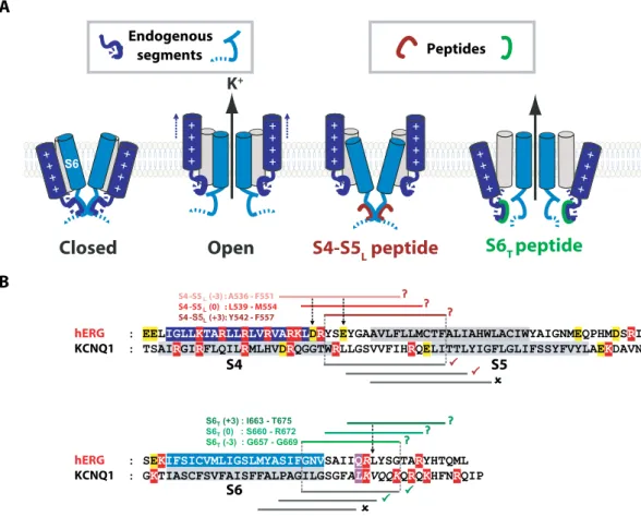
To identify a potential inhibitory S4-S5L peptide, threeS4-S5L encoding plasmids were designed based on sequence alignment with KCNQ1 (Fig. 1B), in which the
ligand/receptor model was first suggested, the S4-S5 linker (S4-S5L) being the inhibiting ligand and the S6
C-terminus (S6T) acting as the receptor11. To study peptide effects on channel activity, each plasmid coding for
one of the S4-S5L peptides was co-transfected with a plasmid encoding the hERG channel.
If the endogenous S4-S5L acts like a ligand to inhibit the activation gate, then a peptide mimicking endogenous
S4-S5L should decrease hERG channel activity. As expected, the S4-S5L (+3) peptide decreased the current
den-sity (Fig. 2A and B, Supplemental Table 1). Western blot experiments revealed that this decrease was not due to a
Closed
Open
K
+S4-S5
Lpeptide
S6
Tpeptide
+ + + + + + + + + + + + + + + + + + + + + + + + + + + + + + + +B
S4 S5 ( 3) : A536 F551 S4 -S5 (0) : L539 M554 S4 - (+3): Y542 F557 S4 S5 hERG KCNQ1 S6 hERG KCNQ1 Endogenous segments PeptidesFigure 1. Hypothetical ligand/receptor model. Alignment used to design S4-S5L and S6T peptides. (A) Scheme
of the hypothetical ligand/receptor model in which S4-S5L (deep blue) binds to S6T (light blue) to stabilize the
channel in a closed state. Upon membrane depolarization, S4 pulls S4-S5L out of the S6T receptor, allowing
channel opening. The S4-S5L peptide (red) mimics endogenous S4-S5L, locking the channel in its closed
conformation. Contrarily, S6T peptide (green) binds to the endogenous S4-S5L and limits its locking effect,
leading to channel up-modulation. (B) Alignment used to design hERG peptides from previously identified KCNQ1 S4-S5L and S6T peptides (based on the multiple alignment obtained using Clustal Omega, presented
in Supplemental Fig. 4). In red are represented the basic residues, in yellow acidic residues, and in purple the position of the narrowest part of the bundle crossing, also named the gating residue (see methods). The color boxes represent the transmembrane segments. Grey lines represent the peptides tested in KCNQ1 while red lines and green lines represent the designed hERG S4-S5L and S6T peptides, respectively. A check sign (✓)
indicates that the KCNQ1 S4-S5L peptide inhibits the channel (red) and that the KCNQ1 S6T peptide activates
www.nature.com/scientificreports/
lower expression of hERG protein in the presence of this peptide, as compared to control conditions (Fig. 3A and C). Thus, these results are consistent with the ligand/receptor model.
However, the S4-S5L (−3) peptide did not show any effect on current density and led to a 5-mV negative
shift in the activation curve (Fig. 2A–C, Supplemental Table 1). The S4-S5L (−3) peptide presents 5 charged
amino-acids, 3 positively charged on one face of the helix and 2 negatively charged on the other face. The absence of any effect on current density suggests a non-specific effect such as membrane charge screening, locally chang-ing the potential detected by the voltage-sensor.
The S4-S5L (0) peptide induced a paradoxical increase in hERG current density without any effect on the
activation curve (Fig. 2A–C, Supplemental Table 1). Western blot experiments revealed that this peptide led to a doubling of hERG protein as compared to control conditions (Fig. 3A and C). Surprisingly, this peptide also induced an increase in GFP expression (by a factor of 7.1, p < 0.01), as shown in Fig. 3A. However expression of endogenous control GAPDH was not altered by S4-S5L (0) (Fig. 3A and C).
This set of experiments shows specific effects of only one of the S4-S5L. The S4-S5L (+3) peptide decreased the
current density without modifying the activation curve, similar to the effects of the S4-S5L inhibiting peptides on
KCNQ1 channel11. This is consistent with the ligand/receptor model.
Figure 2. S4-S5L (+3) peptide inhibits and S6T (−3) peptide activates hERG channels. (A) Representative,
superimposed recordings of the WT hERG current in the absence (0.6 µg hERG plus 1.4 µg GFP plasmids) and in the presence of S4-S5L or S6T peptides (0.6 µg hERG plus 1.4 µg peptide plasmids). Left: schemes of the
hypothetical effects of S4-S5L inhibiting or S6T activating peptide; activation voltage protocol used (one sweep
every 8 s) located above the WT hERG currents; right: data from experiments performed in the presence of various S4-S5L or S6T peptides. (B) Mean hERG tail-current density at −40 mV after a prepulse at +60 mV in
the presence of S4-S5L peptides. (C) Activation curve, obtained from tail currents using the protocol shown
in A, in the presence of S4-S5L peptides (n = 21–65). (D) Inactivation curve in the presence of S4-S5L peptides
(n = 6–24). Inset, Inactivation voltage protocol used (one sweep every 5 s, first pulse = 1 s, second pulse = 15 ms, third pulse = 0.5 s). (E) Through (G) same as B through D, in the presence of S6T peptides (F, n = 28–65; G,
One S6
Tpeptide activates hERG channels.
If S6T is the receptor for S4-S5L, then a peptidemimick-ing endogenous S6T should act as a decoy to the endogenous S4-S5L, preventing its binding to the endogenous
S6T and increasing the channel activity. Indeed, the S6T (−3) peptide up-regulated the WT channel and shifted
the activation curve (and inactivation curve) by 5 mV toward negative potentials (Fig. 2A,E–G, Supplemental Table 1) without any change in the hERG expression level (Fig. 3B and C), which is similar to the effects of the S6T-activating peptides on KCNQ1 channel11 and therefore also consistent with the ligand/receptor model. The
other peptides, namely S6T (0) and S6T (+3) did not produce any effect.
Altogether, the effects of one inhibiting and one activating peptide were similar for both hERG and KCNQ1 channels11. Moreover, both hERG inhibiting and activating peptides are aligned with those of the KCNQ1
chan-nel (Fig. 1). In conclusion, these two similarities suggest that the ligand/receptor model applies to both channels.
A cysteine disulfide bond reinforces the effect of the S4-S5
L(+3) peptide, leading to full
inhibi-tion of hERG channel.
Although consistent with the ligand/receptor model, the S4-S5L (+3) effect ismod-erate, as in KCNQ1, likely because the S4S-5L/S6T interaction has to be loose, as explained in the introduction.
In Xenopus oocytes, introduction of a cysteine pair in the hERG channel (D540C in S4-S5L with L666C in S6T)
has been previously shown to stabilize the channel in a closed conformation through the formation of a disulfide bond6. We reasoned that this disulfide bond could be exploited to covalently bind the mimicking S4-S5
L (+3)
peptide to the S6T channel, which should enhance its inhibitory effect.
First, we evaluated the effects of oxidizing the native cysteines on WT hERG activity. We observed that in COS-7 cells, as previously shown in Xenopus Oocytes, WT currents were unaffected after 2 hours of incubation with 0.2 mM tert-butylhydroperoxide (tbHO2; Fig. 4A,C and D, Supplemental Table 2), showing that oxidizing
the native cysteines does not have any effect on channel gating and trafficking. Such a tbHO2 treatment led to a
permanent closure of the D540C-L666C hERG mutant and this inhibition was reversed by subsequent incubation with 10 mM DTT (Fig. 4B and E, Supplemental Table 2). Finally, the current density of the single mutants D540C and L666C was not altered by incubation with tbHO2 (Supplemental Fig. 2), confirming that the D540C-L666C
mutant inhibition by tbHO2 is specifically due to the disulfide bond formed between D540C and L666C.
Figure 3. S4-S5L (0) peptide increases hERG channel expression. (A,B) western blot analysis of hERG protein
expression in COS-7 cell lysates in the absence or presence of S4-S5L peptides (A) or S6T peptides (B). Top:
stain-free image of total proteins. Bottom: western blot images of hERG, GAPDH, and GFP proteins. The three blots, realized on the same membrane, are cropped. Full-length blots of each tested protein are reported in Supplemental Fig. 1. (C) Histogram of normalized mean intensity of hERG (left) and GAPDH (right) bands in the absence and in the presence of various S4-S5L or S6T peptides. Band intensities are first normalized to
the intensity of the corresponding stain-free membrane lane, and ratios are then normalized to control hERG condition. **p < 0.01 versus control hERG, Mann-Whitney test realized on non-normalized ratios.
www.nature.com/scientificreports/
The inhibiting S4-S5L (+3) peptide does not contain the D540 residue (Fig. 1), but the E544 residue, located
on the same side of the S4-S5L helix as D540, is negatively charged as well. Both D540 and E544 may interact
electrostatically with R665 (next to L666) in S6T when the channel is in the closed state12, 13. Therefore, we tested
if a cysteine introduced in E544 hERG instead of D540 and another cysteine introduced in L666 hERG could also form a disulfide bridge, which would lead to a permanent closure of the channel. COS-7 cells were transfected with the E544C-L666C double mutant hERG, and the generated current was indeed inhibited by incubation with 0.2 mM tbHO2, while the E544C and L666C single mutants were not (Fig. 5A, Supplemental Fig. 2). Since E544C/
L666C can form a disulfide bond within the channel, we tested if this same cysteine pair could also covalently link the E544C mutated S4-S5L (+3) peptide onto the S6T of L666C single mutant hERG channel. Cells were
co-transfected with the E544C S4-S5L (+3) peptide and the L666C channel. Two hours of incubation with 0.2 mM
tbHO2 almost completely inhibited the current (Fig. 5B, Supplemental Table 3), and this effect was specifically
due to the presence of both cysteines (Fig. 5C and D, Supplemental Table 3). In conclusion, covalently binding S4-S5L to S6T inhibited the channel, strongly suggesting that S4-S5L acts as a ligand binding to S6T and locking the
channel in a closed state.
hERG inhibition provoked by E544C S4-S5
L(+3) peptide covalent binding to L666C mutant
channel is reversible.
Cells co-transfected with the E544C S4-S5L (+3) peptide and the L666C channelwere incubated for 2 hours with tbHO2, followed by 2 hours incubation with 10 mM DTT. This double treatment
reversed the effect induced by tbHO2 incubation, as current densities were comparable to the control condition
(Fig. 6). To exclude that the current recovery is due to de novo arrival of hERG at the plasma membrane during the 2 hours DTT incubation, we tested two additional conditions: 1) 10 mM DTT was applied for only 10 minutes or 2) we measured the current in the presence of 10 mM DTT in the pipette tip. In both conditions, current den-sities were similar to control, consistent with DTT application leading to peptide unbinding.
Figure 4. Introduction of 2 cysteines in the S4-S5L and S6T regions of hERG (D540C-L666C hERG) locks the
channel closed in oxidative conditions. (A) Representative, superimposed recordings of the WT hERG current (3.6 µg plasmid plus 0.4 µg GFP plasmids) after 2 h incubation in Tyrode without (control) or with 0.2 mM tbHO2 (tbHO2). (B) D540C-L666C hERG channels, same as in A (3.6 µg plasmid plus 0.4 µg GFP plasmids).
Cartoons: introduction of a cysteine is symbolized by a star. (C) Mean WT hERG tail-current density at −40 mV after a prepulse at +100 mV. (D) Activation curve obtained from the tail currents, using the protocol shown in (B). (E) Mean D540C-L666C hERG tail-current density at −40 mV after a prepulse at +100 mV, after 2 h incubation in Tyrode without (control) or with 0.2 mM tbHO2 (tbHO2), or after 2 h incubation in 10 mM
DTT following tbHO2 incubation (2 h DTT). ***p < 0.001 versus control and ###p < 0.001 versus tbHO2,
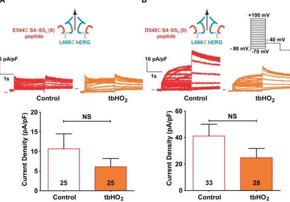
hERG activation by S4-S5
L(0) peptide is not reinforced by its covalent binding through a
disulfide bond.
S4-S5L (0) peptide increased WT hERG current density (Fig. 2A and B), but this effect is dueto an increased expression of hERG protein, suggesting no effect of S4-S5L (0) on channel gating. Consistent with
this hypothesis, the current generated by COS-7 cells co-transfected with E544C S4-S5L (0) peptide and L666C
hERG channel was not increased upon incubation with tbHO2 (Fig. 7A, Supplemental Table 4). Since S4-S5L (0)
also includes the D540 residue (Fig. 1), we also tested the effect of D540C S4-S5L (0) peptide on L666C single
mutant hERG channel. Again, the current was not increased upon tbHO2 application (Fig. 7B, Supplemental
Table 4). Altogether, these observations confirmed that the paradoxical activation effect initially observed when S4-S5L (0) peptide was co-transfected with WT hERG channel was not due to an interaction of this peptide with
the S6T region of hERG.
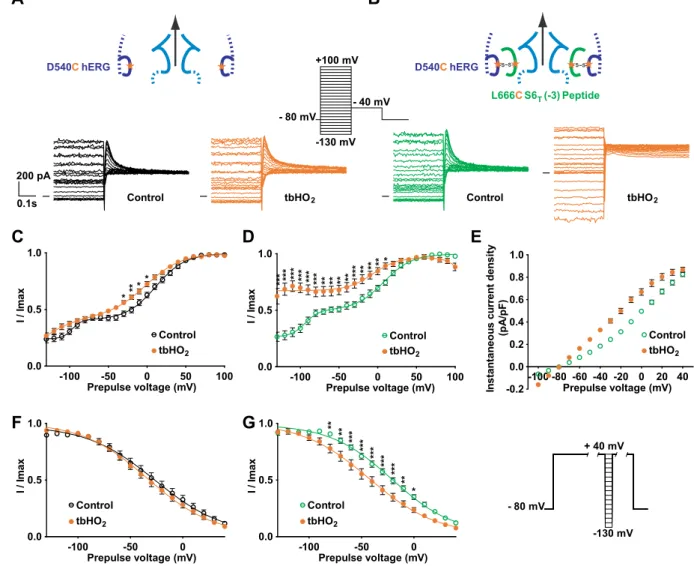
A cysteine disulfide bond reinforces the effect of the activating S6
Tpeptide, rendering the
channel almost voltage independent.
Since the S6T peptide contains the L666 residue, we mutated thisresidue to a cysteine and tested the effect of this mutated peptide on the D540C single mutant hERG channel,
Figure 5. A cysteine disulfide bond reinforces the effect of the S4-S5L (+3) peptide, leading to full inhibition
of the channel. (A) left, representative, superimposed recordings of the E544C-L666C hERG current (3.6 µg E544C-L666C hERG plasmid plus 0.4 µg GFP plasmids) and right, mean tail-current density at −40 mV after a prepulse at +100 mV after 2 h incubation in Tyrode without (control) or with 0.2 mM tbHO2 (tbHO2), both
using the voltage protocol shown in inset. (B) left, representative, superimposed recordings of the single mutant L666C hERG current in the presence of E544C S4-S5L (+3) peptide (1 µg L666C hERG plus 3 µg peptide
plasmids) and right, mean tail-current density measured in the same conditions as A. (C,D) Mean tail-current density in the presence of only one cysteine, i.e. L666C hERG +S4-S5L (+3) peptide (C) or WT hERG +E544C
www.nature.com/scientificreports/
Figure 6. hERG inhibition provoked by E544C S4-S5L (+3) peptide covalent binding to L666C mutant
channel is reversible. Mean hERG tail-current density at −40 mV after a prepulse at +100 mV of L666C hERG channels in the presence of E544C S4-S5L (+3) peptide (1 µg L666C hERG plus 3 µg peptide plasmids) after
2 h incubation in Tyrode without (control) or with 0.2 mM tbHO2 (control and tbHO2, respectively, same
results as in Fig. 5B), and after 2 h or 10 min incubation in 10 mM DTT following tbHO2 incubation, or after
direct application of 10 mM DTT in the pipette tip, following tbHO2 incubation. ***p < 0.001 versus control, ##p < 0.01 and ###p < 0.001 versus tbHO
2, Mann-Whitney test.
Figure 7. Unexpected hERG activation by S4-S5L (0) peptide is not reinforced by its covalent binding though a
disulfide bond. (A) Top, representative, superimposed recordings of the L666C hERG current in the presence of E544C S4-S5L (0) peptide (1 µg L666C hERG plus 3 µg peptide plasmids) after 2 h incubation in Tyrode without
(control) or with 0.2 mM tbHO2 (tbHO2), using the protocol shown in (B). Bottom, corresponding mean hERG
both in the absence and in the presence of tbHO2. As shown in Fig. 8C, the D540C channel presents an atypical
activation curve, with a double Boltzmann and a voltage-independent component, even at very negative poten-tials, unlike the WT channel. This suggests that the mutation D540C by itself alters the role of endogenous S4-S5L
as an inhibiting ligand. This voltage-independent component is similar to the one induced by some mutations in KCNQ1 that were interpreted as decreasing the interaction between S4-S5L and S6T11, 14. Covalent binding of the
L666C S6T (−3) peptide to the D540C hERG channel dramatically increased the voltage-independent component
(Fig. 8B and D). Such a tbHO2-dependent increase in the voltage-independent component was not observed in
absence of the peptide (Fig. 8A and C). The voltage-independent component is potassium selective (Fig. 8E). These observations further confirm the ligand/receptor mechanism by demonstrating that S4-S5L is necessary to
lock the channel gate in a closed state.
Covalent binding of the L666C S6T (−3) peptide to the channel D540C also leads to a 12-mV negative shift in
the inactivation curve (Fig. 8G and Supplemental Table 5). In the hERG channel, inactivation is not directly cou-pled to activation15, 16, suggesting that the observed effect of the L666C S6
T (−3) peptide on the inactivation curve
is not a consequence of a modification of activation. Of note, S6T peptides bind to hERG S4-S5L (Y542-F557)
which is surrounded by regions that are involved in inactivation: double mutant cycle analysis suggests that inter-action between L529, L532, V535 in S4 and I560, L564, I567 in S5 play a role in inactivation gating17. Thus,
cova-lent binding of S6T to S4-S5L may alter this interaction leading to the observed effect on inactivation.
Figure 8. A cysteine disulfide bond reinforces the effect of the activating S6T peptide, rendering the channel
almost voltage independent. (A,B) Representative superimposed recordings of the single mutant D540C hERG current (A, 1 µg D540C hERG plus 3 µg GFP plasmids) and D540C hERG current in the presence of L666C S6T
(−3) peptide (B, 1 µg D540C hERG plus 3 µg peptide plasmids) after 2 h incubation in Tyrode without (control) and with 0.2 mM tbHO2 (tbHO2), using the voltage protocol shown in inset. (C,D) Activation curve, with a
double Boltzmann, obtained from the tail currents in the same conditions as A, in the absence (C) or presence of the L666C S6T (−3) peptide (D). (E) Maximum current density measured during the prepulse, using the
protocol and conditions as in (A). (F,G) D540C hERG inactivation curve obtained using the protocol shown in right inset (same as in Fig. 2), in the absence (F) or presence of the L666C S6T (−3) peptide (G). *p < 0.05,
www.nature.com/scientificreports/
Discussion
In a previous study on KCNQ1, and in the present study on hERG, we proposed a ligand/receptor model of volt-age dependence. In this model, S4-S5L would act as a ligand locking the S6T gate in the closed state. To test this,
we used S4-S5L and S6T mimicking peptides. Our results are in favor of this hypothesis. However, the effects were
moderate. Here, we used another approach to reinforce the peptide effects on the hERG channel by introducing a cysteine pair covalently linking the mutated peptide to the single mutant channel. We observed that covalently linking the E544C S4-S5L (+3) peptide to the L666C hERG almost completely inhibits the channel (Fig. 5B), by
presumably preventing the outward splaying of the S6 helices. Covalently linking the L666C S6T (−3) peptide
to D540C hERG renders the channel almost completely voltage independent (Fig. 8D). We postulate that the covalently bound S6T (−3) peptide blocks the normal interaction between endogenous S4-S5L and S6T which
stabilizes the closed state, and thus the channel remains locked in the open state. For both peptides, either S4-S5L
or S6T, covalent link to the hERG channel clearly reinforces the respective effects of these two peptides when first
tested on WT hERG channel without any introduced cysteine (as in Fig. 2). Altogether, these observations further reinforce the ligand/receptor model of voltage dependence. hERG S4-S5L and S6T peptides that produce the most
potent effects are aligned with the most potent peptides of KCNQ1, strengthening the notion of a similar model for KCNQ1 and hERG activation gating (Fig. 9).
Cysteine introduction either in S4-S5L or S6T endogenous segments leads to alteration of the single mutant
channel activity, as shown in Supplemental Fig. 3. This alteration is indicative of a change in the channel confor-mation. This observation may give rise to an alternative hypothesis: an artificial S4-S5L/S6T interaction is only
due to the cysteine introductions and does not exist in WT conditions. However, (i) S4-S5L (+3) and S6T (−3)
peptides altered WT channel current density, suggesting that the ligand/receptor model is relevant for the WT situation even without the disulfide bond, (ii) despite the alteration of the channel voltage dependence due to the cysteines introduction, covalently linking the peptide to its target leads to effects which are consistent with the ligand/receptor model. Indeed, we observed, on one hand, a complete inhibition by E544C S4-S5L (+3) on L666C
hERG, and, on the other hand, a loss of voltage dependence by L666C S6T (−3) on D540C hERG. In conclusion,
two independent sets of experiments (with or without cysteines) are consistent with the ligand/receptor model. Interestingly, the same combination of cysteine introduction in S4-S5L (0) peptide (E544C) and hERG (L666C),
led to a complete loss of the current increase which was demonstrated to be nonspecific to the channel activity change but rather due to the channel expression increase when probed by western blot experiments. Altogether, our results suggest that cysteine introduction in the channel preserved the ligand/receptor mechanism that was originally observed in WT hERG (and also KCNQ1).
In the absence of structural data of Kv channels in the closed state, molecular mechanisms underlying the
coupling between voltage-sensor movement and pore opening are still under debate. This coupling interaction has been suggested to be electromechanical and categorized as “attractive” or ”repulsive”, depending on the force exerted on the gate by the voltage sensors via the S4-S5L and S6T interaction18. In the “attractive” coupling, the S4
voltage sensors pull the gate to open it, requiring tight binding between S4-SL and S6T. In the “repulsive” coupling,
the voltage sensors push the gate to constrict it. In the present work on hERG channel, the ligand/receptor model corresponds to a third mechanism in which there is no need of a force directly linking the voltage-sensor and the gate, neither “attractive” nor ”repulsive”. Rather, S4-S5L seems to act as a ligand that binds S6T to lock the channel
in a closed state. When S4 segments are in the “down” state at negative potential, S4-S5L binds to S6T, locking the
channel in the closed state. When S4 segments are in the “up” state, they pull S4-S5L out of its binding pocket
(S6T), unlocking the channel. This molecular mechanism is consistent with previous observations made on hERG
channels and other Kv channels:
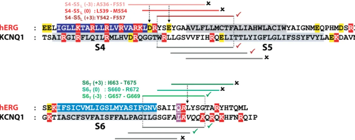
S4 S5 ( 3) : A536 F551 S4 -S5 (0) : L539 M554 S4 - (+3): Y542 F557
S4
S5
hERG KCNQ1S6
hERG KCNQ1Figure 9. hERG inhibiting (S4-S5L (+3)) and activating peptides (S6T (−3)) are aligned with KCNQ1
inhibiting and activating peptides. Same as in Fig. 1B, onto which the results obtained with hERG have been added. A check sign (✓) indicates that the S4-S5L peptide inhibits the channel and that the S6T peptide activates
channel, locks it in a closed state via a disulfide bond6. This is consistent with our hypothesis that S4-S5
L acts
as an endogenous inhibitor, as confirmed by the exogenous peptide approach.
• Split hERG channels expressed as two distinct proteins, one containing the voltage sensor domain and one containing the pore, keep the same voltage dependence as the full-length channel19. In accordance with our
model, where there is no mechanical force transduction from S4 movement to S6T opening, there is no need
for physical continuity between the two parts of the channel for activation transduction.
• The ligand/receptor model also helps in understanding why some hERG mutants, such as D540K, are open by both membrane depolarization and membrane hyperpolarization20. Since specific interactions between
S4-S5L and S6T are needed to keep the channel closed, a charge reversal mutation on S4-S5L may destabilize
the interaction and lead to channel opening when S4-S5L is pulled out of its S6T binding pocket, either toward
the cytosol (hyperpolarization) are toward the membrane (depolarization).
• Of note, it has been recently shown that the structure of the Kv10.1 channel, which is 65% homologous to
hERG, is incompatible with the mechanical lever mechanism21. In the open state structure of K
v10.1, S4-S5L
is a short linker that is not domain swapped and, thus, is not in a position to function as a mechanical lever, leaving the possibility that the Kv10.1 follows the same ligand/receptor model as hERG and KCNQ1.
This ligand/receptor model implies that when S4-S5L is absent, the hERG activation gate should be in the
open state, independent of the potential, as observed in one potassium22 and one sodium23 bacterial channels.
In other words, this gate is intrinsically more stable in the open state. Surprisingly, hERG pore domain construct (S5-S6) alone, isolated from the voltage sensor domain, gives rise to a nonfunctional channel19. One particularity
for hERG, compared to the aforementioned bacterial channels, is that the cytosolic N-terminus deletion greatly accelerates deactivation24–26. Thus, the N-terminus stabilizes the channel in the open state, and ablation of the
hERG voltage sensor domain, which also deletes the N-terminus, may render hERG nonfunctional.
Of note, this ligand/receptor model is completely in frame with the allosteric model of voltage-dependence suggested for KCNQ1 and hERG, in which voltage-dependent movement of S4 is allosterically coupled to pore-opening transitions27, 28. In such a model, the gating of KCNQ1 can be well described by an allosteric model
where the channel can open before all S4 voltage sensors have been activated29, 30. Our ligand/receptor model
suggests that allosteric coupling is realized through the interaction between S4-S5L and S6T (leading to channel
closure), which is favored when S4 segments are in the “down” state at resting potential and not favored when S4 segments are the in the “up” state.
In conclusion, the results that we obtained in the hERG channel with S4-S5L and S6T mimicking peptides, with
or without cysteine introduction, reinforce the ligand/receptor model originally observed in KCNQ1 and suggest that this model applies to other voltage-gated channels. In Shaker channels, sequence complementarity between S4-S5L and S6T has also been shown to be critical to coupling voltage sensor movement and pore opening4, 5.
Testing peptides mimicking S4-S5L and S6T in Shaker-like channels would confirm whether the ligand/receptor
model applies to Shaker-like channels.
Methods
Plasmid constructs.
Oligonucleotides encoding hERG peptides were synthesized by TOP Gene Technologies and contained a XhoI restriction enzyme, a methionine (ATG) for translation initiation, and a glycine (GGA) to protect the ribosome binding site during translation and the nascent peptide against proteolytic degradation31. A BamHI restriction enzyme site was synthesized at the 3′ end immediately following thetransla-tional stop codon (TGA). These oligonucleotides were then cloned into pIRES2-EGFP (Clontech) and sequenced. Three different S4-S5L and S6T plasmids were designed with the same length (16 amino-acids for S4-S5L and
13 for S6T) but different starting positions in order to compensate for the inaccuracy of the alignment due to the
poor homology between hERG and KCNQ1 sequences (Fig. 1 and Supplemental Fig. 4). Multiple alignment was realized with Clustal Omega32. This program aligned the predicted transmembrane domains (Uniprot Q12809 for
hERG, P51787 for KCNQ1) and the predicted position of the narrowest part of the bundle crossing, also named gating residue33, 34, of hERG and KCNQ1. We first designed a hERG S4-S5
L peptide based on the most potent
KCNQ1 S4-S5L (L251-L26611). Since mutagenesis studies suggested a close proximity of D540 in S4-S5L and R665
in S6T6, 13, we chose two peptides shifted by 3 and 6 amino acids to the N-terminus to include the D540 residue.
Names of the peptides were given according to their position along the sequence: S4-S5L (−3) is A536-F551,
S4-S5L (0) is L539-M554, and S4-S5L (+3) is Y542-F557. For S6T peptides, we first selected the hERG sequence
which aligned with the most potent peptide of KCNQ1 (I346-K358). Since in KCNQ1 the effects of the peptides were increased as the sequence was shifted to the C-terminus, we tried two other peptides position-shifted by 3 and 6 amino acids towards the C-terminus. S6T (−3) is G657-G669, S6T (0) is S660-R672, and S6T (+3) is
I663-T675.
Cell culture and transfection.
The African green monkey kidney-derived cell line COS-7 was obtained from the American Type Culture Collection (CRL-1651) and cultured in Dulbecco’s modified Eagle’s medium (GIBCO) supplemented with 10% fetal calf serum and antibiotics (100 IU/ml penicillin and 100 µg/ml strepto-mycin) at 5% CO2 and 95% air, maintained at 37 °C in a humidified incubator. Cells were transfected in 35-mmPetri dishes when the culture reached 50–60% confluence, with DNA (2 to 4 µg total DNA as described below) complexed with Fugene-6 (Roche Molecular Biochemical) according to the standard protocol recommended by the manufacturer. In the screening experiments used to identify the most potent peptides (Fig. 2), COS-7 cells were co-transfected with 0.6 µg pSI-hERG and 1.4 µg pIRES2-EGFP (Clontech) plasmids either encoding or not encoding a peptide. In the experiments with cysteine introduction, some mutations are associated with a decrease in current amplitude (D540C, E544C) or an increase in current amplitude (L666C). Plasmid quantities
www.nature.com/scientificreports/
were thus optimized (i) to maximize the quantity of peptides, as assessed by the amount of florescence, and (ii) to keep the average current amplitude in the same range, in order to avoid undetectable currents or an incorrect voltage clamp. As a result, experiments on channels containing a D540C, E544C or L666C mutation were done with 1 µg mutated pSI-hERG and 3 µg pEGFP. Experiments on double mutant hERG channels (D540C/L666C or E540C/L666C) were performed with 3.6 µg pSI-hERG and 0.4 µg pEGFP. In pIRES2-EGFP plasmids, the second cassette (EGFP) is less expressed than the first cassette, guaranteeing high levels of peptides expression in fluores-cent cells11. Cells were re-plated onto 35-mm Petri dishes the day after transfection for patch-clamp experiments.
Western blot.
20 µg of protein lysate from transfected COS-7 cells were fractionated on one-dimensional polyacrylamide gels and analyzed using western blotting with a goat polyclonal antibody against hERG (Santa Cruz Biotechnology, ref: SC-15968), a rabbit polyclonal antibody against GFP (Invitrogen Molecular Probes, ref: A11122), and a mouse monoclonal antibody against GAPDH (Santa Cruz Biotechnology, ref: SC-32233). Bound antibodies were detected using horseradish peroxidase-conjugated rabbit anti-goat (Santa Cruz Biotechnology, ref: SC-2922), goat anti-rabbit (Santa Cruz Biotechnology, ref: SC-2054), and goat anti-mouse (Santa Cruz Biotechnology, ref: SC-2055) secondary antibodies. Stain free gel technology (Bio-Rad) was used as loading con-trol for protein normalization: band intensities were first normalized to the intensity of the corresponding stain free membrane lane, and ratios were then normalized to control hERG condition35.Electrophysiology.
The day after splitting, COS-7 cells were mounted on the stage of an inverted micro-scope and constantly perfused by a Tyrode solution (cf. below) at a rate of 1–3 ml/min. The bath temperature was maintained at 22.0 ± 2.0 °C. Stimulation and data recording were performed with Axon pClamp 10 through an A/D converter (Digidata 1440A) using an Axopatch 200B (all Molecular Devices). Patch pipettes (tip resist-ance: 2–3 MOhms) were pulled from soda lime glass capillaries (Kimble-Chase). Currents were recorded in the whole-cell configuration. hERG activation and inactivation curves were obtained from the tail currents and fitted by Boltzmann equations.Solutions.
The cells were continuously superfused with a HEPES-buffered Tyrode solution containing (in mmol/L): NaCl 145, KCl 4, MgCl2 1, CaCl2 1, HEPES 5, glucose 5, pH adjusted to 7.4 with NaOH. Patch pipetteswere filled with the following solution (in mmol/L): KCl 100, Kgluconate 45, MgCl2 1, EGTA 5, HEPES 10,
pH adjusted to 7.2 with KOH. The membrane-permeable oxidizing agent tert-butylhydroperoxide (tbHO2)
and dithiothreitol (DTT) were obtained from Sigma. Incubations with tbHO2 and DTT were realized at room
temperature.
Statistics.
All data are expressed as means ± S.E.M. Statistical differences between samples were deter-mined using Student’s t-tests, rank-sum tests (when data were not normally distributed), and two-way analysis of variance associated with a Bonferroni post-hoc test when needed. In the screening experiment (Fig. 2 and Supplemental Table 1), a value of p < 0.10 was considered significant. In the rest of the study, a value of p < 0.05 was considered significant.References
1. Jiang, Y. et al. Crystal structure and mechanism of a calcium-gated potassium channel. Nature 417, 515–522 (2002).
2. Long, S. B., Campbell, E. B. & Mackinnon, R. Crystal structure of a mammalian voltage-dependent Shaker family K+ channel.
Science 309, 897–903 (2005).
3. Grabe, M., Lai, H. C., Jain, M., Jan, Y. N. & Jan, L. Y. Structure prediction for the down state of a potassium channel voltage sensor.
Nature 445, 550–553 (2007).
4. Lu, Z., Klem, A. M. & Ramu, Y. Ion conduction pore is conserved among potassium channels. Nature 413, 809–813 (2001). 5. Lu, Z., Klem, A. M. & Ramu, Y. Coupling between voltage sensors and activation gate in voltage-gated K+ channels. J Gen. Physiol
120, 663–676 (2002).
6. Ferrer, T., Rupp, J., Piper, D. R. & Tristani-Firouzi, M. The S4-S5 linker directly couples voltage sensor movement to the activation gate in the human ether-a’-go-go-related gene (hERG) K+ channel. J Biol. Chem. 281, 12858–12864 (2006).
7. Kasimova, M. A., Zaydman, M. A., Cui, J. & Tarek, M. PIP(2)-dependent coupling is prominent in Kv7.1 due to weakened interactions between S4-S5 and S6. Sci. Rep 5, 7474 (2015).
8. Zaydman, M. A. et al. Domain-domain interactions determine the gating, permeation, pharmacology, and subunit modulation of the IKs ion channel. Elife 3, e03606 (2014).
9. Prole, D. L. & Yellen, G. Reversal of HCN channel voltage dependence via bridging of the S4-S5 linker and Post-S6. J Gen. Physiol
128, 273–282 (2006).
10. Choveau, F. S. et al. Opposite Effects of the S4-S5 Linker and PIP(2) on Voltage-Gated Channel Function: KCNQ1/KCNE1 and Other Channels. Front Pharmacol 3, 125 (2012).
11. Choveau, F. S. et al. KCNQ1 channels voltage dependence through a voltage-dependent binding of the S4-S5 linker to the pore domain. J Biol. Chem. 286, 707–716 (2011).
12. Ng, C. A. et al. The S4-S5 linker acts as a signal integrator for HERG K+ channel activation and deactivation gating. PLoS. One. 7, e31640 (2012).
13. Sanguinetti, M. C. & Xu, Q. P. Mutations of the S4-S5 linker alter activation properties of HERG potassium channels expressed in Xenopus oocytes. J Physiol 514 (Pt 3), 667–675 (1999).
14. Labro, A. J. et al. The S4-S5 linker of KCNQ1 channels forms a structural scaffold with the S6 segment controlling gate closure. J Biol.
Chem. 286, 717–725 (2011).
15. Choveau, F. S. et al. Transfer of rolf S3-S4 linker to HERG eliminates activation gating but spares inactivation. Biophys. J 97, 1323–1334 (2009).
16. Zou, A., Xu, Q. P. & Sanguinetti, M. C. A mutation in the pore region of HERG K+ channels expressed in Xenopus oocytes reduces rectification by shifting the voltage dependence of inactivation. J Physiol 509 (Pt 1), 129–137 (1998).
17. Perry, M. D., Wong, S., Ng, C. A. & Vandenberg, J. I. Hydrophobic interactions between the voltage sensor and pore mediate inactivation in Kv11.1 channels. J Gen. Physiol 142, 275–288 (2013).
18. Chowdhury, S. & Chanda, B. Perspectives on: conformational coupling in ion channels: thermodynamics of electromechanical coupling in voltage-gated ion channels. J Gen. Physiol 140, 613–623 (2012).
domains. Nat. Commun. 6, 6672 (2015).
20. Tristani-Firouzi, M., Chen, J. & Sanguinetti, M. C. Interactions between S4-S5 linker and S6 transmembrane domain modulate gating of HERG K+ channels. J Biol. Chem 277, 18994–19000 (2002).
21. Whicher, J. R. & Mackinnon, R. Structure of the voltage-gated K(+) channel Eag1 reveals an alternative voltage sensing mechanism.
Science 353, 664–669 (2016).
22. Santos, J. S., Grigoriev, S. M. & Montal, M. Molecular template for a voltage sensor in a novel K+ channel. III. Functional reconstitution of a sensorless pore module from a prokaryotic Kv channel. J Gen. Physiol 132, 651–666 (2008).
23. Shaya, D. et al. Voltage-gated sodium channel (NaV) protein dissection creates a set of functional pore-only proteins. Proc. Natl.
Acad. Sci. USA 108, 12313–12318 (2011).
24. Schonherr, R. & Heinemann, S. H. Molecular determinants for activation and inactivation of HERG, a human inward rectifier potassium channel. J. Physiol 493 (Pt 3), 635–642 (1996).
25. Spector, P. S., Curran, M. E., Zou, A., Keating, M. T. & Sanguinetti, M. C. Fast inactivation causes rectification of the IKr channel. J.
Gen. Physiol 107, 611–619 (1996).
26. Wang, J., Trudeau, M. C., Zappia, A. M. & Robertson, G. A. Regulation of deactivation by an amino terminal domain in human ether-a-go-go-related gene potassium channels. J. Gen. Physiol 112, 637–647 (1998).
27. Vardanyan, V. & Pongs, O. Coupling of voltage-sensors to the channel pore: a comparative view. Front Pharmacol 3, 145 (2012). 28. Ma, L. J., Ohmert, I. & Vardanyan, V. Allosteric features of KCNQ1 gating revealed by alanine scanning mutagenesis. Biophys. J 100,
885–894 (2011).
29. Osteen, J. D. et al. Allosteric gating mechanism underlies the flexible gating of KCNQ1 potassium channels. Proc. Natl. Acad. Sci.
USA 109, 7103–7108 (2012).
30. Taylor, K. C. & Sanders, C. R. Regulation of KCNQ/Kv7 family voltage-gated K+ channels by lipids. Biochim. Biophys. Acta (2016). 31. Gilchrist, A., Li, A. & Hamm, H. E. G alpha COOH-terminal minigene vectors dissect heterotrimeric G protein signaling. Sci. STKE.
2002, l1 (2002).
32. Sievers, F. et al. Fast, scalable generation of high-quality protein multiple sequence alignments using Clustal Omega. Mol. Syst. Biol.
7, 539 (2011).
33. Boulet, I. R., Labro, A. J., Raes, A. L. & Snyders, D. J. Role of the S6 C-terminus in KCNQ1 channel gating. J Physiol 585, 325–337 (2007).
34. Thouta, S. et al. Proline scan of the HERG channel S6 helix reveals the location of the intracellular pore gate. Biophys. J 106, 1057–1069 (2014).
35. Es-Salah-Lamoureux, Z. et al. HIV-Tat induces a decrease in IKr and IKsvia reduction in phosphatidylinositol-(4,5)-bisphosphate availability. J Mol. Cell Cardiol. 99, 1–13 (2016).
Acknowledgements
We thank Béatrice Leray and Aurore Girardeau for their technical support. We thank A. Labro, I. Baró and T. James for careful reading of the Manuscript. This work was funded by grants from the Marie Curie European Actions (PIOF-GA-2011-298280). Olfat Malak was laureate of the Line Pomaret-Delalande prize of the Fondation pour la Recherche Médicale (PLP20141031304; FRM). Olfat Malak wishes to personally thank Mrs. Line Pomaret for her generous support. Dr Z. Es-Salah-Lamoureux was supported by grants from the Lefoulon Delalande Foundation, the Fondation pour la Recherche Médicale (SPF20111223614; FRM) and the Fondation Génavie.
Author Contributions
G.L. and Z.L. designed the experiments. O.M. realized the experiments under the supervision of G.L. and Z.L. O.M. prepared the figures. G.L. wrote the manuscript.
Additional Information
Supplementary information accompanies this paper at doi:10.1038/s41598-017-00155-2
Competing Interests: The authors declare that they have no competing interests.
Publisher's note: Springer Nature remains neutral with regard to jurisdictional claims in published maps and
institutional affiliations.
This work is licensed under a Creative Commons Attribution 4.0 International License. The images or other third party material in this article are included in the article’s Creative Commons license, unless indicated otherwise in the credit line; if the material is not included under the Creative Commons license, users will need to obtain permission from the license holder to reproduce the material. To view a copy of this license, visit http://creativecommons.org/licenses/by/4.0/