HAL Id: hal-02496909
https://hal-mines-albi.archives-ouvertes.fr/hal-02496909
Submitted on 17 Jul 2020
HAL is a multi-disciplinary open access
archive for the deposit and dissemination of sci-entific research documents, whether they are pub-lished or not. The documents may come from teaching and research institutions in France or abroad, or from public or private research centers.
L’archive ouverte pluridisciplinaire HAL, est destinée au dépôt et à la diffusion de documents scientifiques de niveau recherche, publiés ou non, émanant des établissements d’enseignement et de recherche français ou étrangers, des laboratoires publics ou privés.
Mechanically Robust Antibacterial Nanopapers Through
Mixed Dimensional Assembly for Anionic Dye Removal
P. Nizam, Vishnu Arumughan, Aloshy Baby, M. Sunil, Daniel Pasquini, Ange
Nzihou, Sabu Thomas, Deepu Gopakumar
To cite this version:
P. Nizam, Vishnu Arumughan, Aloshy Baby, M. Sunil, Daniel Pasquini, et al.. Mechanically Robust Antibacterial Nanopapers Through Mixed Dimensional Assembly for Anionic Dye Removal. Journal of Polymers and the Environment, Springer Verlag, 2020, 28, pp.1279-1291. �10.1007/s10924-020-01681-3�. �hal-02496909�
Mechanically Robust Antibacterial Nanopapers Through Mixed
Dimensional Assembly for Anionic Dye Removal
P. A. Nizam1 · Vishnu Arumughan1,2 · Aloshy Baby1 · M. A. Sunil3 · Daniel Pasquini4 · Ange Nzihou5 · Sabu Thomas1 ·
Deepu A. Gopakumar5
Abstract
There is a piqued interest in development of biobased sorbents for water treatment. Here in we reported, the fabrication of mechanically strong nanopapers by mixed dimensional assembly of 1D Cellulose nanofibers and 2D amino functionalized graphene oxide for water treatment. The fabricated amino functionalized GO/ cellulose nanofiber (AMGO-CNF) nanopaper showed superior antibacterial resistance towards Escherichia coli MTCC 1610 and Klebsiella due to the enhanced surface roughness which was confirmed from SEM and AFM studies. The amino group present in the AMGO enhanced the adsorp-tion efficiency of the nanopaper towards methyl orange dye. The fabricated nanopaper showed an adsorpadsorp-tion of 11.05 mg/gm 30 mg/L concentration at pH 2. Maximum adsorption was observed at pH 2 which was due to protonation of amine group. Moreover, the fabricated membrane showed excellent hydrolytic stability which can be corroborated to the surface rough-ness and reduced hydrophilicity. The investigation into the surface chemistries of cellulose nanofibers beyond the adoption of toxic solvents can enhance the economic usefulness of the process and yield a new eco-friendly adsorbent material that is agreeable to adsorbing various toxic pollutants.
* Deepu A. Gopakumar deepu1789@gmail.com
1 School of Chemical sciences, Mahatma Gandhi University, Kottayam, Kerala 686560, India
2 Department of Chemistry and Chemical engineering, Chalmers University of technology, Gothenburg, Sweden 3 School of Bio sciences, Mahatma Gandhi University,
Kottayam, Kerala 686560, India
4 Chemistry Institute, Federal University of Uberlandia-UFU, Campus Santa Monica- Bloco1D-CP 593, Uberlandia, Brazil 5 Université de Toulouse, IMT Mines Albi, RAPSODEE
CNRS UMR-5302, Campus Jarlard, Albi Cedex 09 81013, France
Graphic Abstract
Keywords Cellulose nanofibers · Graphene oxide · Water purification
Introduction
The rising industrialization has polluted the fresh water resources to such an extent that the survival has become a
threat [1]. With the development of newer varieties of mod-ern industries, the pollutants like toxic textile dyes, bacte-ria, suspended minute particles entering into fresh water resources are bound to go on enhancing [2]. Therefore the
removal of dyes, bacteria and suspended minute particles has become an integral discussion of our technological society. Another major effect due to industrial effluents is the rise in pathogenic bacteria [3]. These bacteria can be distinguished into two categories, gram negative and gram positive, gram negative being reported as most hospital-acquired infectious species [4]. Escherichia coli and Klebsiella pneumoniae are two gram-negative bacteria and their presence in water is ultimately pointing towards water pollution [5]. E. coli is considered as an indicator of the bacteriological quality of water [6].
Biological processes, chemical processes, operation of electromagnetic radiation etc. are the common removal tech-niques [7]. From these techniques ultrafiltration (UF), micro-filtration (MF), and nanomicro-filtration (NF) with pressure driven techniques have achieved substantial attention owing to their remarkably high performance and cost effective in nature [8]. Now a days, numerous scientists across the world have been extensively using electrospinning technology to fabri-cate water filtration membranes with high strength and uni-form pore size [9]. However, the electrospinning technique is too costly to commercialize at industrial level. In this context an eco-friendly, cost effective and efficient method is very crucial since the commercially available synthetic polymer membranes eventually end up as non-degradable waste.
Biopolymers, due to its excellent biodegradability and physiochemical properties has gained immense attention and presumably replacing all the other commercial mate-rials same time offering best prospects [10]. Among the numerous biopolymers, isolated cellulose stands foremost due to its excellent chemical, physical and biodegradable properties [11]. Cellulose are bio polymers mostly isolated from wood [12, 13]. Other sources include algae, bacteria and tunicate, the only animal resource [14]. The extracted cellulose nanofibers have inherent properties such as good mechanical properties and high specific surface area [15]. One dimensional (1 D) cellulose nanofibers (CNFs) are one of the promising adsorbent materials for water purification due to their low cost, abundant hydroxyl groups, natural abundance, and ecofriendly nature [16]. Additionally, the CNFs have the immense amount of surface hydroxyl (OH) groups, which facilitates diverse surface modifications via incorporation of chemical moieties that may lead to the adsorption toward the various pollutants in water [17]. On the other hand, Graphene oxide (GO) is another class of cost effective two dimensional (2D)nanomaterial emerged in recent years which could be effectively used for water purification [18]. The highly reactive nature of GO due to hydroxy, epoxy and carboxylic groups enables various sur-face chemistries which can be explored for the removal of various pollutants from water [19].
Until, there has been no studies reported on amino func-tionalized GO/cellulose nanofiber (AMGO-CNF) nanopaper
for water purification. The study carried out by Zhu et al. reported a cellulose-GO hybrid membrane for water purifi-cation [20]. The demonstrated membrane had a synergistic property of adsorption of Cu (II), flexibility, hydrolytic sta-bility and mechanical robustness. Another work reported by Fryczkowska et al. investigated the physico-chemical and transport properties of GO/Cellulose membranes [21]. Meng et al. shredded light on toughening mechanism of cellulose nanopaper by developing a multiscale crack-bridging model [22]. Meng et al also developed a theoretical model to under-stand the effect of nano-fiber alignment in fracture toughness of CNF nanopaper [23]. A review by Meng et al. discusses the effect of orientation, polymerization degree, density, porosity and humidity of nanopaper, lignin on the mechani-cal properties of CNF nanopaper [24]. In this context, we are intended to fabricate a novel functional nanopaper by mixed dimensional assembly of 1D Cellulose nanofibers (CNFs) and 2D Amino functionalized graphene oxide (AMGO) via simple vacuum filtration process for removing toxic textile dyes. We have functionalized the GO to introduce amine groups on the surface of nanopaper in order to enhance the adsorption towards anionic dyes as well as introduce excel-lent antibacterial property.
Experimental Section
Materials
Cellulose nanofibers (CNFs) were procured from SUZANO, Brazil with diameter in the nano range of 20–30 nm. Gra-phene Oxide was synthesised in lab using Improved Hum-mers Method. Methyl Orange, sulphuric acid, phosphoric acid, KMnO4 were purchased from Sigma Aldrich.
Synthesis of Graphene Oxide
Graphene oxide (GO) was prepared by modified hummers method using KMnO4 as oxidising agent. 3:1 ratio of
sul-phuric acid and phosphoric acid were taken, onto which 0.09 gm of graphite powder were added under constant stirring. Oxidising agent KMn04 0.9 gm was added when uniform
dispersion was achieved, and the mixture was stirred for 7 h. 15 ml of hydrogen peroxide was added to react any remain-ing reactant. The resultant GO was washed with ethanol and water till the pH was adjusted to 7 [25].
Amino Functionalization of Graphene oxide
A hydrothermal method was employed for functionalization of Graphene Oxide [26]. 0.5 gm of hydrous ferric chloride, 3 gm of Sodium acetate and functionalising agent Diethyl-ene Triamine were stirred vigorously for 1 h on a magnetic
stirrer. The mixture was transferred to a stainless-steel enclosed Teflon autoclave and reacted at 200 °C for 6 h. After cooling to room temperature, the product was washed with ethanol and deionized water for several times to remove any unreacted product. The final product, amine function-alised Graphene oxide AMGO was dried at 60 °C for 24 h.
Fourier Transform Infrared Spectroscopy, Raman Spectroscopy and XRD Analysis
The prepared graphene oxide and amine functionalised gra-phene oxide were characterised using Fourier Transform infrared spectroscopy from 4000 to 400 cm−1 wavelengths
to confirm the materials. FT-IR on fabricated nanopapers were also carried out. Raman imaging of graphene oxide and amine functionalised graphene oxide were recorded from 200 to 2000 cm−1 using Spectroscopy Model Alpha
300R. The X-ray diffraction (XRD) diffraction patterns were obtained at diffraction angles between 5° and 40° with a scanning rate of 0.4°/min at room temperature using Rigaku MiniFlex600 XRD analyser.
Scanning Electron Microscopy and Atomic Force Microscopy
The morphology of cellulose and AMGO-CNF nanopa-pers were examined using Scanning Electron Microscopy JEOL-Model JSM 6390. Samples were gold sputtered in an argon atmosphere. Analysis was carried out at 20 KV with 20 µm magnification. Surface roughness of nanopapers was analysed using Atomic Force Microscopy (A.P.E Research, Italy). The nanopapers were cut into 0.25 cm2 size area and
attached to plate of area 50 cm2 using a double-sided tape.
The nanopapers were characterized by a size of 2.5 × 2.5 µm with an aid of tapping tool.
UV–Visible Spectrophotometer
Anionic dye adsorption was evaluated using Thermo Scien-tific Evolution 201 UV–Vis Spectrophotometer by absorb-ance technique. Samples were tested at different time inter-vals by pouring the solution to UV cuvette. Calibration of the sample for absorbance was done using water as refer-ence. The absorbance was measured at 507 nm. Concen-tration of starting and final solutions were determined by UV–Vis spectroscopy.
Evaluation of Porosity of CNF and CNF‑AMGO Nanopaper
The porosity of CNF and CNF-AMGO nanopaper were determined using gravimetric method using following Eq. (1),
where Ww and Wd is wet and dry weight of nanopaper
respectively, A is the nanopaper effective area, L the thick-ness of the membrane and p is the density of water.
The average nanopaper pore radius was calculated by Guerout Elford-Ferry Equation given in Eq. (2)
where n is the water viscosity [(8.9 × 10⁻⁴ Pa s) Q is water
flux ( m3/s−1) and ΔP operational pressure which is 1.5 bar
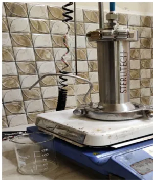
pressure. The flux was calculated using dead end filtration technique using Sterlitech model HP4750 stirred cell shown in Fig. 1.
Evaluation of Anionic Dye Adsorption by AMGO‑CNF Nanopaper
Methyl orange (MO) was used as a model compound for the anionic dye adsorption study. Various concentration of dyes was prepared for this study 5, 10, 20 and 30 mg/l respectively. Initially, 0.5 gm of AMGO-CNF nanopaper was weighed and immersed in 50 ml of dye solution, placed on a shaking bed at ambient temperature. At different time intervals the samples were collected to evaluate the concen-tration of dye. The study was carried out at varying pH of 2, 5, 7 and 10. The adsorption efficiency of the AMGO-CNF nanopaper was assessed from change in concentration of (1) Porosity 𝜀= Ww− Wd∕ A × L × p
(2) √ (2.9 − 1.75𝜀)8nLQ ∕ 𝜀 × A × ΔP
Fig. 1 Dead end filtration technique using Sterlitech model HP4750 stirred cell
MO solution before and after the test using UV spectros-copy at 507 nm. Absorbance of the nanopaper was calcu-lated using beer lambert’s equation and adsorption capacity were evaluated by following equation [27].
where C0 is initial concentration, C is the concentration at
time T, L is volume of solution taken for test and G is weight of nanopaper (g).
Antibacterial Action of the Fabricated AMGO‑CNF Nanopaper
Antibacterial studies were carried out on cellulose and modi-fied nanopapers via Kirby Bauer Disc diffusion method[28]. A 4 mm thick Mueller–Hinton Agar plates were prepared after incubation for 20 min at 121 °C. Test organisms such as
E. Coli MTCC 1610 and Klebsiella, (Gram Negative) were
used in this study. Inoculum preparation was done using a log phase method where four to five well isolated colonies of bacteria were transferred from an agar plate using a loop to a Muller-Hinton broth. This was then incubated at 35 °C until a turbidity of 0.5 McFarland standard was achieved (approx. 1 to 2 × 10⁸CFU ml−1). BaSO
4 turbid solution was used as
a standard for inoculum density. This solution was equiva-lent to 0.5 McFarland standard. The bacterial suspension was uniformly inoculated to a prepared MHA plate using a sterilise swap. The cellulose and AMGO-CNF nanopaper were placed on to this agar plate using a sterilise forceps. These discs were gently pressed to get complete contact with the agar surface. Streptomycin (25 µg per disc) were used (3) Adsorption Capacity(mg∕g) =(C0− C) L∕G
as positive control for all the bacteria. These plates were incubated at 37 °C for 20 h. After 20 h zone of inhibition was measured.
Contact Angle of Fabricated AMGO‑CNF Nanopaper
In order to determine the extent of hydrophilicity, the contact angle test was carried out using SEO-Phoenix 300 model. The source light was focused on instrument on hand camera and deionized water was used as probe liquid. Three contact angles were measured at different places of the sample and the average value is reported.
Mechanical Property of AMGO‑CNF Nanopaper
Mechanical test was conducted using UTM model Tinius Olsen H50KT. Samples were prepared with a thickness of 5 mm and the test was conducted with a deformation of 5 mm min−1 using 100 N load cell.
Result and Discussion
Characterisation of GO and AMGO Using FT‑IR and Raman Spectroscopy
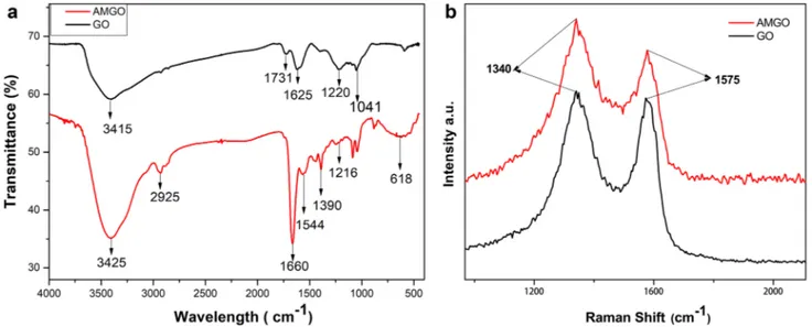
In order to confirm the amine functionalization on GO, FTIR and Raman spectroscopy of both graphene oxide (GO) and amino functionalized graphene oxide (AMGO) were studied [26]. Figure 2a shows the FTIR and Raman spectra of GO and AMGO. From the Fig. 2, GO shows peak at 1041 cm−1
(C–O epoxy stretching), 1220 cm−1 (C–OH stretching),1625
cm−1 (C=C stretching), 1731 cm−1 (C=O stretching) and
3415 cm−1 (–OH stretching). The presence of these groups
confirmed the formation of GO. In the case of AMGO, a new peak has emerged at 1390 cm−1 indicating the C–N
stretching vibration. The band at 1544 cm−1 was attributed
to the formation of stretching and bending vibration of N–H Bond. Medium peaks or bands at free region can be described as asymmetric C–N vibration coupled with NH2 or N–H modes. The disappearance of peak at 1731 cm−1 (C=O
stretch of COOH) of FTIR spectrum in AMGO confirms the successful amino functionalization of GO[29]. Raman spec-troscopy showed the presence of defects on graphite layer. Figure 2b shows the Raman spectra of GO and AMGO. Figure 2b shows two peaks at 1340 cm−1 and 1575 cm−1
for both GO and AMGO which can be attributed to D band and G band respectively. D band represents the defect band (SP3 carbon) and G band contributes to graphitic structure
(SP2 carbon in Graphite sheet). The ratio of D and G band
gives the defect ratio. Compared with GO, the defect ratio of AMGO was increased which confirmed the successful amine functionalization on GO [30].
Fabrication of AMGO‑CNF Nanopaper
Nanopapers were prepared using vacuum filtration technique [31]. 20 gm of cellulose (3 wt%) and 10% AMGO (w.r.t cel-lulose wt%) were mixed and dispersed using a homogenizer (IKA-25 ULTRA TURRAX). When proper homogeneous dispersion was achieved, the dispersion was casted into nan-opaper using vacuum filtration. The fabricated nannan-opapers were peeled off from the filter and dried on a hot press for 30 min at 60 °C. Figure 3 shows the fabrication of AMGO-CNF nanopaper via vacuum filtration and possible interaction of cellulose and AMGO. The AMGO nanosheets effectively interact with cellulose nanofibers through strong hydrogen bonding interactions as shown inFig. 3.
FTIR and XRD Studies of CNF and AMGO‑CNF Nanopaper
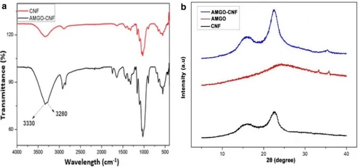
CNF and AMGO-CNF nanopaper were characterised using ATR-FTIR [32]. Figure 4a shows the FTIR spectra of CNF and AMGO-CNF nanopaper. The peaks of CNF nanopa-per show a typical saccharide structure. The peaks at 3335
cm−1 (–OH vibration), 2910 cm−1 (C–H stretching), 1362
cm−1 (–CH
3 vibration), 1159 cm−1 (C–O–C anti- symmetric
stretching), 1030 cm−1 (C–C–O stretching), 890 cm−1 (C–C
stretching), confirmed the cellulosic structure of nanopaper. In the case of AMGO-CNF nanopaper, two distinct sharp peaks at 3330 cm−1 and 3280 cm−1 attributed to NH stretch,
which overlapped with the broad peak arisen from carbox-ylic acid stretch. Also, compared to CNF the band width at 3330 cm−1 and intensity of all peaks attributed to OH
func-tional group decreases remarkably which indicates effective reduction of oxygen species during the amination process.
Figure 4b shows the XRD spectrum of AMGO, CNF and AMGO-CNF nanopaper. It was clearly evident that, the XRD spectrum of CNF nanopaper showed peaks at 2θ = 16.3° and 22.6° corresponding to (110) and (200) planes. These are typically attributed to cellulose type I structure. AMGO dis-played a broad peak at 2θ = 25.9 corresponding to the (002) plane of graphitic phase, indicating high extend of reduction [33]. The incorporation of AMGO into CNF matrix affects the crystallinity of CNF. At lower concentration AMGO loading, the AMGO aligned in parallel and facilitated an ordered alignment of CNF molecules leading to an increased crystallinity. These findings were well agreement with Phiri et al. where they showed the enhancement of crystallinity of micro fibrillated cellulose (MFC) upon the incorporation of reduced graphene oxide (RGO) [34]. The enhancement in crystallinity could influence the mechanical properties and porosity.
Morphology and Porosity of Fabricated CNF and AMGO‑CNF Nanopaper
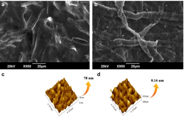
Figure 5 shows SEM images of CNF and AMGO-CNF nanopaper [20]. SEM image of CNF displays a nonporous nanofiber arrangement. They are randomly arranged and have empty spaces between fibres. With the addition of AMGO, the empty spaces were filled, and a compact struc-ture could be seen from the SEM image. 3D topological image of CNF and AMGO-CNF nanopaper were shown in Fig. 5b. Surface roughness value mainly depends on three factors, root mean square (Rq), arithmetic mean deviation of roughness (Ra), and height difference between five maxi-mum height peak (Rmax) and five minimaxi-mum height peaks (Rmin). Usually Ra is considered to scrutinize the rough-ness nature. From Table 1 the cellulose nanopaper showed a Ra value of 7.34, without any addition of AMGO. Further, with addition of AMGO, AMGO-CNF nanopaper showed a Ra value of 15.50. The addition of AMGO resulted in the enhanced surface roughness to a greater value. This higher value of Ra might be due to the uniform dispersion of AMGO nanoparticles on the surface
The porosity of CNF and CNF-AMGO nanopaper were determined using gravimetric method using Eq. (1) and the average nanopaper pore radius was calculated by Guerout Elford-Ferry equation given in Eq. (2) in the experimental sec-tion [35]. The porosity percentage was found to be 32% and 51% for AMG-CNF and CNF nanopaper respectively whereas the average pore radius was found to be 1.9 nm and 3.2 nm for AMG-CNF and CNF nanopaper respectively.
Evaluation of Adsorption Capacity of AMGO‑CNF Nanopaper Against Anionic Methyl Orange Dye
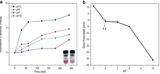
UV spectroscopy was employed to determine the adsorp-tion capacity of AMGO-CNF nanopaper against anionic dye [36]. Here, we used methyl orange as a model for negatively charged dye. Figure 6 illustrates the adsorption capacity of AMGO-CNF nanopaper as a function of time with differ-ent pH conditions. Result shows that, maximum adsorption was observed at pH 2 with an adsorption value of 7.62 mg/g at 20 mg/L concentration. Figure 7 illustrates the proposed removal mechanism of methyl orange (MO) dye by AMGO-CNF nanopaper. Amine groups in AMGO either could be protonated at lower pH to form NH3+ (R–NH2 + H+→NH3+)
or deprotonated at lower pH to form NH2...OH−. Zeta
poten-tial of different pH were carried out and found the value of pHzpc (Zero Point Charge Fig) of 5.9. pH below the pHzpc,
AMGO had positive charge and anionic dyes were adsorbed on to the surface of AMGO-CNF nanopaper by electrostatic interaction. The negatively charged anionic dyes were easily
adsorbed by positively charged surface of AMGO in the fab-ricated nanopaper. At higher pH, the demonstrated AMGO-CNF nanopaper showed a decreased adsorption capacity which might be due to the decrease in extend of protonation.
Adsorption Isotherm and Adsorption Kinetics
Adsorption isotherm describes the equilibrium relation-ship between adsorbent and adsorb ate [37]. This equi-librium data which describes the interaction of dyes with adsorbent can be expressed via a series of models. Some of the commonly investigating models are Langmuir and Freundlich [38].
where Ce, Qe, Q0 and Klang are equilibrium concentration of dye solution (mg/L), amount of dye adsorbed by nanopaper (mg/g), maximum adsorption capacity of nanopaper (mg/g) and Langmuir constant.
Kf is the Freundlich constant and n the heterogeneity fac-tor. n describes absorbent’s adsorption intensity.
(4) Langmuir Equation: Qe= Q0KlangCe∕(1 + KlangCe)
(5) Linear Form Ce∕Qe= 1∕Q0Klang+ Ce∕Q0
(6) Freundlich Equation∶ log Qe= log Kf+ (1∕n) log Ce
Fig. 5 SEM and AFM images of CNF and AMGO-CNF nanopaper
Table 1 Surface roughness of nanopapers
Nanopaper Ra Rq Rz Rmax CNF 7.34 9.51 0.00 78.21 AMGO-CNF 15.5 19.70 0.00 141.40
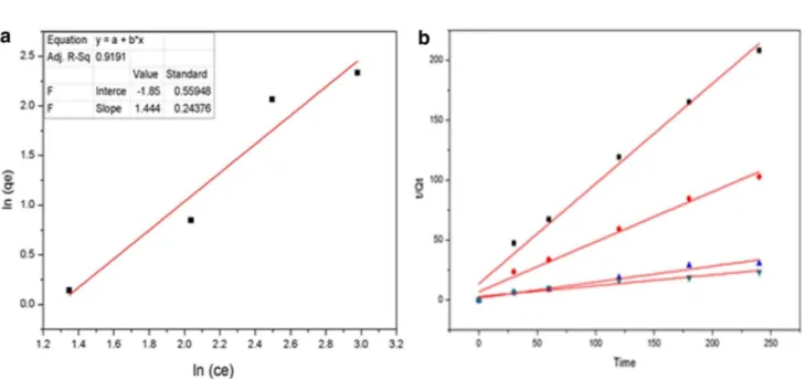
Adsorption isotherm data were plotted using above equa-tions. Other parameters were calculated from linear regres-sion plotting. Langmuir and Freundlich models were plotted out of which Freundlich model apparently fitted better. Fig-ure 8a shows, the Freundlich model fitting with a correlation coefficient of R2 = 0.919 and shows that, the adsorption of
methyl orange to AMGO-nanopaper via multilayer adsorp-tion. Freundlich theory is based on parameter of nf value. Here we got 1/nf value of 1.44 which indicates the adsorp-tion was S-type isotherm. This type of isotherm has been
observed in low concentration ranges with a polar functional group [39].
Adsorption kinetics explains the rate of chemical reaction and factors affecting the reaction rate [37] Pseudo first order kinetic model and pseudo second order kinetic model were used for kinetic studies.
A linear form of both equations are as follows:
(7) Pseudo First order∶ log (Qe − Qt) = log Qe − K1t∕2.303
Fig. 6 Adsorption capacity AMGO-CNF nanopaper with a varying pH and b zeta potential
Fig. 7 Proposed removal mech-anism of MO dye by fabricated AMGO-CNF nanopaper
Rate constants and equilibrium adsorption capacity were tested for Pseudo-First order (PFO) and Pseudo-Second order (PSO). Methyl orange adsorption did not follow the pseudo first order (correlation coefficient less). Moreover, pseudo second order showed good correlation for the experi-ment data (Fig. 7b). Plots of t/qt and t showed good linearity. The PSO rate constant K2, calculated adsorption and the linear regression correlation coefficients value R2 are given
in Table 2. The calculated Qe values were in well agreement with experimental adsorption value and correlation coef-ficient value R2 are higher and close to one.
Antibacterial Action of Nanopapers
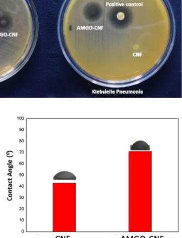
Biofouling due to the action of microbes such as bacteria and viruses is a major challenge for designing materials for water purification. Figure 9 shows the digital image of anti-bacterial action of AMGO-CNF and CNF nanopapers. The fabricated AMGO-CNF nanopaper showed superior antibac-terial activity against E.Coli MTCC 1610 and Klebsiellar. The contact active antibacterial action of the AMGO-CNF nanopaper can be attributed to a combined effect of rough-ness and surface amino groups of AMGO in AMGO-CNF nanopaper. The characteristic outer membranes of gram-negative bacteria are responsible for their resistance towards antibiotics. The lipopolysaccharides in outer membrane con-tains phosphate group which would susceptible to interact with amino groups which resulted in the rupture of the bac-terial membrane. Table 3 illustrates the zone of inhibition data for different bacteria.
(8)
Pseudo Second order∶ t∕Qt= 2∕KeQ2e+(1∕Qe) t Hydrophilicity of the Fabricated AMGO‑CNF
Nanopaper
Figure 10 shows the contact angle of CNF and AMGO-CNF nanopaper. Contact angle of CNF and AMGO-CNF nanopa-per were found to be 43° and 71° respectively. The enhance-ment in contact angle of AMGO-CNF nanopaper was due to the decrease in the hydrophilicity of nanopaper. The OH groups of CNF could be interacted with amine groups of AMGO in AMGO-CNF nanopaper which resulted in the reduction of OH groups. This reduction in OH groups makes the nanopaper less hydrophilic compared with CNF nano-paper. Furthermore, surface roughness also could enhance the contact angle of AMGO-CNF nanopaper which was con-firmed from the AFM studies.
Hydrolytic Stability of CNF‑AMGO Nanopaper
Since the fabricated CNF-AMGO nanopaper are intended to use in water purification, it is very relevant to study the hydrolytic stability of these fabricated nanopapers. To inves-tigate the water stability and reusability, the nanopaper were kept in water for 60 h. The pristine CNF nanopaper lost its
Fig. 8 a Freundlich model plot and b pseudo second order plot for experiment data
Table 2 Kinetic parameter for pseudo second Order
Concentration qe, cal (mg g− 1) K
2 (g mg− 1 min− 1) R2 5 mg 1.19731 0.05169 0.98602 10 mg 2.3980 0.02554 0.9838 20 mg 7.5318 0.01087 0.96103 30 mg 11.0485 0.002879 0.9366
stability in water and structurally degraded after 60 h. Fig-ure 11a shows and AMGO-CNF nanopaper before and after soaking in water. There was a weight loss of 28% in case of CNF nanopaper, whereas in AMGO-CNF nanopaper had a weight loss of 2%. In order to further investigate the hydro-lytic stability, both nanopapers were subjected to sonication in water. Figure 11b illustrates the hydrolytic stability of both CNF and AMGO-CNF nanopapers after sonication in water for 30 min. The CNF nanopaper started to collapse within 1 min and completely dispersed in water within 4 min, whereas AMGO-CNF nanopaper had good hydrolytic stability even after the probe sonication for 30 min. The mechanical stability of the nanopaper depends on the struc-tural integrity of the fibre network. The pristine cellulose nanopaper is hydrophilic compared to the fabricated AMGO CNF nanopaper which would undergo rapid swelling and became mechanically poor whereas decreased hydrophilicity in fabricated AMGO-CNF nanopaper reduced the interac-tion with the water molecules and there by enhanced the wet strength.
Wet Strength of the CNF‑AMGO Nanopaper
Mechanical properties are crucial for nanopaper to work under stress [40]. The nanopapers those works under the pressure driven technology should have sufficient strength for their optimum performance. Table 4 shows the tensile
strength of CNF and AMGO-CNF nanopaper under normal condition and under wet condition. Figure 12 depicts the stress by strain graph of wet CNF and wet AMGO-CNF nanopaper. Pristine CNF nanopaper had a tensile strength of 49.3 Mpa whereas AMGO-CNF nanopapers tensile strength reduces to 38.1 Mpa. Both fabricated nanopapers were immersed in water for 30 min in order to evaluate the wet strength. It was found that, pristine CNF nanopaper had a wet strength of 17.5 MPa, whereas AMGO-CNF nanopaper had wet strength of 25.9 MPa. The enhanced wet strength of AMGO-CNF nanopaper was due to the reduction of hydrophilicity after the incorporation of AMGO. The inher-ent hydrophilic nature of pristine CNF nanopaper resulted in the swelling of the cellulose nanofibers when comes in contact with water. Additionally, CNF nanopaper had 80% of increase in weight after soaking in water for half hour, whereas AMGO-CNF nanopaper only showed 30% increase
Table 3 Inhibition zone of fabricated AMGO-CNF nanopaper
Bacterium Diameter (mm) Zone of inhibition (mm)
Escherichia Coli 11 14
Klebsiella pneumonia 11 15
Fig. 9 Digital image illustrating the antibacterial activity of CNF and AMGO-CNF nanopaper
in weight. Amine functionalised graphene oxide (AMGO) being a strong material could interact with cellulose nanofib-ers which resulted in the decreased active sites for hydrogen bonding and thereby reduced the swelling behaviour.
Reusability of the AMGO‑CNF Nanopaper
Figure 13 illustrates the reusability of CNF-AMGO nano-paper with 10% concentration[38]. The reusability of the nanopaper were investigated using 99% ethanol as desorp-tion agent. These nanopapers were washed and dried at 50 °C for 12 h. It was found that, around 74% of adsorption retention was achieved after four times of reuse.
Conclusion
Herein we have fabricated a hybrid cellulosic and amine functionalized graphene oxide nanopaper via a simple vacuum filtration technique. Graphene oxide was prepared using modified hummers method and successfully amine functionalised under a controlled hydrothermal condition.
This was confirmed by FT-IR and Raman Spectroscopy. The addition of AMGO on to cellulose nanofiber resulted in the enhanced surface roughness which was evident from SEM and AFM images. The increased surface roughness aids the anti-bacterial action of the nanopaper against
E.Coli MTCC 1610 and Klebsiella. The demonstrated
AMGO-CNF nanopaper exhibited excellent antibacterial action and good anionic dye adsorption without compro-mising the strength factor. At 30 mg/l of MO dye, the fabricated AMGO-CNF paper showed an excellent adsorp-tion of 11.05 mg/g at pH 2. This adsorpadsorp-tion was due to the protonation of amine group. Moreover, it was found that, the protonation decreased with increase in pH. The adsorption data were plotted for adsorption isotherm and
Fig. 11 Hydrolytic stabil-ity of CNF and AMGO-CNF nanopaper
Fig. 12 Stress v/s strain graph of normal and wet nanopapers
Table 4 Mechanical strength of CNF and AMGO-CNF Nanopapers Specimen Dry sample (Mpa) Wet
sample (Mpa) CNF nanopaper 38.1 17.48 AMGO-CNF nanopaper 49.3 25.9
its kinetics were investigated. Additionally, the fabricated AMGO-CNF nanopaper exhibited good wet strength of 25.9 with excellent hydrolytic stability even after sonica-tion for 30 min. Moreover, simple fabricasonica-tion technique with enhanced antibacterial action and adsorption capabil-ity using less amount of AMGO opens new platform for the dye adsorption.
References
1. Pathan TS, Thete PB, Shinde SE, Sonawane DL, Khillare YK (2009) J Bot Res Int 21:71–78
2. Ebenstein A (2012) Rev Econ Stat 94:186–201
3. Haq I, Raj A (2018) Emerging eco-friendly approaches for waste management. Springer, Singapore, pp 121–142
4. Pfaller MA, Flamm RK, Mendes RE, Streit JM, Smart JI, Hamed KA, Duncan LR, Sader HS (2018) Antimicrob Agents Chemother 63:1–12
5. Koksal E, Tulek N, Sonmezer MC, Temocin F, Bulut C, Hatipo-glu C, Erdinc FS, Ertem G (2019) Invest Clin Urol 60:46–53 6. Odonkor ST, Ampofo JK (2013) Microbiol Res (Pavia) 4:2 7. Yadollahpour A, Rashidi S, Ghotbeddin Z, Jalilifar M, Rezaee
Z (2014) J Pure Appl Microbiol 8:3711–3719
8. Castaing JB, Massé A, Pontié M, Séchet V, Haure J, Jaouen P (2010) Desalination 253:71–77
9. Liang Y, Kim S, Kallem P, Choi H (2019) Chemosphere 221:479–485
10. Andreeßen B, Lange AB, Robenek H, Steinbüchel A (2010) Appl Environ Microbiol 76:622–626
11. Brinchi L, Cotana F, Fortunati E, Kenny JM (2013) Carbohydr Polym 94:154–169
12. Zhao J, Zhang W, Zhang X, Zhang X, Lu C, Deng Y (2013) Carbohydr Polym 97:695–702
13. Valencia L, Arumughan V, Jalvo B, Maria HJ, Thomas S, Mathew AP (2019) ACS Omega 4:4330–4338
14. Zhao Y, Moser C, Lindstrom ME, Henriksson G, Li J (2017) ACS Appl Mater Interfaces 9:13508–13519
15. Espino-Pérez E, Bras J, Ducruet V, Guinault A, Dufresne A, Domenek S (2013) Eur Polym J 49:3144–3154
16. Nechyporchuk O, Belgacem MN, Bras J (2016) Ind Crop Prod 93:2–25
17. Xu X, Zhao G, Wang H, Li X, Feng X, Cheng B, Shi L, Kang W, Zhuang X, Yin Y (2019) J Power Sources 409:123–131 18. Compton OC, Nguyen ST (2010) Small 22:711–723 19. Kim F, Cote LJ, Huang J (2010) Adv Mater 22:1954–1958 20. Zhu C, Liu P, Mathew AP (2017) ACS Appl Mater Interfaces
9(24):21048–21058
21. Fryczkowska B, Wiechniak K (2017) Polish J Chem Technol 19:41–49
22. Meng et al (2017) J Mech Phys Solids 103:22–39 23. Meng et al (2018) Eng Fract Mech 194:350–361 24. Meng, Wang (2019) Appl Mech Rev 71(4):040801
25. Marcano DC, Kosynkin DV, Berlin JM, Sinitskii A, Sun Z, Slesarev A, Alemany LB, Lu W, Tour JM (2010) ACS Nano 4(8):4806–4814
26. Zhao D, Gao X, Wu C, Xie R, Feng S, Chen C (2016) Appl Surf Sci 384:1–9
27. Annadurai G, Juang RS, Lee DJ (2002) J Hazard Mater 92:263–274
28. Elias E, Chandran S, Zachariah AK, Vineesh Kumar VK, Sunil MA, Bose S, Souza FG, Thomas S (2016) RSC Adv 6:85107–85116
29. Shanmugharaj AM, Yoon JH, Yang WJ, Ryu SH (2013) J Col-loid Interface Sci 401:148–154
30. Zhang W, Ma J, Gao D, Zhou Y, Li C (2016) Prog Org Coat 94:9–17
31. Gopakumar DA, Pai AR, Pottathara YB, Pasquini D, Carlos L, De Morais M, Luke N, Kalarikkal Y, Grohens, Thomas S (2018) ACS Appl Mater Interfaces 10:20032–20043
32. Garside P, Wyeth P (2006) Stud Conserv 51:205–211 33. Mahdavi H, Kahriz PK, Rajbar HG, Shahalizade T (2017) J
Mater Sci Mater Electron 28:4295–4305
34. Phiri J, Johansson LS, Gane P, Maloney TC (2018) Nanoscale 10:9569–9582
35. Guo J, Kim J (2017) RSC Adv 7:33822–33828
36. Gopakumar DA, Pasquini D, Henrique MA, De Morais LC, Gro-hens Y, Thomas S (2017) ACS Sustain Chem Eng 5:22026–22033 37. Chakraborty S, Chowdhury S, Das Saha P (2011) Carbohydr
Polym 86:1533–1541
38. Zhou Y, Zhang M, Hu X, Wang X, Niu J, Ma T (2013) J Chem Eng Data 58:413–421
39. Limousin G, Gaudet JP, Charlet L, Szenknect S, Barthès V, Kri-missa M (2007) Appl Geochem 22:249–275
40. Wu YB, Yu SH, Mi FL, Wu CW, Shyu SS, Peng CK, Chao AC (2004) Carbohydr Polym 57:435–440