Université de Montréal
Nitrate metabolism in the dinoflagellate Lingulodinium
polyedrum
par
Steve Dagenais Bellefeuille
Département de sciences biologiques Faculté des arts et sciences
Thèse présentée à la Faculté des études supérieures et postdoctorales en vue de l’obtention du grade de Ph.D
en sciences biologiques
Décembre, 2015
Résumé
Les dinoflagellés sont des eucaryotes unicellulaires retrouvés dans la plupart des écosystèmes aquatiques du globe. Ces organismes amènent une contribution substantielle à la production primaire des océans, soit en tant que membre du phytoplancton, soit en tant que symbiontes des anthozoaires formant les récifs coralliens. Malheureusement, ce rôle écologique majeur est souvent négligé face à la capacité de certaines espèces de dinoflagellés à former des fleurs d'eau, parfois d'étendue et de durée spectaculaires. Ces floraisons d'algues, communément appelées "marées rouges", peuvent avoir de graves conséquences sur les écosystèmes côtiers, sur les industries de la pêche et du tourisme, ainsi que sur la santé humaine. Un des facteurs souvent corrélé avec la formation des fleurs d'eau est une augmentation dans la concentration de nutriments, notamment l’azote et le phosphore. Le nitrate est un des composants principaux retrouvés dans les eaux de ruissellement agricoles, mais également la forme d'azote bioaccessible la plus abondante dans les écosystèmes marins. Ainsi, l'agriculture humaine a contribué à magnifier significativement les problèmes associés aux marées rouges au niveau mondial. Cependant, la pollution ne peut pas expliquer à elle seule la formation et la persistance des fleurs d'eau, qui impliquent plusieurs facteurs biotiques et abiotiques. Il est particulièrement difficile d'évaluer l'importance relative qu'ont les ajouts de nitrate par rapport à ces autres facteurs, parce que le métabolisme du nitrate chez les dinoflagellés est largement méconnu. Le but principal de cette thèse vise à remédier à cette lacune. J'ai choisi Lingulodinium polyedrum comme modèle pour l'étude du métabolisme du nitrate, parce que ce dinoflagellé est facilement cultivable en laboratoire et qu'une étude transcriptomique a récemment fourni une liste de gènes pratiquement complète pour cette espèce. Il est également intéressant que certaines composantes moléculaires de la voie du nitrate chez cet organisme soient sous contrôle circadien. Ainsi, dans ce projet, j'ai utilisé des analyses physiologiques, biochimiques, transcriptomiques et bioinformatiques pour enrichir nos connaissances sur le métabolisme du nitrate des dinoflagellés et nous permettre de mieux apprécier le rôle de l'horloge circadienne dans la régulation de cette importante voie métabolique primaire.
Je me suis tout d'abord penché sur les cas particuliers où des floraisons de dinoflagellés sont observées dans des conditions de carence en azote. Cette idée peut sembler contre-intuitive, parce que l'ajout de nitrate plutôt que son épuisement dans le milieu est généralement associé aux floraisons d'algues. Cependant, j’ai découvert que lorsque du nitrate était ajouté à des cultures initialement carencées ou enrichies en azote, celles qui s'étaient acclimatées au stress d'azote arrivaient à survivre près de deux mois à haute densité cellulaire, alors que les cellules qui n'étaient pas acclimatées mourraient après deux semaines. En condition de carence d'azote sévère, les cellules arrivaient à survivre un peu plus de deux semaines et ce, en arrêtant leur cycle cellulaire et en diminuant leur activité photosynthétique. L’incapacité pour ces cellules carencées à synthétiser de nouveaux acides aminés dans un contexte où la photosynthèse était toujours active a mené à l’accumulation de carbone réduit sous forme de granules d’amidon et corps lipidiques. Curieusement, ces deux réserves de carbone se trouvaient à des pôles opposés de la cellule, suggérant un rôle fonctionnel à cette polarisation.
La deuxième contribution de ma thèse fut d’identifier et de caractériser les premiers transporteurs de nitrate chez les dinoflagellés. J'ai découvert que Lingulodinium ne possédait que très peu de transporteurs comparativement à ce qui est observé chez les plantes et j'ai suggéré que seuls les membres de la famille des transporteurs de nitrate de haute affinité 2 (NRT2) étaient réellement impliqués dans le transport du nitrate. Le principal transporteur chez Lingulodinium était exprimé constitutivement, suggérant que l’acquisition du nitrate chez ce dinoflagellé se fondait majoritairement sur un système constitutif plutôt qu’inductible. Enfin, j'ai démontré que l'acquisition du nitrate chez Lingulodinium était régulée par la lumière et non par l'horloge circadienne, tel qu'il avait été proposé dans une étude antérieure.
Finalement, j’ai utilisé une approche RNA-seq pour vérifier si certains transcrits de composantes impliquées dans le métabolisme du nitrate de Lingulodinium étaient sous contrôle circadien. Non seulement ai-je découvert qu’il n’y avait aucune variation journalière dans les niveaux des transcrits impliqués dans le métabolisme du nitrate, j’ai aussi constaté qu’il n’y avait aucune variation journalière pour n’importe quel ARN du transcriptome de
Lingulodinium. Cette découverte a démontré que l’horloge de ce dinoflagellé n'avait pas
besoin de transcription rythmique pour générer des rythmes physiologiques comme observé chez les autres eukaryotes.
Mots-clés : acclimatation, amidon, boucles de rétrocontrôle transcription-traduction, carbone,
corps lipidiques, dinoflagellé, floraisons d'algues, horloge circadienne, Lingulodinium
polyedrum, photosynthèse, rythmes journaliers, nitrate, transcriptome, transporteurs de nitrate
Abstract
Dinoflagellates are unicellular eukaryotes found in most aquatic ecosystems of the world. They are major contributors to carbon fixation in the oceans, either as free-living phytoplankton or as symbionts to corals. Dinoflagellates are also infamous because some species can form spectacular blooms called red tides, which can cause serious damage to ecosystems, human health, fisheries and tourism. One of the factors often correlated with algal blooms are increases in nutrients, particularly nitrogen and phosphorus. Nitrate is one of the main components of agricultural runoffs, but also the most abundant bioavailable form of nitrogen in marine environments. Thus, agricultural activities have globally contributed to the magnification of the problems associated with red tides. However, bloom formation and persistence cannot be ascribed to human pollution alone, because other biotic and abiotic factors are at play. Particularly, it is difficult to assess the relative importance of nitrate addition over these other factors, because nitrate metabolism in dinoflagellate is mostly unknown. Filling part of this gap was the main goal of this thesis. I selected Lingulodinium
polyedrum as a model for studying nitrate metabolism, because this dinoflagellate can easily
be cultured in the lab and a recent transcriptomic survey has provided an almost complete gene catalogue for this species. It is also interesting that some molecular components of the nitrate pathway in this organism have been reported to be under circadian control. Thus, in this project, I used physiological, biochemical, transcriptomic and bioinformatic approaches to enrich our understanding of dinoflagellate nitrate metabolism and to increase our appreciation of the role of the circadian clock in regulating this important primary metabolic pathway.
I first studied the particular case of dinoflagellate blooms that occur and persist in conditions of nitrogen depletion. This idea may seems counterintuitive, because nitrogen addition rather than depletion, is generally associated with algal blooms. However, I discovered that when nitrate was added to nitrogen-deficient or nitrogen-sufficient cultures, those that had been acclimated to nitrogen stress were able to survive for about two months at high cell densities, while non-acclimated cells died after two weeks. In conditions of severe nitrogen limitation, cells could survive a little bit more than two weeks by arresting cell division and reducing photosynthetic rates. The incapacity to synthesize new amino acids for
these deprived cells in a context of on-going photosynthesis led to the accumulation of reduced carbon in the form of starch granules and lipid bodies. Interestingly, both of these carbon storage compounds were polarized in Lingulodinium cells, suggesting a functional role.
The second contribution of my thesis was to identify and characterize the first nitrate transporters in dinoflagellates. I found that in contrast to plants, Lingulodinium had a reduced suite of nitrate transporters and only members of the high-affinity nitrate transporter 2 (NRT2) family were predicted to be functionally relevant in the transport of nitrate. The main transporter was constitutively expressed, which suggested that nitrate uptake in Lingulodinium was mostly a constitutive process rather than an inducible one. I also discovered that nitrate uptake in this organism was light-dependent and not a circadian-regulated process, as previously suggested.
Finally, I used RNA-seq to verify if any transcripts involved in the nitrate metabolism of Lingulodinium were under circadian control. Not only did I discovered that there were no daily variations in the level of transcripts involved in nitrate metabolism, but also that there were no changes for any transcripts present in the whole transcriptome of Lingulodinium. This discovery showed that the circadian timer in this species did not require rhythmic transcription to generate biological rhythms, as observed in other eukaryotes.
Keywords : acclimation, carbon, circadian clock, daily rhythms, dinoflagellate, harmful algal
blooms, high-affinity nitrate tranporters, Lingulodinium polyedrum, lipid bodies, nitrate, nitrate uptake, nitrogen stress, photosynthesis, starch, transcription-translation feedback loops, transcriptome
Table of contents
Résumé ... i
Abstract... iv
Table of contents... vi
List of tables ... ix
List of figures...x
List of abbreviations ... xii
Remerciements ... xviii
Chapter 1- Introduction...1
1.1. Overview of dinoflagellates...2
1.1.1. Phylogeny ...2
1.1.2. Plastid origin and distinctive features...2
1.1.3. A unique mitochondrial genome ...5
1.1.4. Particularities of nuclear structure and biology ...6
1.1.5. Ecological functions ...8
1.2. Publication # 1 ...12
Putting the N in dinoflagellate...13
1.2.1. Abstract...14
1.2.2. Introduction...15
1.2.3. Overview of the marine N cycle...17
1.2.4. Uptake of nitrogen using transporters...19
1.2.5. Uptake of nitrogen by feeding ...24
1.2.6. Nitrogen assimilation and metabolism ...28
1.2.7. Adaptations to nitrogen stress...32
1.2.8. Conclusion ...36
1.2.9. Acknowledgments ...43
1.4. Lingulodinium polyedrum: a circadian model for the study of nitrate metabolism...51
Chapter 2- Publication # 2 ...52
The dinoflagellate Lingulodinium polyedrum responds to N depletion by a polarized deposition of starch and lipid bodies ...53
2.1. Abstract...54
2.2. Introduction...55
2.3. Material and methods ...57
2.3.1. Cell culture...57
2.3.2. Cell density measurements ...57
2.3.3. Elemental analysis ...57
2.3.4. Protein and amino acid quantification ...58
2.3.5. Photosynthetic measurements...58
2.3.6. Starch quantification...59
2.3.7. Nile red quantification of neutral lipid ...59
2.3.8. Microscopy ...60 2.3.9. Statistical analyses ...61 2.4. Results...62 2.5. Discussion...66 2.6. Acknowledgments ...87 Chapter 3- Publication # 3 ...88
The main nitrate transporter of the dinoflagellate Lingulodinium polyedrum is constitutively expressed and not responsible for daily variations in nitrate uptake rates ...89
3.1. Abstract...90
3.2. Introduction...91
3.3. Material and methods ...94
3.3.1. Cell culture...94
3.3.1.1. Initial conditions ...94
3.3.1.2. Daily and circadian nitrate uptake measurements ...94
3.3.1.3. Expression of LpNRT2.1 protein ...94
3.3.3. BLAST searches, phylogeny and bioinformatic analyses ...95
3.3.4. Relative transcript abundance of Lingulodinium NRT2 sequences ...96
3.3.5. Electrophoretic analyses ...96 3.3.6. Statistical analysis...97 3.4. Results...98 3.5. Discussion...101 3.6. Conclusion ...105 3.7. Acknowledgements...125 Chapter 4- Publication # 4 ...126
The Lingulodinium circadian system lacks rhythmic changes in transcript abundance...127
4.1. Abstract...128 4.2. Background...129 4.3. Results...131 4.4. Discussion...134 4.5. Conclusions...137 4.6. Methods ...138 4.6.1. Cell Culture...138
4.6.2. RNA extraction and RNA-Seq ...138
4.6.3. Transcription Inhibition ...139
4.6.4. Rhythm Measurements ...140
4.7. Acknowledgements...153
Chapter 5 - General discussion and perspectives ...154
5.1. General discussion and perspectives ...155
5.2. Conclusion ...158
References... i
Appendix 1... xxxiv
List of tables
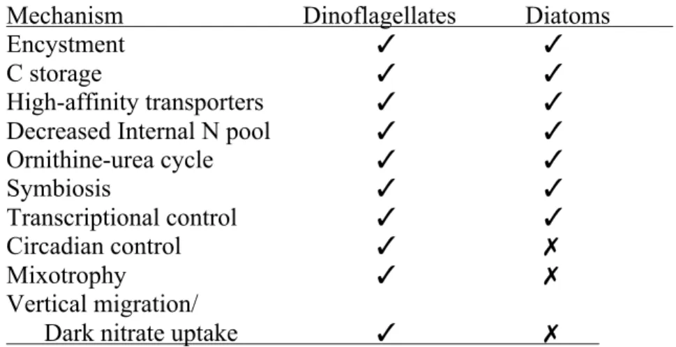
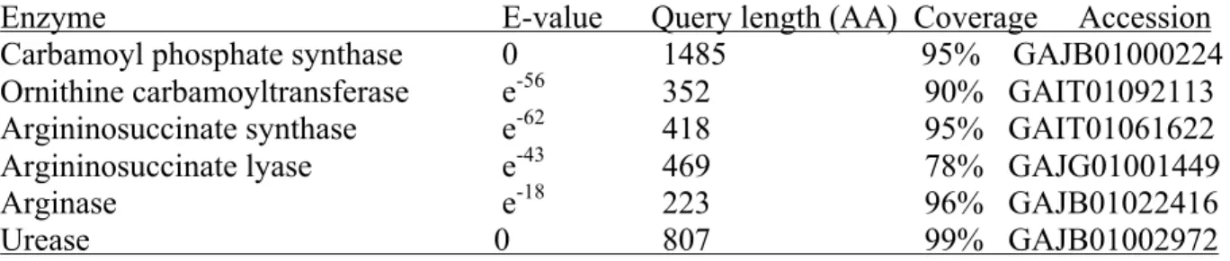
Table 1.2.1. Comparison of adaptation mechanisms to N stress between dinoflagellates and diatoms...37 Table 1.2.2. Nitrogen Metabolizing Enzymes similar to those in diatoms in the Transcriptome of Alexandrium tamarense...38 Table 3.1. BLAST searches for putative nitrate transporters in Lingulodinium polyedrum...106
List of figures
Figure 1.1.1. Distinctive features of dinoflagellate evolution within the Alveolata...10
Figure 1.2.1. Nitrogen metabolism in dinoflagellates. ...39
Figure 1.2.2. Ornithine-urea cycle...41
Figure 1.3.1. Illustration of a Lingulodinium polyedrum cell...47
Figure 1.3.2. Circadian rhythms of bioluminescence and photosynthesis in Lingulodinium polyedrum. ...49
Figure 2.1. Growth of N-replete and N stressed cultures. ...71
Figure 2.2. Elemental analysis shows a decreased N content in N stressed cells without a change in their C content. ...73
Figure 2.3. Changes in the total protein content and free amino acid profile in N stressed cells are consistent with a decrease in N assimilation. ...75
Figure 2.4. Photosynthesis decreases in N stressed cells...77
Figure 2.5. Starch accumulates in N stressed cells...79
Figure 2.6. TAGs accumulate in N stressed cells...81
Figure 2.7. Polarized localization of lipid bodies and starch granules visualized by transmission electron microscopy...83
Figure 2.S1...85
Figure 3.1. Nitrate uptake in Lingulodinium is light dependent. ...107
Figure 3.2. Transcripts of NRT2.1 are the most abundant, relative to all of Lingulodinium putative nitrate transporter sequences...109
Figure 3.3. Lingulodinium NRT2 sequences are found in two different clades. ...111
Figure 3.4. All sites essential for nitrate transport are conserved in Lingulodinium NRT2 sequences. ...113
Figure 3.5. Mature dinoflagellate NRT2 share the 12 predicted transmembrane domains...115
Figure 3.6. LpNRT2.1 is constitutively expressed. ...117
Figure 3.S1. Absolute nitrate uptake in Lingulodinium is the same over a 24 h period in ZT or CT. ...119
Figure 3.S3. Transcripts of NRT2.1 are the most abundant, relative to all of Lingulodinium
putative nitrate transporter sequences...123
Figure 4.1. Transcript abundance does not change between midday (ZT 6) and midnight (ZT 18). ...141
Figure 4.2. Lingulodinium does not have rhythmic transcripts over a 24 hour cycle...143
Figure 4.3. Rhythms continue in the presence of transcription inhibitors...145
Figure 4.S1. Analysis using the Velvet assembly...147
Figure 4.S2. Analysis using duplicate ZT6 and ZT/CT18 samples...149
List of abbreviations
A: adenineADP: adenine diphosphate AMT: ammonium transporter An: Aspergillus nidulans Arg: arginase
Asl: argininosuccinate lyase AsuS: argininosuccinate synthase At: Arabidopsis thaliana
Ata: Alexandrium tamarense ATP: adenosine triphosphate ATX: Ataxin-2
BLAST: Basic Local Alignment Search Tool bp: base pair
C: carbon
CA: carbonic anhydrase CaCl2: calcium chloride
CCM: CO2-concentrating mechanisms cDNA: complementary DNA
CHAPS: 3-[(3-Cholamidopropyl)dimethylammonio]-1-propanesulfonate hydrate CK2: casein kinase 2
Cl: chloride
CLCs: chloride channels CO2: carbon dioxide
CPS: carbamoyl phosphate synthase Cr: Chlamydomonas reinhardtii cRNA: complementary RNA CT: Circadian Time
DEPC: diethylpyrocarbonate
DIN: dissolved inorganic nitrogen DMSO: dimethyl sulfoxide DNA: deoxyribonucleic acid
DNRA: dissimilatory NO3- reduction to ammonium DON: dissolved organic nitrogen
DPM: disintegrations per minute DTT: dithiothreitol
DVM: diurnal vertical migration DW: dry weight
EDTA: ethylenediaminetetraacetic acid ER: endoplasmic reticulum
FAA: free amino acid
FAV: flavine adenine dinucleotide Fd: ferredoxins FW: fresh weight G: guanine G6PDH: glucose-6-phosphate dehydrogenase Gln: glutamine Glu: glutamate
GOGAT: glutamine oxoglutarate aminotransferase (also known as glutamate synthase) GS: glutamine synthetase
HABs harmful algal blooms HCl: hydrogen chloride
HEPES: 4-(2-hydroxyethyl)-1-piperazineethanesulfonic acid HK: hexokinase
HLP: histone-like proteins
HPLC: high performance liquid chromatography HTDs: heterotrophic dinoflagellates
I2: iodine
IEF: isoelectric focusing K: potassium
Kb: kilobase
KCl: potassium chloride kD: kilodalton
KI: potassium iodide KOH: potassium hydroxide LBP: luciferin-binding protein LCF: luciferase
LD: light/dark
Lp: Lingulodinium polyedrum
Mes: 2-(N-morpholino) ethanesulfonic acid MFS: major facilitator superfamily
mg: milligram
MgSO4-: magnesium sulfate MIPs: major intrinsic proteins mL: milliliter
mM: millimolar
MPSS: massively parallel signature sequencing mRNA: messenger RNA
MS: mass spectrometry
MSX: L-methionine sulfoximine MTDs: mixotrophic dinoflagellates MYA: millions years ago
N: nitrogen N2: dinitrogen gas N2O: nitrous oxide Na: sodium
NaCl: sodium chloride
NAD(P)H: nicotinamide adenine dinucleotide (phosphate) NaHCO3: bicarbonate
NaNO3: sodium nitrate
NAR2: nitrate assimilation related NH3: ammonia
NH4+: ammonium NiR: nitrite réductase nm: nanometer NO2-: nitrite NO3-: nitrate
NPF: nitrate transporter 1/ peptide transporter NR: nitrate reductase
NRT1: nitrate transporter 1 NRT2: nitrate transporter 2 NS: nitrate signature O2: oxygen
ORF: open reading frame
OTC: ornithine carbamoyltransferase OUC: ornithine-urea cycle
P: phosphorus
PAS: Periodic acid-Schiff PBS: phosphate-buffered saline PCP: peridinin-chlorophyll a-protein Per: Period
pg: picogram
PMF: proton motive force
PMSF: phenylmethanesulfonyl fluoride PO43-: phosphate
PON: particulate organic nitrogen PSII: photosystem II
PST: paralytic shellfish toxin
RACE: rapid amplification of cDNA ends RNA: ribonucleic acid
RNAi: RNA interference RNA-seq: RNA sequencing rRNA: ribosomal RNA
RPKM: Reads Per Kilobase per Million mapped reads RuBisCO: ribulose-1,5-bisphosphate-carboxylase-oxygenase SD: standard deviation
SDS: sodium dodecyl sulfate SE: standard error
Si: silicium SL: spliced leader
SLAC/SLAH: slow anion channel-associated 1 homologues Ssp: Symbiodinium sp. Freudenthal
T: thymine
TAGs: triacylglycerols
TCA: tricarboxylic acid cycle (also known as Krebs cycle and citric acid cycle) TEM: transmission electron microscope
TM: transmembrane domain ToL: Tree of Life Web Project
Tris: 2-amino-2-hydroxymethyl-1,3-propanediol tRNA: transfer RNA
TSA: Transcriptome Shotgun Assembly
TTFL: transcriptional/translational feedback loops TYF: Twenty-four µg: microgram µL: microliter µM: micromolar µmol: micromole Ure: urease v/v: volume/volume ZT: Zeitgeber Time
Je dédie cette thèse à ma grand-mère Lucie Bouchard et à mon grand-père Laurent Bellefeuille, qui ont tous les deux contracté l'Alzheimer durant mes études doctorales. En souvenir de votre acharnement au travail, de votre indépendance et de votre tendresse sans équivoque
Remerciements
Je prends souvent des décisions sur le vif. Cette stratégie bien qu'elle amène beaucoup de surprises dans ma vie, vient également avec son lot de déceptions. Chose certaine, je n'ai jamais regretté avoir choisi David Morse comme directeur de recherche et ce, en me basant strictement sur la qualité de sa performance en tant qu'orateur lors des 15 premières minutes d'un cours d'introduction à la biologie cellulaire, il y a près de 8 ans. C'est pourquoi, j'aimerais tout d'abord offrir mes plus sincères remerciements à ce professeur qui m'a accueilli dans son laboratoire dès ma deuxième année de baccalauréat et qui a suivi ma progression depuis toutes ces années. J'apprécie qu'il m'ait donné la liberté de piloter mon projet de recherche dans ses montées, ses vrilles et ses descentes. Dans tout le processus, j'ai reçu conseils, encouragements et critiques constructives, qui ont solidifié ma réflexion et mon autonomie scientifiques. Aussi, je tiens à souligner le plaisir que j'ai eu à écrire conjointement avec le professeur Morse. Au passage de sa plume, les idées conservent leur essence tout en devenant plus concrètes, plus colorées et tout simplement plus attrayantes. Travailler dans son laboratoire a été pour moi une expérience remarquable qui a accentué ma compréhension et mon amour de la biologie.
Cette expérience n'aurait pas été la même si elle n'avait pas été accompagnée d'excellents collègues comme Mathieu Beauchemin et Sougata Roy. Par leur intelligence et leur créativité, ils ont contribué à enrichir mon projet de recherche. Je leur suis également reconnaissant pour les nombreuses discussions stimulantes que nous avons eues sur la science en générale et sur nos vies au quotidien.
J'aimerais aussi remercier Sonia Dorion et le professeur Jean Rivoal avec qui j'ai eu la chance de travailler dans le cadre de l'article présenté au chapitre 2 de cette thèse. J'ai bénéficié de leur expertise en biochimie dans une ambiance à la fois rigoureuse et conviviale. Toujours ils m'ont offert une aide technique précieuse et un intérêt honnête dans mes démarches scientifiques
Sur un plan psychologique et plus personnel, je remercie tous les membres de ma famille pour leur patience et leur soutien, surtout dans les derniers mois de rédaction. En particulier, je souligne la présence dans ma vie de mon amoureux Alexis Laurin, qui par son
tempérament calme, son humour créatif et sa curiosité aigüe, tant pour la science que pour les arts, a atténué de beaucoup le stress de mes études.
Finalement, je remercie le FQRNT et le CRSNG qui m'ont soutenu financièrement durant la majeure partie de mon doctorat.
1.1. Overview of dinoflagellates
1.1.1. Phylogeny
Dinoflagellates are unicellular organisms that can be found in most aquatic ecosystems of the world. They display a tremendous diversity in morphology, nutritional strategies and ecological functions. Phylogenetically, dinoflagellates cluster with ciliates and apicomplexans within the well-supported superphylum Alveolata (Fig. 1.1.1), named for the flattened vesicles (cortical alveoli) that form a continuous layer just under the plasma membrane in these organisms [1]. The apicomplexans are sister to dinoflagellates and molecular clock analyses estimated their divergence at about 800-900 million years ago [2,3]. These analyses are consistent with the discovery of the oldest dinoflagellate fossils in late Mesoproterozoic rocks [4]. Molecular phylogenetics has divided the dinoflagellates in three groups: the Oxyrrhinales, the Syndiniales and the core dinoflagellates (Fig. 1.1.1) [5,6]. While not a true dinoflagellate, the oyster pathogen Perkinsus marinus has been useful in inferring ancestral conditions in dinoflagellates, because this organism possesses some distinctive dinoflagellate features, while retaining more general eukaryotic characteristics (Fig 1.1.1). The free-living Oxyrrhinales and parasitic Syndiniales are also heterotrophic and localized at the base of the tree [7,8,9,10]. The core dinoflagellates contain the majority of characterized species, which are heterotrophic, phototrophic or mixotrophic. Fensome et al, initially classified the core dinoflagellates into 14 orders based on morphological data, such as the patterns of thecal plates or the absence of a theca altogether [11]. However, molecular and ultrastructural phylogenies showed that thecal characteristics evolved multiple times within the core dinoflagellates [5,9] and as a result, many of the orders previously described were poly- and paraphyletic. Thus, relationships among dinoflagellates are still being investigated and future classifications will most likely introduce major changes [5].
1.1.2. Plastid origin and distinctive features
About half of the ~2000 dinoflagellate species that have been described are photosynthetic [12]. Most of these have distinctive chloroplasts surrounded by three membranes and contain chlorophylls a and c, in addition to the xanthophyll peridinin, which is
a photopigment unique to dinoflagellates and responsible for the characteristic red colour of most species [13]. The peridinin chloroplast is thought to result from an endosymbiotic event involving a red alga. However, it was a matter of debate if this event occurred ancestrally, through a single secondary endosymbiotic event or rather recently through, serial tertiary endosymbiosis [14,15], the latter occurring by engulfment of an alga containing a secondary plastid. On one hand, the "Chromoalveolata hypothesis" suggested that all members within this supergroup, including the cercozoans, the foraminiferans, the radiolarians, the Alveolates and the Stramenopiles, were derived from a common bikont ancestor that had engulfed a rhodophyte [16]. The main rationale for the Chromoalveolata monophyly was that plastid gain was thought to be much more evolutionary difficult than plastid loss [17]. Thus, it was more parsimonious to hypothesize a single secondary endosymbiotic event in a common ancestor to all Chromoalveolata, and subsequent loss of plastids in ciliates, some apicomplexans and early-diverging members of many chromalveolate lineages [14], than to propose multiple plastid acquisitions by horizontal transfer. On the other hand, tertiary endosymbiosis is observed in multiple dinoflagellate taxa that have chloroplasts of different origins [6,18]. For example, two dinoflagellate genera, Karenia and Karlodinium, have permanent haptophyte-derived plastids [19,20], while species of the Dinophysis obtain temporary chloroplasts from ingestion of cryptophytes [21]. Another problem with the Chromoalveolata hypothesis was that the very few phylogenies based on nuclear DNA and including all groups within the chromoavleolates were incongruent with plastid-based phylogenies [6,20,22]. Thus, it was suggested that the red algal-derived plastid in chromoalveolates was more likely acquired horizontally by tertiary endosymbiosis than by vertical inheritance from a common ancestor [15,18]. However, this alternative was recently challenged by the discovery of Chromera
velia, a free-living photosynthetic alga closely related to apicomplexans and sharing a
common plastid origin with heterokonts (Stramenopiles) [23]. Combined with the observation of a vestigial chloroplast in some apicomplexans [24], and the discovery of several plastid-targeted genes in the nuclear genome of the heterotrophic dinoflagellate Oxyrrhis marina [25], it now seems more likely that the common ancestor of dinoflagellates and apicomplexans was photosynthetic rather than heterotrophic.
Chloroplasts of peridinin-containing dinoflagellates also possess multiple distinctive features. They have replaced the typical form I
ribulose-1,5-bisphosphate-carboxylase-oxygenase (RuBisCO) by a form II RuBisCO [26], which is usually found in proteobacteria growing in high-CO2, low-O2 environments [27]. The reason for this is that form II RuBisCO has a significantly lower affinity to CO2 than the form I enzyme [27]. However, dinoflagellates typically live in low-CO2, high-O2 environments, and thus, it was suggested that these organisms needed novel CO2-concentrating mechanisms (CCM) to compensate for the imperfections of form II RuBisCO [6]. One such CCM was identified in Lingulodinium
polyedrum, where a δ-type carbonic anhydrase (CA), which catalyzes the rapid
interconversion of CO2 and HCO3, was found to be exclusively localized at the plasma membrane [28]. Treating the cells with acetazolamide, a non-membrane-permeable CA inhibitor, resulted in decreases in photosynthetic rates [28]. This suggested that the action of the CA at the vicinity of the cell membrane increased CO2 uptake, leading to higher photosynthetic rates [28]. Interestingly, Lingulodinium also showed a circadian variation in the localization of RuBisCO within individual chloroplasts that correlated with the CO2- fixation rhythm [29]. During the day, where carbon fixation rates are highest, RuBisCO was sequestered near the center of the cell in the pyrenoid within the chloroplast, while the enzyme distributed evenly within the chloroplasts at night. Thus, the strategic localization of RuBisCO in day-phased cells was proposed to optimize carbon fixation by limiting access of oxygen to the active site of the enzyme, rather than increasing CO2 concentrations within the chloroplasts [29].
Unlike most genes that are typically encoded in the plastids of photosynthetic eukaryotes, the form II RuBisCO is nuclear-encoded in peridinin dinoflagellates [26]. In fact, these organisms have the smallest known functioning plastid genomes, with only 16 genes described so far [30], in contrast to the ~100 genes typically found in chloroplasts of other photosynthetic eukaryotes [6]. This dramatic reduction is thought to be the result of massive gene transfer from the chloroplast to the nucleus in dinoflagellates [31]. The DNA structure in their plastids is also peculiar. Whereas conventional plastid genomes have their genes distributed on a single circular DNA molecule, the plastid genome of peridinin dinoflagellates has been broken down in multiple small 2- to 3-kb plasmids, termed "minicircles" [32]. Each of these minicircles encodes one to several genes and possesses a noncoding core sequence proposed to act as an origin of replication [32,33]. Transcription in the chloroplasts was suggested to occur in a "rolling circle" fashion where the minicircular DNA is transcribed
continuously to generate polycistronic transcripts larger than the minicircle itself [34]. These transcripts are then cleaved by an endonuclease to produce long RNA precursors, which are further processed into mature RNAs by processes including 3'-polyuridylylation [35].
On an evolutionary perspective, it is interesting that all the usual plastid features that were mentioned above (i.e. peridinin, form II RuBisCO, small genomes, minicircles, 3'-polyuridylylation of mRNAs) were lost in the dinoflagellates that have replaced this ancestral peridinin plastid with plastids from other algal sources [6]. The term "replacement" is used, because many nuclear-encoded genes that are targeted to the plastids of non-peridinin dinoflagellates have chimeric origins. For example, the fucoxanthin dinoflagellates Karenia and Karlodinium possess a plastid proteome derived from many genes that originated from the recent haptophyte endosymbiont, while also retaining some that are remnants of the peridinin-containing ancestor [36,37]. This suggests that the ancestors of non-peridinin dinoflagellates lived with at least two plastids of different origins in the same cell [37].
1.1.3. A unique mitochondrial genome
Similar to the plastid genome, gene content of the mitochondrial genome in dinoflagellates is highly reduced when compared to other eukaryotes. Only 3 protein-coding genes (cob, cox1, cox3) and 2 rRNAs sequences have been identified [38,39,40], which are the same as those reported for the apicomplexans [41,42]. However, dinoflagellates mitochondrial gene arrangements and expression are unique. In contrast to apicomplexans where the mitochondrial genome is compactly arranged on a contiguous and linear stretch of DNA [41,42], dinoflagellate mitochondrial genomes are highly fragmented, contain multiple copies of the same genes and are characterized by large amounts of inverted repeats and noncoding DNA [43]. The cox3 gene is encoded by two genomic elements that are each transcribed and polyadenylated, and the two RNA fragments are joined by trans-splicing to produce a mature
cox3 mRNA[38]. Trans-splicing is a particular RNA process where exons of two pre-mRNAs
are ligated to produce a single mature mRNA molecule. Mitochondrial transcripts also lack canonical start and stop codons [38,40,44], and they are extensively edited with a bias toward G or C [38,39,40]. While the function of mRNA editing is mostly unknown, dinoflagellates generally possess GC-rich nuclear sequences, and thus, reducing the AT content of
mitochondrial transcripts could potentially make them better suited for imported nucleus-encoded tRNAs [45].
1.1.4. Particularities of nuclear structure and biology
It is impossible to describe dinoflagellates without mentioning their nuclear biology since this is among the most divergent in the eukaryotic domain. It is so divergent that before molecular phylogenetics confirmed their membership within the Alveolates, dinoflagellates were considered mesokaryotes, an intermediate between prokaryotes and eukaryotes [46]. All core dinoflagellates possess a "dinokaryon" that is characterized by permanently condensed chromosomes attached to the nuclear envelope and lacking the nucleosomes necessary for DNA packing [47]. Instead of histones, the organization of chromatin in these organisms relies on histone-like proteins (HLP), which are structurally similar to bacterial DNA-binding proteins [48]. However, the HLP/DNA ratio in dinoflagellates is roughly one tenth of the histones/DNA ratio found in other eukaryotes, and this low level of nucleoproteins is thought to favor the liquid crystalline form of DNA that is observed in core dinoflagellates [49,50]. While histones have long been postulated to be absent in these organisms, this idea has been rejected with the finding of all core nucleosomal histones along with histone modifying enzymes and a nucleosome assembly protein in dinoflagellate transcriptomes [51,52]. However, the relevance of these histones in chromatin organization is still questioned, because no proteins have yet been detected on immunoblots [52].
In contrast to their organellar genomes, some dinoflagellate nuclear genomes are among the largest found in nature. For example, Prorocentrum micans contains 250 pg of DNA per haploid cell [53], which is about eighty-fold more than what is found in a haploid human cell. While regression models can predict gene content in most organisms based on their genome size [54], genes in many dinoflagellate genomes are present in several copies organised in tandem arrays [54,55,56,57]. Thus, based on sizes of known dinoflagellate genomes, the estimation of 37000 to 87000 unique genes in these organisms is bound to be an exaggeration of the actual gene content [54]. The reason why single cell eukaryotes like dinoflagellates accumulate such large amounts of DNA and how they manage to conserve and express the relevant sequences is still being investigated.
A major breakthrough in dinoflagellate biology was made by the discovery that every nuclear transcript contained an identical 22-nucleotides spliced leader (SL) sequence on the 5' end [58,59]. This SL sequence was found to be added to mature mRNAs by 5'-trans-splicing. The leader sequences themselves were found to be encoded in tandem gene arrays of unknown number and size [58,59]. Interestingly, the same SL sequence was found in all dinoflagellate species studied, including those of the Oxyrrhinales [60], and Syndiniales [61] orders, which suggest that 5' trans-splicing arose early in the evolution of dinoflagellates. Kinetoplastids, which include the trypanosome parasites, are another group that make extensive use of
SL-trans-splicing for mRNA maturation. In these organisms, mRNAs are first transcribed as
polycistrons, where trans-splicing of the SL sequence serves to delineate and excise individual open reading frames (ORFs) [62]. This observation led to the hypothesis that dinoflagellate mRNA could also be transcribed as polycistrons [58]. However, a recent study invalidated this hypothesis [63], and the function of SL-trans-splicing in dinoflagellates still needs to be clarified.
Transcriptional regulation in dinoflagellates is another particularity that sets them apart from other eukaryotes. First, the canonical eukaryotic TATA box promoter element is absent in dinoflagellate [6,64]. However, it was suggested to have been replaced by a TTTT motif, because an intermediate TATA box binding protein with a higher affinity for TTTT was identified in Crypthecodinium cohnii [65]. While this prediction was made more than 10 years ago, it was only recently confirmed experimentally with the analysis of Symbiodinium
kawagutii genome, which is the first genome sequenced for dinoflagellates [66]. A global
search revealed that the TTTT motif was present in the upstream regions of 94% of the predicted genes in the genome [66]. Moreover, the strategic localization of the TTTT ~30 bp upstream of potential transcriptional sites strongly suggested that it served as a bona fide dinoflagellate core promoter motif [66].
A second difference to typical eukaryotes, is the infrequent use of transcriptional regulation in dinoflagellates. For example, microarray analysis in Pyrocystis lunula revealed that only 3% of transcripts showed a circadian variation [67], while 4% responded to oxidative stress [68]. Another microarray study in Karenia brevis also found that 3% of genes showed circadian changes at the mRNA level [69]. Massively parallel signature sequencing in
conditions [70,71]. A likely explanation for this limited use of transcriptional control is that dinoflagellates might show a reduction in the number protein factors necessary for transcriptional regulation, when compared to other eukaryotes. This hypothesis was confirmed in Lingulodinium where it was found that DNA-binding proteins were greatly underrepresented when compared to diatoms, ciliates and green algae [63]. Analysis of sequences common to Lingulodinium, Karenia and Alexandrium also showed that DNA-binding proteins were depleted, while translational factors, protein kinases and protein phosphatases were enriched [63]. These findings support the idea that dinoflagellates favor posttranscriptional mechanisms to control their gene expression over transcriptional regulation.
1.1.5. Ecological functions
If the oddities of dinoflagellate biology inspired many cellular, molecular and evolutionary researchers, dinoflagellates have also had a tremendous impact on past and modern ecology. Dinoflagellates have one of the most extensive fossil records among microscopic eukaryotes, due to the ability of many species to form resting cysts [72]. These dormant cells can resist unfavourable conditions and remain viable for hundred of years [73]. Assemblages of cysts from different species deposit in sediments and form stratum. Changes in these dinoflagellate assemblages reflect variations in productivity [74,75], water temperature [76,77], salinity [76,78] and ice cover [79,80], and are used extensively by paleoecologists to reconstruct the history of past seas and oceans, particularly those of the Quaternary Period. Cysts of freshwater dinoflagellates were also shown to be useful indicators of productivity, temperature and pH for paleoecological reconstructions [81].
In present marine environments, dinoflagellates are major contributors to primary production, either as free-living phytoplankton or as symbionts to reef-forming corals [6,82]. Some heterotrophic and mixotrophic species are prolific grazers that can prey on multiple planktonic groups [83]. Dinoflagellates are in turn excellent prey for some protists and metazoans. Thus, dinoflagellates play diverse roles in marine food webs. However, their ecological value is often overshadowed by the propensity of some species to form harmful algal blooms (HABs), commonly described as red tides. HABs often kill fishes and other marine wildlife and microorganisms through oxygen depletion, irradiance reduction, physical
damage and/or secretion of biotoxins [84]. Some of these toxins can also accumulate within shellfish and fish, and can cause serious diseases in humans such as paralytic shellfish poisoning, neurotoxic shellfish poisoning and ciguatera fish poisoning [85]. Thus, red tides have serious negative impacts on marine ecosystems, human health and economic activities.
While the development and persistence of HABs involve multiple factors, an increase in nutrient inputs is often correlated with bloom outbreaks [86,87,88]. Nitrogen is one of these essential nutrients that is commonly found in agricultural fertilizers in the form of nitrate, ammonium or urea. Nitrogen runoffs contribute to the eutrophication of many coastal regions of the world and are sometimes directly linked to the development of HABs dominated by dinoflagellates [89]. Anthropogenic addition of nitrogen also modifies the elemental ratios that have been reported to influence the composition and abundance of phytoplankton [90,91,92]. For example, decreases in the Si:N [91,93], and increases in the N:P [92], ratios in coastal waters have globally reduced the diversity and abundance of diatoms to the benefit of flagellate species. Thus, dinoflagellates and the problems associated with HABs are becoming more prevalent in coastal ecosystems.
Figure 1.1.1. Distinctive features of dinoflagellate evolution within the Alveolata
Schematic representation of the Superphyllum Alveolata, based on molecular phylogenetic analyses [6].
Putting the N in dinoflagellate
Steve Dagenais Bellefeuille, David Morse
Review article published in Frontiers in Microbiology, 2013, 4: 369 DOI:10.3389/fmicb.2013.00369
Author contributions:
S. Dagenais Bellefeuille wrote the first version of the text, which was revised and corrected by D. Morse. Figures were drafted by S. Dagenais Bellefeuille.
In this section, I review nitrogen metabolism in dinoflagellates. I present the multiple strategies that these organisms have developed to meet their nitrogen demands while competing for this resource with other phytoplankton, particularly diatoms. I also present the adaptation mechanisms dinoflagellates have evolved to cope with nitrogen stress.
1.2.1. Abstract
The cosmopolitan presence of dinoflagellates in aquatic habitats is now believed to be a direct consequence of the different trophic modes they have developed through evolution. While heterotrophs ingest food and photoautotrophs photosynthesize, mixotrophic species are able to use both strategies to harvest energy and nutrients. These different trophic modes are of particular importance when nitrogen nutrition is considered. Nitrogen is required for the synthesis of amino acids, nucleic acids, chlorophylls, and toxins, and thus changes in the concentrations of various nitrogenous compounds can strongly affect both primary and secondary metabolism. For example, high nitrogen concentration is correlated with rampant cell division resulting in the formation of the algal blooms commonly called red tides. Conversely, nitrogen starvation results in cell cycle arrest and induces a series of physiological, behavioral and transcriptomic modifications to ensure survival. This review will combine physiological, biochemical and transcriptomic data to assess the mechanism and impact of nitrogen metabolism in dinoflagellates and to compare the dinoflagellate responses with those of diatoms.
Key words: Dinoflagellates, diatoms, nitrogen metabolism, nitrogen stress, autotrophy, mixotrophy, heterotrophy
1.2.2. Introduction
Dinoflagellates are unicellular eukaryotes that appeared ∼ 400 MYA and still thrive today in most marine and freshwater ecosystems [94]. They have evolved various life styles, which has enabled them to populate a great diversity of ecological niches. Many dinoflagellates are found within the phytoplankton, and are important contributors to oceanic primary production. Others, such as Pfiesteria or Protoperidinium, are predators that are known to feed on a wide array of prey. Still other dinoflagellates can be symbiotic, as exemplified by the endosymbiotic associations formed between Symbiodinium and some anthozoans. This mutualistic symbiosis is of immense ecological importance because many tropical reef corals live in nutrient-poor water and the photosynthetic products supplied by the zooxanthellae symbionts are essential for growth and survival of the host [82]. The order Syndiniales is comprised exclusively of parasitic species that infect tintinnid ciliates, crustaceans, dinoflagellates and fish [95,96,97,98,99,100]. Curiously, some dinoflagellate genera, such as Gambierdiscus, Ostreopsis or Prorocentrum, can live fixed to a substrate. They can be found both in epiphytic associations with macroalgae and in benthic sediments [101]. The benthic zone also contains dinoflagellate temporary or resting cysts. It is now believed that one explanation for the ecological versatility of dinoflagellates comes from the three trophic modes, autotrophy, mixotrophy and heterotrophy, they have evolve to harvest energy.
Traditionally, dinoflagellates have been categorized as either photoautotrophic or heterotrophic, based on the presence or absence of chloroplasts. Over the past 30 years, however, it became evident that these two trophic modes were actually the extremes of a continuum, with the middle region being composed of mixotrophic species. Mixotrophs combine photosynthesis and food ingestion to harvest both energy and nutrients, and are quite common in marine phytoplankton, with the diatoms being a noteworthy exception [102]. Mixotrophy can be found in all dinoflagellate orders, even if evidence is stronger in some taxa [103]. Most dinoflagellates have complex life cycles, and in some cases mixotrophic behavior is only apparent in some life stages. For example, P. piscicida lacks chloroplasts and is heterotrophic for most of its life, except in its flagellated zoospore stage where the cells contain functional kleptochloroplasts stolen from ingested cryptophytes [103,104].
Although dinoflagellate life styles are diverse, all species require carbon (C), phosphorus (P) and nitrogen (N). Of these, N nutrition is of particular interest, because high concentrations of various N sources are often correlated with the appearance of harmful algal blooms (HABs) dominated by dinoflagellates [86,89,105,106,107]. There is a general scientific consensus that HAB events have globally increased in frequency, magnitude and geographic extent over the last 40 years [105]. Concurrently, the impacts of HABs on public health, tourism, fisheries and ecosystems have also increased. Some HABs are toxic, such as those caused by the widespread Alexandrium genus, as they can synthesize a suite of paralytic shellfish toxins (PST) [108,109]. These toxins accumulating within filter-feeding mollusks can cause paralytic shellfish poisoning. PSTs all contain N and their concentration within Alexandrium cells can increase up to 76% following N-enrichment [109,110]. A better understanding of N metabolism in dinoflagellates could help to better predict the occurrence of HABs and limit their impact.
This review will cover nitrogen metabolism in dinoflagellates of various marine life styles. Unfortunately, even though some physiological and transcriptional studies are available, there is little known about the molecular components involved in N metabolism for these organisms. However, it is known that most dinoflagellates species with permanent chloroplasts can live in medium supplemented strictly with various inorganic nutrients, nitrate (NO3-) being the predominant N form [83]. This implies that N assimilation genes are present in these species and, because this process is remarkably well conserved in plants and algae, it is likely that parallels can be made with dinoflagellates (Fig. 1.2.1, see further sections for details). This review will first address these parallels by describing what is currently reported about the molecular components involved in N metabolism in plants and eukaryotic algae. We will then discuss the particular case of mixotrophy for the uptake of N in dinoflagellates. This nutrient being essential for synthesis of amino acids, nucleic acids, chlorophylls, and toxins, it is a major factor limiting growth. In conditions of N-stress dinoflagellate cells either die or modify their metabolism and trophic behavior to ensure their survival. We will finish by presenting the various adaptations used by dinoflagellates to cope with N stress. Throughout the text, we will compare the dinoflagellate N responses to those of diatoms to examine which environmental conditions could favor one group of organisms over the other.
1.2.3. Overview of the marine N cycle
The marine N cycle is probably the most complex of the biogeochemical cycles, as it involves various chemical forms and multiple transformations that connect all marine organisms. In this section and the following one, we will begin by giving a brief overview of the N cycle to better understand how N flux in the oceans and what chemical forms are the most relevant for dinoflagellates. We will then describe the molecular mechanisms of N transport in plants and algae. We will finish with what is currently known about physiological N uptake in dinoflagellates and how our understanding has been helped by genomic and functional studies in diatoms.
About 94% of the oceanic N inventory exists as biologically unavailable dissolved nitrogen gas (N2) [111]. This gas can be made bioavailable through N2- fixation, a process carried out by photoautotrophic prokaryotes, mainly cyanobacteria, using iron-dependent nitrogenases to catalyze reduction of N2 to NH4+. N2-fixation thus provides a counterbalance to the loss of bioavailable N through denitrification (NO3- to N2) and anaerobic ammonium oxidation (Annamox, NH4+ to N2), both of which are anaerobic reactions catalyzed by bacteria. The 6% of biologically available N exists primarily as NO3- (~88%) and dissolved organic nitrogen (DON, ~11.7%) [111]. The remaining 0.3% is found in other chemical forms, such as NO2-, NH4+, nitrous oxide (N2O) and particulate organic nitrogen (PON) [111].
The distribution and composition of fixed N forms vary with depth as direct consequences of combined biological and physical processes. In the euphotic zone, C fixation by photosynthesis drives the assimilation of inorganic N in order to sustain growth. If the resulting organic N produced is released into the seawater in the euphotic zone, most can be directly reassimilated, remineralized into inorganic N or respired for energy production. However, some organic N will sink down to the aphotic zone where ammonification and nitrification will remineralize it back to inorganic N. Finally, ocean circulation and mixing can return this remineralized N to the euphotic zone where it can be used to sustain new growth. The direct consequences of this biogeochemical loop are that surface waters are generally depleted in inorganic N while the deep oceans are enriched [111]. Conversely, concentrations of PON and DON are much higher near the surface than deeper in the oceans [111]. While this scenario holds in the open ocean, anthropogenically-derived N additions have significantly
changed the nutrient states in the coastal regions of the world [112,113]. Agricultural runoffs and aquaculture industries bring sizeable amounts of new inorganic and organic N which tend to accelerate the N cycle in coastal ecosystems [107,111,112,114]. The main engine driving this accelerated N cycle is the ability of phytoplanktonic species to take up nitrogen directly from the environment using very efficient transport systems.
1.2.4. Uptake of nitrogen using transporters
The work of Epstein and Hagen on ionic transport in plant rhizodermal cells was among the first to describe the dynamics of transporters [115]. They found that the kinetics of ion uptake shared all the characteristics of classic Michaelis-Menten enzyme catalyzed reactions, although uptake of potassium (K) and rubidium was later shown to have a low Km or a high Km depending if the external ionic concentrations were low (µM) or high (mM), respectively [116]. In plants and eukaryotic algae, physiological import of inorganic and organic N is also generally dependent on environmental concentrations [109,117,118,119,120,121,122,123]. Expression of transport proteins in heterologous systems such as yeast and Xenopus oocytes has greatly helped to determine their biochemical properties [121,124]. At the molecular level, dual affinity can be explained by the presence of a group of transporters, which individually have either a high or a low affinity for their substrates. However, dual affinity can also result when an individual transporter is able to switch between the two affinities. The best example of a switching transporter is CHL1/AtNRT1.1 in Arabidopsis, where phosphorylation of threonine 101 (T101) changes its activity from a low-affinity to a high-affinity NO3 -transporter [125]. Interestingly, T101 was also shown to be involved in NO3- sensing, as assessed by the ability of NO3- to induce expression of genes involved in NO3- metabolism. Mutants mimicking the phosphorylated form of the transporter were unable to elicit a low-affinity NO3- response, whereas mutants mimicking the dephosphorylated form had an increased NO3- response at all concentrations of N [126]. CHL1/AtNRT1.1 was thus named a “transceptor”. Finally, a last group of transporters is made up of channel-like proteins such as the major intrinsic proteins (MIPs), a family which also contains aquaporins. MIPs provide facilitated diffusion of NH4+ and urea into plant cells and tonoplasts and have only low affinity for their substrates [127,128,129,130].
N transporters and channels are often multi-selective and differentially regulated. For example, the Chlamydomonas reinhardtii transporters CrNRT2.1 and CrNRT2.3 transport both NO3- and NO2- with identical or different affinities, respectively. CrNRT2.1 has a high affinity for both NO3- and NO2-, while CrNRT2.3 has a low affinity for NO3- and a high affinity for NO2- [131]. Generally, however, most transporters of the same family or subfamily share similar substrate selectivity and affinity. As for the regulation patterns, some transporters
are constitutively expressed while others are influenced by different conditions such as pH, light and level of substrates in the environment.
Facilitated diffusion such as carried out by MIPs follows down the concentration gradient and thus has no requirement for energy. However, for the majority of inorganic and organic N compounds, transport goes against a concentration gradient and thus needs a source of energy. Plants and algae produce a proton motive force (PMF) using H+-ATPases at their plasma membranes and other cell compartments. The PMF is an electrochemical gradient exploited by transporters, such as those for NO3-, NO2-, the high-affinity system of urea, some amino acids and peptides. These transporters work either by the symport or antiport of H+, and their activities are thus dependent on pH [121,124,131,132,133,134]. Others, such as the high-affinity NH4+ uniporter AMT1.1 from Arabidopsis, exploit the electrical gradient generated by the PMF, and thus do not require H+ transport [120]{Ninnemann, 1994
#129}.
One problem concerning the use of a PMF in marine organisms is that seawater is typically alkaline (pH ≥ 7.8). Fortunately, seawater also contains abundant sodium (Na+, 450-500 mM), and as in animal cells, many marine algae can exploit these Na+ gradientsfor uptake of nutrients such as NO3-, PO43-, glucose, amino acids and silica [135,136,137,138]. Moreover, existence of P-type Na+-ATPases in marine algae was confirmed in Tetraselmis viridis and
Heterosigma akashiwo [139]. The Na+ versus H+ powered-transport in, respectively, marine and freshwater/terrestrial organisms, is however not an absolute rule. For example, the freshwater chlorophyte Ankistrodesmus braunii requires Na+ for the transport of PO43- while Charales living in brackish waters typically use H+ symporters for nutrient transport [140,141]. The strong negative membrane potential of characean cells is thought to enable H+-symport even in alkaline environments [140]. Notably, Charales are also able to use Na+-coupled transport for PO43-, urea and Cl- [142,143,144]. To date, no comparisons of the energetics of transport have been made between dinoflagellates living in freshwater and those living in seawater.
Physiological uptake of NO3-, NO2-, NH4+, urea and other DON by chloroplast-containing dinoflagellates has been reported in both field and laboratory studies [145,146,147,148,149,150,151,152,153,154]. Most describe the uptake kinetics in relation to cell growth, and the variability in the kinetics has emerged as an important feature. Depending
on the experimental sampling conditions, different intraspecific half-saturation constant (Km) values for NO3-, NH4+ and urea were reported in Lingulodinium polyedrum and Alexandrium
catenella [84,145,148]. In the latter species, Km for NH4+ and Km for urea varied from 0.2 to 20 µM and 0.1 to 44 µM over a 4-year period, respectively [145]. In the same study, the authors’ measured Km values range from 0.5 to 6.2 µM NH4+ within a 3-day interval, showing how fast transport kinetics can change within the same dinoflagellate population. Similar variations were also noted in L. polyedrum [147]. In a 2005 review, Collos et al. noted a linear relationship between Km for NO3- and ambient NO3- concentrations for various freshwater and marine unicellular algae in the field [155]. They proposed that most phytoplankton possess an ability to physiologically acclimate to different NO3- concentrations. The variations of Km observed for NH4+ and urea in dinoflagellates suggest that acclimation could be generalized to various N forms, not only NO3-. From a molecular perspective, these results suggest that different combinations of transporters each with particular kinetics and level of expression/activity will be found in dinoflagellates.
Generally, when growing in presence of various different N compounds, dinoflagellates (as well as plants and algae) prefer to take up NH4+. However, there is a concentration threshold above which NH4+ becomes toxic to the cells, and this threshold seems to be species-specific. For example, in A. minutum, NH4+ concentrations of 25 µM and higher lead to growth inhibition while for A. tamarense and Cochlodinium polykrikoides, this threshold was not observed until 50 µM [149,150,156]. Another tendency in dinoflagellates is inhibition of NO3- uptake when in the presence of NH4+. In A. minutum, this inhibition was found to be greater when the cells were in N-sufficient compared to N-deprived conditions [153]. This suggests that when N is limiting, uptake of different forms will be favored over strict assimilation of NH4+ which has a reduced energy cost. Curiously, different blooming populations of dinoflagellates were found to have high uptake rates for urea and/or amino acids, and these rates were always higher than the rates for NO3- uptake [145,146,148]. In L.
polyedrum, the urea uptake rate was also about 2 times more than that of NH4+, even if environmental urea concentrations were less than NH4+ [148]. Taken together, these observations suggest that dinoflagellates possess a full suite of transporters for inorganic N and organic N forms, that they have the biochemical means to assimilate these N forms, and
that they show a great physiological plasticity in response to external N types and concentrations.
Dinoflagellates are able to store large amounts of inorganic and organic N forms. Comparison of N uptake and assimilation rates at various growth rates in A. minutum showed that most of the NO3- and NH4+ taken up in 1 h was not assimilated, and it was hypothesized that the unassimilated N was stored within the cell [153]. This species was also found to have a great storage capacity for amino acids [157], and a similar storage capability was also described in A, catenella, A. tamarense, and Amphidinium cartarae as well as other unicellular algae [158,159,160]. One recent study, using Nanoscale Secondary-Ion Mass Spectrometry (NanoSIMS) and transmission electron microscopy, showed that Symbiodinium spp. temporarily stored N in the form of uric acid crystals after sudden increases in environmental N [161]. Indeed, pulses of 15N-labeled NH4+, NO3- or aspartic acid promoted accumulation of cytosolic crystalline uric acid inclusions in the zooxanthellae, which were formed in only 45 minutes in the case of NH4+. After 24 h of chasing with unlabeled-NH4+ seawater, the inclusions completely dissolved and were remobilized uniformly within the cell. These results suggest that dinoflagellates might store their N within the cytosol, in contrast to plants where up to 50 mM of NO3- can be stored in the vacuoles [162]. The chemical nature of the long-term storage of N in dinoflagellates is still unclear.
Another interesting feature of N transport in dinoflagellates is the ability of some species to take up substantial amounts of various N forms in the darkness (Table 1.2.1.). Dinoflagellates often display a diurnal vertical migration (DVM) in the water column and, because NO3- concentrations increase with depth, dark NO3- uptake was first described as a means to sustain uninterrupted growth by meeting their N requirements under conditions where the cells cannot photosynthesize [147]. It was further suggested that the DVM of dinoflagellates gave them a competitive advantage for N uptake over the non-motile diatoms [84,147]. Paasche et al. showed that uptake efficiency and the N form preferentially taken up in the dark were species-specific. At one end of the spectrum, P. minimum took up NH4+ and NO3- at similar rates in the light or in the dark, while at the other end, Gyrodinium aureolum in the dark had smaller rates of uptake for NH4+ and did not take up NO3- in N-sufficient conditions [163]. Recently, dark uptake of NO3-, NH4+ and urea was confirmed in A.
In contrast to dinoflagellates where molecular characteristics of transporters are mainly based on predictions from physiological studies, the three presently available diatom genomes have helped considerably to better appreciate the full extent of N transport and assimilation in these organisms [164,165,166]. Consistent with their fast growth rate and high productivity, diatom genomes were found to contain multiple transporters for NO3-, NH4+, urea and other organic N forms [164,167]. Analysis of genomes and studies in Cylindrotheca
fusiformis revealed that diatoms seemed to possess twice as many transporters for NH4+ compared to NO3- [168,169,170]. It was suggested that these numbers reflect the fact that marine phytoplankton generally face low concentrations of NH4+, while NO3- concentrations are higher [168]. Amino acid sequence analysis of five NH4+ transporters of C. fusiformis showed that they shared 40% similarity with the vascular plant NH4+ transporters AMT1 and AMT2 [169]. Furthermore, rescue by functional complementation of a yeast strain missing all three of its native NH4+ transporters not only confirmed the functionality of the identified transporters, but also showed that AMT1 in diatoms were much more efficient transporters than AMT2 [169]. Identification and characterization of NO3- transporter sequences were also made in C. fusiformis [170]. As a general rule, genomic data and functional characterization showed that marine N transporters share sequence homology with N transporters of terrestrial and freshwater organisms, and that these tools can be used to better understand the responses of an organism to different N forms and concentrations. In dinoflagellates, the immense sizes of their genomes and the high gene copy number have long hindered sequencing projects. However, efforts are now being made to sequence the smallest of dinoflagellate genome,
Symbiodinium [171]. There is no doubt that the presently available transcriptomic data for Alexandrium, Karenia, Lingulodinium and Symbiodinium, as well as the upcoming genome
sequences will help in unraveling the complexity of N transport and assimilation in dinoflagellates [63,172,173,174].