S966 ESTRO 35 2016 _____________________________________________________________________________________________________ furthermore prolonged the latency to epithelial ulceration
and reduced ulcer duration. Proliferation measurements with BrdU did not show any substantial effects of DS. The adherens junction protein β-catenin did significantly increase during irradiation, which occurred earlier with additional DS treatment. The hypoxia markers HIF-1α and GLUT-1 showed a progressive increase during irradiaton alone, which, however, was also not influenced by DS. IL-1β and NF-κB as markers of inflammation were dramatically increased during irradiation. While DS treatment abolished the radiation-induced increase of IL-1β, however, no systematical effect on the expression of NF-κB was observed.
Conclusion: DS has a significant mucoprotective effect. This is not based on stimulation of epithelial proliferation nor on modulation of radiation-induced hypoxic changes. In contrast, reduced or modulated inflammatory processes and/or increased/modified function of adherens junctions may have a mechanistic role. This hypothesis, however, needs to be validated in further studies.
Electronic Poster: Radiobiology track: Biomarkers and biological imaging
EP-2047
1H NMR based metabolomic approach to monitoring of the head and neck cancer treatment toxicity
L. Boguszewicz
1Maria Sklodowska-Curie Memorial Cancer Center and
Institute of Oncology, Department of Medical Physics, Gliwice, Poland
1, A. Hajduk2, J. Mrochem-Kwarciak3, A.
Skorupa1, M. Ciszek1, A. Heyda2, M. Sokol1, K. Skladowski2
2Maria Sklodowska-Curie Memorial Cancer Center and
Institute of Oncology, I Radiotherapy Clinic, Gliwice, Poland
3Maria Sklodowska-Curie Memorial Cancer Center and
Institute of Oncology, Analytics and Clinical Biochemistry Department, Gliwice, Poland
Purpose or Objective: Anticancer treatment affects composition and concentrations of metabolites in body fluids. In case of head and neck (HNC) cancers the acute radiation syndrome (ARS) was studied only at the genomic, proteomic and lipidomic levels. We aimed to identify and investigate molecular processes of treatment toxicity in HNC patients using high resolution NMR and NMR-based metabolomics. Material and Methods: Forty five patients with HNC were treated with radiotherapy (RT) or cisplatin-based chemoradiotherapy (CHRT). Blood samples were collected within a week after RT/CHRT completion. The ARS was evaluated using Multi-parameter Monitoring (MPM) – an original evaluation system designed by the study investigators. The patients were divided into two classes (of high and low ARS) on the basis of the highest individual ARS value observed during the treatment. The NMR spectra of the serum samples were acquired on 400.13 MHz Bruker spectrometer at 310 K. The referenced to alanine and bucketed to 0.002 ppm spectra were analyzed using principal component analysis (PCA) and orthogonal partial least squares discriminant analysis (OPLS-DA). Additional statistical analyses (Mann-Whittney test, Pearson correlation) were performed on quantified metabolites.
Results: In the high ARS group we observed the increased signals of N-acetyl-glycoprotein – the NMR marker of inflammation, and acetate – a product of beta-oxidation of adipose tissue fatty acids. The high ARS group showed also the decreased signals of metabolites involved in energy metabolism: branched chain amino acids (BCAAs), alanine, creatinine, carnitine and glucose as well as decreased choline containing compounds reflecting disturbed membrane metabolism. Furthermore, we observed the positive correlations between C-reactive protein (CRP) and N-acetyl-glycoprotein as well as acetate and a percentage weight loss during the treatment. CRP was also negatively correlated with alanine and BCAAs.
Conclusion: 1H NMR is an efficient tool for detection of RT/CHRT toxicity markers in human serum. The results indicate at least three concomitant processes related to high treatment toxicity (high ARS): inflammation, altered energy metabolism and disturbed membrane metabolism. The combination of clinical and molecular approaches could deliver comprehensive information on treatment response, allowing monitoring and/or prediction of tolerance/toxicity of therapy as well as its outcome. Such approach gives a step forward into personalized therapy.
EP-2048
Serum cytokines as a predictive factor in hepatoma patients treated with radiotherapy
J. Seong
1Yonsei Cancer Center- Yonsei University Health System,
radiation oncology, Seoul, Korea Republic of
1, H. Cha1, E.J. Lee1
Purpose or Objective: Cytokines, which are involved in chronic inflammation, are also related to tumor aggressiveness and resistance to treatment in many cancers. However, there are limited reports on the significance of cytokines in tumor response to radiotherapy (RT). The aim of this study was to analyze serum cytokine levels and identify their association with treatment outcome in patients with hepatocellular carcinoma (HCC) treated with RT.
Material and Methods: Patients with HCC who treated with RT were eligible for this prospective study. Blood samples were collected before and after completion of the whole RT course. Serum cytokine levels measured using Cytokine Bead Array kits were analyzed with respect to patients’ clinical profiles and treatment responses.
Results: Between September 2008 and October 2009, 51 patients were included in the analysis. Median follow-up duration was 12.3 months (range, 0.5-62.3). Forty-seven patients were diagnosed with modified UICC stage III or IV disease at the time of RT. Baseline serum IL-8 level increased with increasing stage and the IL-6 level was highest in patients with a history of pre-RT treatment (treatment-non-naïve). A higher baseline serum IL-6 level was also observed in patients with treatment failure, including overall, infield, and outfield failure, than in those without treatment failure. In subgroup analysis, a significant difference in serum IL-6 level was observed only in treatment-non-naïve versus treatment-naïve patients. Median overall survival and progression-free survival (PFS) were 13.9 and 7.7 months, respectively. Elevated serum IL-6 level was significantly associated with PFS for patients with infield failure (HR 1.011, p<0.0001).
Conclusion: The current findings suggest that assessment of baseline serum IL-6 level may be helpful to predict treatment outcome after RT for HCC, especially in patients who undergo treatment before RT.
EP-2049
Diffusion MRI for following tumor modifications after neoadjuvant radiotherapy
F. Lallemand1, N. Leroi2, M. Bahri3, E. Balteau3, A. Noel4, P.
Coucke5, P. Martinive
1University of Liège and CHU, Radiotherapy and Cyclotron
Research Centre and Laboratory of Tumor and Development, Liège, Belgium
5, A. Plenevaux3
2University of Liège and CHU, Radiotherapy Laboratory of
Tumor and Development, Liège, Belgium
3University of Liège, Cyclotron Research Centre, Liège,
Belgium
4University of Liège, Laboratory of Tumor and Development
Biology, Liège, Belgium
5University of Liège and CHU, Radiotherapy, Liège, Belgium Purpose or Objective: Neoadjuvant radiotherapy (NeoRT) improves tumor local control and tumor resection in many cancers. The timing between the end of the NeoRT and surgery is driven by the occurrence of side effects or the tumor downsizing. Some studies demonstrated that the
ESTRO 35 2016 S967
________________________________________________________________________________
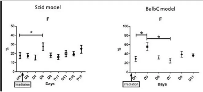
timing of surgery and the RT schedule could influence tumor dissemination and subsequently patient overall survival. We demonstrated the impact of NeoRT on metastatic spreading in a Scid mice model. After an irradiation of 2x5gy, we show more metastasis in the lung when the mice are operated at day 4 compared to day 11 (1). Here, our aim is to evaluate with functional MRI (fMRI) the impact of the radiation treatment on the tumor microenvironment and subsequently to identify non-invasive markers helping to determine the best timing to perform surgery for avoiding tumor spreading. Material and Methods: We used two models of NeoRT in mice we have previously developed: MDA-MB 231 and 4T1 cells implanted in the flank of mice (1). When tumors reached the planned volume, they are irradiated with 2x5 Gy and then surgically removed at different time points after RT. Diffusion Weighted (DW) -MRI was performed every 2 days between RT and surgery. For each tumors we acquired 8 slices of 1 mm thickness and 0.5 mm gap with an “in plane voxel resolution” of 0.5 mm. For DW-MRI, we performed FSEMS (Fast Spin Echo MultiSlice) sequences, with 9 different B-value (from 40 to 1000) and B0, in the 3 main directions. We also performed IVIM (IntraVoxel Incoherent Motion) analysis, in the aim to obtain information on intravascular diffusion, related to perfusion (F: perfusion factor) and subsequently tumor vessels perfusion.
Results: With the MBA-MB 231 we observed a significant increase of F at day 6 after irradiation than a decrease and stabilization until surgery. No other modifications of the MRI signal, ADC, D or D* were observed. We observed similar results with 4T1 cells, F increased at day 3 than returned to initial signal (fig 1). The difference in the peak of F can be related to the difference in tumor growth between MBA-MB 231 in four weeks and 4T1 in one week.
Figure 1: Graphs representing F factor in tumor bearing mice before and after radiotherapy in MDA-MB 231(n=6) (Scid model) and in 4T1 (n=4) (BalbC model); (*=p<0, 05)
Conclusion: For the first time, we demonstrate the feasibility of repetitive fMRI imaging in mice models after NeoRT. With these models, we show a significant difference between the pre-irradiated acquisition and day 6 or day 3 for perfusion F. This change occurs between the two previous time points of surgery demonstrating a difference in the metastatic spreading (1). These results are very promising for identifying noninvasive markers for guiding the best timing for surgery.
Reference: (1) The timing of surgery after neoadjuvant radiotherapy influences tumor dissemination in a preclinical model Natacha Leroi et al. (2015) Oncotarget vol. 5
EP-2050
The assessment of fractal dimension with Dual Energy CT gives information on lung cancer biomarkers
V. González-Pérez
1Fundación Instituto Valenciano de Oncología, Servicio de
Radiofísica y Protección Radiológica, Valencia, Spain
1, E. Arana2, A. Bartrés1, S. Oliver1, B.
Pellicer1, J. Cruz3, M. Barrios2, L.A. Rubio4
2Fundación Instituto Valenciano de Oncología, Servicio de
Radiología, Valencia, Spain
3Fundación Instituto Valenciano de Oncología, Servicio de
Anatomía Patológica, Valencia, Spain
4Fundación Instituto Valenciano de Oncología, Servicio de
Biología Molecular, Valencia, Spain
Purpose or Objective: To assess whether texture analysis of images obtained with Dual Energy CT (DECT) is related to
KRAS and Ki-67 lung cancer biomarkers.
Material and Methods: A retrospective review (May 2013 - January 2015) of 125 lung cancer patients with lung GSI (Gemstone Spectral Imaging) and perfusion CT imaging on a DECT was fulfilled. For 25 of them, the fraction of Ki-67 positive-tumour cells was analysed and for 19 patients KRAS-positive (mutation detected) or KRAS-negative (mutation not detected) character was evaluated (11 positive, 8 negative). DECT examination was performed on a Discovery CT 750 HD scanner (GE Healthcare, USA).
For the perfusion exam, blood volume, blood flow and permeability-surface studies were analyzed. At GSI exam, images related to absorption in Hounsfield units (HU), iodine concentration and monochromatic virtual images reconstructed at 40, 60, 80, 100, 120 and 140 keV were assessed. Tumour fractal dimension was measured with the use of Mapfractalcount plug-in for ImageJ (National Institute of Health, USA) software.
After extraction of DNA from paraffin embedded tissue using QIAamp DNA Investigator Kit (Qiagen), analysis of the KRAS gene exons 2 (codons 12/13) and 3 (codon 61) were performed in order to identify possible associated mutations with real-time PCR kit cOBAS® KRAS Mutation Test (Roche Diagnostics, SL).
T-Student test or U Mann-Whitney test were used to compare differences between KRAS-positive from KRAS-negative cohorts. Pearson correlation coefficient was used to study linear relationship between fractal dimension and the fraction of Ki-67 positive-tumour cells.
Results: Best result (p=0.02) for distinguishing KRAS-positive cohort was obtained for lesion fractal dimension at 140 keV virtual image. This parameter showed an AUC=0.80. It was predictive of KRAS-positive with 90.9% sensitivity and 75.0% specificity for a fractal dimension threshold of 2.352. There was a correlation of lesion fractal dimension in blood volume image and the fraction of Ki-67 positive-tumour cells (p= 0.04).
Conclusion: Ki-67 positive-tumour cells and KRAS-positive biomarkers lead to tumour heterogeneity that modify radiographic image. Fractal dimension parameter quantifies such imaging heterogeneity and could allow to differentiate them.
A higher fractal dimension (higher heterogeneity) of lesion at virtual monochromatic images is measured for KRAS-positive mutation, while a higher fraction of Ki-67 positive-tumour cells is associated with a more homogeneous blood volume at perfusion.
EP-2051
Hsp70 as a tumor specific biomarker in primary glioblastoma multiforme patients
F. Laemmer
1Klinikum rechts der Isar- TU Muenchen, Radiation Oncology,
Muenchen, Germany
1,2, C. Delbridge2, K.A. Kessel1,3, S. Stangl1, J.
Hesse1, B. Meyer4, J. Schlegel2, D. Schilling1,3, G. Multhoff1,3,
T.E. Schmid1,3, S.E. Combs1,3
2Institute of Pathology- TU Muenchen, Neuropathology,
Muenchen, Germany
3Institute of Innovative Radiotherapy- Helmholtz Zentrum
Muenchen, Radiation Sciences, Muenchen, Germany
4Klinikum rechts der Isar- TU Muenchen, Neurosurgery,