UNIVERSITE DE GENEVE
Faculté de MédecineSection de Médecine Clinique Département de Médecine Interne
Laboratoire de Biologie du Vieillissement
Thèse préparée sous la direction du Professeur Karl-Heinz Krause
et Dr Irmgard Irminger-Finger
Expression aberrante de BARD1 est associée
aux pronostiques défavorables pour les cancers
du sein et de l’ovaire
Thèse
présentée à la Faculté de Médecine
de l´Université de Genève
pour obtenir le grade de Docteur en médecine
par
Jian-Yu WU
De HuNan, Chine
Thèse n° Méd. 10433
Genève, 2005
Dedicated to
my parents, my husband, and my daughter,
who gave me understanding and great support with love
in these years
List of contents
Page A. List of abbreviations 3
B. Résumé et introduction (en français) 4 C. Abstract 9
D. Introduction 10
1. Epidemiology and prognostic factors of cancer 10
2. Genetic factors affecting cancer 11
3. Introduction of tumor suppressor BARD1 13
3.1 Structure of BARD1 13
3.2 Expression and subcellular localization of BARD1 13
3.3 Biological functions of BARD1 16
E. Materials and methods 19
1. Clinical data 19
2. Immunohistochemistry 20
3. RNA isolation from frozen tissue 21
4. RT-PCR 21
5. Cloning and sequencing 22
F. Results 24
1. BARD1 expression in sporadic ovarian cancers 24
2. BARD1 expression in breast cancers 30
2.1 BARD1 expression in sporadic breast cancers 30
2.2 BARD1 expression in breast cancers carrying BARD1 mutations 30 2.3 BARD1 expression in breast cancers with BRCA1 mutations 31
3. BARD1 expression in NSCLC 31
4. Aberrant form of BARD1 expressed in cancers 37
G. Discussion 39
H. Conclusions 44
I. References 45
List of abbreviations
AJCC American Joint Committee on Cancer
ANK Ankyrin domain
BARD1 BRCA1-associated RING domain protein 1 BRCA1 Breast cancer susceptibility protein 1
BRCT BRCA1 carboxy-terminal repeat
DAB Diaminobenzidine tetrahydrochloride DCI Invasive ductal breast cancer
DFS Disease free survival DSB Double strand break EOC Epithelial ovarian cancer
EtB Ethidium bromide
GAPDH Glyceraldehyde-3-phosphate dehydrogenase LCI Invasive lobular breast cancer
NES Nuclear export signal NLS Nuclear locating signal
NSCLC Non-small-cell lung cancer RFS Relapse free survival
RING RING (really interesting new gene) domain RT-PCR Reverse transcription polymerase chain reaction SCLC Small-cell lung cancer
Résumé et introduction
1. Introduction
1.1 Epidémiologie et facteurs pronostiques de cancer
Le cancer se place en deuxième position, derrière les maladies cardiovasculaires, comme facteur mortalité en occident. Pour l'année 2004 les projections aux Etats Unis sont de 1,368,030 nouveaux cas de cancer (699,560 hommes et 668,470 femmes) et 563,700 décès. Le cancer du poumon reste la majeure cause de mortalité cancéreuse chez les deux sexes (32% des hommes, 25% des femmes), et représente 12% des nouveaux cas attendus pour cette année. Considérant les femmes uniquement le cancer du sein et des ovaires sont les deux plus répandus. Parmi les 668,470 nouveaux cas attendus chez les femmes la proportion de cancer du sein sera de 32% et celle de cancer ovarien sera de 4%.
La majorité des cancers ovariens sont d'origine épithéliale (Epithelial Ovarian Cancers, EOC) et englobent les tumeurs séreuses, les tumeurs mucineuses, les tumeurs de l'endomètre,
les carcinome à cellules claires, les tumeurs de Brenner, les tumeurs indifférenciées et enfin
les tumeurs épithéliales mixtes parmi lesquelles le carcinome à cellules claires est connu comme indicateur de mauvais pronostic. Quelques facteurs de risque ont été identifiés dans les cancers ovariens comme Cycline D, P53, P21 et CA125; cependant la valeur pronostique de ces facteurs de risque reste controversée.
Le cancer du sein est largement le plus répandu chez les femmes. Plusieurs facteurs sont des indicateurs de pronostic, tels que la taille de la première tumeur, l'implication d'un nodule lymphatique, le type et degré pathologique de cette tumeur, le statut des récepteurs aux œstrogènes à la progestérone.
Les cancers du poumon restent toutefois les cancers les plus fréquents également chez les femmes. Ils ont été divisés en deux classes: "small cell lung cancer" (SCLC) and non small cell lung cancer (NSCLC). Les facteurs pronostiques pour les NSCLC sont le degré pathologique et le stade clinique. Le gène le plus souvent muté -tous types de cancer du poumon confondus- est P53, qui est fortement lié à leur développement. Une mutation dans ce gène représente un indicateur de mauvais pronostic, quel que soit le stade de progression du NSCLC.
Malgré les progrès récents dans les traitements anti-cancéreux, l'incidence de la maladie et sa mortalité changent très peu, les pronostics souvent peu clairs, et surtout le taux de survie sur 5 ans n'a pas significativement augmenté. Par conséquent l'accent a été mis sur des études
portant sur des patients, le diagnostic précoce et la prévention, et qui ont amené au développement d'analyses génétiques.
L'identification de BRCA1 (BReast CAncer susceptibility gene 1) a constitué un pas important vers l'élucidation de ces mécanismes moléculaires. En effet des mutations dans BRCA1 ou BRCA2 sont impliquées dans 40-50% des cancers familiaux précoces du sein, et de plus en plus d'évidences vont dans le sens d'un rôle de ces deux gènes également dans les cancers sporadiques du sein et des ovaires. Toutefois ces deux seuls gènes ne suffisent pas à expliquer tous les cas de cancers ovariens, et dès lors il paraît nécessaire de se tourner vers leurs partenaires fonctionnels. BARD1 (BRCA1 Associated Ring Domain 1) est une protéine pouvant former un dimère avec BRCA1.
1.2 Structure de la protéine BARD1
BARD1 fut découverte lors d'un criblage 2H (two hybrid screening) comme protéine interagissant avec BRCA1. Son gène, BARD1 , se trouve sur le chromosome 2 en position 2q34-q35 et code pour une protéine de 777, 765 et 768 acides aminés chez l'homme, la souris et le rat respectivement. Trois régions sont hautement conservées: un domaine N-terminal RING (86,7% d'homologie), trois répétitions internes en tandem d'ankyrines (90.1% d'homologie) qui sont des motifs (ANK) que l'on trouve fréquemment impliqués dans des régulations transcriptionnelles, et deux motifs BRCT en C-terminal (79,8% d'homologie). Les domaines RING de BARD1 et BRCA1 se combinent pour former une quadruple hélice stable avec leurs structures en directe apposition. L'hétérodimère ainsi formé est plus résistant à la protéolyse que les homodimères respectifs. Bien que la structure en solution des domaines ANK et BRCT ne soit pas bien connue, ces trois domaines structuraux conservés pourraient bien être essentiels aux fonctions de BARD1.
1.3 Expression et localisation intracellulaire de BARD1
Le niveau d'expression de BARD1 est très haut dans la rate et les testicules, mais pas dans le cœur, le cerveau, le foie, les poumons, le muscle squelettique ou le rein. Dans la plupart des cas, BARD1 et BRCA1 sont exprimés en même temps dans les tissus, mais l'expression de BARD1 reste relativement constant pendant le cycle cellulaire, ce qui n'est pas le cas de BRCA1 qui augmente en phase G1 tardive, et atteint un maximum pendant la phase S. Dans les organes contrôlés hormonalement et spécialement dans l'utérus, l'expression de BARD1
augmente de la phase diestrus à postestrus , par opposition à BRCA1 dont l'expression augmente de la phase diestrus à estrus précoce, et diminue pendant l'estrus et le postestrus. Bard1 est exprimé à tous les stades de la spermatogenèse alors que BRCA1 l'est seulement dans les spermatocytes meiotiques et les spermatides rondes précoces. Tous ces éléments portent à croire que BARD1 a des fonctions indépendantes de BRCA1.
Bard1 a d'abord été décrite en tant que protéine nucléaire, trouvée dans des extraits de noyaux et colocalisant avec BRCA1 en "nuclear dots" durant la phase S. En cas de dommage sur l'ADN, les points formés par BARD1 et BRCA1 se dispersent, mais une partie se reforme spécifiquement dans des sites contenant PCNA, ce qui suggère une implication de ces deux protéines dans la réparation de l'ADN. Les données actuelles en fait que BARD1 est capable de faire la navette entre noyau et cytoplasme où elle se trouverait impliquée dans ses fonctions apoptotiques.
1.4 Fonctions biologiques de BARD1
Bard1 joue un rôle important dans le maintient de la stabilité génomique et du phénotype. Dans des cellules d'épithélium mammaire de souris réprimées pour ce gène des changements notables dans le phénotype cellulaire peuvent êtres observés, incluant des altérations de forme, de taille, une haute fréquence de cellules multinucléées ainsi qu'une progression aberrante du cycle cellulaire. La perte de BARD1 résulte en une instabilité chromosomique et une mort embryonnaire précoce a cause d’un dysfonctionnement du cycle cellulaire, mais qui n’est pas accompagnée par une augmentation d’apoptose.
Le complexe BARD1-BRCA1 est une structure essentielle à certaines fonctions biologiques. Les deux protéines se stabilisent entre elles, participent à la réparation de l'ADN, sont impliquées dans des régulations transcriptionnelles, le RNA processing et l'ubiquitination d'autres protéines en vue de leur dégradation. On peut voir BARD1 colocaliser avec BRCA1 et RAD51 en petits points nucléaires durant la phase S ou avec PCNA dans des sites qui se révèlent lors d'une lésion à l'ADN. BARD1 interagit avec CstF-50 (Cleavage stimulation Factor), inhibe la polyadénylation in vitro, et prévient d'un mauvais processing du RNA. Le complexe BARD1-BRCA1 a une activité d'ubiquitine ligase beaucoup plus haute que des préparations individuelles, dans les quelles un mutant de BRCA1 dérivé d'une tumeur (C61G) s'est même trouvé avoir perdu complétement cette capacité. En conditions de stress génotoxique, l'expression de BARD1 augmente et induit une apoptose indépendante de
BRCA1. La transfection ou la surexpression de BARD1 induit une mort cellulaire présentant les signes caractéristiques de l'apoptose, alors que des cellules réprimées pour ce gène sont incapables d'une réponse apoptotique à un stress génotoxique. Le mécanisme présumé d'activation de la voie apoptotique par BARD1 se fait par la voie de signalisation de P53. En contraste avec la localisation nucléaire essentielle au complexe BARD1-BRCA1 pour la réparation de l'ADN, L'induction de la voie apoptotique est corrélée avec une localisation cytoplasmique de BARD1.
Le mutant de BRCA1 C61G, trouvé dans une tumeur, se trouve dans l'incapacité de lier BARD1, suggérant un rôle de BARD1 dans la suppression tumorale mediée par BRCA1. De plus en plus de résultats indiquent que BARD1 pourrait bien être un suppresseur de tumeur à lui-seul. La répression de son expression dans les cellules épithéliales mammaires de souris résulte en des changements phénotypiques rappelant ceux de la pré-malignité. Son expression se trouve également diminuée dans des lignées de cellules cancéreuses. Plusieurs mutations génétiques et altérations somatiques ont été trouvées dans les cancers humains du sein, des ovaires et de l'utérus.
Alors que les études in vitro montrent clairement que la protéine elle-même est un suppresseur de tumeur, ses fonctions dans le cancer et la carcinogenèse restent à élucider. Cette étude a pour objectif d'analyser l'expression de BARD1 en rapport avec son impact sur les tumeurs cliniques et par là-même mettre la lumière sur son rôle dans la tumorigenèse.
2. Résumé
Des mutations dans le gène de prédisposition de tumeur, BARD1, ont été trouvées dans de nombreux cas de cancer du sein, de l’ovaire ou de l’utérus, que ceux-ci soient hérités ou spontanés. La protéine BARD1 joue un rôle dans la réparation de l’ADN et l’ubiquitination comme protéine associée de BRCA1. BARD1 et BRCA1 se co-localisent dans des focis nucléaires. Indépendamment de BRCA1, BARD1 peut induire l'apoptose (la mort cellulaire) en présence de p53, et en réponse à un stress gènotoxique. La réparation de l’ADN et l'apoptose sont des fonctions anti-tumorales importantes qui sont souvent défectueuses dans des cellules cancéreuses. Pour expliquer pourquoi certaines cellules cancéreuses ont échappées à l’apoptose, nous avons posé l’hypothèse que BARD1 et/ou p53 puissent être défectueux. Nous avons donc déterminé par immunohistochimie, les niveaux d'expression de BARD1 et de p53 dans des tissues de cancer de l’ovaire, du sein et du poumon (non-small-cell). Pour cela,
les anticorps primaires N19 et C20, qui reconnaissent respectivement les épitopes N- et C- terminaux de BARD1, ont été employés.
L'expression de BARD1 est fortement élevée dans le cytoplasme de la plupart des cellules cancéreuses, tandis qu’une coloration nucléaire faible est observée dans le tissu sain entourant la tumeur. L'expression maximale de BARD1 est associée au cancer ovarien le plus invasif, le
carcinome à cellules claires. Dans le cancer du sein, l'expression de BARD1 est corrélée avec
une tumeur peu différentiée, de grande taille et une baisse de la survie des patientes. Les concentrations de protéines BARD1 sont semblables dans les tumeurs présentant des mutations soit de BARD1 soit de BRCA1 ou dans des cancers sporadiques. Cependant, les concentrations protéiques de BARD1 sont élevées dans des cancers présentant des mutations de BRCA1 et de p53. Par opposition aux cancers du sein ou de l’ovaire, dans les cancers du poumon, aucune corrélation n’est mise en évidence entre l’expression de BARD1 et le stade ou le grade de la tumeur.
Nos données suggèrent que dans des cellules cancéreuses il existe une forme de BARD1 qui a perdu sa fonction suppresseur de tumeurs. Des expériences de RT-PCR, exécutées sur 10 cas de cancer ovarien, ont révélé l'absence de la portion 5' de l'unité de transcription de BARD1 dans sept de ces tumeurs et le séquençage des autres trois cDNA a permis d'identifier une mutation missense A1291G aboutissant à un changement d'acide aminé de glutamine 406 en arginine. Ces données suggèrent donc que des changements génétiques et épigénetiques puissent mener à l’accumulation de BARD1 dans le cytoplasme des cellules tumorales. De plus, la présence cytoplasmique de BARD1 pourrait être un facteur pronostique défavorable pour les cancers du sein et de l’ovaire.
Abstract
Mutations in tumor suppressor gene BARD1 have been found in cases of inherited and spontaneous breast, ovarian and uterine cancers. The BARD1 protein plays a role in DNA repair and ubiquitination as binding partner of BRCA1 with which it colocalizes to nuclear dots. Independently of BRCA1, BARD1 can induce p53-dependent apoptosis in response to genotoxic stress. DNA repair and apoptosis are tumor suppressor functions that are often defective in cancer cells. In cancer cells that escaped from apoptosis, we expected that either BARD1 or p53 might be defective. We therefore determined expression levels of BARD1 and p53 in ovarian, breast, and non-small-cell lung cancers by immunohistochemistry. For this, the primary antibodies N19 and C20, which recognize epitopes at the N- and C-terminus of BARD1 respectively, were used. BARD1 expression was highly upregulated in the cytoplasm in most cancer cells, while a weak nuclear staining was observed in the surrounding normal tissue. A maximum of BARD1 expression was associated with the most malignant ovarian cancer, clear cell carcinoma. In breast cancer, BARD1 expression was correlated with poor differentiation, large tumor size, and short disease-free-survival time. Tumors with either BARD1 or BRCA1 mutations showed similar BARD1 protein levels as sporadic cancers. However, BARD1 levels were elevated in cancers with BRCA1 and p53 mutations. In contrast to breast and ovarian cancers, no correlation of BARD1 expression with either grade or stage could be determined for lung cancer. Our data suggested that an aberrant form of BARD1 deficient of its tumor suppressor function might be expressed in cancer cells. RT-PCR, performed on 10 cases of ovarian cancers, revealed the absence of the 5’portion of the BARD1 transcript in seven tumors, and sequencing of the remaining three identified a missense mutation A1291G resulting in an amino acid change of glutamine 406 to arginine. These data suggest that genetic and epigenetic changes might lead to elevated cytoplasmic expression of BARD1, and that cytoplasmic BARD1 might be a poor prognostic factor for breast and ovarian cancers.
Introduction
1. Epidemiology and prognostic factors of cancer
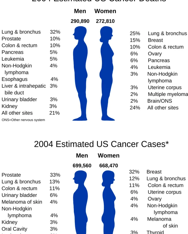
Cancer has been the second leading cause of death (the first is heart disease) in western countries. A total of 1,368,030 new cancer cases and 563,700 deaths areexpected in the United States in 2004. Lung cancer, with high incidence, is still the leading cause of cancer death in both sexes (32% for men and 25% for women). Breast cancer and ovarian cancer are 2 of the major cancers in females. In 2004 the estimated new cancer cases in the United States will be approximately 699,560 for men and 668,470 for women, among which lung cancer accounts for 12-13% for men and women, and breast cancer and ovarian cancer accounts approximately for 32% and 4% in women (Figure 1) 1.
Majority of ovarian cancers is of epithelial origin. Epithelial ovarian cancers (EOC) include serous tumors, mucinous tumors, endometrioid tumors, clear cell tumors, Brenner tumors, undifferentiated tumors and mixed epithelial tumors (Scully RE, in: Young RH, Clements PB eds. Atlas of tumor pathology, 3rd series. Washington DC. Armed Forces Institute of Pathology, 1996:27), among which clear cell carcinoma is reported to have specific biological features and worst prognosis 6-10. Few prognostic factors have been identified in ovarian cancer so far, such as cyclin D, p53 and p21, and CA125; for some of these their prognostic values are controversial 11-23. The frequency of over-expression of a mutant p53 is found to be significantly higher in advanced stage III/IV ovarian cancer as compared to stage I cases (10–20%). This may indicate that p53 inactivation is a late event in ovarian carcinogenesis (reviewed in Feki and Irminger-Finger, 200424).
Breast cancer is the most common cancer in females. Many factors have been reported to influence the prognosis for breast cancer, such as the primary tumor size, the lymph node involvement, pathological type and grade, status of estrogen and progesterone receptors and other biomarkers such as HER-2, p53, bcl-2, Bfl-1, Ki-67, VEGF-C 25-31. Tumor suppressor p53 is often inactivated and overexpressed in breast cancers. Although there are some reports that p53 mutations have a negative correlation with DFS (disease-free survival) or RFS (relapse-free survival), especially in older patients32-34. The prognostic value of p53 is still controversial.
Lung cancer is by far the most frequent type of cancer, roughly divided into small cell lung cancer (SCLC) and non small cell lung cancer (NSCLC). Prognostic factors for NSCLC include the pathological grade and clinical stage. However, the most frequently mutated gene in all types of lung cancer is p53, and has been linked to lung cancer development 35 and was described as an unfavorable factor for prognosis in any stage of NSCLC 36-38.
Despite the recent progress in treatment of cancers, the incidence and mortality has changed very little and the prognosis for the cancers remains unclear and the 5-year-survival rate for cancer has not improved a lot. From 1974 to 1999, the 5-year survival rate for breast cancer and lung cancer was 78-87% and 12-15%, respectively. Seemingly, the focus was more on the subjects, on early diagnosis, and on prevention, and has lead to progress in the development of genetic studies.
2. Genetic factors affecting cancer
Cancer is a genetic disease. In 1914 Boveri hypothesized that cells become malignant either because of over-activation of a gene that promotes cell division or because of loss of function of a gene that normally restrains cell growth. This hypothesis is largely correct, although defects in DNA repair genes are also involved. Genes that promote normal cell growth are referred to as proto-oncogenes, and activation of such genes by point mutation, amplification, or dysregulation converts them to oncogenes. Genes that normally restrain cell growth are called tumor suppressors, and unregulated cell growth arises if their function is lost.
Till now, many genes having been studied, as mentioned above, and mutations of the p53 gene are by far the most common genetic abnormalities found in all types of human cancer. Some tumor suppressor genes are responsible for familial cancer syndromes, for example: p16 mutation for familial melanoma syndrome 39, 40, BRCA1 for familial breast/ovarian cancer syndrome 41, 42. The identification of BRCA1 was an important breakthrough. BRCA1 is known to be one of the breast cancer susceptibility genes, because mutations in BRCA1 or BRCA2 have been reported to account for 40-50% of the early onset familial breast cancers 41, 42
. There is accumulating evidence for a potential role of BRCA1 and BRCA2 in sporadic
breast and ovarian cancers 43-45. However, mutations of BRCA1 and BRCA2 do not account for all breast ovarian cancers. The functional partners of BRCA1 or BRCA2 could be the
candidates for those cancers without BRCA1 or BRCA2 mutations. BARD1 is one of the proteins dimerzing with BRCA1.
2004 Estimated US Cancer Deaths*
Men
290,890Women
272,810 Lung & bronchus 32%Prostate 10%
Colon & rectum 10% Pancreas 5% Leukemia 5% Non-Hodgkin 4%
lymphoma
Esophagus 4% Liver & intrahepatic 3%
bile duct
Urinary bladder 3%
Kidney 3% All other sites 21%
ONS=Other nervous system
25% Lung & bronchus 15% Breast
10% Colon & rectum 6% Ovary 6% Pancreas 4% Leukemia 3% Non-Hodgkin lymphoma 3% Uterine corpus 2% Multiple myeloma 2% Brain/ONS
24% All other sites
Men
699,560Women
668,4702004 Estimated US Cancer Cases*
32% Breast
12% Lung & bronchus 11% Colon & rectum 6% Uterine corpus 4% Ovary 4% Non-Hodgkin lymphoma 4% Melanoma of skin 3% Thyroid 2% Pancreas 2% Urinary bladder 20% All Other Sites Prostate 33%
Lung & bronchus 13% Colon & rectum 11% Urinary bladder 6% Melanoma of skin 4% Non-Hodgkin lymphoma 4% Kidney 3% Oral Cavity 3% Leukemia 3% Pancreas 2% All Other Sites 18%
Figure 1 Estimated US cancer death and new cancer cases in 2004. The leading cause of cancer
death in both sexes is lung cancer. *Excludes basal and squamous cell skin cancers and in situ carcinomas except urinary bladder. Source: American Cancer Society, 2004; adapted from 1.
3. Tumor suppressor gene BARD1
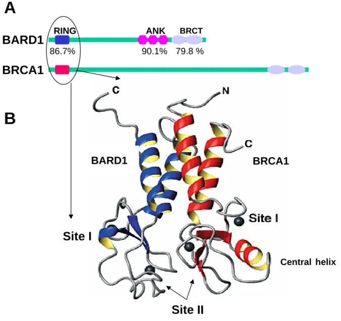
3.1 Structure of BARD1 (BRCA1 associated RING domain protein)
BARD1 was first identified by yeast two-hybrid screening as a protein interacting with BRCA146. The BARD1 gene locates to chromosome 2 at 2q34-q35, it encodes a protein of 777 46, 765 47, and 768 48 amino acids in human, mouse and rat, respectively. Unlike other tumor suppressor genes with a homology from 78% to 98% 49, human BARD1 and mouse BARD1 share only 70% identity of amino acids. But highly conserved structures can be found in three regions: a N-terminal RING domain (homology 86.7%), three internal tandem ankyrin repeats (ANK) (homology 90.1%), which are found in many proteins involved in transcriptional regulation 50 and two C-terminal BRCT motifs (homology 79.8%) 4. The physical state of full length BARD1 is not known yet. The RING domain is the most recognizable structure in the primary sequence of BARD1. It is structurally homologous to that of BRCA1, characterized by a short antiparallel three-stranded β-sheet and two large Zn2+ binding loops, but lacks the central α-helix between the third and fourth pair of Zn2+ ligands 3. Although the regions comprising RING domains of BARD1 (residues 26-119) and BRCA1 (1-109) can form homodimer in vitro, they are not stable structures, they preferentially form heterodimer 51 and only BARD1-BRCA1 heterodimeric complexes have been found in vivo 46. The paired RING domains of BARD1 and BRCA1 combine together to form a stable four-helix bundle with the core elements of the two RING domains in direct apposition to one another 52 (Figure 2). This structure is more resistant to proteolysis than homodimers. The solution structures of ANK and BRCT domain of BARD1 are not well known. However, these three highly conserved structures might be essential for BARD1 functions.
3.2 Expression and subcellular localization of BARD1
BARD1 is highly expressed in spleen and testis 4, but not in heart, brain, liver, lung, skeletal muscle or kidney 4. In most cases, BARD1 and BRCA1 are coordinately expressed in the tissues, but BARD1 expression remains relatively constant during the cell cycle 53, although increases during mitosis is also observed 54. This differs from BRCA1 which increases in late G1 and reaches a maximum during S phase 53. In hormonally controlled organs, especially in the uterus, BARD1 expression is increasing from diestrus phase through postestrus phase whereas BRCA1 expression increases from diestrus to early estrus and decreases during estrus
and postestrus 47. During spermatogenesis, BARD1 is expressed in all stages while BRCA1 is only expressed in meiotic spermatocytes and early round spermatids55. This suggests that BARD1 has some functions independent of BRCA1.
BARD1 was first described as a nuclear protein. It was found in nuclear extracts 46 and localizes to BRCA1 ´nuclear dots’ in the nucleus during S phase 56. Upon DNA damage, BRCA1 and BARD1 nuclear dots disperse, but part of them re-aggregate focally at sites containing PCNA, suggestive of a role in DNA repair 53. BARD1 regulates the subcellular location of BRCA1 by masking the nuclear export signal (NES) of BRCA1 57. However, emerging data show that BARD1 is a protein shuttling between nucleus and cytoplasm. Rodriguez et al identified NES near the RING domain of BARD1, which may be masked by co-expression of BRCA1 and results in nuclear retention. Its cytoplasmic location is associated with its apoptotic function which is markedly reduced by BRCA1 54.
The mechanism of BARD1 degradation is not known. However, a 67kDa proteolytic cleavage product has been found associated with apoptosis in cancer cells 48. A shorter splice form of BARD1β, which lacks RING finger but retains proapoptotic activity, was found in later stages of spermatocyzes precursors55. Auto-ubiquitination on RING domain maybe one of the mechanisms of protein degradation 54.
BARD1
BRCA1
79.8 %BARD1
B
RING 86.7%A
90.1% ANK BRCTSite I
Central helixBRCA1
Site II
Figure 2 Structure of BARD1 and BRCA1. (A) The highly conserved structures of BARD1 and
BRCA1: RING motif and BRCT domain in both BRCA1 and BARD1, 3 ankyrin repeats (ANK) in BARD1 but not in BRCA1. The numbers underneath indicate the identity of the regions of human and mouse BARD1. (B) Solution structure of BARD1-BRCA1 heterodimer. The paired RING domains of BARD1 (in blue) and BRCA1(in red) combine together to form a stable four-helix bundle with the core elements of the two RING domains in direct apposition to one another. Site I and site II indicate the Zn2+ binding site (ref. 3-5).
3.3 Biological functions of BARD1
BARD1 plays an important role in maintaining genomic stability and phenotype. In BARD1-repressed murine mammary epithelial cells, marked phenotypic changes have been found, including altered cell shape, increased cell size, high frequency of multinucleated cells and aberrant cell cycle progression 47. Loss of BARD1 resulted in chromosomal instability and early embryonic lethality which caused severe impairment of cell cycle proliferation but not accompanied by increased apoptosis 58.
BARD1-BRCA1 complex is an essential structure for biological functions. BARD1 and BRCA1 stabilize each other, participate in DNA repair, transcriptional regulation, RNA processing, ubiquitination, and apoptosis. Cotransfection of BARD1 with BRCA1 reduces BARD1-induced apoptosis 2, 59. Similarly, over-expression of BARD1 reduces BRCA1 dependent apoptosis 60. These data suggest that the BARD1-BRCA1 complex contributes to DNA repair and cell survival. A tumor-associated mutation in BRCA1 C61G resulted in a deficiency in binding to BARD1, suggesting a role of BARD1 in BRCA1 mediated tumor suppression. BARD1 co-localizes with BRCA1 and Rad51 (a major participant in double-strand break repair and homologous recombination) in discrete nuclear dots during S phase 53 or with PCNA in DNA damage-inducible nuclear foci 56. BARD1 also participates with BRCA1 in homology-directed repair of chromosomal breaks (DSB). In this case, nuclear localization of BARD1 and BRCA1 is not compromised, suggesting a direct effect on repair 61. A fragment of BARD1 comprising half of ANK through BRCT domain (residues 464-777) binds in vitro to the ankyrin repeats domain of Bcl-3 and modulates the transcriptional activity of NF-κB and NF-κB driven gene expression62.
BARD1 interacts also with CstF-50 (cleavage stimulating factor), inhibits polyadenylation in vitro, and prevents inappropriate RNA processing 63. A tumor-associated germline mutation in BARD1 (Q564H) exhibits reduced binding to CstF-50 and abrogated inhibition of polyadenylation 64. These findings are supportive of a BARD1 function in DNA repair and indicate a link between RNA processing, DNA repair and tumor suppression.
Ubiquitination is nowadays recognized as a multifunctional signaling mechanism with regulatory significance comparable to that of phosphorylation. The functional consequence of ubiquitination varies, including protein degradation, repair activation, transcriptional regulation and cell cycle control. Ubiquitin conjugates to target protein requiring the actions of
ubiquitin–activating enzyme (E1), ubiquitin-conjugating enzyme (E2) and ubiquitin-ligase (E3). Although BARD1 and BRCA1 have very low ubiquitin ligase activity (E3), the BARD1-BRCA1 complex shows dramatically higher ubiquitin ligase activity than individual preparations of BARD1 or BRCA1, while a tumor-derived mutation of BRCA1 C61G lost ubiquitin ligase activity 65. Although the exact target protein of BARD1-BRCA1 ubiquitination is not clear, RNA polymerase II holoenzyme (Pol II), reported to be one of the BRCA1-associated proteins66, might be suspected. Upon DNA damage, BARD1-BRCA1 ubiquitination activity increases and degradation of RNA polymerase II and transcription arrest is observed. BARD1 RING domain or BARD1-BRCA1 complexes also have autoubiquitination function 67 which may serve as a signaling event, such as in DNA repair or in regulating the BARD1-mediated inhibition of mRNA polyadenylation after DNA damage 64, instead of serving as target for proteasomal degradation. Or, perhaps the BARD1-RING domain is the target of protein degradation by the ubiquititin pathway, as speculated for BRCA1 68.
Upon genotoxic stress, BARD1 expression increases and induces BRCA1-independent apoptosis. BARD1 transfection or overexpression induces cell death, which displays features of apoptosis while BARD1-repressed cells are defective for apoptotic response to genotoxic stress2. A proteolytic product, 67kDa protein, found in apoptotic bodies of rat colon cancer cells was identified as an apoptotic cleavage product of BARD1 and antibodies against this product were detected in rats immunized with apoptotic bodies against experimentally induced colon cancer 48. In prostate cancer, treatment with Camptothecin causes BARD1 and NF-κB upregulation and induces apoptosis 69. All these suggest that BARD1 participate in apoptotic response. The mechanism of BARD1 induced apoptosis is presumed to act through a p53 pathway2. In contrast to the nuclear retention of BARD1-BRCA1 complex in DNA repair, BARD1-induced apoptosis is reported to be associated with cytoplasmic location of BARD1 54, 59
.
A tumor associated mutation in BRCA1 C61G resulted in deficiency in binding to BARD1, suggesting a role of BARD1 in BRCA1 mediated tumor suppression. There is accumulating evidence that BARD1 itself is a tumor suppressor. Repression of BARD1 expression in murine mammary epithelial cells resulted in the phenotypic changes reminiscent of premalignancy 47. BARD1 expression is reported to be some reduced in breast cancer cell lines 70. Thai et al have
described the BARD1 somatic and germ-line mutations in a subset of primary breast, ovarian and uterine cancers 71. Ishitobi reported a germline mutation in Japanese familial breast cancers 72. Mutation of BARD1 was also found with elevated frequency in Finnish breast cancer families 73. Particularly BARD1 germline mutations were identified in breast and breast/ovarian cancer families without BRCA1 or BRCA2 alterations 74.
While in vitro studies provide evidence that BARD1 itself is a tumor suppressor, the function of BARD1 in cancer and in carcinogenesis remains unclear. The aim of this study was to investigate the BARD1 expression and its significance in the clinical tumors and to shed light on the role of BARD1 in tumorigenesis.
Materials and methods
1. Clinical data
Ovarian cancer and sporadic breast cancer specimens were obtained from the Pathological Division of Department of Gynecology and Obstetrics of Geneva University Hospitals. Non-small-cell lung cancer specimens were offered friendly by Dipartimento Scienze Cardio-Toraciche e Respiratorie, Napoli, Italy. The samples of BARD1-mutant breast/ovarian cancer and BRCA1-mutant breast/ovarian cancer were from University of Pisa, Italy. The pathological diagnosis were made by experienced pathologists blinded of the experiments, according to World Health Organization (WHO) classification of tumors and the American Joint Committee on Cancer (AJCC) TNM (AJCC Cancer staging handbook. TNM classification of malignant tumors. Sixth edit. Springer 2002).
44cases of sporadic ovarian cancer, from women of 26 to 83 years of age, were analyzed, comprising 16 cases of serous carcinoma, 11 cases of mucinous carcinoma, 10 cases of endometrioid carcinoma, and 7 cases of clear cell carcinoma, graded from G1 to G3, according to the Silverberg system75.
54 cases of sporadic lung cancer aged from 39 to 77 years old (mean age 62.9ys), 20 cases of squamous cell carcinoma, 21 cases of adenocarcinoma, 8 cases of large cell carcinoma, 4 cases of adenosquamous carcinoma and 1 case of anaplastic change. Cases of pathological grade 1 to grade 3 was2, 18, 21 respectively, and 12cases were variable in G1 to G3. 29 cases in stage I; 8 cases in stage II; 15 cases in stage III, 1 case in stage IV. (Table 1)
In total 10 cases of sporadic breast cancer, 9 invasive ductal carcinoma (DCI), 1 invasive lobular carcinoma (LCI), and 13 cases of breast/ovarian cancers with BRCA1 mutations from 11 families (10 DCI of the breast, 3 ovarian carcinoma), and 5 cases of breast cancers with BARD1 mutations from 4 BARD1-mutant families (DCI) were included in our study.
Table 1. Clinical data of ovarian carcinoma and NSCLC
Ovarian carcinoma Number of cases Total
Histology Serous carcinoma Mucinous carcinoma Endometrioid carcinoma Clear cell carcinoma
16 11 10 7 44 Grade G1 G2 G3 21 19 4 44
NSCLC Number of cases Total
Histology Squemous cell carcinoma Adenocarcinoma
Adenosquemous carcinoma Large cell carcinoma Anaplastic change 20 21 4 8 1 54 Grade G1 G2 G3 G1-G3 2 18 21 12 53 Stage I II III IV 29 8 15 1 53
2. Immunohistochemistry
5µm-thick sections of formalin-fixed and paraffin-embedded tissue were deparaffinized with xylene and rehydrated through descending alcohol (100% alcohol x2, 95% alcohol x2, 75% alcohol x1, H2O x1); the sections were boiled 5 minutes in microwave oven for antigen retrieval, and then blocking the endogenous peroxidase. The slides were incubated over night at 4°C in a humidified chamber with the primary antibody after BSA (bovine serum albumin) blocking the nonspecific proteins. The primary antibodies we used for detecting BARD1 were BARD1-N19 (sc-7373) and BARD1-C20 (sc-7372; Santa Cruz Biotechnology) which recognize the epitope of N-terminus and C-terminus of BARD1 protein. Secondary antibodies (rabbit-antigoat) with HRP were applied in dilution 1:100 at room temperature 1 hour. Then, DAB staining was performed 15 minutes at room temperature. The slides were counterstained with hematoxyline before dehydration and mounting. To quantify expression of BARD1 or p53, four different regions were chosen from each stained tumor section, the total number of cells and the number of positively stained cells were counted. The average was calculated to
determine the percentage of BARD1 positive cells. These quantifications were repeated independently by three persons.
Table 2. Primary antibodies used in the study
Specificity Dilution Incubation source product BARD1-N19 1:25 Over night; 4ºC goat sc-7373 BARD1-C20 1:20 Over night; 4ºC goat sc-7372
p53 1:20 Over night; 4ºC rabbit sc-6243
3. RNA isolation from the frozen tissue sections
TRIzol Reagent was used to isolate RNA from the frozen tissue. 4-5 10µm-thick sections were used for each sample. 100-150µl of TRIzol Reagent were added onto the slides (the total amount of sections and TRIzol Reagent depended on tissue size) and incubated 5 minutes at room temperature. Pipetted carefully several times to homogenize the tissue and transferd the homogenized tissue into a RNase-free eppendorf tube. Proceeding extractions were operated following the protocol of RNA isolation with TRIzol Reagent. Added 0.1ml of chloroform and centrifuge 14,000g 15minutes at 4ºC to separate the phases. The aqueous phase was transferred to a clean RNase-free eppendorf tube and equal volume of isopropanol was added. After centrifugation at 4ºC, the pellet was washed with 75% ethanol. 20µl of RNase-free water was added to elute RNA. RNA concentration was measured and the quality was assessed by D260/D280 ratio.
4. RT-PCR (reverse transcription and polymerase chain reaction)
For reverse transcription, 1µg of RNA was used in 21µl of reverse transcription buffer containing 1µl of dNTP(10mM), 1µl of oligo dT, 2µl of DTT(0.1M), 4µl of 5x First Stand Buffer. The reaction took place at 65ºC 5 minutes followed by 42ºC 2mins and 1µl of Superscript II (Invitrogen) was added, then incubations were 42ºC 50 minutes and 70ºC 15 minutes. 2µl of cDNA was used as a template for PCR with different primers (Table 1). 40 cycles of stepwise PCR (annealing temperature from 65 ºC to 56ºC, each cycle decrease 1ºC,
and then 55ºC 30 cycles) was performed with Taq polymerase in a final reaction volume of 50µl. Primary denaturation (94 ºC, 3minutes) and final extension (72ºC, 10minutes) were the same for each PCR reaction. 10ul of PCR product was used for analysis in 1% of agarose/TAE gel with ethidium bromide (EtB) and the band was visible under ultraviolet light.
Table 3. Primers and conditions
forward primer site reverse primer site PCR product size (bp) Extension 72ºC 5’ccatggaaccggatggtc3’ 74 5’aacaccaccgggtatcaaaa3’ 1481 1407 100s 5’ccatggaaccggatggtc3’ 74 5’cgaaccctctctgggtgata3’ 2252 2178 120s 5’agcaagtggctccttgacag3’ 783 5’cgaaccctctctgggtgata3’ 2252 1469 100s 5’ctccagcataaggcattggt3’ 1441 5’cgaaccctctctgggtgata3’ 2252 811 60s
5. Cloning and sequencing
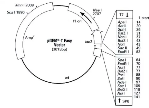
QIAEX II kit was used for DNA purification. The purified DNA was cloned into pGEM-T® Easy vector (Promega, Figure 3). The ligation and transformation were performed according to the manufacturer’s instructions. The insert/vector ratio was 3:1. 2µl of the ligation reaction mixed with 50µl of JM109 High Efficiency Competent Cells (Cat#L2001) in LB was plated onto the LB/ampicillin/PTG/X-Gal plate and incubated at 37ºC over night. The recombinant clones could be identified by color screening on indicator plates. 5 white colonies in each plate were chosen and incubated in 3ml of LB with ampicillin at 37ºC over night. Recombinant plasmid DNA was isolated using Miniprep kit (Sigma) and followed by sequencing with primers T7 and SP6 (Figure 3).
Figure 3 pGEM-T easy vector circle map and sequence reference points.
Results
1. BARD1 expression in sporadic ovarian cancer
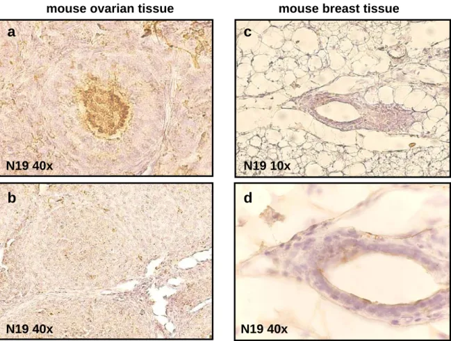
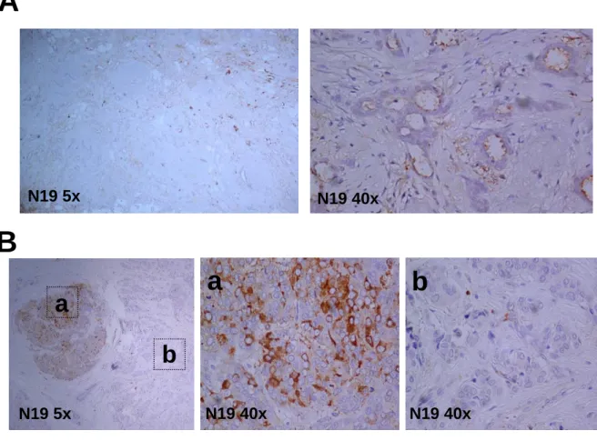
We used two antibodies for testing BARD1 expression, N19 and C20, which recognize epitope at the N-terminus and C-terminus of BARD1 protein, respectively. These antibodies had been used in previous studies and their specificity had been tested2, 76. From expression studies in the mouse, we knew that BARD1 is expressed in the mouse ovary2, 47 with maximal expression in germ cells but not in granulosa cells and sparsely in theca cells (figure 4).
mouse ovarian tissue
N19 40x
N19 40x
b
a
mouse breast tissue
N19 10x
c
N19 40x
d
Figure 4. BARD1 expression in normal mice tissue. In ovarian follicles before ovulation, BARD1
stained the germ cells positively in the nucleus (a), but not in the granulosa cells or theca cells. After ovulation, only a few cells were stained in the nucleus (b). In breast tissue, BARD1 stained a few mammary gland epitheliums in the nucleus.(c,d)
Interestingly, BARD1 was highly expressed in the cytoplasm of tumor cells. In the normal tissue surrounding the tumor, however, little staining and nuclear localization was observed
(Figure 5). This results contrast with the expression of BRCA1, which is generally down regulated in ovarian and breast carcinoma.
N19 40x
a N19 40x b N19 5x a bFigure 5. BARD1 expression in cancer and normal tissue. BARD1 is highly expressed in the
cytoplasm of ovarian carcinoma cells (a), while little staining in peripheral normal ovarian tissue (b).
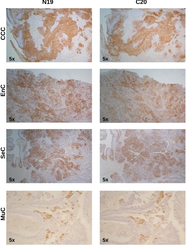
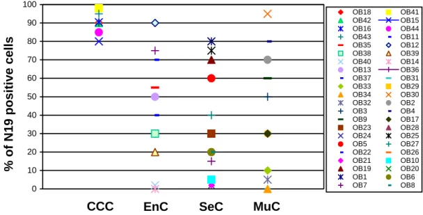
We investigated the BARD1 protein expression in four types of ovarian cancers: clear cell, serous, endometrioid, and mucinous carcinoma. The extend of BARD1 expression varied considerably between the different histological types of ovarian cancer. In clear cell carcinoma, all cells were stained homogeneously and all cases had high positivity of BARD1 staining (90% of the cells were stained in average), while in the other types of ovarian carcinomas, BARD1 expression varied from 0 to 95% of positive cells and showed a mosaic expression pattern, which was most prominent in serous carcinoma (Figure 6).To quantify the expression of BARD1, the proportion of positive cells was determined in tumor tissue (see Material and Methods). Clear cell carcinoma had highest percentage of BARD1 positive cells. The variation within each histological type of tumor was from 0 to 90 percent, but in clear cell carcinoma from 80 to 100 percent.This difference of expression was statistically significant (p<0.005) (Figure 7).
In the majority of the cases, N19 and C20 antibodies stained the same regions when tested on adjacent tissue sections (Figure 6,8A), but differences in N19 and C-20 staining were observed in endometrioid, mucinous, and serous carcinoma (Figure 8B). In most of these cases N-19 staining was lost and C-20 staining was retained. This loss of N-terminal epitopes could be explained by changes on the gene, the transcript, or the posttranslational level.
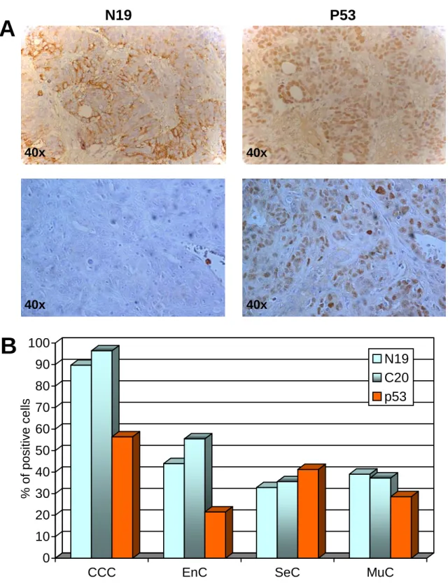
Since BARD1 can act as apoptosis inducer by stabilizing the tumor suppressor p532, we also determined p53 expression levels in the same cases. Consistent with observations made in other types of cancer 24, p53 showed nuclear localization in the ovarian cancer cells while BARD1 was localized to the cytoplasm (Figure 9A), which excluded a BARD1-p53 interaction. In some cases, BARD1 and p53 co-localized in the same region, in others BARD1 and p53 expression showed completely different distributions in the same region. Similarly to BARD1, p53 expression levels varied in different histological types of carcinoma. In clear cell carcinoma, p53 expression was slightly higher than in the other types of cancer, but the variations were not as striking as observed with BARD1 staining and not statistically significant. Comparison of the expression levels of BARD1 and p53 proteins showed no obvious correlation (Figure 9B).
In summary our data indicate that BARD1 expression in ovarian cancer is correlated with histological tumor type, but not with histological differentiation, tumor size, serum CA125 level, or age of the patient (data not shown). Since clear cell carcinoma, where maximal BARD1 expression was found, have a lower reported five year survival, it is suggestive that BARD1, rather than p53 positivity, is correlated with a poor prognosis.
C20
N19
5xCCC
5xEnC
5x 5xSeC
5x 5xMuC
5x 5xFigure 6. Different expression levels of BARD1 in different pathological type of ovarian
carcinoma. In clear cell carcinoma, N19 and C20 stained diffusely the tumor cells, while in other type of ovarian carcinoma, it showed mosaic staining. CCC: clear cell carcinoma; EnC: endometrioid carcinoma; SeC: serous carcinoma; MuC: mucinous carcinoma
0 10 20 30 40 50 60 70 80 90 100 OB18 OB41 OB42 OB15 OB16 OB44 OB43 OB11 OB35 OB12 OB38 OB39 OB40 OB14 OB13 OB36 OB37 OB31 OB33 OB29 OB34 OB30 OB32 OB2 OB3 OB4 OB9 OB17 OB23 OB28 OB24 OB25 OB5 OB27 OB22 OB26 OB21 OB10 OB19 OB20 OB1 OB6 OB7 OB8 % of N19 positive cells
CCC EnC SeC MuC
Figure 7. Distribution of BARD1 (N19) expression in different type of ovarian carcinoma. The
expression of BARD1 in CCC was significantly different with that in other type of ovarian carcinoma (p<0.005). CCC: clear cell carcinoma; EnC: endometrioid carcinoma; SeC: serous carcinoma; MuC: mucinous carcinoma
C20
N19
A
5x 5x 40x 40xB
5x 5x 40x 40xFigure 8. Comparison of N19 and C20 staining. (A) Homogenous and identical staining with N19
and C20 was observed in clear cell carcinoma. (B) Representative example of diverging staining obtained with N19 and C20, as observed in some cases of serous carcinoma.
40x
A
N19 P53
40x 40x 40x 0 10 20 30 40 50 60 70 80 90 100CCC EnC SeC MuC
N19 C20 p53
% of positive cells
B
Figure 9. Comparison of BARD1 (N19 and C20) and p53 expression in ovarian and breast carcinoma.
(A) BARD1 staining was cytoplasmic, p53 staining was nuclear. In some cases, BARD1 and p53 co-localized in the same region (upper panels), in others BARD1 and p53 expression showed completely different distributions in the same region (lower panels). (B) Comparison of BARD1 and p53 expression in four pathological types of ovarian carcinoma. BARD1 positivity decreased from clear cell carcinoma to mucinous. This tendency was less pronounced for p53 positivity. CCC: clear cell carcinoma; EnC: endometrioid carcinoma; SeC: serous carcinoma; MuC: mucinous carcinoma.
2. BARD1 expression in breast cancer
2.1 BARD1 expression in sporadic breast cancerWe analyzed BARD1 expression in 10 cases of sporadic breast cancer. As observed in ovarian cancers, BARD1 was localized in the cytoplasm. The expression levels of BARD1, detected with N19 and C20 were variable. Mosaic staining patterns were observed in some cases (Figure 10), as it was observed in serous ovarian carcinoma. Comparing the expression of BARD1 with tumor differentiation and tumor size, we found that considerably more staining was observed in G3 grade tumors than in G1, and more in T4 staged tumors than in T1(Figure 10 C).
We analyzed the expression of p53 in the same samples, but could not establish any correlation between p53 and BARD1 expression. Unlike BARD1, p53 levels did not increase with tumor size or undifferentiated state. Since tumor differentiation, size, and stage are prognostic factors for breast cancer, it therefore seems likely that BARD1 rather than p53 staining is associated with a poor prognosis for breast cancer and it maybe a prognostic marker of breast cancer.
2.2 BARD1 expression in breast cancers carrying BARD1 mutations
Several mutations of BARD1 have been reported associated with breast and ovarian cancer. We analyzed BARD1 expression in 5 cases of 4 different BARD1 mutations of breast cancer families. The mutation sites were A957G, A1009T, G1743C, 1144del21, respectively74. Since these mutations are associated with breast cancer, one could speculate that they might affect BARD1 protein function. Using antibody BARD1-N19, however, we found, similar to what we observed in sporadic breast and ovarian cancer, intensive cytoplasmic staining (Figure 11A). The percentage of BARD1 positive cells was also similar to that of sporadic breast cancer. The staining intensity and the percentage of positive cells were not correlated with a specific mutation site. It was unclear at this point whether all BARD1 mutations investigated result in a cytoplasmic localization of the protein, or whether the cytoplasmic localization of BARD1 is dictated by other tumor factors and not influenced by specific mutations.
It can be concluded that aberrant location of BARD1 is a negative prognostic factor, but not necessarily associated with a specific mutation.
2.3 BARD1 expression in breast cancer with BRCA1 mutations
We analyzed BARD1 expression in 13 cases of BRCA1 mutant breast/ovarian cancer including 10 cases of invasive ductal breast cancer (DCI) and 3 cases of ovarian cancer. In BRCA1-mutant cancers, BARD1-N19 staining was similar to that in sporadic breast cancer and BARD1-mutant breast cancer (Figure 11B). The mutation type of BRCA1 didn’t correlate with the extent of BARD1 staining. Thus BRCA1 mutations did not influence the pattern of BARD1 expression nor the stability of BARD1 protein expressed in tumors, as expected, based on previous reports54, 77.
In four cases, patient outcome data were available and BARD1 expression levels could be correlated with the disease free survival (DFS). High BARD1 expression was observed in patients with shorter DFS than in those with longer DFS, suggestive that BARD1 expression was negatively correlated with DFS (Figure 11C) in patients with BRCA1 mutant tumors. It was interesting to know whether BARD1 expression in cancers with BRCA1 mutations correlated with p53 mutation status. RT-PCR was performed and p53 cDNA was cloned and sequenced. Interestingly higher BARD1 expression levels were found correlated with tumors carrying p53 mutations. In p53 wild type tumors low levels of BARD1 expression were found and in p53 mutant tumors BARD1 expression was elevated (Figure 12). These data further support the conclusion that BARD1 expression in tumors is not indicative of its apoptosis function, which depends on a functional p53.
3. BARD1 expression in non-small-cell lung cancer (NSCLC)
To determine whether BARD1 expression was limited to tumors of hormonally regulated tissues, we investigated its expression in lung cancers. 54 cases of sporadic NSCLC were analyzed for BARD1 (N19 and C20 antibody staining) and p53 protein expression. BARD1 was found in the cytoplasm in the lung cancer cells similar to what we observed in the ovarian and breast carcinoma. We compared the expression of BARD1 (N19 and C20) in tumors of various histological type, pathological grade, and clinical stage. In large cell carcinoma, BARD1 expression was higher than in other types of lung cancer. BARD1 levels were increasing from adeno to squamous and large cells, however, no correlation of BARD1 with p53 expression levels were found (Figure 13). However, the expression of p53 was clearly increasing with stage of tumors and was elevated in poorly differentiated cancers as compared
to well-differentiated NSCLC with a statistical significance of p<0.05 (Figure 13B). This finding is consistent with the observation that more that 50% of lung cancers exhibit p53 mutations and that p53 is the major target for mutations during malignant transformation35.
A
N19 5x N19 40xB
b
a
N19 5xa
N19 40xb
N19 40xC
Correlation of BARD1 and p53 with differentiation in sporadic breast cancer
Correlation of BARD1 and p53 with tumor size in sporadic breast cancer
0 10 20 30 40 50 60 70 T1 T2 T3 T4 N19 C20 p53 % of positive cells 0 5 10 15 20 25 30 35 G1 G2 G3 N19 C20 p53 % of positive cells
Figure 10. The expression of BARD1 in sporadic breast cancer. BARD1 (N19) staining was less in well
differentiated G1 tumor (A) than that in poorly differentiated G3 tumor (B). In some cases, it showed mosaic staining as that in serous ovarian carcinoma. (C) Correlation of BARD1 (N19/C20) and p53 with pathological differentiation and tumor size. BARD1 expression was highly elevated in grade 3 and T4 tumors.
A
N19 10x N19 40x
C
Figure 11. The expression of BARD1(N19) in BARD1-mutant or BRCA1-mutant breast cancers. BARD1 stained in the cytoplasm of BARD1-mutant breast cancer (A) and BRCA1 mutant cancer (B). The left case in (B) had less staining than the right one. (C) Expresssion of BARD1 had negative association with disease free survival (DFS) in the BRCA1-mutant breast cancers.
N19 40x 5x N19 40x
B
5x 80 60 40 20 BRCA1 G1354X BRCA1 1100del AT BRCA1 1100del AT BRCA1 1499 insA % of N19 positive cellsdisease free survival (y)
case4 case3 case2 case1 100 0 18 16 14 12 10 8 6 4 2 0
BRCA1mt/p53mt
BRCA1mt/p53wt
5x 40x 5x 40x 5x 40x 5x 40xA
B
0 10 20 30 40 50 60 70 80 p53mt p53wt N19% of N 19 positive cells
Figure 12. Relationship between BARD1 expression and status of p53 mutations in BRCA1-mutant
breast/ovarian cancers. (A) BARD1 (N19) expression was higher in BRCA1-mutant breast cancers with mutated p53 (p53mt) than in those with wild type p53 (p53wt) (upper panel). Similarly, BRCA1-mutant ovarian cancers with mutant p53 (p53mt) had more BARD1 (N19) expression than those with wild type p53 (p53wt) (lower panel). (B) Comparison of BARD1 expression levels (% positive cells) with p53 mutation status in BRCA1-mutant breast/ovarian cancers.
5x
A
N19 40x p53 40x 5x 0 10 20 30 40 50 60 70 80 90 100 aden o squa m aden osqu large 6159 10356 6609 8040 8315 8341 1708 12740 12748 1176 1659 8532 8664 12278 11638 12756 6634 8075 9977 12767 11601 1103 1531 8366 8930 10362 7987 9018 10319 6661 7723 8650 12111 9844 6699 8100 8981 11400 7993 11203 11605 12500 357 1506 1154 7982 1505 1492 870 6827 4 11281% of N19 positive cells 0 10 20 30 40 50 60 70 80 90 100 aden o squa m aden squ large N19 C20 P53
% of positive cells 0 10 20 30 40 50 60 70 80 90
C
0 G1 G2 G3 G1-3 N19 C20 p53 10B
0 10 20 30 40 50 60 70 80 90 100stage I stage II stage III stage IV
N19 C20 P53
% of positive cells
% of positive cells
Figure 13. The expression of BARD1-N19/C20 and p53 in NSCLC. (A) ) Staining of BARD1 (N19) and
p53 in NSCLC. BARD1 was cytoplasmic, p53 nuclear. (B) The expression of BARD1 and p53 compared in pathological differentiation and clinical stage. No correlation was found for BARD1, but p53 expression was correlated with grade and stage. (C) BARD1 and p53 expression and distribution of BARD1-N19 expression in different histological types of NSCLC. adeno:adenocarcinoma; squam: squamous cell carcinoma; adenosqu: adenosquemous carcinoma; large: large cell carcinoma
4. Aberrant form of BARD1 expressed in tumors
Several reported functions of BARD1 suggest a role in tumor suppression. Its upregulation in tumors therefore led to speculate that the form of BARD1 expressed in tumors might be an aberrant form deficient of its tumor suppressor function. To test this hypothesis, RT-PCR amplified and cloned and sequenced BARD1 cDNAs from frozen tissue sections of ovarian tumor samples. Control RNAs, from HeLa cells and from human cytotrophoblasts, lead to similar amplification of the 5' and 3' portions of the BARD1 transcript. However, in 7 of 10 tumor samples, , RT-PCR showed a complete loss of the N-terminal half of the protein coding region (nucleotides 74 to 1481), or the segment including nucleotides 783 to 1441 (Figure 14). Similar amplification of the 5' and 3' halfs of BARD1, as performed with control RNAs, was only possible in three cases, OB23, OB35, and OB37. In these three cases, we found the same missense mutation A1291G, which translates into an arginine instead of glutamine (Q406R) (Figure 14B). In some cases the lack of the N-terminal half of the BARD1 transcript was consistent with the observed loss of N-19 but not C-20 staining by immunohistochemistry. We concluded that the form of BARD1 which is overexpressed in cancer cells and localized to the cytoplasm, presumably presents an aberrant form of BARD1. Interestingly, BARD1 expression is highly elevated in clear cell ovarian carcinoma and poorly differentiated, large sized breast cancers which have the worst prognosis for five year survival. This expression pattern of BARD1 is similar to p53, which shows elevated expression in tumors, correlated with poor prognosis, and when expressed in tumors, is mostly mutated. Whether similar aberrant forms of BARD1 exist in lung cancer remains to be determined.
N-terminus 74-1481
A
C+ 8 8* 9 21 23 24 24* 25 33 33* 34 35 37 C- 1500bp C-terminus 1441-2252 800bp HGAPDH 300bpB
G2354ABARD1
RING
ANK
74 1441 1481 783BRCT
G2156C G1765C G1743C A1009T 1144del21 A1482G G1592A A957G 2252 A1291GFigure 14. Transcriptional expression of BARD1 in ovarian cancers. (A) RT-PCR analysis of 10
ovarian cancers. Control RNA from HeLa cells (C+) and RNA from ovarian cancers were tested using primers amplifying the N-terminal (74-1481) or C-terminal (1441-2252) part of the transcript. The results obtained with forward primers 783 and reverse primer 1481 were similar to that of 74-1481 (data not shown). Samples 8, 24 and 33 were performed on two doublicate samples for the same tumor (*). In 7 of 10 cases the N-terminal part could not be amplified, and was weaker than the C-terminal part in the remaining ones. (B) Mutation A1291G found in tumors 23, 35, and 37 is indicated (red arrow) on the schematic diagram of the BARD1 protein structure. RING, ankyrin (ANK) and BRCT domains of BARD1 are presented with known mutations indicated by arrows. Primers used for RT-PCR are shown underneath.
Discussion
Originally BARD1 has been described as a tumor suppressor that participates in DNA repair, transcriptional regulation, RNA processing, and ubiquitilation by binding to BRCA1 46, 63, 78. Repression of BARD1 expression in murine mammary epithelial cells resulted in phenotypic changes reminiscent of premalignancy 47 and to genomic instability 47, 58, 77, 79 and overexpression leads to apoptosis 2 suggesting a function in tumor suppression independently of BRCA1.
Consistent with its presumed role as tumor suppressor, the transcriptional expression of BARD1 is reduced in breast cancer cell lines 70.Furthermore, tumor associated mutations of BARD1 were found for both somatic and germ-line mutations in a subset of primary breast, ovarian and uterine cancers 71. Ishitobi reported a germ line mutation in Japanese familial breast cancers 72. Mutations of BARD1 were also found with high frequency in Finnish breast cancer families 73. Ghimenti et al. identified BARD1 germline mutations in breast and breast/ovarian families that were negative for BRCA1 and BRCA2 alterations 74.
It is generally assumed that genetic mutations could induce structural changes of the protein product, leading to a loss of function due to a presumed decrease of protein stability. However, we observed an important upregulation of BARD1 protein expression in cancer tissue as compared to normal tissue, in many tumors of non-familial origin and unknown genetic status of BARD1 or BRCA1as well as in tumors associated with four specific mutations of BARD1. Similarly, we found BARD1 upregulation in tumors with BRCA1 mutations while BARD1 destabilization would be expected in the absence of BRCA1 by a reduced mutual stabilization of BARD1-BRCA1 77 .
The protein expression of BARD1 in tumors has not been investigated before and there is no reported immunohistochemical study of BARD1 in human cancer tissue. BARD1 was described as a nuclear protein 46, 56 , and in association with apoptosis its translocation to the cytoplasm is observed 54, 60. However, here we report that BARD1 localizes to the cytoplasm in cancer cells that are not apoptotic. We conclude that BARD1 expressed in cancers corresponds to an aberrant form and/or a form that lacks tumor suppressor functions due to posttranslational modifications. It even is possible that BARD1 acquires novel (dominant negative) functions in favor of tumor cell growth. Alternatively, factors influencing the intracellular localization of BARD1 and/or its stability could be modified in the tumor cells. In fact, elevated expression in
tumors has been reported for p53, which when upregulated in cancer cells is derived from abnormal protein stability and/or long half -life of mutated form80.
Interestingly, BARD1-N19 and BARD1-C20 staining, with antibodies recognizing N-terminal and C-terminal epitopes, respectively, did not always overlap, and a loss of the N-terminal but not C-terminal epitopes was observed in several tumors, suggesting incomplete expression of the transcript or protein and/or introduction of modifications affecting the N-terminus. Interestingly, cloning and sequencing of RT-PCR products from a number of such tumors revealed an aberrant form of BARD1. In seven of ten cases the 5'half of the transcript could not be amplified, consistent with the loss of the N-terminal epitope in some tumors. In the remaining three cases, which apparently expressed 5'and 3' portions of the transcript, the same missense mutation A1291G was found which gives rise to an amino acid substitution Q406R. This mutation localizes within a region that already harbors many reported mutations71-74 as indicated in Figure 14. In fact, the BARD1 A1291G mutation localizes close to a potential nuclear location signal (NLS)54, which might influence the proper function of this signal and explain the aberrant cytoplasmic localization. From the 10 cases sequenced in this report and from the immunohistochemical analysis of tumors with previously reported BARD1 mutations, it can be concluded, that loss of the 5'portion of BARD1 transcript, or mutations in the region upstream of the ankyrin motif are associated with loss of nuclear localization. Interestingly, the A1291G mutation is different from the mutations reported before, but appears in 3 of 10 cases and might be a specific mutation for cancers from Geneva.
Although the loss of 5'half of BARD1 transcript could be due to genomic rearrangements, the fact that in 7 of 10 cases the 5’part of the transcript is missing, is suggestive of a mechanism which is not the result of random genomic instability. It is possible that new transcription initiation sites are used or that most of the 5’end of the BARD1 transcript is deleted due to alternative splicing or to translocations. Indeed, a recent finding of a new splice variant of BARD1 in a rat ovarian cancer cell line, missing exons 2 through 7 (Feki et al., submitted) is in support of the argument that aberrant transcripts missing the coding region that comprises regions upstream of the ANK repeat might be associated with ovarian and breast cancer. It has been discussed in several studies6-10
that clear cell carcinoma is distinct from other types of ovarian cancers with poor prognosis and insensitivity to platinum-based chemotherapy 81, 82.We find that BARD1 expression levels are correlated with pathological type of ovarian cancer, and it is in clear cell carcinoma where nearly 100 percent of the cells express BARD1,
while in other types of ovarian carcinomas the expression varies from 0 to 90 percent. This suggests that BARD1 might be correlated with negative outcome. This hypothesis was confirmed for cases of sporadic breast cancer, where BARD1 expression was correlated with poor differentiation and large tumor size. Even more so, in familial breast cancers with BRCA1 mutations, the expression of BARD1 is inversely correlated with disease-free-survival. Taken together, we propose that immunohistochemical staining of BARD1 might be a negative prognostic factor for human breast and ovarian cancer.
Interestingly, a differed scenario is observed in NSCLC. Only moderate increase of BARD1 expression in large cell carcinoma as compared to other types of lung cancer can be observed. BARD1 expression is neither correlated with pathological differentiation nor stage of NSCLC. In contrast, p53 protein expression is distinctly increased in grade 3 and stage IV cancers, confirming the role of p53 as primary mutation target during malignant transformation in NSCLC 83. From these data it can be concluded that mutations of p53 are the main predisposing factor in NSCLC, while in cancers of hormone-responsive organs, BARD1 mutations present a significant factor which maybe used as a prognostic factor.
Upregulation of BARD1 in tumors was observed in DNA array analyzes, investigating gene expression profiles of various cancers. In prostate cancer cells BARD1 was observed upregulated in response to treatment with 7-substituted lipophilic camptothecin 69.BARD1 upregulation was also found in responsive to oncogenic RAS signaling 84.Finally, consistent with our observations for breast and ovarian cancers, BARD1 expression was described as one of the markers for treatment failure in embryonic central nervous system tumors 85.
BARD1 was also identified as component of the TGFβ signaling pathway in a study of breast cancers. Slightly reduced expression was found correlated with a more malignant and invasiveness phenotype of breast cancers, albeit only in conjunction with expression of other marker genes 86. These results are in contrast to our observations, however, it remains to consider that in most DNA array studies the 3’end portions of cDNAs are used as probes, and 5’portions of transcripts are not represented.
Several reports support the notion that BARD1 is an important target gene for mutations and epigenetic modifications during tumorigenesis. BARD1 is one of the genes in 2q35-36, a region often showing LOH or amplification in cervical cancer 87. No changes in gene expression of BARD1 or neighboring genes were found and it was concluded that none of these genes was implicated in cervical cancer. Again it cannot be excluded that expression of