TABLE OF CONTENTS
ABBREVIATIONS... 3
LIST OF FIGURES... 7
INTRODUCTION... 11
1. VERTEBRATE SKELETAL TISSUES... 11
A. CARTILAGE... 11
B. BONE... 13
2. ZEBRAFISH... 15
3. SKELETOGENESIS IN ZEBRAFISH... 16
I. Zebrafish chondrocranial structures...16
II. Chondrogenesis in the zebrafish head skeleton...19
i. Homeobox transcription factors...22
ii. Transcription factor AP (Activating enhancer binding Protein)-2 alpha...23
iii. Distal-less related homeobox transcription factor...24
iv. SRY (Sex determining Region Y) Box 9 transcription factors...24
v. Runt-related transcription factor 2...27
III. Pharyngeal endoderm patterns cNCC and is required for zebrafish head development...28
i. casanova and van gogh mutants...28
ii. SRY (Sex determining Region Y) Box 9 transcription factor b...29
iii. Runt-related transcription factor 3...30
IV. Ectoderm...30
V. Cell signaling in cartilage and bone...31
i. Bone morphogenic protein...31
ii. Hedgehogs...34
4. ZINC FINGER TRANSCRIPTION FACTORS... 37
A. EGR FAMILY... 38
B. EGR1...40
I. General...40
II. Promoter...40
III. Transcriptional regulation...42
IV. Expression pattern...43
V. Functions...43
OBJECTIVES OF THIS STUDY... 47
RESULTS... 49
1. EGR1 IS ESSENTIAL FOR ZEBRAFISH PHARYNGEAL CARTILAGE DEVELOPMENT...49
2. EGR1 REGULATES LATE CHONDROGENESIS IN PHARYNGEAL SKELETON...51
3. EGR1 REGULATES MYOGENIC GENE MYOD...55
4. EGR1 IS EXPRESSED IN PHARYNGEAL ENDODERM AND ORAL EPITHELIUM...56
5. EGR1 IS REQUIRED FOR PHARYNGEAL ENDODERM EXPRESSION OF SOX9B...59
6. RUNX3 CONTROLS CARTILAGE DEVELOPMENT BY REGULATING EGR1 AND SOX9B EXPRESSION IN PHARYNGEAL ENDODERM... 62
7. DOWN-REGULATION OF EGR1 DOES NOT REGULATE HH OR BMP LIGAND EXPRESSION OR FGF
SIGNALING IN THE PHARYNGEAL REGION... 66
8. EGR1 DOWN REGULATES FOLLISTATIN A EXPRESSION IN PHARYNGEAL ENDODERM AND CARTILAGE.. .67
9. BMP SIGNALING IS REQUIRED FOR PHARYNGEAL CARTILAGE FORMATION AND RUNX2B EXPRESSION IN CARTILAGE... 70
10. EGR1 IS REQUIRED FOR BMP SIGNALING IN THE PHARYNGEAL REGION...74
11. EGR1 IS INVOLVED IN EARLY BONE FORMATION...76
12. MUTATION OF THE EGR1 GENE CAUSES DEFECTS SIMILAR TO THOSE IN EGR1 MORPHANTS...77
DISCUSSION & CONCLUSIONS... 79
1. ENDODERMAL EXPRESSION OF THE TRANSCRIPTION FACTOR EGR1 IS ESSENTIAL FOR CHONDROGENESIS AND OSSIFICATION... 79
2. PHARYNGEAL ENDODERM HOSTS A REGULATORY CASCADE OF TRANSCRIPTION FACTORS REQUIRED FOR FORMATION OF THE VISCEROCRANIUM...80
3. ENDODERMAL SIGNALING CONTROLS THE BMP PATHWAY IN CARTILAGE PRECURSOR CELLS...82
4. MULTIPLE ROLES OF THE PHARYNGEAL ENDODERM IN CRANIAL CARTILAGE FORMATION...85
5. MULTIPLE SIGNALING PATHWAYS CONVERGE ON DEVELOPING CHONDROCYTES...86
6. PRELIMINARY RESULTS OF THE ANALYSIS OF EGR1 MUTANT ARE SIMILAR TO EGR1 MORPHANTS...87
7. EGR1 IN MUSCULOSKELETAL DEVELOPMENT...88
8. FINAL CONCLUSIONS... 90
MATERIALS & METHODS... 93
1. FISH AND EMBRYO MAINTENANCE... 93
2. LOSS OF FUNCTION AND RESCUE EXPERIMENTS...93
3. WHOLE-MOUNT IN SITU HYBRIDIZATION AND IMMUNOHISTOCHEMISRTY...94
4. IMAGE ACQUISITION... 95
5. ALCIAN BLUE STAINING... 95
6. ALIZARIN RED STAINING... 95
7. HEAT SHOCK CONDITIONS... 96
8. DORSOMORPHIN TREATMENTS... 96
BIBLIOGRAPHY... 97
ABBREVIATIONS
A
Alk: anaplastic lymphoma kinase Aml: acute myeloid leukemia protein Ap2: Activating enhancer binding Protein
2 alpha (Tfap2)
B
Bmp: bone morphogenic protein BmpR: bone morphogenic protein
receptor
C
Cas: casanova
Cbfa: core-binding factor subunit alpha
cNCC: cranial neural crest cells
Col: collagen
CRE: cyclic AMP (adenosine
monophasphate) response element
D
Disp: dispatched
Dlx: distal-less homeobox DMSO: dimethylsulfoxid
dnBMPR: BMPR dominant negative dpf: days post fertilization
E
Ebs: egr1 binding site
Egf: epidermal growth factor Egr1: early growth response 1
EMC: extracellular matrix
Erk: extracellular-signal-regulated kinase
F
Fgf: fibroblast growth factor Fli1: friend leukemia integration Fsta: follistatin A
G
GAG: glucoaminoglycans GFP: green fluorescent protein Gh: growth hormone
I
Ihh: indian hedgehog
H
Hh: hedgehog
hpf: hours post fertilization Hox: homeobox
Hsp: heat shock protein
J
Jef: jellyfish
K
kb: kilo-base pair KO: knock-out Krox: krüppel box
L
Low: lockjaw
M
M-csf: Macrophage colony-stimulating
factor
Map: Mitogen activated protein
Mek: Mitogen-activated protein kinase
kinase mM: millimolar Mmp: matrix metalloproteinase MO: morpholino MO spl: splicing morpholino MO tr: translation morpholino MOcon: control morpholino
Moz: monocytic leukemia zinc finger mRNA: messenger ribonucleic acid MyoD: myogenic différentiation
N
NAB: Ngfi A binding NCC: neural crest cells
NCD: NAB conserved domain
Nfb : nuclear factor
kappa-light-chain-enhancer of activated B cells
ng: nanogram
Ngf: nerve growth factor
Ngfi A: nerve growth factor induced clone
A
Nkx: NK homeobox
O
OA: osteoarthritis Osx: osterix
Otx: orthodenticle homolog
P
pb: pair base
Pdgf: Plateled-Derived Growth Factor pg: picogram
Ptc/Ptch: patched
R
RA: rheumatoid arthritis
RT-PCR: reverse transcriptase –polymerase
chain reaction
Runx: runt-related
S
Smad: Mothers against decapentaplegic
homolog
SRE: serum response element Srf: serum response factor SSD: sterol-sensing domain Shh: sonic hedgehog Smo: smoothened
Sox: SRY (Sex determining Region Y) Box Sp1: specific protein 1
T
Tbx: T box
Tcf: ternary complex factor
tgf: Transforming growth factor beta
Timp: tissue inhibitors of
metalloproteinase
V
Z
LIST OF FIGURES
Introduction
PageFigure 1. Histological sections of hyaline, fibrous and elastic cartilage. 12 Figure 2. Schematic representation of endochondral bone formation. 14 Figure 3. Schematic representation of intramembranous bone formation. 15 Figure 4. Schematic representation of larval (A-C) and juvenile (D-F) zebrafish
skull. Lateral (A, D) and ventral (B, C, E, F) views of the viscerocranium (A, C, D, F) and neurocranium (B, E).
16
Figure 5. Ventral scheme of zebrafish pharyngeal tissue structure at 24 hpf. 18 Figure 6. Schematic representation of neural crest cell formation and
migration from the neural plate. 20
Figure 7. Schematic representation of cranial neural crest cell differentiation in
zebrafish. 21
Figure 8. Schematic representation of the relation between posterior brain
segmentation and migration of neural crests. 22 Figure 9. Sox9a and Sox9b mutation causes cartilaginous defects in zebrafish
development. 25
Figure 10. Sox9a and Sox9b are essential for bone formation and Sox9b
regulates runx2b expression. 26
Figure 11. A. BMP pathway. 32-33
Figure 11. B. BMP pathway. 32-33
Figure 12. Hedgehog (Shh) signaling pathway. 35-36 Figure 13. Schematic representation of a C2H2 type zinc finger. 38
Figure 14. Schematic representations of EGR proteins. 40-41 Figure 15. Schematic representation of the regulatory region of the mouse
EGR1 gene 42
Table 1. Zebrafish viscerocranium and neurocranium composition at 6 dpf. 18-19 Table 2. EGR family members in various vertebrate species. 40-41
Results
Figure 1. Injection of various concentration of egr1 splicing (spl) morpholino
Figure 2. Knock-down of egr1 severely affects head cartilage formation at 4
dpf. 51-52
Figure 3. Only late chondrogenic and osteogenic marker genes display
decreased expression in egr1 morphants between 24 and 48 hpf. 53 Figure 4. Egr1 knock-down does not alter Fli1a expression during
chondrogenesis. 54
Figure 5. Transcription factor Egr1 regulates expression of myoD. 56 Figure 6. Expression of egr1 in the pharyngeal region between 30 hpf to 5 dpf
is restricted to endoderm and epithelium. 57
Figure 7. casanova mutants, lacking endoderm, do not express egr1 at 48 hpf. 59 Figure 8. casanova mutants, lacking endoderm, display head cartilage deffects
at 4 dpf. 60
Figure 9. Egr1 is required for expression of sox9b in pharyngeal endoderm. 61 Figure 10. Endodermal Sox9b is essential for runx2b expression pharyngeal
arches. 62
Figure 11. Runx3 is required for pharyngeal egr1 and sox9b expression at 48 hpf.
63 Figure 12. Runx3 depleted embryos can be rescued by runx3 and egr1 mRNA. 65 Figure 13. egr1 expression does not regulate shh expression at 48hpf. 66 Figure 14. egr1 morphants do not display a variation in expression of Bmp
ligands. 67
Figure 15. casanova mutants, lacking endoderm, do not express sox9b or fsta
at 48 hpf. 68
Figure 16. Expression of fsta is increased in runx3 and egr1 morphants and
sox9b mutants. 69
Figure 17. egr1 and fsta knock-down do not affect ventralisation of cranial
neural crest cells. 70
Figure 18. BMP signaling is required between 27 and 37 hpf for runx2b
expression and head cartilage development. 72-73
Figure 19. Bmp signaling is down-regulated in egr1 morphants. 75 Figure 20. Egr1 regulates bone formation at 5 dpf. 76 Figure 21. Homozygous egr1 mutant embryos present cartilaginous defects
similar to egr1 morphants. 77
Discussion and Conclusions
Figure 1. Runx3, Egr1 and Sox9b form a regulatory cascade required to modulate Bmp-signaling during cranial cartilage development in zebrafish. 84
INTRODUCTION
1. Vertebrate skeletal tissues.
The term skeleton derives etymologically from the Greek word skeletos which means « dried ». The skeleton is a rigid animal structure that supports organs, muscles and helps to maintain a certain shape despite Earth’s gravity. The main roles of vertebrates’ endoskeleton are protection, support, locomotion, metabolism of calcium and phosphates, food intake and breathing. Skeletal tissues include different types of specialized connective tissues such as bone, cartilage, ligaments and tendons. Functional differences between these tissues are essentially linked to the nature and proportion of the components of their extracellular matrix (ECM) (Weather 2001).
a. Cartilage.
Cartilage is an avascular semi-rigid tissue that combines flexibility and firmness. These properties are due to the predominance of an extracellular matrix that is primarily composed of glucosaminoglycans (GAGs), chondroitin sulphates and proteoglycans, but also elastic fibers and collagen fibers. The major type of collagen is type II and in lesser amounts type IX, X and XI.
Cartilage can be subdivided into three different types according to their histology: Hyaline: This type of cartilage is the most common cartilage type and is
mainly composed of chondrones (group of isogenic chondrocytes) encased into an ECM that is predominantly composed of GAGs and collagen fibers (Fig.1A). Hyaline cartilage also represents the precursor matrix to bone during embryogenesis and endochondral bone formation. This type of cartilage is resistant to deformation. In adult vertebrates, hyaline cartilage can be found in joints, the respiratory system, the epiphyseal growth plate and the most ventral parts of the ribs.
Fibrous: Also called fibrocartilage, it is a cartilage type where the web of collagen fibers (type I) is much more developed than in hyaline cartilage (Fig.1B). Fibrous cartilage combines the properties of hyaline cartilage,
resistance to deformation and the properties of connective tissue, resistance to traction. This type of cartilage is mostly found in intervertebral discs, insertion points of ligament and tendons, meniscus, pubic symphysis and in the temporomandibular joint.
Elastic: This cartilage is enriched in collagen fibers (type II) and elastin in the ECM and is found in intervertebral discs, joints, outer ear, epiglottis and larynx cartilage. Elastin confers to elastic cartilage the ability to resist to pressure and bending. (Fig.1C)
Figure 1. Histological sections of hyaline, fibrous and elastic cartilage.
(A) Hyaline cartilage is characterized by chondrocytes encaged in lacunae and surrounded by a large amount of extracellular matrix. Clusters of chondrocytes in their lacunae form isogenic groups. (B) Fibrous cartilage is formed by a large amount of collagen fibers, chondrocytes are less numerous and more widespread than in other cartilage types. (C) Elastic cartilage is like hyaline cartilage but differs from it due to its high amount of elastic fibers.
http://www.vetmed.vt.edu/education/curriculum/vm8054/labs/Lab7/lab7.ht m
Originally, cartilage derives from cranial neural crest cells (cNCCs) and mesoderm. Cartilage arising from cNCCs forms cartilage of the head, while cartilage arising from the mesoderm mainly forms limb and trunk cartilage (Kuo and Erickson 2010). Formation of cartilage begins by the differentiation of primitive mesenchymal cells into chondroblasts. These cells divide and proliferate while synthesizing the fundamental substance and fibrous
components of the ECM. Chondroblasts are progressively separated from each other by ECM and are finally isolated in a space called lacuna. They continue their division within this lacuna and form isogenic groups of cells called chondrones. This type of division is called interstitial growth and is only possible if the ECM is not too rigid. When a single, postmitotic chondroblast is encaged in a lacuna, it is called a chondrocyte. Later on, the cartilage only grows by appositional growth on its surface, also called perichondral growth. Perichondrium is a dense irregular connective tissue surrounding the cartilage. The perichondrium is composed of two layers, the inner chondrogenic layer that forms chondroblasts and chondrocytes and the outer, fibrous layer composed of fibroblasts that produce collagenous fibers.
b. Bone.
Bone is composed of cells and an extracellular collagen matrix called osteoid. Its mineralization is due to a deposit of hydroxyapatite cristals which confers to bone rigidity and considerable solidity. Like in cartilage, the periosteum, which is a fibrous and dense connective tissue, surrounds the external surface of the bone.
In terms of ontogeny, two types of bone are distinguished, endochondral bone and dermal bone.
Endochondral ossification concerns bones that are preceded by a cartilaginous matrix (Fig.2). The perichondrium acquires an osteogenic potential and plays the role of periosteum. This connective tissue produces osteoblasts secreting unmineralized bone matrix, called osteoid. Osteoblasts that get trapped within the matrix are called osteocytes. A bone collar is being formed.
Meanwhile, chondrocytes within the cartilage matrix become hypertrophic and resorb the surrounding cartilage, only leaving thin perforated trabeculae in the matrix. This matrix progressively calcifies and chondrocytes degenerate. Blood vessels and mesenchymal cells colonize the spaces left by chondrocytes, the latter differentiate into osteoblasts. These osteoblasts produce osteoid that becomes mineralized.
At the beginning, the bone is called woven bone because it is characterized by a random orientation of collagen fibers and is mechanically weak. Later, woven bone will undergo reorganization by osteoclasts and osteoblasts into lamellar bone (haversian bone), which has a regular parallel alignment of collagen into sheets (lamellae) and is mechanically strong. The inner part of the bone is composed of spongy bone, which contains the red marrow and is the center of hematopoiesis. Note that teleost fish (like zebrafish) do no form bone marrow and their hematopoietic center is located in the kidney (Song, Sun et al. 2004).
Figure 2. Schematic representation of endochondral bone formation.
1. A bone collar forms around a cartilaginous scaffold. 2.Mature chondrocytes degenerate, leaving a perforated trabeacula cavitation. 3. Blood vessels invade the cavitation, osteoblasts colonize it and secrete osteoid. 4. In long bones, secondary ossification centers form in the epiphyses. 5. The first ossification step is completed.
http://classes.midlandstech.edu/carterp/Courses/bio210/chap06/lecture1.h tml
Intramembranous ossification gives rise to dermal bones, which are not built on a cartilaginous matrix (Fig.3). Dermal bones are formed within the derma. The connective tissue forms sheets and is highly irrigated with blood. Some of
the mesenchymal cells in this tissue differentiate into osteoblasts and form an ossification center. These osteoblasts secrete bone matrix (osteoid) and will form spongy bone. Osteoblasts getting trapped within the matrix are then called osteocytes. The surrounding connective tissue forms the periosteum and produces more and more osteoblasts. These osteoblasts accumulate at the surface and also produce matrix, which will form compact bone that surrounds the spongy bone. Compact bone is denser and stronger than spongy bone, which contains red marrow. Bones that undergo this type of ossification are flat bones that are usually found on the skull and clavicles.
Figure 3. Schematic representation of intramembranous bone formation.
1. Mesenchymal cells within the derma differentiate into osteoblast and form ossification centers. 2. Osteoblasts secrete bone osteoid and osteoblasts captured into this matrix differentiate into osteocytes. 3. Blood vessels colonize the osteoid and periosteum forms. 4. Bone becomes compact on the outer part and red bone marrow develops centrally of the bone.
http://classes.midlandstech.edu/carterp/Courses/bio210/chap06/lecture1.h tml
2. Zebrafish.
The zebrafish, or Danio rerio, is a tropical freshwater fish belonging to the Cyprinidae family and to the order of Cypriniformes. The zebrafish is a fabulous vertebrate model organism in scientific research due to its remarkable characteristics such as fully sequenced genome, transparency of the embryos developing outside of the mother, rapid development, availability of mutants and easy manipulation and drug administration.
3. Skeletogenesis in zebrafish.
I. Zebrafish chondrocranial structures.
74 bones compose the cephalic skeleton of the adult zebrafish, with 45 endochondral bones of the neurocranium and viscerocranium and 29 dermal bones of the dermatocranium. When the head skeleton is cartilaginous, we talk about the chondrocranium and after ossification, we talk about osteocranium (Nüsslein-Volhard C. 2002).
Figure 4. Schematic representation of larval (A-C) and juvenile (D-F) zebrafish skull. Lateral (A, D) and ventral (B, C, E, F) views of the viscerocranium (A, C, D, F) and neurocranium (B, E).
anterior basicranial commisure (abc), auditory capsule (ac), basibranchials (bb), basihyal (bh), basioccipital (boc), basal plate (bp), ceratobranchials (cb), ceratohyal (ch), dentary (d), epibranchial (eb), epihyal (eh), ethmoid plate (ep), epiphysial bar (epb), hypohyal (hh), hyosymplectic (hs), lateral commissure (lc), Meckel’s cartilage (mc), notochord (n), posterior basobranchial commissure (pbc), palatoquadrate (pq), parasphenoid (ps), trabeculae (t), tectum synopticum (ts). (Nüsslein-Volhard and Dahm 2001)
The neurocranium is formed by four capsules; ethmoid, orbital, otic and occipital, which protect the major subdivisions of the brain and the sensory organs (Fig.4B,E).
The viscerocranium (Fig.4A,C,D,F), also called pharyngeal arches, is composed of seven pairs of cartilaginous and bony elements surrounding the pharynx. The two most anterior pairs of arches are called the mandible and the hyoid. The four most posterior pairs
carry the gills, while the fifth carries the only teeth of the zebrafish. These five posterior pairs of arches are also known as the branchial arches. Once the embryo has become juvenile (thirty days of development), each branchial arch can contain up to five elements, including basi-, hypo-, cerato-, epi- and pharyngobranchials (table 1) (Vandewalle, Parmentier et al. 1998). At the larval stage, the different elements that compose each branchial arch are not separated and form one single element called the ceratobranchial. It is important to mention that these structures form when the yolk sac is reducing. After hatching, the larvae will be able to feed themselves and the viscerocranium will be vital for predatory life. The viscerocranium of the zebrafish is completely ossified at 21 dpf (days post fertilization).
It is important to mention that the viscerocanium is formed by the interaction of cranial neural crest cells (ectodermal origin), endoderm, mesoderm and ectoderm. Cellular communication via signaling pathways and physical interactions between these tissues are required for correct cartilage formation. Each arch is constituted by a core made of cranial neural crest cells (cNCC) that will differentiate into skeletal cells (Fig.5). Central of each cNCC core, there is a mesodermal core, which will form musculature and endothelial cells. An outer layer of ectoderm surrounds the cNCC; it will form the epidermis and neuronal tissues. The inner endodermal layer will form endodermal pouches, pharyngeal epithelium and gills.
Figure 5. Ventral scheme of zebrafish pharyngeal tissue structure at 24 hpf. Each pharyngeal arch is constituted by an
inner core of
mesoderm (green), surrounded by a core of cranial neural crest cells (grey). The outer layer is formed of ectoderm (blue) and the inner layer is
composed of
endoderm (yellow) that forms the endodermal pouches (red, arrows).
(Kopinke, Sasine et al. 2006)
The dermatocranium forms the external surface of the skull and surrounds the neurocranium and the two first pairs of pharyngeal arches.
Table 1. Zebrafish viscerocranium and neurocranium composition at 6 dpf (Nüsslein-Volhard and Dahm 2001).
Region Cartilage elements
Viscerocranium or pharyngeal arches Pharyngeal arch 1 or mandible Meckel’s cartilage Palatoquadrate Pharyngeal arch 2 or hyoid Hyosymplectic Interhyal Ceratohyal Basihyal Pharyngeal arch 3 or branchial arch 1 Ceratobranchial 1 Hypobranchial 1 Basibranchial 1 Pharyngeal arch 4 or branchial arch 2 Ceratobranchial 2 Hypobranchial 2 Basibranchial2 Pharyngeal arch 5 or branchial arch 3 Ceratobranchial 3 Hypobranchial 3 Basibranchial 3 Pharyngeal arch 6 or branchial arch 4 Ceratobranchial 4 Hypobranchial 4 Basibranchial 4 Pharyngeal arch 7 or branchial arch 5 Ceratobranchial 5 Basibranchial 5 Neurocranium
Anterior Ethmoid plate
Trabeculae cranii
Anterior basicranial commissure Orbital cartilage
Posterior Posterior basicranial commissure Basal plate
Parachordal cartilage Otic capsule
II. Chondrogenesis in the zebrafish head skeleton.
The chondrocranium derives from two types of tissues, cranial neural crest cells (cNCCs) and mesoderm, while the viscerocranium derives exclusively from the cNCCs (Schilling and Kimmel 1994).
In vertebrates (Fig.6), neural crest cells (NCCs) form bilaterally at the dorsal edge of the neural tube (neuroepithelium) by the end of gastrulation. Later, they loose their adhesive properties, they separate from the neural tube (neural keel in zebrafish) and migrate ventrally to finally give rise to cartilage, neurons, glial cells and pigment cells. NCCs are sometimes referred to as ectomesenchyme, forming the “fourth germ layer” in addition to endoderm, mesoderm and ectoderm.
Figure 6. Schematic representation of neural crest cell formation and migration from the neural plate.
The neural plate border (green) is induced by signaling between the neuroectoderm (purple) and the non-neural ectoderm (blue) and from the underlying paraxial mesoderm (yellow). During neurulation, the neural plate borders (neural folds) elevate, causing the neural plate to roll into a neural tube (neural keel in zebrafish). Neural crest cells (green) delaminate from the neural folds or the dorsal neural tube (shown), depending on the species and axial level (Gammill and Bronner-Fraser 2003).
During chondrogenesis, the cNCCs differentiate into chondrocytes and express various genes coding for transcription factors or signaling molecules (Fig.7). Throughout the entire process, they also receive different extracellular signals, which are essential for proper chondrogenesis and morphogenesis of skeletal pieces.
Figure 7. Schematic representation of cranial neural crest cell differentiation in zebrafish.
During chondrogenesis of the viscerocranium, the different cartilaginous elements arise from the cranial neural crest cells (cNCCs). These progenitors differentiate and express various genes such as ap2α3 and
hoxa2, which are essential for arch segmentation specification. Later on,
cNCCs lose their adhesive properties and express genes such as dlx2a. After their migration, those cells will express other genes crucial for chondrogenesis. Among these, runx2b, sox9a and col2a1. Afterwards, chondrocytes will proliferate and become hypertrophic and finally undergo endochondral ossification.
Based on their original anterior-posterior position along the posterior brain, cNCCs exhibit a segmental organization and specification to form three clusters of cells that migrate and separate into the pharyngeal arches (Fig.8) (Schilling and Kimmel 1994). The first cluster gives rise to the mandible, the second to the hyoïd and the third will separate into five clusters of cells giving rise to the five branchial arches.
Figure 8. Schematic representation of the relation between posterior brain segmentation and migration of neural crests. (A) The zebrafish posterior brain is segmented into seven rhombomeres (r1-7). Branchial motor nerves V, VII and IX (orange ellipses) gather the axons of cellular bodies (orange circles) that are located in the different rhombomeres. Cranial neural crest cells (cNCCs) (green circles) migrate laterally (green arrows) from rhombomeres r1, r2, r4, r6 and r7 to pharyngeal arches forming the first three clusters (ba 1-3) (Trainor and Krumlauf 2001). (B) Cranial neural crest cells migrate in three distinct waves. The code of colors on this scheme indicates where the cNCCs originate from the rhombomeres and to which cartilage element they will give rise at 4 dpf (Piotrowski and Nusslein-Volhard 2000). neural crest cells (ncc), otic vesicle (ov), trigeminal nerve (V), facial nerve (VII), glossopharyngeal nerve (IX), rhombomere 1 to 7 (r 1-7), pharyngeal arches 1 to 7 (p 1-7).
i. Homeobox transcription factors.
Among the genes involved in the anterior-posterior positioning of the diverse skeletal elements, there are the homeobox transcription factors Hox (Homeobox) and Otx (Orthodenticle homolog) (Santagati and Rijli 2003). Like in drosophila and in all vertebrates, the overlapping expression domains of the Hox and Otx genes delimit positional identity (Lumsden and Krumlauf 1996). For example, migrating cNCCs of the second stream (hyoid) are the most anterior cNCCs to express hox genes and derive from the hox2-expressing region of the hindbrain. In contrast, the first stream (mandibular) gives rise to jaws, cells do
not express hox genes and derive from hox-negative regions of the anterior hindbrain and midbrain. In chicken, grafts of NCCs from a HOX-negative region of the neural tube to a more posterior HOX-positive region give rise to ectopic mandibular structures in an inappropriate anterior-posterior (A-P) position (Noden 1983). In zebrafish and Xenopus, ectopic expression of hox genes in cNCCs of the first stream leads to ectopic hyoid skeletal elements instead of the mandible (Pasqualetti, Ori et al. 2000; Hunter and Prince 2002).
In most cases, each Hox protein controls the setting of a limited number of skeletal elements and its absence causes dysostosis (alteration of one single or several bones) (Kimmel, Miller et al. 2001). A similar phenotype is also observed in lockjaw (low) zebrafish mutants, where the expression of the transcription factor Tfap2 in cNCC is disrupted. The
hox2a gene harbors response elements for the transcription factor Tfap2 and is required in
cNNC for hyoid formation. Loss of expression of each of these two, hox2a or tfap2, genes leads to partial transformation of the mandibular and hyoid elements, especially in the dorsal region of the arch (O'Brien, d'Alencon et al. 2004). In contrast, mutations in the histone acetyltransferase moz (monocytic zinc finger) disrupt the expression of both hox2a and
hox2b in the second cNCC stream, but not in the brain. This disruption results in a partial
transformation of hyoid elements into mandibular elements (Miller, Maves et al. 2004). These studies in low and moz support the paradigm of independent regulation of hox2 genes in cNCC and in the hindbrain and reveal that the final fate of the cNCC is not dictated only by its positional origin in the hindbrain, but requires signals from adjacent tissues.
ii. Transcription factor AP (Activating enhancer binding Protein)-2 alpha
Long before starting their migration process, cNCCs express among others tfap2, starting at 12 hpf (8-10 somites) (Knight, Javidan et al. 2005). tfap2 is also expressed in very early ectoderm during late blastula and late gastrula stages. Mutation of this gene (lockjaw) causes defects in all cNCCs derivatives, including craniofacial cartilages and pigment cells. Later on, diverse cartilaginous elements are absent (hyoid) or reduced and pigmentation is decreased. Indeed, in addition to the before-mentioned Hox genes hoxa2 and hoxb2, the transcription factor Tfap2 regulates the expression of dlx2a in cNCCs and kit in pigment cell
precursors. Tfap2 is required for melanocyte and iridophore differentiation but also neuronal and glial cell survival. Transplantation of wildtype cNCC into lockjaw embryos rescues hyoid arch outgrowth, but never completely rescues development of all cartilage elements, suggesting that tfap2 may have functions in pharyngeal ectoderm for cartilage development (Knight, Nair et al. 2003; Knight, Javidan et al. 2004).
iii. Distal-less related homeobox transcription factors.
cNCCs lose their adhesive properties and start their migration during neurulation around 12 hpf. Throughout their migration, these cells express members of the Dlx (distal-less-related homeobox) family, which play a major role in proximo-distal polarity within each single pharyngeal arch (Yanagisawa, Clouthier et al. 2003; Sperber, Saxena et al. 2008). The zebrafish has three bi-gene clusters, containing dlx1a and dlx2a, dlx3b and dlx4b, dlx5a and
dlx6a (Stock, Ellies et al. 1996). Both genes of each cluster more or less coexpress in the
same domains of the pharyngeal cartilage and they have functional redundancies (Ellies, Stock et al. 1997; Qiu, Bulfone et al. 1997). In this family, the most studied and used as marker is dlx2a, which is the first gene of the Dlx family to be expressed in cNCC (14 hpf); its down-regulation leads to an increase of apoptosis and a down-regulation of sox9a expression in cNCCs (Sperber, Saxena et al. 2008).
iv. SRY (Sex determining Region Y) Box 9 transcription factors.
In zebrafish, the Sox9 gene is duplicated into sox9a and sox9b. These two orthologs have been described as presenting redundant as well as distinct expression patterns during embryonic development. Before cNCC migration, sox9a is expressed in the otic vesicle, while transcripts of sox9b are observed in cNCCs, NCCs of the trunk and also in the otic vesicle. After cNCC migration into the arches, sox9a starts to be expressed in cNCCs (Yan, Willoughby et al. 2005). At this stage, cNCCs are considered as pre-chondrocytes. The sox9b gene is expressed first in premigratory cNCC but is downregulated shortly after cNCC migration and starts to be expressed in pharyngeal endoderm (Yan, Willoughby et al. 2005).
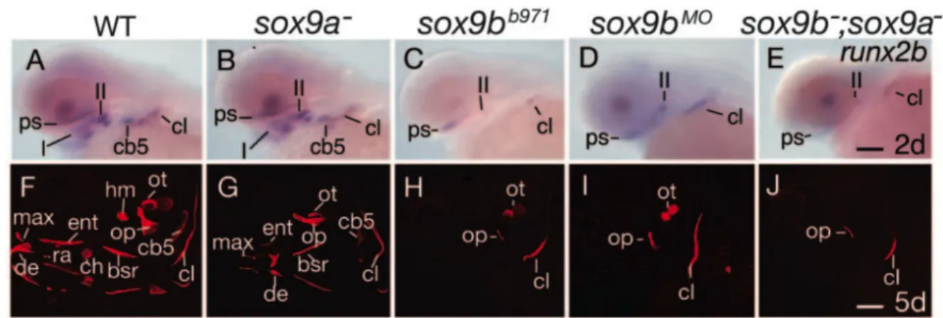
Loss of function studies revealed that transcription factor Sox9a is not required for cNCC specification, nor migration. However, Sox9a is absolutely essential for chondrogenesis (Fig.9B,G,L,Q). Indeed, Sox9a directly controls the expression of the col2a1 gene coding for collagen type II, the major collagen type in cartilage (Bell, Leung et al. 1997; Yan, Willoughby et al. 2005). Yan has described the jellyfish mutant, deficient in the sox9a gene (Yan, Miller et al. 2002; Yan, Willoughby et al. 2005) whose phenotype results in a complete absence of pharyngeal cartilages. Concerning osteogenesis (Fig.10G), sox9a disruption results in a complete absence of many endochondral bones (hyomandibular, ceratohyal) and some smaller, dermal bones (dentary, opercular, maxillary), while others remain unaffected (cleithrum) at 5 dpf.
Figure 9. Sox9a and Sox9b mutation causes cartilaginous defects in zebrafish development.
Lateral views of living larvae (A-E), lateral views (F-J) and ventral views of flatmounts (K-O: viscerocranium; P-T: chondrocranium) of Alcian Blue stained larvae. All animals are 4 dpf old. Scale bars 100 m. anterior basicranial commissure (abc), ethmoid plate (ep), ceratobranchials (cbs), ceratohyal (ch), hyosymplectic (hs), jaw (j), Meckel’s cartilage (m), notochord (no), otic vesicle (ov), parachordal (pc), trabecula.(tr) (Yan, Willoughby et al. 2005).
Yan et al. described a sox9bb971 mutant, characterized by cartilaginous and bone
mutant larvae preserve portions of cranial cartilages. sox9b-disrupted larvae display a reduction in size of the two first arches (mandible and hyoid), the ceratobranchials are absent, whereas the neurocranium is merely reduced (Fig.9C,D,H,I,M,N,R,S). Like the sox9a gene, sox9b does not influence tfap2 expression in premigratory cNCC, however, only
sox9bb971 mutation leads to smaller pre- and postmigratory dlx2 gene expressing cell
population domains. Both mutants, sox9a-/-and sox9b-/-, exhibit a severe reduction of col2a1
expression in postmigrating cNCC. sox9b deficient embryos express runx2b only in the cleithrum (Fig.10C) and in a small portion of the neurocranium, while this expression remains unaffected in jellyfish mutants (Fig.10B). Bone analyzes by Alizarin red staining reveals that Sox9b is absolutely required for development of endochondral bone and dermal bone in cephalic and pectoral fin skeletons (Fig.10H). Indeed, sox9b deficiency leads to a total absence of any bony elements; only drafts of the opercula and the cleithrum are present (Yan, Willoughby et al. 2005).
Figure 10. Sox9a and Sox9b are essential for bone formation and Sox9b regulates runx2b expression.
(A-E) In situ hybridizations of runx2b on sox9a mutants, sox9b mutants and morphants and double sox9a and sox9b mutants at 48 hpf. sox9b
-/-(C), sox9b morphants (D) and double mutants sox9a-/-;sox9b-/- do not
express runx2b in pharyngeal arches. (F-J) Alizarin Red staining of the calcified bones on 5 dpf old larvae. Sox9b deficient larvae (H-J) do not develop dermal, nor endochondral bone, while sox9a deficient larvae only fail to develop endochondral bones (Yan, Willoughby et al. 2005). first pharyngeal arch (I), second pharyngeal arch (II), brachiostegal rays (bsr), ceratobranchial arch 5 (cb5), ceratohyal (ch), cleithrum (cl), dentary (de), entopterygoid (ent), hyomandibular (hm), maxilla (max), opercle (op), otolith (ot), parasphenoid (ps), retroarticular (ra). Scale bar: in J, 100 µm.
The sox9a;sox9b double-mutant phenotype is additive: chondrocytes do not stack in
sox9a mutants and do not expand properly in sox9b mutants, whereas compound mutants
fail to do either, resulting in more severe craniofacial defects (Yan, Miller et al. 2002). These double mutants completely fail to form cartilage (Fig.9E,J,O,T) and bones (Fig.10E,J), only partial cleithrum and opercula are observable at 5 dpf. Taken together, these results suggest that Sox9a is more important for endochondral bone formation, while Sox9b plays a role for both endochondral and dermal bone formation in skeletal head and pectoral fin formation.
v. Runt-related transcription factor 2.
The RUNX family, RUNX1 (AML1), RUNX2 (CBFA1) and RUNX3 (AML2), is composed of DNA-binding transcription factors that regulate the expression of genes involved in cellular differentiation and cell cycle progression. RUNX1 plays a crucial role in hematopoiesis and its loss of function is frequently observed in leukemia (Speck and Gilliland 2002). RUNX3 is best characterized in neurogenesis of the dorsal root ganglia (Inoue, Ozaki et al. 2002; Kramer, Sigrist et al. 2006) and is considered as tumor suppressor in stomach cancer (Bae and Choi 2004; Ito, Chuang et al. 2011). In humans and mouse, RUNX2 haploinsufficiency causes a hereditary congenital bone disorder called cleidocranial dysplasia (Lee, Thirunavukkarasu et al. 1997; Mundlos, Otto et al. 1997; Otto, Thornell et al. 1997). The absence of Runx2 causes a failure of endochondral and intramembranous ossification (Otto, Thornell et al. 1997). Affected mouse embryos do not develop any bones and die at birth after respiratory failure. Detailed investigations have shown that these mice fail to express alkaline phosphatase, an early marker of osteoblast differentiation. These mice lack osteoblasts and vascularization of the bone marrow. Humans who are carrying this disease are viable but have a delay in the ossification of midline structures. People with RUNX2 haploinsufficiency have severe skeletal defects, such as hypoplasia or aplasia of clavicles, permanent teeth leading to supernumerary teeth, prognathic with a protrusive mandible due to hyperplasia of facial bones, frontanelles fail to close, bones and joints are underdeveloped and their stature is short (Mundlos, Otto et al. 1997). The loss of functional RUNX2 affects both intramembranous and endochondral ossification, like in mice mutants.
In zebrafish, the Runx family is represented by four genes, runx1, orthologs runx2a and runx2b and runx3. In pharyngeal skeletal development, runx1 is expressed in the
epithelium, runx2a and runx2b in mesenchymal condensations (cNCC) and runx3 in endoderm (Flores, Lam et al. 2006). The function in skeletogenesis of each gene was studied by morpholino microinjection: runx1 and runx2a knock-down only caused a mild delay in chondrogenesis of pharyngeal arches, while runx2b and runx3 expression have proven to be absolutely required for pharyngeal cartilage development. runx2b down-regulation causes absence of all pharyngeal arches and defects in the neurocranium, whereas loss-of-function of runx2a has no influence on zebrafish cranial chondrogenesis. Runx2b plays a key role in chondrocyte maturation but also in osteoblast differentiation (Flores, Lam et al. 2006). Embryos lacking Runx2b fail to develop hypertrophic chondrocytes and osteoblasts do not differentiate. Flores et al. have shown that the expression of runx2b is dependent of Runx3, but they did not show by which signaling pathway (Flores, Lam et al. 2006). runx2a and
runx2b are differentially expressed throughout zebrafish head skeletal development. During
their maturation, the mesenchymal condensations resulting from migration of cNCCs express
runx2a and runx2b. Starting at 44 hpf, runx2a is expressed at low levels in operculum and
cleithrum, which are the two first intramembranous bones to form in zebrafish. runx2a transcripts are also observed at higher levels in the mandible from 40 hpf until at least 3 dpf and starting at 44 hpf in basicranial cartilage such as trabecula cranii, ethmoid plate and parachordal. runx2b expression starts much earlier, at 34 hpf, in the basicranial cartilage, more specifically in the trabecula cranii. At 40 hpf, this expression extends to all pharyngeal cartilages, operculum, ethmoid plate and cleithrum and continues at least until 3 dpf.
III. Pharyngeal endoderm patterns cNCC and is required for zebrafish head development.
During development, pharyngeal endoderm plays a crucial role in patterning of the cNCC-derived skeleton. Indeed, like in all vertebrates, a series of pharyngeal pouches or slits subdivide the pharyngeal arches of the embryo; these pouches are derived from endoderm. The cNCC will differentiate between these endodermal pouches into cartilage (David, Saint-Etienne et al. 2002). It has been proven that the endoderm physically interacts with cNCCs and that its absence, reduction or malformation causes severe cartilage alterations or absence of diverse cartilaginous elements (David, Saint-Etienne et al. 2002). Endodermal pouches guide cNCC migration into arches and missing or malformed endodermal pouches lead to streams of cNCC that often do not separate from each other.
i. casanova and van gogh mutants.
Screening of mutants during mutagenesis programs have led to the identification of many mutants with craniofacial defects due to absence or alteration of endodermal pouches (Piotrowski, Schilling et al. 1996; Schilling, Piotrowski et al. 1996). Their detailed study has allowed identification of the mutated genes and led to a better understanding of their function in cartilage development. The endoderm-deficient mutant casanova (cas, mutation of the sox32 gene) fails to develop pharyngeal cartilages entirely, while neurocranial cartilage remains unaffected. These specific defects can be restored by transplantation of wild-type endoderm into casanova mutants, suggesting that cNCCs contributing to pharyngeal cartilages depend on the presence of pharyngeal endoderm (Alexander, Rothenberg et al. 1999; David, Saint-Etienne et al. 2002). Disruption of tbx1 (in the van gogh mutant) does not result in the absence of endoderm, but in a failure of endodermal pouch formation and a disorganization of mesodermal cores, which leads to an absence of cNCC segmentation into distinct elements. Pharyngeal cartilage is reduced or lost. Similar to casanova mutants, a transplantation of wild-type endoderm can rescue the van gogh defects (Piotrowski, Ahn et al. 2003). These mutant analyzes clearly reveal the importance of endoderm in ventral head skeletal differentiation and patterning.
ii. SRY (Sex determining Region Y) Box 9 transcription factor b. Studies of other mutants and genes expressed in endoderm support the fact that pharyngeal endoderm plays a crucial role in cranial cartilage development in zebrafish, similar to other vertebrates. As referred above (section 3.a.II.iv), the sox9b gene is expressed first in premigratory cNCC but is downregulated shortly after cNCC migration and starts to be expressed in pharyngeal endoderm (Yan, Willoughby et al. 2005). The sox9bb971 mutant is
characterized by an absence of ceratobranchials, reduced mandible and hyoid elements and a slightly reduced neurocranium. As mentioned previously, this mutant does not express
runx2b in the viscerocranium, but still in the cleithrum and neurocranium at two days of
development. Sox9b is also essential for development of endochondral and dermal bone in cephalic and pectoral fin skeleton (Yan, Willoughby et al. 2005). The signaling pathway that links endodermal Sox9b to Runx2b expression in cartilage has not yet been identified.
iii. Runt-related transcription factor 3.
An additional member of the runt-related factor family, Runx3 is also expressed in pharyngeal endoderm and plays a crucial role in cranial cartilage development (section 3.a.II.v). runx3 is expressed in pharyngeal endoderm starting at 34 hpf until at least 3 dpf, but at 4 dpf it starts also to be expressed in mature chondrocytes. Knock-down experiments using morpholino injection have shown its role on runx2b expression in cartilage precursor cells, down-regulation of runx3 results in a complete absence of the viscerocranium and of the anterior part of the neurocranium. Thus, Runx3 has a crucial function in chondrogenesis, but also in osteogenesis by regulating runx2b expression. On the other hand, runx3 morphants have no morphological abnormality in pharyngeal endoderm, thus it can be concluded that Runx3 has no action on formation and survival of endodermal cells (Flores, Lam et al. 2006).
IV. Ectoderm.
A second set of epithelial-mesenchymal interactions occurs in cranial skeletal development between cNCC and the surface ectoderm, which forms epidermis.
Studies of the previously described tfap2 (section 3.a.II.ii) and its close relative
tfap2 support a role for the ectoderm in craniofacial patterning in zebrafish. tfap2 is
expressed in cNCC and in the ectoderm, while tfap2 mRNA is found exclusively in ectoderm. Depletion of Tfap2ß has no or little effect on the cranial skeleton, probably due to functional redundancy with ectodermal Tfap2a, while the deficiency in expression of both factors leads to an absence of both pharyngeal and neurocranial cartilages. The defect in this double deficient animals is also much more important than in lockjaw mutants (tfap2 gene mutation), indicating that ectoderm is involved in cartilage development. The facial ectoderm shows a high level of apoptosis in embryos lacking both orthologs and transplantation of wild type ectoderm rescues cartilage development (Schorle, Meier et al. 1996; Knight, Nair et al. 2003; Knight, Javidan et al. 2005).
V. Cell signaling in cartilage and bone.
As mentioned above, cartilage and bone interact with the different surrounding tissues both physically and molecularly. Different signaling pathways are well known for their crucial role in cartilage and bone development. These signaling pathways are composed of secreted ligands that bind to and activate a membrane receptor. In this manuscript, we will cover two major signaling pathways, the bone morphogenic protein (BMP) and hedgehog (Hh) pathwaysthat are very conserved among vertebrates and which interact together to form and maintain the skeleton.
i. Bone morphogenic protein.
Bone morphogenic proteins (BMP), as their name suggests, were originally discovered for their function in bone and cartilage formation. BMPs are found in vertebrates as well as in invertebrates and have a broad spectrum of biological activities in various tissues such as kidney, liver, lungs, neuronal and hematopoietic development, tooth and of course bone and cartilage. They are members of the BMP/transforming growth factor (TGF) family and more than a dozen of these factors have been classified in the BMP subfamily. BMP ligands are secreted (autocrine, paracrine, endocrine) and bind to type II serine/threonine kinase receptor dimers (homo- or heterodimers), causing the recruitment of type I (Alk-X) serine/threonine kinase receptor dimers (homo- or heterodimers) to form a hetero-tetrameric complex with the ligand. Type I receptors transduce their signal inside the cell by phosphorylating receptor-regulated Smads (R-Smad) (Wrana, Attisano et al. 1992; Kim and Choe 2011). There are five R-Smads: Smad1, Smad2, Smad3, Smad5 and Smad9 (also referred as Smad8). Once phosphorylated, R-Smad has a high affinity to a co-Smad (Smad4) and forms a complex with it. This complex enters the cell’s nucleus and initiates transcription of its target genes by binding to the promoter and cofactors. Two other Smads complete the Smad family, Smad6 and Smad7, which are inhibitory Smads (I-Smad). I-Smads either compete with R-Smads for type I receptor binding and prevent their phosphorylation, or bind to co-Smads and inhibit their binding to R-Smads. A further complexity is brought to the system by the existence of extracellular inhibitors, such as Noggin, Chordin or Follistatin, which bind to BMP ligands and prevent their binding to their receptors. These inhibitors block activation of Bmp signaling.
Figure 11. A. BMP pathway.
A secreted BMP dimer binds to its receptor complex. Once the BMP dimer is bound to its receptors, the active kinase domain of receptor II phosphorylates receptors I and III. In turn, receptor I then activates the Smad pathway by phosphorylating regulatory R-Smads. The phosphorylated R-Smad associates with a co-Smad and forms a complex
that translocates into the nucleus to initiate transcription of various genes. BMPs can also signal through non-Smad pathways, notably via MAPK, ERK, JNK, NF-B etc.
B. BMP signaling inhibitors.
Extracellular inhibitors such as Noggin, Chordin, Follistatin, etc. can block receptor activation by binding to the BMP ligands. Various molecules and proteins such as inhibitory I-Smads (Smad6 and 7), intracellular inhibitors (Smurfs) and drugs such as dorsomorphin can suppress phosphorylation of R-Smads. All these inhbitors are able to block the BMP signaling pathway.
BMP signaling plays an essential role for chondrocyte and osteoblast differentiation and maturation, especially during endochondral ossification. Among the BMP ligands that stimulate chondrocyte and osteoblast differentiation are Bmp2, Bmp4, Bmp6 and Bmp7 (Nishimura, Hata et al. 2008). Various studies in mice have shown that BMPs regulate transcription factors such as Runx2, Osterix, Msx2 and Sox9, crucial for cartilage and bone formation (Nishimura, Hata et al. 2008). Bmp2 and Bmp7 possess an osteoinductive signaling capacity and are required for Runx2 expression in mouse (Lee, Hong et al. 2002; Tou, Quibria et al. 2003). They are expressed in prehypertrophic and hypertrophic chondrocytes during endochondral bone formation and are essential for cell proliferation and maturation (Shu, Zhang et al. 2011). Analyses of Bmp7 have revealed its essential role in cartilage repair after injury (Chubinskaya, Hurtig et al. 2007; Che, Zhang et al. 2010). Bmp receptors (BmpR) are similarly required for skeletal development (Fujii, Takeda et al. 1999; Zhao, Harris et al. 2002). Studies on transgenic mice expressing a truncated, dominant-negative BmpR-IB under the control of the type I collagen promoter have shown that BmpRs are essential for postnatal bone growth in mice (Zhao, Harris et al. 2002). R-Smads (Smad1, Smad5, Smad8) and co-Smad (co-Smad4) similarly play a critical role as transcriptional regulators in osteoblastogenesis and chondrogenesis (Fujii, Takeda et al. 1999). However, Smad proteins and Bmp receptors are ubiquitously expressed in many tissues and it is therefore difficult to conceive regulatory models for osteoblastogenesis and chondrogenesis that involve BMP-Smad signaling.
Like in mice, Bmps play a crucial role in zebrafish cartilage and bone development. Various Bmp genes are transcribed in endodermal pouches of the pharyngeal arches, such as
bmp2a, bmp2b, bmp4, and bmp5 (Holzschuh, Wada et al. 2005). Bmp signaling has been
inhibitor, Smith et al. have demonstrated that there is an alteration in the maturation of bone-secreting cells, reduction of bone-matrix deposition but also a downregulation of
runx2a/b, sox9a/b and col10a1, all of which are crucial for chondrocyte and osteoblast
maturation. Bmp receptor type I, Alk8, has been clearly identified to be required for cranial neural crest cell (cNCC) formation (Payne-Ferreira and Yelick 2003). Analyses of alk8 mutants,
laf (lost-a-fin) and dominant-negative alk8 transgenic lines revealed that an altered alk8
function results in patterning defects of premigratory cNCC and, as a consequence defects in cartilage development. Recent studies have shown that Bmps play an essential role in dorso-ventral patterning of the craniofacial skeleton (Alexander, Zuniga et al. 2011; Zuniga, Rippen et al. 2011). These studies have shown that Bmp signaling is also required for ventral arch development in cNCCs just after their migration. By blocking Bmp signaling using dominant-negative Bmp transgenics, specific ventral markers, such as edn1, hand2 and dlx6a were reduced while, on the other hand dorsal marker expression domains were enlarged, such as that for jag1b. These effects lead to the loss or reduction of ventral and intermediate cartilage elements and the misshaping of dorsal elements. On the other hand, an overexpression of Bmps causes a dorsalization of ventral elements, specific ventral markers expand in a more dorsal position.
ii. Hedgehogs.
The Hedgehog (Hh) protein was first discovered in the fruitfly, Drosophila
melanogaster as a determinant for segment antero-posterior polarity in the developing
larvae. Its mutation causes fruitfly embryos to be covered with small projections, making them look like a hedgehog. In vertebrates, three different homologs have been discovered; sonic hedgehog (Shh), indian hedgehog (Ihh) and desert hedgehog (Dhh). The hedgehog signaling pathway is involved in numerous functions, such as patterning the nervous system, limbs, teeth, notochord, lungs, etc.
In vertebrates, Hh ligands are cleaved and a cholesterol molecule is added to the carboxyl end of the N-terminal domain (Fig.12). This cleavage is absolutely required for proper Hh signaling, while the cholesterol residue allows the Hh ligand to bind to cell membranes and is essential for secretion of the Hh ligand. Disruption of the cleavage leads to
developmental disorders such as holoprosencephaly, which is characterized by a failure of the brain to form two hemispheres and craniofacial defects, including midline facial cleft and cyclopia (Roessler, Belloni et al. 1996; Maity, Fuse et al. 2005; Roessler, Ma et al. 2009). Diffusion of Hh is also restricted through sequestration by its receptor Patched (Ptch or Ptc). Hh can signal either in an autocrine or in a paracrine way. During paracrine signaling, the Hh ligand requires participation of the dispatched protein (Disp). The Disp protein modifies the cholesterol residue to release the Hh ligand coupled to its cholesterol-modified molecule from the membrane (Burke, Nellen et al. 1999; Kawakami, Kawcak et al. 2002). Hh reaches its target cell and binds to its receptor Patched (Ptch1, Ptch2). In absence of Hh ligands, Ptch inhibits Smoothened (Smo). Ptch has a sterol-sensing domain (SSD), which has been shown to be essential for suppression of Smo activity. It is suggested that Ptch regulates Smo by removing oxysterols from Smo like a pump (Strutt, Thomas et al. 2001). Upon binding of a Hh protein or a mutation in the SSD of Ptch, the pump is turned off, thereby allowing oxysterols to accumulate around Smo (Corcoran and Scott 2006). This accumulation of oxysterols leads to the activation of Smo, which in turn activates the zinc finger proteins Gli. Activated Gli accumulates in the nucleus and controls the transcription of hedgehog target genes.
Figure 12. Hedgehog (Shh) signaling pathway.
(1) Sonic hedgehog (Shh), the Hh ligand, is cleaved and linked to a cholesterol molecule at its N-terminus. (2) Dispatched (Disp) enables Shh to be secreted. (3) Shh binds to Patched (Ptch) and (4) Ptch
inhibition on Smo is lifted. (5) Smo activates Gli, which translocates into the nucleus (6) and activates transcription of target genes (7). (Figure from Peter Znamenskiy, donated to public domain)
Hh signaling is known to have a critical role in vertebrate skeletal development. Mice lacking Ihh display abnormal chondrocyte proliferation and maturation and an absence of mature osteoblasts in prenatal enodochondral bone formation (St-Jacques, Hammerschmidt et al. 1999). Ihh controls chondrocyte hypertrophization by forming a negative feedback loop with parathyroid hormone related protein (PTHrP). Pthrp binds to its receptor Pthr1 and inhibits Ihh expression, with the consequence to keep chondrocytes in a proliferative state and not becoming hypertrophic. If Pthrp is absent, chondrocytes become hypertrophic. More recent studies have revealed that Ihh regulates chondrocyte and osteoblast development independently of Pthrp expression as well (Long, Zhang et al. 2001; Kobayashi, Soegiarto et al. 2005; Maeda, Nakamura et al. 2007; Mak, Kronenberg et al. 2008). shh-/- KO mice (Chiang,
Litingtung et al. 1996; Yamagishi, Yamagishi et al. 2006) also present a loss of craniofacial elements. Shh is essential for mesenchymal cell survival in pharyngeal arches.
In zebrafish, various mutants have confirmed that Hh signaling is required for proper chondrocranium formation. The chameleon mutant, lacking functional Disp, displays reduced mandible, hyoid arches and a complete lack of ceratobranchials (Schwend and Ahlgren 2009). This mutant succeeds in cNCC specification and migration, however the cNCCs differentiate into fibrous connective tissues rather than into chondrocytes. Chameleon larvae do not express key-genes for cartilage formation such as dlx2 and sox9a, but also bapx1 and
gdf5 that are essential for joint formation. smo-/- receptor mutants in zebrafish (Chen,
Burgess et al. 2001) also present a loss of craniofacial elements. Treating zebrafish embryos with the Hh signaling inhibitor cyclopamine (binds to Smo) leads to cranial defects (Schwend and Ahlgren 2009). Treatments between 4 and 12 hpf cause defects in cNCC patterning and between 32-48 hpf inhibit differentiation of cNCC into chondrocytes (Schwend and Ahlgren 2009). The Hh ligand Ihh is also involved in endochondral bone formation. Ihh promotes hypertrophy of chondrocytes and is essential for maintenance of the growth plate and trabecular bone (Maeda, Nakamura et al. 2007; Mak, Kronenberg et al. 2008). Skeleton development involves multiple signaling pathways that also interact with one another. Studies have demonstrated that the Gli2 protein, mediating Hh signaling, directly regulates
transcription of bmp2 during osteoblast maturation (Zhao, Qiao et al. 2006). Hammond et al. have recently shown that an increased Hh signaling leads to premature mineralization of endochondral bones (Hammond and Schulte-Merker 2009). Detailed analyses of Hh receptor
ptc1/2 mutants showed that their early-mineralized bone was characterized by an
upregulation of osterix (osx), a master regulator of osteoblast differentiation, when compared to wild-type siblings. On the other hand, disruption of Ihh in cranial skeletogenesis leads to a severe delay of endochondral bone formation. Together with Fgfs, Hhs are also well studied in limb growth. Shh signaling is required for anterior-posterior patterning of the limb bud in vertebrates; Shh expression in the posterior limb bud is regulated by Fgf signaling also controlling proximal-distal limb outgrowth (Towers, Mahood et al. 2008; Zhang, Verheyden et al. 2009).
4. Zinc finger transcription factors.
The molecular processes that control skeletal development are various, involving hormones, extracellular factors, membrane receptors and transcription factors. Among the transcription factors, binding specifically to DNA through their binding site, the zinc finger family is the largest, but only a few of them are involved in cartilage and bone development.
Zinc fingers are small protein structural motifs that are highly conserved and frequently involved in protein-nucleic acid interaction (Pellegrino and Berg 1991). Each zinc finger is composed of a group of conserved amino acids, four of which bind to one or more zinc ions to help stabilize their folds. There are different types of zinc finger domains, among which the C2H2 type is by far the best-characterized (Fig.13). Actually, the first described zinc finger was a C2H2 type. It was identified in the transcription factor TFIIIA in Xenopus laevis, which is involved in transcription of the 5S RNA gene contributing to the large subunit of the ribosomes. This motif is composed of 25 to 30 amino acids and contains two conserved cysteines and two conserved histidines. Its consensus sequence is (Tyr, Phe)-X-Cys-X2-4-Cys-X3
-Phe-X5-Leu-X2-His-X3-His-X5, where each X represents a non-conserved amino acid (Pelham
and Brown 1980; Ginsberg, King et al. 1984; Miller, McLachlan et al. 1985; Pellegrino and Berg 1991; Wilson, Day et al. 1992). Nuclear magnetic resonance studies have established that the C2H2 motif forms a C-terminal helix including the two conserved histidine
residues and an N-terminal, antiparallel sheet containing the two conserved cysteine residues (Gibson, Postma et al. 1988; Lee, Gippert et al. 1989; Klevit, Herriott et al. 1990) . The C2H2 zinc finger motif is found in numerous transcription factor families, among those the EGR family.
Figure 13. Schematic representation of a C2H2 type zinc finger.
The C-terminal α helix contains the two conserved histidine residues (in blue), while the anti-parallel β sheet in N-terminal contains the two conserved cysteine residues. These four residues chelate a zinc ion.
a. EGR family.
Early growth response (EGR) family members are immediate early genes coding for transcription factors involved in transmission of various stimuli that initiate cellular responses such as mitosis, cellular differentiation and apoptosis (Swirnoff and Milbrandt 1995) . Activation of immediate early genes is very fast, transient and independent of de novo protein synthesis (Lemaire, Revelant et al. 1988; Cao, Koski et al. 1990).
Table 2. EGR family members in various vertebrate species. Gene
s
Synonyms Full name Organisms References
EGR1 Nfgi-A Nerve growth
factor-induced clone A
Zif268 Zinc finger clone 268 Mouse (Lemaire, Revelant et al. 1988)
Krox-24 Krüppel box 24 Mouse (Sukhatme, Cao et al. 1988)
EGR1 Early growth response 1 Human (Suggs, Katzowitz et al. 1990)
egr1 Early growth response 1 Zebrafish (Drummond, Rohwer-Nutter et al. 1994)
EGR2 Krox-20 Krüppel box 20 Mouse (Chavrier, Lemaire et al. 1988)
EGR2 Early growth response 2 Human (Joseph, Le Beau et al. 1988)
Krox-20 Krüppel box 20 Chicken (Nieto, Bradley et al. 1991)
Krox-20 Krüppel box 20 Xenopus (Bradley, Snape et al. 1993)
krox-20 Krüppel box 20 Zebrafish (Oxtoby and Jowett 1993)
EGR3 EGR3 Early growth response 3 Human (Patwardhan, Gashler et al. 1991)
Egr3 Early growth response 3 Mouse (Patwardhan, Gashler et al. 1991)
Egr3 Early growth response 3 Rat (Yamagata, Kaufmann et al. 1994)
EGR4 Ngfi-C Nerve growth
factor-induced clone A
Rat (Crosby, Puetz et al. 1991)
Egr4 Early growth response 4 Mouse (Barrow, Simin et al. 1994)
pAT133 Clone pAT133 Human (Muller, Skerka et al. 1991)
The EGR family is composed of four genes, EGR1, EGR2, EGR3 and EGR4, which were all described under different names depending on the animal species where they were identified (Table 2). Their translated proteins have several characteristics in common, such as a DNA binding domain in the C-terminus that is composed of three C2H2 type zinc fingers. This binding domain recognizes the nonameric consensus sequence (5‘-GCG T/GGG GCG-3’ which can be found in the promoter of numerous genes (Fig.14A,B) (Christy and Nathans 1989; Wilson, Day et al. 1992; Cao, Mahendran et al. 1993). Each zinc finger binds through hydrogen bonds and Van der Waals interaction to a subunit of the binding site composed of three base pairs (Pavletich and Pabo 1991; Elrod-Erickson and Pabo 1999). EGR transcription factors possess a transactivation domain at the N-terminus, but also a repression domain R1 (Fig.14C) (O'Donovan, Tourtellotte et al. 1999). This repression domain is located between the DNA binding and the transactivation domains, but is absent in EGR4 (Fig.14C) (Crosby, Veile et al. 1992). By binding to this repression domain R1, the NAB (NGFI-A binding) co-repressors NAB1 and NAB2 inhibit the transcriptional activity of EGR1, EGR2 and EGR3 (Russo, Sevetson et al. 1995; Svaren, Sevetson et al. 1996; Swirnoff, Apel et al. 1998) . NAB proteins have two conserved domains, the N-terminal NCD1 (NAB conserved domain 1) domain and the C-terminal NCD2 (NAB conserved domain 2) domain (Fig.14D). The first one is responsible for co-repressive binding to EGR family members, while the second one is responsible for EGR protein repression (O'Donovan, Tourtellotte et al. 1999). NAB1 is a
constitutive gene and is expressed in almost all cell types, while NAB2 is only induced by the same stimuli that control EGR expression (Svaren, Sevetson et al. 1996; Swirnoff, Apel et al. 1998). This suggests that NAB2 is under dependence of EGR factors and acts as a negative feed-back loop on EGR activity (Ehrengruber, Muhlebach et al. 2000).
A
B
C
D
Figure 14. Schematic representations of EGR proteins.
(A) Bond between an EGR member to its specific response element (RE). Each zinc finger (black circles) binds to a sub-unit composed of three base pairs in an anti-parallel conformation. Zinc finger I binds to the triplet in 3’ and zinc finger III to the triplet in 5’. (B) Representation of the molecular structure of the zinc finger transcription factor EGR1. The EGR1 protein is composed of an N-terminal transactivation domain (grey) and a C-N-terminal DNA