Université de Montréal
Expression des récepteurs EphA dans le raphé dorsal
néonatal et adulte
par
Moogeh Baharnoori
Département de pathologie et biologie cellulaire
Faculté de médecine
Mémoire présenté à la Faculté des études supérieures
en vue de l’obtention du grade de maîtrise
en sciences neurologiques
April 2007
© Moogeh Baharnoori, 2007
fc cPi I \cCE ç, c)
Direction des bibliothèques
AVIS
L’auteur a autorisé l’Université de Montréal à reproduite et diffuser, en totalité ou en partie, par quelque moyen que ce soit et sur quelque support que ce soit, et exclusivement à des fins non lucratives d’enseignement et de recherche, des copies de ce mémoire ou de cette thèse.
L’auteur et les coauteurs le cas échéant conservent la propriété du droit d’auteur et des droits moraux qui protègent ce document. Ni la thèse ou le mémoire, ni des extraits substantiels de ce document, ne doivent être imprimés ou autrement reproduits sans l’autorisation de l’auteur.
Afin de se conformer à la Loi canadienne sur la protection des renseignements personnels, quelques formulaires secondaires, coordonnées ou signatures intégrées au texte ont pu être enlevés de ce document. Bien que cela ait pu affecter la pagination, il n’y a aucun contenu manquant.
NOTICE
The author of this thesis or dissertation has granted a nonexclusive license allowing Université de Montréal to reproduce and pub)ish the document, in part or in whole, and in any format, solely for noncommercial educational and research purposes.
The author and co-authors if applicable retain copyright ownership and moral rïghts in this document. Neither the whole thesis or dissertation, nor substantial extracts from it, may be printed or otherwise reproduced without the author’s permission.
In compliance with the Canadian Privacy Act some supporting forms, contact information or signatures may have been temoved fcom the document. While this may affect the document page count, it does flot represent any loss of content from the document.
Ce mémoire intitulé
Expression des récepteurs EphA dans le raphé dorsal
néonatal et adulte
présenté par:
Moogeh Baharnoori
a été évalué par un jury composé des personnes suivantes:
Dr Serge Rossignol
président-rapporteur
Dr Guy Doucet
directeur de recherche
Dr Luc DesGroseillers
co-directeur de recherche
Dr Karl Fernandes
membre du jury
Résumé français
Des travaux antérieurs réalisés in vitro, dans notre laboratoire, ont montré que des neurones sérotoninergiques (5-HT) dissociés du raphé dorsal (DR) peuvent reconnaître des signaux spécifiques de guidage axonal dans des membranes cellulaires extraites à partir de diverses régions du cerveau néonatal, comme le cortex cérébral (Ctx), le striatum ($tr) et le mésencéphale ventral (VM). Cette activité de guidage a été diminuée dans les membranes traitées avec la phospholipase-C spécifique du phosphatidylinositol (PI-PLC), qui enlève les protéines à ancrage membranaire par lien lipidique glycosylphosphatidylinositol (GPI). Un traitement des membranes avec la protéine de fusion EphA3-Fc, qui bloque les
éphrines-A a eu un effet similaire, quoique moins grand. Les éphrines-A sont des protéines membranaires à ancrage GPI ayant une haute affinité pour les récepteurs EphA, qui ont des rôles reconnus dans le guidage axonal. Elles représentent ainsi des candidates appropriées pour cette activité de guidage des axones 5-HT. Les objectifs du travail actuel étaient d’examiner la présence d’éphrines-A dans les extraits de membrane de Ctx, de Str et de VM néonatals de même que l’expression de récepteurs EphA dans le DR d’embryons, de nouveau-nés et d’adultes murins. Des transferts western avec des anticorps spécifiques ont démontré la présence des éphrines-A4 et -A5 dans les membranes extraites du Ctx, Str et VM néonatals. D’autre part, l’hybridation insituavec des ribosondes marquées à la
digoxygénine a montré l’expression d’EphA4, EphA5 et EphA7 dans la région du DR de rats foetaux ou néonatals et de souris adultes. D’autres expériences, avec double marquage de la région du DR par immunocytochimie de la 5-HT et hybridation in situ des récepteurs EphA, seront nécessaires pour confirmer si les neurones marqués dans la région du DR comprennent des neurones 5-HT, et si tous les neurones 5-HT sont marqués uniformément
avec les 3 sondes. L’hybridation in situ en fluorescence (fISH), avec des ribosondes d’EphA pourrait aussi permettre dans le futur d’examiner l’expression de ces récepteurs
dans des neurones 5-HT dissociés, en culture.
Mots clés:
Neurobiologie, développement, guidage axonal, raphé dorsal, sérotonine, cortex cérébral, striatum, mésencéphale ventral, éphrines, récepteurs Eph, hybridation in situ, transfert western
Résumé anglais
Previous in vitro experiments in our laboratory have showii that serotonergic (5-HI) neurons dissociated from the dorsal raphe (DR) region recognize specific axon guidance signais in celiular membranes extracted from various target brain regions, i.e. neonatal cerebral cortex (Ctx), striatum ($tr) and ventral rnidbrain (VM). This axon guidance activity was decreased in the membranes treated with the phosphatidylinositol-specific phospholipase-C (PI-PLC), which removes the glycosylphosphatidylinositol (GPI) anchors, as well as in membranes tTeated with the fusion protein, EphA3-Fc, which blocks
ephrin-As. Ephrin-As, being the GPI-anchored membrane ligands ofEphA receptors, represent proper candidates for this axon guidance activity.
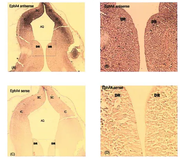
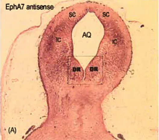
The objectives of the present work were to examine the presence of ephrin-A iigands in membrane extracts from neonatal Ctx, Str and VM, as well as the expression of EphA receptors in the DR of embryonic, neonatal and aduit rodents. Western blot experiments with specific antibodies showed the presence of ephrin-A4 and ephrin-A5 in membrane extracts of Ctx, Str and VM. In situ hybridization experiments with digoxygenin-labelled riboprobes showed a high expression of EphA4, EphA5 and EphA7 in the DR of fetai or neonatal rat, as well as of aduit mouse.
Further experiments, with double labelling ofthe DR with 5-HT
irnmunocytochemistiy combined with in situ hybridization for EpliA receptors, will tell us if the ceil bodies labelled in DR include 5-HT neurons, and whether ail 5-HT neurons are uniformly labelled with the 3 riboprobes. Fluoroscent in situ hybridization (FISH) with EphA riboprobes could also be used to determine the expression of these receptors in dissociated 5-HT neurons in culture.
Key words:
Neurobiology, developrnent, axon guidance. dorsal raphe, serotonin fleurons, in situ
Table of contents:
RÉSUMÉ FRANÇAIS III
MOTS CLÉS IV
RÉSUMÉ ANGLAIS V
KEY WORDS VI
TABLE 0F CONTENTS VII
LIST 0F FIGURES IX
LIST 0F ABBREVIATIONS- X
DEDICATIONS- XII
ACKNOWLEDGEMENTS XIII
f. INTRODUCTION
1.1 Serotonin metabolism and neurotransmission 2
1.1.1 Biosynthesis of5-HT 2
1.1.2 Serotonin storage and release 3
1.1.3 Inactivation ofreleased 5-HT 3
1.2 Serotonin impairments in neurological or psychiatric diseases 6
1.2.] Migraine 6
1.2.2 $erotonin in sleep disorders 7
1.2.3 Depression 9
1.2.4 $chizophrenia 11
1.2.5 Down Synd*orne 13
1.2.6Autism 14
1.3 Rotes of 5-HI in development 17
1.4 Organization of the 5-HI systems 19
1.5.1 Sonic hedgehog and Fibroblast Growth Factors .20 1.5.2 Gli transcription factors in the dfferentiation of 5-HT fleurons 21 1.5.3 Other transcription factors involved in5-HTneuron development 22
1.5.4 Molecules that influence the growth of5-HT axons 24
1.6 Eplirins and Eph receptors 26
1.6.1 Ephrin and Eph signalingpathways 27
1.6.2 functions ofephrin/Eph in CNS 29
1.7 Objectives of present work 33
2. MATERIALS AND METHODS 35
2.1 Animais 35
2.2 Detection of EphA4, EphA5 and EphA7 mRNA byin situ hybridization 36
2.2.] Preparation ofconstructs and insertion into pÏasmids 36
2.2.2 Synthesis ofantisense and sense riboprobes 42
2.2.3 In situhybridization 44
2.3 Detection of 5-HT neurons by SERT immunohistochemistry 45
2.4 Cortical, striatal, and ventral midbrain ccli membrane extraction ami Western
blotting 45
3.RESULTS 48
3.1. In situ hybridization in hippocampus 4S
3.2. Expression ofEphA4, EpItAS and EphA 7 in the DR region of rat embryos 52
3.3. Expression of EpItA5 in the DR of neonatai rat brain 56
3.4. Expression of EphA4, EphAS and EpitA 7 in the DR of aduit mouse brain 56 3.5. Detection of ephrin-A4 and ephrin-A5 in membranes extracted from the Ctx, Str,
andVM 62
4. DISCUSSION 64
4.1. EphA4, EphA5 and EphA7 are expressed in the DR at ail stages examined 64
4.2. Ephrin-A expression in Ctx, Str and VM of neonatal rat brain 67
5. CONCLUSION 68
Lïst of Figures:
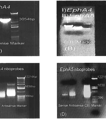
Figure 1. Production of the EphA4 and EphA5 riboprobes 39
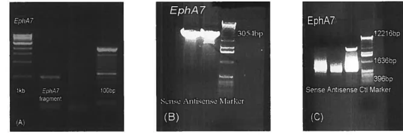
Figure 2. Production of EphA7 riboprobes 43
Figure 3. Dissection of VM, Str and Ctx tissues from neonatal rat brain (modified
from Petit et aI., 2005) 47
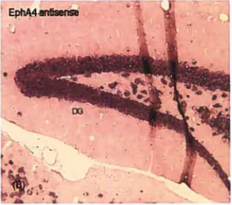
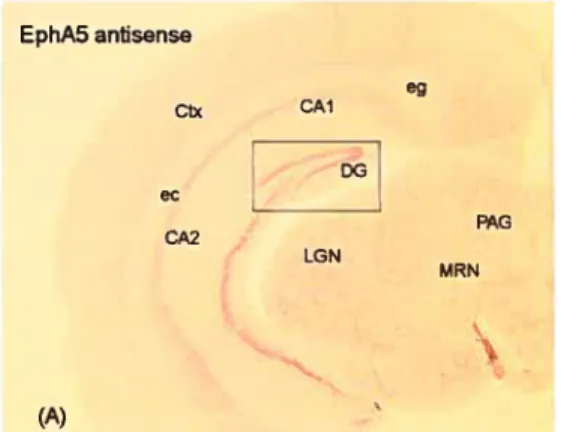
Figure 4. EphA4 in situ hybridization in aduit mouse hippocampal tissue 49 Figure 5. EphA5 in situ hybridization in aduit mouse hippocampal tissue 50 Figure 6. EphA7 in situ hybridization in aduit mouse hippocampal tissue 51 Figure 7. Expression of EphA4 in the DR of E15 rat brainstem 53 Figure 8. Expression of EphA5 in the DR of E15 rat brainstem 54 Figure 9. Expression of EphA7 in the DR of E15 rat brainstem 55 Figure 10. Expression of EphA5 in the DR of P0 rat braïnstem 58
Figure 11. Serotonin neurons in DR of aduit mouse 58
Figure 12. Expression of EphA4 in the DR of P60 aduit mouse brainstem 59 Figure 13. Expression of EphA5 in the DR of P60 aduit mouse brainstem 60 Figure 14. Expression of EphA7 in the DR of P60 aduit mouse brainstem 61 Figure.15. Ephrin-A4 protein in membranes extracted from Ctx, Str, and VM 63 Figure.16. Ephrin-A5 protein in membranes extracted from Ctx, Str, and VM 63
List of Abbreviations:
5-HT: 5-hydroxytryptamine, (sérotonine)
CNS: central nervous system (système nerveux central) CSF: cerebrospinal fluid (liquide cérébro-spinal) Ctx: cerebral cortex (cortex cérébral)
DR: dorsal raphe (raphé dorsal) Eph: Eph receptor (récepteur Eph)
F0F: fibroblast growth factor (facteur de croissance des fibroblasts) GPI: glycosylphosphatidylinositol
GTP: guanosine tri-phosphate
MAO: monoamine oxidase (monoamine oxydase)
PET: positron-emitting tomography (tomographie à emission de positons)
PI-PLC: phosphatidylinositol-specific phospholipase C (phospholipase-C spécifique du phosphatidylinositol)
REM: rapid eye movement (mouvements rapides des yeux) SCZ: schizophrenia (schizopbrenia)
SERT: Serotonin transporter (transporteur de la sérotonine) Shh: sonic hedgehog
SSRI: selective serotonin re-uptake inhibitor (inhibiteur spécifique de la recapture de sérotonine)
SWS slow wave sleep (sommeil à ondes lentes) $tr: striatum
VM: ventral midbrain (mésencéphale ventral)
Dedîcations:
To my dear father and mother; Nasy and Fariba
My lovely brother and sister; Nazan and Mahbod
Acknowledgements
I wish to thank Dr. Guy Doucet for the supervision ofthis project and
Dr. Luc Desgroseillers for his co-supervision and kind collaboration,
Frédérique Badeaux for her help in making riboprobes and Maria Sanchez for
her advice on
in situhybridization experiments, Bahram SharifAskari who
did the serotonin neuron culture experiments and David Bouvier for his
assistance in Western blot studies. I am also grateful to the Groupe de
recherche sur le système nerveux central (GRSNC), the Canadian Institutes
for Health Research (CIHR), and the Scottish Rite Charitable Foundation of
Canada (SRCF) for their financial support.
1. Introduction
Serotonin (5-hydroxytryptarnine or 5-HT) is an important neurotransmitter, taking part in diverse functions during development and at maturity, in neuronal and non-neuronal celis, including pain perception, sleep/wake cycles and neurodevelopment. Likewise, as exemplified below, disruptions in 5-HT neuronal development or 5-HT neurotransmission appear to be involved in mood disorders, cognitive dysfttnction, mental retardation, anxiety and aggression.
Therefore, a better knowledge ofthe molecules involved in the development ofthe 5-HT neurons is currently needed. There has been some progress, recently, in the
identification of factors influencing the differentiation of 5-HT neurons (see below), but littie is currently known about the molecuies that guide 5-HT axons to their multiple innervation targets (SharifAskari, 2006).
Recent work in our laboratory show that 5-HT neurons ofthe dorsal raphe (DR) can recognize axon guidance signals in membrane purified from different brain regions, i.e., cerebral cortex (Ctx), striatum (Str) and ventral midbrain (VM) (Petit et al., 2005; Sharif Askari, 2006). Some ofthese signais were sensitive to prior treatment ofthe membranes with phosphatidylinositol-specific phospholipase-C (PI-PLC), removing GPI-anchored membrane proteins. Arnong those signals, some were inducing the branching of 5-HT axons. This latter effect of the membranes was inhibited by a prior treatment with a fusion protein comprising the extracellular dornain of the EphA3 receptor and the fc domain of human immunoglobulin (EphA3-Fc); an agent interfering with the binding of ephrin-As with EphA receptors. Thus, these resuits suggest a role for the ephrin-A and EphA guidance molecules in the branching of 5-HT axons.
The present work was undertaken to further test this hypothesis, by examining the presence of ephrin-A molecules at the level of 5-HT innervation targets (Ctx, Str and VM), as well as the expression of EphA receptors by 5-HT neurons ofthe DR.
In the following paragraphs, we will first review briefly the basic current knowledge on 5-HT neuronal systems, their roles in some neurological diseases, and the molecules involved in their development.
1.1 Serotonin metabolism and neurotransmission 1.].] Biosynthesis of 5-HT
In 1930, Erspamer and colleagues identified a molecule from enterochromaffin celis in the gastrointestinal tract that they named “enteramine”. In 194$, Page and colleagues isolated a vasoconstrictor substance in blood serum that they named serotonin (Lozeva Thomas, 2004; Pucadyil et aÏ., 2005).
Only about 1-2% of total body 5-HT is present in the central nervous system (CN$). Serotonin itself cannot pass the blood brain barrier, and must be synthesized locally. In the CNS, it is synthesized by 5-HT neurons, from the precursor amino acid L-tryptophan, through two enzymatic steps. The first step is hydroxylation of tryptophan that takes place in the neuronal cytosol, catalyzed by the enzyme tryptophan hydroxylase. It is the rate limiting step in 5-HT synthesis and produces 5-hydroxytryptophan. The second step is catalyzed by aromatic L-amino acid decarboxylase, which decarboxylates
5-hydroxytryptophane to 5-HT. Tryptophan is present in blood at high concentrations depending on dietary supply. The availability oftryptophan in the CNS depends on the balance between cerebral demand and the rate oftryptophan active transport through the blood brain barrier. for this specific transport, tryptophan has to compete with other neutral
amino acids, like phenylalanine, leucine and methionine (Russo et al., 2003). In normal conditions, tryptophan hydroxylase is flot completely saturated by tryptophan, so any change in the transport ofthis aminoacid across the blood-brain barrier may affect the synthesis rate of 5-HT and 5-HT-dependant behaviors or functions (Frazer & Hensler, 1994).
1.1.2 $erotonin storage and release
Like other neurotransmitters, 5-HT is primarily stored in storage vesicles. It is accumulated into the vesicles by the vesicular monoamine transporters (VMAT- 1, pre sent in endocrine and paracrine ceils in peripheral organs, or VMAT-2, the predorninant forrn in CNS). VMAT-2 may transport 5-HT, dopamine, adrenaline, noradrenaline or histamine. It uses the proton gradient that is present across the vesicular membrane, as the motive force.
The release of 5-HTinto the synaptic clefi depends on the firing rate of 5-HT
neurons, but is also regulated by somatodendritic and terminal autoreceptors (Squire LR, 2003).
Other molecules may also regulate 5-HT release, such as Inhibitory Protein Factor, or IPF, which has been shown to inhibit the vesicular uptake of 5-HT -or other
neurotransmitters -, and thereby influence the size of its quantal release (Tamura et al.,
2001).
1.1.3 Inactivation ofreÏeased 5-HT
Serotonin neurotransmission also depends on its reuptake from the synaptic clefi. Eighty percent of released 5-1-IT is inactivated by reuptake (active membrane transport) and recycling. The 5-HT transporter (SERT) belongs to a family ofNa7CF-dependent
transporters with 12 transmembrane domains (Iversen, 2006). Inside the nerve terminal,
5-HT is catabolized by mitochondrial monoamine oxidase (MAO) into 5-hydroxyindole acetaldehyde, and then oxidized to 5-hydroxyindoleacetic acid, the main metabolite of 5-HT in CNS (Cooper JR, 1996). Monoamine oxidase exists in two forms, MAO-A and MAO-B, that have different distributions, substrate selectivities and structures (Saura et al.,
1992). Monoarnine oxidase-A preferentially oxidizes 5-HT and norepinephrine, whereas MAO-B preferentially oxidizes phenyl ethylamine (see Nishi et al., 2006). Interestingly, it is MAO-B, which has a lower affinity for 5-HT, which is expressed in 5-HT neurons (and glia).
1.1.4 $erotonin receptors
There are at least 14 different 5-HT receptors, classified into 7 major types, named
5-HT1 to 5-HT7 with subtypes for 5-HT1 (5-HTJA, 5-HTIB, 5-HT1D, 5-HTIE, 5-HTIF),
5-HT2 (5-HT2A, 5-HT23, 5-HT2c), and 5-HT5 (5-HT5A, 5-HT53) receptors. With the exception of 5-HT3, which is an ionic channel receptor, the others are G-protein-coupled receptors with 7 transmembrane-spanning domains. The 5-HT1 subtypes and 5-HT4 activate or inhibit adenylate cyclase, whereas 5-HT1c and 5-HT2 stimulate the phospholipase-C (PLC) pathway that increases the intracellular levels ofdiacylglycerol and inositol 1,4,5-triphosphate (Oh et al., 2001).
Among the 5-HT receptors, 5-HT1A has been the rnost extensively studied. It has been stably expressed into several neuronal and non-neuronal celi lines, and it is the earliest receptor appearing during development, with its highest peak during the prenatal period (Whitaker-Azmitia, 2005). It is encoded by a gene lacking introns. It is thought to have an active role during neural development and a neuroprotective activity against apoptosis and degeneration. It has its highest expression in hippocampus, raphe nuclei, amygdala,
hypothalamus, and cortex and its lowest expression in the basal ganglia, substantia nigra and cerebellum. This pattemof expression is consistent with 5-HT roles in
thermoregulation, aggressive and sexual behaviour, mood, appetite and sleep-wake cycle. At the sub-cellular level, 5-HT1Ais located in the somato-dendritic region ofraphe fleurons,
where it acts as an autoreceptor inhibiting 5-HT celi firing tbrough inhibition of cAMP. In hippocampus, it is found on postsynaptic fleurons, acting as a heteroreceptor. Because of the various known functions of 5-HTlA.this receptor has been considered as a
pharmacological target for several disorders, notably neuro-developmental impairments such as autism ($tamford et ai., 2000; Pucadyil et al., 2005).
The 5-HT2 family subtypes, 5-HT2A, 5-HT2B and 5-HT2, are similar in structure, pharmacology and signal transduction pathway. The genes of 5-HT2A and 5-HT2B receptors have 2 introns, whereas there are 3 for 5-HT2. Ail 3 bind to phospholipase-C and mobilize intracellular calcium. Antagonists of 5-HT2 have been considered as potential treatment for schizophrenia, anxiety, sleep disorders and migraine. Receptor 5-HT3 is a ligand-gated ion channel. It is highly expressed in the dorsal vagus complex, in brainstem, a center initiating and coordinating the vomiting reflex; and thus, 5-HT3 antagonists are used as antiemetic medication. Its overali expression is low in the forebrain. The 5-HT4 receptor, modulates dopamine and acetylcholine release in CNS, and its activation increases cognitive performance, with also some adverse reactions, like anxiety (Bames & $harp, 1999).
Receptors 5-HT5, 5-HT6, and 5-HT7 were discovered and cloned about ten years ago. but there is stiil littie known about their functions or signaling pathways. There is some evidence that 5-HT6 might be involved in appetite, cognition, leaming. psychosis, or convulsions (Glennon, 2003).
1.2 Serotonin impairments in neurological or psychiatric diseases
Serotonin, being an ubiquitous neurotransmitter in the CNS, has naturally been involvedinvarious neurological and psychiatric disorders. We will briefly discuss the
current state of knowledge for a few of these disorders.
1.2.1 Migraine
Migraine is a chronic neurological disorder manifesting as attacks of severe unilateral and pulsatile headaches associated with nausea, photophobia and increased reactivity to sensory stimuli. The crisis is aggravated by physical activity, and may be accompanied by an aura. It has a one year prevalence of 13% and is more prevalent in women (Linde, 2006).
In migraine subjects, the plasma levels of 5-HTare lower, and those of
5-hydroxyindoleacetic acid (5-HIAA) higher than in normal individuals between aftacks, whereas the reverse occurs during migraine attacks. It thus seems that blood 5-HT metabolism increases in migraine subjects during headache-free periods, but decreases during attacks (ferrari et al., 1989).
Tryptophan depletion in migraine patients increases headache and nausea episodes in comparison with normal individuals (Drummond, 2006). Receptors 5-HTIBand 5-HT1D
play important roles in migraine modulation, via mechanisms known to mediate vascular smooth muscle constriction and to inhibit vasoactive peptide synthesis. Treatment with 5-HT1B,1D receptor agonists, like sumatriptan and zolmitriptan, decreases the attack rates in migraine headache subjects. Serotonin can also produce dilatation of intra and extra cranial vessels via 5-HT7 receptors. The mRNA of 5-HT7 is highly expressed in cranial vessels and 5-HT7 activation may induce excitatory activity in the nervous system and cause
hyperalgesia and neurogenic inflammation. Most ofthe 5-HT receptor antagonists used in prophylaxis of migraine have a high affinity for 5-HT7 receptors. Reduction ofthis receptor can also decrease the rates of migraine attacks in susceptible individuals (Terron, 2002).
1.2.2 Serotonin in sleep disorders
For the first time, in 195$, Bradley found out that injection of 5-HT in the lateral ventricles of a cat, induced a short period of arousal foïÏowed by drowsiness and sleep. Jouvet, in 1967, found that lesions of the raphe nuclei region induced a lack of sleep, while the size of the lesions correlated positively with sleep loss and negatively with remaining brain 5-HT (Portas et aÏ., 2000).
$erotonin is viewed as a sleep and a wake neurotransmitter. The levels of extra cellular 5-HT, and the discharge rates of 5-HT neurons in DR increase during waking periods, and decrease progressively during slow wave sleep ($WS) to reach a minimum level during paradoxical sleep, or rapid eye movernent (REM) sleep. During SWS, the decrease in 5-HT levels is mediated by an activation of inhibitory GABA in the DR region. In REM phase, histamine and phenylephrine antagonists reverse the inhibition of 5-HT neurons, but GABA antagonists are without effect during this phase (Adeil et al., 2002).
Different mechanisms take part in the activation or inhibition of 5-HT neurons during the sleep/wake cycle (Portas et al., 2000). The inhibitory effect of cholinergic neurons is mediated via5-HT1Areceptors on 5-HT neurons ofthe DR, during the REM
phase. Local activation of 5-HTlAin DR region augments REM sleep in both rats and cats.
Agonists of 5-HT18, dose-dependently increase the waking periods and reduce REM sleep, without effect on the SWS phase. Receptors 5-HT18 are located on cholinergic,
receptors located on thalamic reticular nucleus neurons, inducing arousal. The 5-HT2 receptor is also involved in the regulation of SW$ and respiratory control during sleep. Serotonin has a facilitatory effect on SW$, via5HT2B receptors, and an inhibitory effect
rnediated by 5-HT2A. Agonists of 5-1112 also increase sleep apnea attacks during SWS. (Popa et al., 2005)
Narcolepsy, a well-recognized type of hypersomnia, starts with excessive daytirne sleepiness that can lead to cataplexy in a few years. Cataplexy is defined as a skeletal muscle atonia, usually triggered by emotional stimuli, that usually lasts about 2 min. The inhibitory pathways of descending motor neurons are excessively activated during cataplexy attacks (Young & Suber, 2006). PET scan experirnent using a S-HTI1A
radioligand in narcoleptic patients showed that the binding potential ofthis receptor was increased significantly during sleep versus wakeftilness, in whole brain and, specifically, in temporal, mesial temporal, and cingulate cortex. This finding demonstrates that there is increased availability of 5-HTIIA receptor in the sleep phase in narcoleptic humans. (Derry et al., 2006).
Sleepwalking (somnambulism) is a special type ofparasomnia characterized by several motor behaviours, including walldng,runningand aggressive behaviours that start during stages 3- 4 of SWS. Sleepwalking is more common in chiidren, and could thus be assurned to be a development disorder. During sleepwalking periods, the firingrate of 5-HT neurons increases and, interestingly, paroxetine (a SSRI), which increases SWS, can also induce sleepwalking episodes. febrile illness can be a precipitating factor for
somnambulism. During fever, the total brain concentration of 5-HT increases, andcould be the trigger for sleepwalking episodes (Juszczak & Swiergiel, 2005).
1.2.3 Depression
Major depressive disorder is an important clinical problem with lifetime risk of 15-20% ofthe general population. The prevalence is twice in women than in men and is associated with a high risk of suicidai behaviour (15%) (Albert & Lemonde, 2004). Symptoms include depressed mood or reduced interest, non reactive mood, helplessness, somatic and psychic anxiety, hopelessness, change in sleep and appetite (decrease or increase), impaired concentration and overexpression of anger.
The relationship between 5-HT and depression was realized for the first time in 1975 when reserpine, a monoamine depleting agent (used to decrease blood pressure) induced depressive symptoms in susceptible patients. Altered 5-HIAA concentration in the C$F ofdepressed cases and suicide victims supported the 5-HT hypothesis. Tryptophane free diet is also known to induce a relapse of depressive symptoms in patients that have been treated with an SSRI.
Detection of susceptible genes for affective disorders, like major depression, is complicated by the multiple phenotypes, smail effect of individual genes and interaction between environmentai and genetic factors. Genetic poiymorphism of 5-HT-associated molecules, like SERT or MAO, are considered as candidates in affective disorders, including major depressive disorders (Cryan & Leonard, 2000).
The 5-HT1Areceptor has been implicated also in the pathology of major depression.
Positron-emitting tomographic (PET) imaging using a 5-HT1A radioligand showed that this receptor’ s binding potential is reduced in depression, particularly in midbrain raphe
The hypothalamic-pituitary-adrenal axis is dysregulated in a large proportion of depressed patients, leading to increased secretion of cortisol. This hypercortisolemia may contribute to aetiology of depressive symptoms. Chronic administration of corticosterone in rats induced reduction in sensitivity of5-HTIAin DR, whereas such effect was flot reported
in rats with acute administration corticosteroids (Fairchild et al., 2003).
Serotonin dysfunction is also implicated in suicidai behaviour, in depressed cases. A decrease in 5-HTlAsignal transduction (coupling with adenylate cyclase) was reported in
suicide victims (Hsiung et al., 2003).
Sorne transcription factors might potentially be involved in altering the levels of brain 5-HT1A. For example, Freud-l is a transcription factor with a helix-loop-helix DNA binding domain, a calcium-dependent binding domain, a PKC conserved region and a phospholipid binding domain. Its rnRNA and protein have been detected in the raphe nuclei, cerebral cortex, hippocampus, and hypothalamus and they co-localize with 5-HuA. An intact calcium binding domain is necessary for the function of Freud-1. Alteration in the level of Freud-1 expression, or activation of Freud-1 by decreased levels of intracellular calcium, down regulates the expression of 5-HTIA receptors. Thus, altered expression of such factors might be implicated in depressive disorders, even if the 5-HT1A gene itself is not affected (Albert & Lemonde, 2004).
Sleep deprivation is an effective treatment for mood swings in depressive patients. There are several mechanisms underlying the effects of sleep deprivation on depressed cases. In rat, sleep deprivation causes a down regulation ofnorepinephrine and 5-HT transporters, and increases the availability of NE and 5-HT in the synaptic space. This effect is the same that has been observed during intake of certain antidepressants in several brain regions (Hipolide et aÏ., 2005). Also consistent with an antidepressant effect of sleep
deprivation is that sleep deprivation also increases 5-HT levels in hippocampus (a region higffly implicated in the pathophysiology of depression) in rat models (Lopez-Rodriguez et aÏ., 2003). Afler 24 hours of sleep deprivation, the highest 5-HT increase, in comparison with normal controls, was observed in the hypothalamus, in une with the known role of this area in regulating sleep/wake cycles and with the participation of 5-HT in this function (Senthilvelan et al., 2006).
1.2.4 $chizophrenia
Schizophrenia (SCZ) is a catastrophic disease with a prevalence of 0.5 to 1.0 percent of the population. The profound and pervasive cognitive and emotional
djsturbances that characterize SCZ suggest a serious underlying brain disease affecting multiple functions and systems. Clinical symptoms of SCZ are divided in two categories: positive symptoms like delusion, hallucination, disorganized speech and bizarre behaviour, and negative symptoms such as anhedonia, poverty of speech, flat affect, social withdrawal and psychomotor retardation. The etiology ofthe disorder is complex. Genetic, early environmental risk factors (matemal infection, obstetric complications, later spring/winter birth) and late environmental factors (immigration status, chronic cannabis use, stress), uniquely or in combination, are believed to give rise to the disorder (Winograd-Gurvich et al., 2006).
Several genes have been potentially associated with SCZ, including neuregulin-1 dysbendin-1, and Disrupted-in-Schizophrenia-1 (DISC-1) (Ishizuka et al., 2006; Ross et al., 2006).
The classical hypothesis for SCZ is the dopamine hypothesis, implicating a hyperactivity of dopamine receptors (D2) in the mesencephalic projections to the limbic
striatum. Nevertheless, a 5-HT hypothesis of SCZ has emerged, when it was found that LSD (d-lysergic acid diethylamide) had a strong inhibitory effect on 5-HT neurons ofthe DR, and that the clinical presentation of LSD-like hallucinations was very similar to SCZ. There is a potent correlation between the affinity ofhallucinogenic drugs for 5-HT2A receptors and the level of hallucination in human subjects. Hallucinogens increase ifie rate of glutamate release in the neocortex, via activation of5-HT2Areceptors. Glutamate release
activates NMDA-type receptors and induces excitatory activity in cerebral cortical neurons, a pattem similar to that observed in the hallucinating phase of $CZ (Aghajanian & Marek, 2000). Moreover, the amounts of 5-HT1A and 5-HT2A are altered in SCZ (respectively increased and decreased) and certain polymorphisms for 5-HT2A gene have been associated with SCZ (Miyamoto etal., 2003).
Positron-emitting tomographic studies, in at risk individuals, showed decreased expression of 5-HT2A in prefrontal cortex. This pattem of alteration was also observed in psycho-affective disorders, indicating that, perhaps, these two categories share common 5-HT impairments in prefrontal cortex (Hurlemann etal., 2005). The typical antipsychotics
that block D2 receptors only decrease the positive symptoms of SCZ, whereas atypical antipsychotics, like Clozapine whose effect is mediated by 5-HT2A, is a potent medication for the control of negative symptoms. It thus seems that there is a correlation between 5-HT2A receptors and negative symptoms in SCZ (Akhondzadeh, 2001; Sawa & Snyder, 2002).
Post-mortem studies showed altered levels of other 5-HT receptors in the cerebral cortex of subjects with SCZ, but it is not always clear that this alteration resuits from the antipsychotic medication, or participates in the pathophysiology of SCZ. A recent study showed a significant decrease in the binding capacity of a radioligand, SB269970, specific
for 5-HT7, in Brodmann’s area 9 of SCZ patients that were neyer treated with such medication. The same study reported no significant differences in the binding of
surnatryptan, which specifically binds both 5-HT1Dand 5-HT1F.It was thus concluded that
5-HT7 rnight be involved in pathophysiology of $CZ (Dean et al., 2006).
1.2.5 Down Syndrome
Down syndrome is a common chromosomal anomaly, with a prevalence of 1 in $00 live births. li resuhs from the trisomy of chromosome 21, and is the most common genetic cause of mental retardation. The risk of having an affected child increases with maternai age. The affected chiidren show earÏy mental retardation and aberrant behavior, as weli as cognitive dysfunction with Alzheimer’s disease-type neurodegeneration in early adulthood.
Down syndrome is accompanied with lower levels of 5-HT in blood, CSf and brain (post-rnortem). The gene coding for 5-HTIAis located on chromosome 21. In normal
fetuses, the levels of5-HTAI reach a peak in hippocampus and cerebral cortex, at
approximately 24 weeks of gestation. In affected fetuses, the levels of 5-HT1A are below those of the normal individuals at 24 weeks of gestation and remain low in the brain of neonates (Okado et al., 2001; Stasko et al., 2006).
In affected aduits, there are region-specific anomalies in 5-HT innervation. Serotonin levels are decreased in caudate nucleus and temporal cortex, whereas higher amounts were reported in occipital and frontal cortex (Seidi et al., 1999; Whitaker-Azmitia, 2001). The level of SERT is also significantly increased in prefrontal cortex, but flot in cerebellum (Gulesserian et al., 2000).
Si 0013 is an astroglial-derived, calcium-binding protein that was shown to have trophic effects on 5-HT neurons. The gene coding for $10013 is also located on
chromosome 21, and the levels of this protein are elevated in the biood and brain of Down syndrome cases. Transgenic mice over-expressing Si 003 show some morphological changes similar to Down syndrome cases, like overdeveiopment and eariy loss of dendrites in the hippocampus. In behaviourai experiments, these animais show ieaming and mernory deficits and exhibit more approaches to novel and harmfui objects in comparison with normai mice. Recentiy, the amniotic leveis of $1 003 have been proposed for prenatal diagnosing of Down syndrome (Whitaker-Azmitia, 2001; Beil et aÏ., 2003).
The brain in Down syndrome is characterized by decreased numbers of cortical neurons, deformed dendritic trees and spines, defects in cortical larnination and abnormal synapses. In normal individuais, spine density and size of pyramidal dendritic trees increase with postnatal development, reach to peak at around 1 year, and then decrease to reach the aduit value. Synaptogenesis follows a similar pattem in cerebral cortex. In Down syndrome patients, the overproduction of spines does not happen and spine density decreases
graduaily in the postnatal period. Because of this neuro-deveiopmentai defect, brain size in Down syndrome is normal at embryonic ages, and near normal in neonates, but decreases during, as well as afler the first year of life (Okado et al., 2001; Nelson et al., 2006).
1.2.6 Autisrn
Autism is a neuro-developmental disorder with a prevalence of 1-2 per 1000 with a large range of severity. There are more than 15 candidate genes associated with autism. However, environmental factors are also important, as concordance of disease in
monozygotic twins is less than 100%, and the phenotypic expression of the disease differs widely, even between monozygotic twins (Santangeio & Tsatsanis, 2005).
Autism is characterized by a triad of behavioral defects in social skills, language development and stereotyped behaviour. Cognitive deficits, affective flatness, lack of interest and perseveration are usually present (Boylan et aÏ., 2006).
Early findings ofhyperserotoninemia have been reported in 30% of autistic patients. On the other hand, trytophan depletion in patients can increase stereotyped autistic
behaviour, although measurement of 5-HIAA in cerebrospinal fluid (CSf) showed no difference between autistics and normal subjects. functional neuro-imaging studies showed irnpaired regional 5-HT synthesis in the cerebral cortex of autistics, compared with normal subjects. Positron-ernitting tomographic studies in boys with autism showed unilateral decreases in 5-HT synthesis in the frontal cortex and thalamus, and augmented levels in contralateral dentate nucleus of cerebellum. These 3 regions are anatomically connected via the dentato-thalamo-cortical pathway (taking part in sensory integration and speech skills) that is apparently impaired in autistic patients. These regional anomalies in 5-HT synthesis could explain why total 5-HIAA concentrations in CSF are flot different between normal subjects and autistic cases. Altogether, the evidence supports that impairment in 5-HT systems could participate in autistic behaviour and phenotype (Scott & Deneris, 2005; Lam et al., 2006).
There are both environmental and genetic hypotheses for autism. There is a higher prevalence of autistic disorders in chiidren that have been exposed to intrauterine drugs that increase 5-HT levels, like cocaine and possibly alcohol (Lam et al., 2006). It has also been reported that plasma 5-HT levels in mothers of autistic chiidren are significantly lower than those ofmothers of normal newboms. Thus, low maternai plasma 5-HT, during fetal deveiopment ofthe CN$, can itseifbe a risk factor for autism (Connors et al., 2006).
One of the most likely candidate genes for autism is the SERT gene (locus
SLC6A4). The 5-HT transporter is a transmembrane protein with a high affinity for 5-HT. A polymorphism has been reported for the promoter ofthis gene: a short allele (with a frequency of 43%) and a long allele (with a frequency of 57%). The short allele was associated with severely autistic patients, while the long allele was associated with rnild/moderate cases (Tordjrnan et al., 2001). Specific 5-HT reuptake inhibitors (SSRI), which block SERT, reduce the hyperactive, compulsive and stereotyped behaviours in autism. Risperidone, an antipsychotic that blocks dopamine and 5-HT post-synaptically also reduces anxiety, aggression and self-injury behaviours in autistic patients.
Another candidate gene is ReeÏin, coding for a protein, that acts as an extracellular matrix protein responsible for correct lamination of the cerebral cortex during the
embiyonic period and involved in ceil signaling and synaptic plasticity in the adult life (Fatemi, 2002). Post-rnortem studies showed that Reelin signaling is impaired in frontal cortical and cerebellar areas of autistic brains in comparison with control subj ects (Fatemi et al., 2005). The anatomical defects in Reelin knockout mouse resemble those reported in individuals with autisrn (such as cerebellar hypoplasia and decrease in number of Purkinje cells).
Engrailed is still another candidate gene. It is a transcription factor taking part in the development of the midbrain and cerebellum, during the embryonic and early postnatal period. Engrailed genes (1, 2) control the development of 5-HT neurons in the DR and noradrenergic neurons in locus coeruleus. In Engrailed double knockout mice, the
population of 5-HT and noradrenergic neurons of midbrainlhindbrain are highly reduced. Mutations of Engrailed produces a cerebellar phenotype similar to that described in autism (Bartlett et al., 2005; Simon et aÏ., 2005).
Brain imaging studies in affected children showed that they have smaller than normal brain size at birth, but that the brain grows faster in the first years of life, resulting in increased cortical volumes. This abnorrnal growth then slows down, so in late
adolescence the autistic brain has the sarne size as the normal. Magnetic resonance
imaging, in newboms autistics showed increases in cerebral cortical gray matter, as well as cerebrai and cerebeilar white matter (Boylan et al., 2006). In SERT knockout mice, the excessive levels of 5-HT induce aiterations in arborisation and segregation of
thalamocortical terminais. In this mode!, a reduction in 5-HT clearance, and an over stimulation of5-HTIBreceptor were reported as potentially responsibie for a decrease in
thickness of cortical layer IV. The SERT mutant animals also showed some other
neuroanatomical abnormalities reminiscent of those found in autistic brains, like increased neuronal packing densities in hippocampus, amygdaia and entorhinal cortex, and
augmented numbers ofneurons in cerebrai cortex. The association between increased cortical gray matter and reduction in SERT also supports the role for extracellular 5-HT in stirnulating abnormal brain growth and macrocephaly in autistic patients (Altamura et al., 2006).
1.3 Roles of 5-HT in development
Evidence from biochemical, pharmacologicai and ciinical studies indicate a roTe for the 5-HT systems in CNS development. A direct involvement of 5-HT has been disclosed in the development ofthe architecture ofthe somatosensory cortex. Indeed, 5-HT depletion in newbom rat caused a significant reduction in the size of vibrissae-related barrels of thalamocortical afferents. But this reduction was not associated with a reduction of the brain or cortical size, or a decrease in somatosensory cortex dimensions (Benneft-Clarke et
al., 1994; Gu, 2002). These changes are reminiscent of defects described in the autistic brain (Bauman & Kemper, 2005).
Reelin is among the genes potentially associated with autism (see above). Now, 5-HT axons innervate the marginal zone during the prenatal period and make synaptic contacts with the soma and proximal dendrites of reelin-producing Cajal-Retzius celis, as eariy as E17 (Janusonis et al., 2004). Blockade ofthe 5-HT input, using
5-methoxytryptamine, a 5-HT1 and 5-HT2 receptor antagonist, led to altered reelin levels in the brain ofnewborn pups and resulted in a disruption ofthe laminar and columnar
organization of the cerebral cortex, also similar to defects described in autistic cerebral cortex (Bauman & Kemper, 2005).
An excess of 5-HT (in knock-out MAO-A mice) also causes abnormal branching of thalamocortical axons in layer IV ofthe somatosensory cortex. hi double TrkB and MAO-A knock-out mice, there was an even more severe phenotype than in either mutant animais (a widespread tangential and radial expansion ofthalamocortical axons). These
observations indicate that 5-HT and TrkB signalings may act together to cluster thalarnocortical axons in layer IV (Vitalis et al., 2002).
Chronic tryptophan restriction in rat was reported to have adverse effects on the density of dendritic spines in pyramidal neurons of hippocampal CAl region, and on the dendritic arborization and number of dendritic spines of pyramidal neurons in the third layer of prefrontal cortex. These changes correlated with defects in short term mernory (Feria-Velasco et al., 2002). Serotonin depletion by para-chloroamphetarnine (pCA), in P14 rats, also induced reductions in the number and length of dendritic spines of dentate gyms granule ceils, suggesting that 5-HT has a neurotrophic action on these neurons (Yan et al.,
1997; Faber & Haring, 1999). This neurotrophic action of 5-HT was apparently mediated by 5-HTJAreceptors, since subcutaneous injection ofthe 5-HTIA antagonist, NAN-190,
also resulted in permanent loss of dendritic spines. This resuit shows that the first two weeks of life constitute a critical period for the action of 5-HT on developing granule ceils.
In the cerebellum, 5-HT axons and receptors appear early after birth, and influence dendrite formation on Purkinje celis. In vitro studies on cerebellar suces suggested that the dendritic growth ofPurkinje ceils was promoted by a 5-HTIA receptor agonist and inhibited by a 5-HT2A receptor agonist (Kondoh et al., 2004).
1.4 Organization of the 5-HT systems
Studies on the development of 5-HT neurons showed that they develop early in fetal life, in two separate clusters in brainstem: a rostral one, just caudal to the mesencephalic flexure, that gives rise to almost ai! ascending 5-HT fibers, and a caudal cluster in the medulla oblongata that gives rise to the majority of descending fibers. The 5-HT neurons in brainstem have been classified into 9 groups, Bl-B9. Tlie Bi, 32/4 and 33 groups
constitute the caudal cluster, whereas the B5/$, B6/7 and B9 groups form the rostral cluster. Group Bi is located in nucleus raphe pallidus, which is the most ventrally placed 5-HT neuron group, in the medulla oblongata. Groups 32 to B4 are in nuciei raphe obscurus and raphe magnus and in the periventricular gray of medulla oblongata. Groups B5 to B$ are located in the midbrainlpontine medial raphe and dorsal raphe nuclei, and B9 is a lateral extension ofB$, in the media! lemniscus (see Harding et al., 2004; Cordes, 2005).
From the brainstem, 5-HT neurons have extensive projections to virtually ail areas of brain and spinal cord. The present work concems the ascending projections of the DR, notably those that innervate the cerebral cortex, the striatum and the ventral midbrain.
These axons initially enter the medial forebrain bundie and then divide into several pathways to innervate their target structures, including the olfactory bulbs, hypothalamus, thalamus, septal area, striatum, hippocampus and cerebral cortex. The basal ganglia receive dense 5-HT projections from the B7 group, more concentrated in the posteromedial
striatum and globus pallidus. The same 5-HT neurons also send collaterals to the ventral midbrain. Most cytoarchitectural areas of cortex receive their 5-HT innervations from B7 and B9, which innervate the entire cerebral cortex, with particular subdivisions ofneurons innervating specific cortical or subcortical regions. For example, 5-HT neurons in the lateral wing of DR innervate the primary visual cortex, as well as the superior colliculus or lateral geniculate thalamic nucleus. In contrast, group B9 innervates the outer cortical layers of most cortical regions (Harding et aÏ., 2004).
1.5 Molecules influencing the differentiation of 5-HT neurons 1.5.1 $onic hedgehog and Fibroblast Growth factors
The differentiation of 5-HT- and dopamine -neurons is controlled by several
signals expressed by dorso-ventral or antero-posterior compartments. Sonic hedgehog (Shh) is a signalling protein that is expressed by the notochord and floor plate, as well as
prechordal mesoderrn, and has a major role in the dorso-ventral patteming of the neural tube, early in the development of the CNS (Cordes, 2005). In vitro experiments, using explants of the midbrainlbindbrain, or of more caudal hindbrain have shown that the expression and signalling of both $hh and FGF$ are necessary for the differentiation of the rostral 5-HT cdl groups, whereas, differentiation of the caudal groups, which are farther from the brainstem isthmus, the source of FGF$, only depends on Shh. FGF2 and FGF4 were also shown to induce the differentiation of 5-HT neurons in more rostral, midbrain
explants, perhaps to the expense of dopamine neurons, suggesting that FGF4 in midbrain may change the fate of neuronal progenitors to become 5-HT fleurons. These findings suggest that these 3 signals act in concert to produce rostral 5-HT neurons (Ye et al., 199$).
1.5.2 Gli transcrztionfactors inthe djfferentiation of5-HT neurons
One of the downstream components of the $hh signailing pathway, during
embryogenesis, is the Gli transcription factor famiiy. Gli 1, -2 and -3 are the major members ofthis group that are expressed throughout the neural plate prior to floor plate induction (although G1i3 is weak or absent from the midline). In G1i2 knockout mice, the floor plate does flot form in the midbrain, hindbrain and spinal cord, although the notochord is present and expresses ShK Despite the absence of a floor plate and lack of Shh expression in the ventral midline ofthe neural tube, the pattem of cell differentiation along the dorsoventral axis, outside the ventral midbrain appears normal in Gli2 homozygotes, stiggesting the signais from the notochord can be sufficient for the early dorso-ventral differentiation of the ventral spinal cord and hindbrain. In the Gli2 knockout mice, the number of 5-HT and dopaminergic neurons that were generated in the region flanking the floor plate, was markedly reduced, indicating either a direct requirement for G1i2 in the precursors of these cells or a requirement for a normal floor plate for a normal development ofthese neurons (Matiseet aï., 199$).
At early ages, Glu is expressed tbroughout the neural tube, including ventral midline cells and receives the Shh signaIs from notochord. Ectopic expression of Gli 1 in the dorsal midbrain and hindbrain ieads to activation of ventral neural tube markers and to the formation of ectopic dorsal clusters of 5-HT and dopaminergic neurons in the neural tube (Hynes et al., 1997).
1.5.3 Other transcription frictors involved in 5-HT neuron development
Transcription factors involved in 5-HT neuron deveiopment may 5e divided into 2 groups: I) the factors that are necessary to generate 5-HT neuron precursors, like Nkx2.2, Nkx6. 1, or Mashi; and II) factors that are required for 5-HT subtype selection and ultimate differentiation of 5-HT fleurons, like Mashi, Gata2, Gata3, Lmxib, Peti, and Otx2.
Nkx2.2 (a homeodomain transcription factor that acts downstream of Shh signaling) is necessary for initiating the specification of ail 5-HT neurons in the raphe, except the DR. In Nkx2.2 knockout mice, only DR 5-HT neurons are present and ail others are missing (Cordes, 2005).
The earliest 5-HT neurons that develop adjacent to the mid-hindbrain boundary at the ventral midline are in rhombomere 1 (ri). This group of 5-HT neurons sends ascending projections to the forebrain. During 5-HT neuron development in chick embryo, Nkx2.2 cooperates with another transcription factor, Nkx6. 1, to induce the expression of the zinc finger transcription factor Gata2 in ventral ri. Gata2 is sufficient to activate transcription factors Gata3, Lmxib and Peti and induce 5-HT neurons in ri. Gene Imockout smdies showed that 5-HT neurons in ri are specified normally in Gata3 mutant embryos, but are completely absent in mid-hindbrain explants derived from Gata2 nuli mice. These data suggest that Gata3 is unable to specify 5-HT neurons in the absence of Gata2. The loss of 5-HT neurons in the caudal raphe nucleus of Gata3 knockout mice indicates that Gata3 has a role in the development of caudal 5-HT populations (Craven et al., 2004; Scott &
Deneris, 2005)
Lmxlb is a LIM homeobox-containing gene that is expressed bilaterally in the ventral part ofhindbrain, including the floor plate, at EiO in mouse. At E13.5 and later
stages, Lmxlb is present in the raphe nuclei and adjacent reticular formation. The expression pattem ofLmxlb is similar to the 5-HT expression pattem in the hindbrain, during embryonic deveiopment. In Lmxlb knockout mice, the differentiation of 5-HT neurons is completely interrupted. The expression of Shh and Nkx2.2 are normal in these animais, supporting the idea that Lmxlb acts downstream oftheses two genes. However, Gata3 expression is largeiy lost in Lmx lb mutant mice in the caudal raphe nuclei, indicating that it acts downstream ofLmxlb in the development of some caudal 5-HT neurons (Cheng et al., 2003; Ding et aÏ., 2003).
The transcription factor Otx2 determines the mesencephalic, versus metencephalic (cerebellumlpons) territories during embryogenesis. It is involved in positioning and maintaining the isthmic organizer at the border between midbrain and anterior hindbrain. Otx2 expression regulates the location and size ofdopamine and 5-HT neuron populations. If Otx2 is expressed more caudally, the isthmic organizer also shifis caudally, and the number of midbrain dopamine neurons augments, at the expense ofrostral 5-HT neurons of B7, or DR. If Otx2 expression is reduced, the isthmic organizer shifis rostrally and the rostral 5-HT population expands into the rostral region (Brodski et al., 2003).
Otx2 expression is maintained long afier this organizer is established. Experiments on Otx2 knockout mice revealed that Nkx2.2 and Shh are abnormally co-expressed in midbrain and hindbrain neuronal pro genitors of these mice and give rise to ectopic 5-HT neurons (Nkx2.2 expression being normally limited to bindbrain progenitors). Ectopic Nkx2.2 may be responsible for the abnormal formation of 5-HT neurons in the midbrain. In agreement with this hypothesis, removal ofNkx2.2 in Otx2 mutant mice, resulted in the disappearance ofectopic midbrain 5-HT neurons. These resuits suggested that Otx2
normally prevents 5-HT development in the midbrain by limiting the expression ofNkx2.2 (Vemay et al., 2005).
Ascii (or Mashl) is a basic helix-loop-helix protein that is expressed along with Nkx2.2 in fleurai precursors of hindbrain that generate 5-HT neurons later during development. Ascii together with Nkx2.2, is also required for the phenotypic specificatïon of 5-HT neurons in the neuroepithelium. Experiments on Gata3 mutant mice also showed that Gata3, Lmx lb and Peti act downstream of these transcription factors (Pattyn et aï., 2004).
1.5.4 Molecules that influence the growth of 5-HT axons
Although major progress has been made recently in the identification of factors specifying the fate of 5-HT neurons, liffle is currently known about the molecules that guide 5-HT axons to their targets. This subject has been reviewed by my colleague, B. Sharif Askari, in his own Master’s thesis (Sharif Askari, 2006). I will thus only present a brief summary of the question..
Some trophic factors have been shown to have some effect on the 5-HT axonal projections in the forebrain. One of them is brain-derived neurotrophic factor (BDNf), wfflch may promote terminal 5-HT sprouting in the hippocampus and whose receptor, TrkB, mRNA is expressed in DR 5-HT neurons (Madhav et al., 2001). Brain-derived neurotrophic factor was shown to promote the sprouting of 5-HT axons afier local chemical insult in cerebral cortex, and to induce ifie expression of 5-HT markers, such as tryptophan hydroxylase (Rios et al., 2006).
Another trophic factor, S1003, a glia-derived calcium binding protein, was also shown to have a trophic activity on 5-HT neurons in ceil culture, where it enhanced the
neurite extension and 5-HT uptake capacity of neurons dissociated from the mesencephalic raphe (Azmitia et aÏ., 1990). However, experiments on mice with mutant Si 00E3 showed no significant difference in the number or morphology of 5-HT neurons compared with wild type animais (Nishiyama et al., 2002).
Glial ceil line-derived neurotrophic factor (GDNf) family was also shown to have a trophic activity on 5-HT neurons from the ventral mesencephalic area, increasing their soma size, number of primary neurites and iength of 5-HT axons (Ducray et aÏ., 2006).
Nevertheless, the mechanisms of 5-HT axonal guidance to their various targets remain largely unknown. Previous in vitro experiments in our iaboratory suggested that GPI-linked celi membrane proteins may influence the guidance of 5-HT axons. Some candidate membrane proteins that could act as guidance cues include the ephrins (see below), semaphorins (Tamagnone et al., 1999), netrins (Baraliobre et al., 2005), ceil adhesion molecules like cadherins or immunoglobulin CAMs (Litwack et al., 2004) and Siits (Bagri et aL, 2002)
Up to now, Sut-1 and Sut-2 are the only axon guidance cues to have been involved in the guidance of ascending 5-HT axons to forebrain. Sut proteins, in combination with their receptors, the roundabouts or Robos, are known as a midiine repelients reguiating midiine commissural formation. In sut] and slit2 double mutant animais, the ascending fibers of DR 5-HT neurons normally projecting to the forebrain, are displaced ventrally as they course through the diencephalon. These data suggest that the loss of Sut function affects the development of the 5-HT axons, while they grow rostrally into the forebrain (Bagri et al., 2002). In addition, 2 Robo receptors, Robo2 and Robo3 interact with the zinc transcription factor, eagle, to regulate 5-HT neuron differentiation. Loss of robo2/3
ftmnction causes a loss of SERT expression in 5-HT neurons in embryos (Couch et al., 2004).
Recent neurochemical studies showed that the 5-HT levels in the striatum of mice expressing a dominant-negative fonn of the EphA5 receptor, were significantly reduced in comparison with wild type animais. No change was detected, however, in the frontal cortex or hippocampus of these animais. These mutants aiso dernonstrated behavioural deficits (two-way active avoidance leaming) compared to controls (Halladay et aÏ., 2004). Thus, with the recent observations of Sharif Askari, in our laboratory, this evidence suggests that ephrins and their receptors might be involved in 5-HT axon guidance or growth in forebrain.
1.6 Ephrins and Eph receptors
Eph receptors belong to the superfamily oftyrosine kinase receptors and take part in various aspects of morphogenesis, angiogenesis and tumorigenesis in different organs. Eph receptors and their ephrin iigands are widely expressed in the developing and aduit nervous system. They are implicated in contact repulsion and attraction between adjacent ce!! surfaces and in the guidance of migrating neurons and growing axons. Ephrins are
subdivided into 2 subciasses: ephrin-As (A1-A6) are attached to the outer celi surface by a glycosylphosphatidylinositoi (GPI) anchor and ephrin-Bs (31-33) are transmembrane proteins. Ephrin-As in general have a high affinity for EphA receptors (Al -A 10), whereas ephrin-Bs prefer EphB receptors (EphBl-B6). The latter rule lias 2 exceptions: EphA4 having aiso a high affinity for ephrin-B2 and -33, and EphB2 for ephrin-A5 (Klein, 2001; Blits-Huizinga et aï., 2004; Hirnanen et al., 2004).
1.6.1 Ephrin and Eph signaling pathii’ays
Eph receptors are type-1 transmembrane proteins. Their extra-cellular part contains a highly conserved N-terminal dornain, followed by a cysteine-ricli region. and then by 2 fibronectin type III repeats (involved in receptor dimerization and Cis interactions with other proteins like NMDA receptors). Their intraceilular region contains ajuxta-membrane domain. a conseiwed tyrosine kinase domain, a sterile Œ-motif ($AM) domain and a PDZ binding domain (Himanen & Nikolov, 2003).
There are 3 types of signaling pathways associated with Eph receptors: forward signaling, reverse signaling, and cross-talk signaling.
In forward signaling, the first step is the binding of an epbrin ligand and an Eph receptor located on closely apposed ceil surfaces. The activated Eph starts downstream signaling through autophosphorylation that regulates actin dynamics via small guanosine tri-phosphatases (GTPase) ofthe Rho family (Martinez & Soriano, 2005). These GTPases act as moiecular switches, being inactive in the GDP-bound state, and active in the GTP bound state. Guanine nucÏeotide exchange factors (GEFs) that promote the exchange of GDP for GTP, facilitate this activation pathway, whereas this process is inhibited by GTPase activating proteins (GAPs). The most important members ofthe Rho famiiy are RhoA, Rac I and Cdc42. RhoA regutates stress fiber and focal adhesion formation and ccli contractibility, whereas Rad and Cdc42 respectively take part in the formation ofthe protrusive structures, lamellipodia and filopodia (Noren & Pasquale, 2004).
Ephexin is a GEF, iinking EphA to RhoA. When an EphA binds to an ephrinA, the catalytic activity of ephexin increases and induces activation of RhoA and down regulation ofRaci and Cdc42 and leads to ccli morphology changes (Shamah etai., 2001).
EphB receptors bind to other exchange factors, like intersectin or kalirin that activate Rac2 and Cdc42, respectively. In hippocampal neurons, EphB interactions with these exchange factors regulate dendritic spine morphogenesis. EphB2 also cooperates with neural Wiscotte-Aldrich syndrome protein (N-WA$P), to activate intersectin. N-WASP binds to actin polymerising complex AP 2/3 in the presence of activated Cdc42 and induces actin filament assembly and branching. Kalinn is predominantly expressed in the nervous system. In immature hippocampal neurons, attachment of ephrin-B 1 to EphB induces accumulation of kalirin in the postsynaptic region to induce dendritic spine morphogenesis. Phosphorylated kalirin itself regulates the activation ofRaci and Pak (a Rad downstream effector). Activated kalirin, Rad and ?AK are necessary factors for the induction of dendritic spine maturation via ephrin-B signaling. Eph receptors also regulate the activity of the Ras family of GTPases that are involved in several developmental processes, including axon guidance and ceil adhesion. In most ceils, Eph receptors negatively control phosphorylation ofthe Ras-MAP kinase pathway (Noren & Pasquale, 2004).
The reverse signalling pathway is induced following the activation of an ephrin ligand (then acting as a receptor) by an Eph receptor. for ephrin-Bs, this pathway is phosphotyrosine- or PDZ-dependent. Ephrin-3 phosphorylation can be mediated by a tyrosine kinase of the Src family. Ephrin-A ligands, although deprived of any intraceliular domain, also have reverse signalling potentials. for exampte, activation of ephrin-A2 or -A5 by EphA3 resuits in a 2—integrin-dependent increase in laminin adhesion capahility of the ceil expressing the ephrin (Martinez & Soriano, 2005).
Cross-talk signaling. or interaction between Ephrin/Ephand other proteins on the same ccli surface, in Cis, is thought to be important in synaptogenesis and ccli adhesion.
For example, during embryonic life in mice, EphB2 and -B3 cooperate with the tyrosine kinase Ryk to bind to the ccli junction-associated, PDZ-domain-containing protein, Af6. This attachment is important in ccli binding. Ryk knockout mice present a clefi palate phenotype similar to that observed in EphB2/B3 double knockout mice; indicating a cooperation among these proteins in palate formation (Murai & Pasquale, 2003). EphB receptors aÏso induce aggregation and phosphorylation ofNMDA receptors in synapses and initiate postsynaptic maturation during development (Martinez & Soriano, 2005).
1.6.2 Functions ofephrin/Eph in CNS
1.6.2.1 Segmentation and tissue formation:
Severai Ephs and ephrins are expressed in a segmented pattem in hindbrain rhombomeres and in somites, and have active roles in the pattem formation of these areas by both forward and reverse signaling pathways. EphA4 and EphB2-33 are highly
expressed in rhombomeres r3 and r5, and their potential ligands, ephrins-B l-33, are present in r2, r4 and r6. Reverse signaling keeps EphA4 expressing celis at the boundary ofr2, r4 and r6 by a repulsion mechanism. The ligandlreceptor interaction at the interface of
adjacent rhombomeres has an important role in establishing the precise border between celi populations and organization ofeach segment subunit. Disruption ofephrin!Eph
interactions or signalisation, causes major defects in hindbrain segmentation (Martinez & Soriano, 2005).
1.6.2.2 Cet!migration and adhesion:
Ephrins and Ephs control both cranial and trunk fleurai crest ceil migration. In early stages of development the trunk neural crest ceils of rodents and chick migrate through the
anterior, but flot the posterior haif of each sornite. The presence of ephrin-B 1 and -B2 in the posterior haif of each sornite represents a repulsive signal for neural crest ceils that express EphB receptors. In Xenopus, forward signaling of EphA4 acts as a repulsive signal and guides branchial neural crest ceils: in a dominant negative mutant of EphA4, only the EphA4 expressing neural crest ceils are affected, not those expressing the ephrins (Davy et al., 2004).
L 6.2.3 Synaptogenesis:
Ephrins and Ephs are expressed in aduit as well as developing nervous system, and play critical roles in modulating synapse maturation and function. They are located in both axonal and dendritic compartments (Bouvier et aï., 2007; Tremblay et al., 2007). Ephrins may also be present in pre- and post-synaptic compartments, as well as in astrocytes. Expression of ephrins on glia may have an active role in neuron-glia cross-talk (Murai & Pasquale, 2004). Dendritic spines have a very dynamic structure, they go tbrough
remodelling even in mature neurons. Actin neurofilaments have a leading role in the shape and motility of spines and, as mentioned before, several Eph signaling pathways modulate actin neurofilaments remodelling and can take part in structure formation of dendritic spines in aduit and developing neurons (Murai & Pasquale, 2004).
EphB receptors regulate the maturation of filopodia into dendritic spines. They induce the clustering and also modulate NMDA-dependant calcium influx (by activating a pathway via Src kinase farnily) and have important roles in synaptic transmission (Martinez et al., 2005).
Triple EphB 1, -82 and -B3 knockout mice fail to form mature spines by postnatal day 21, and instead retain immature filopodial processes. These triple knockout mice
displays altered ciustering ofboth f-actin and post synaptic density-95 protein (PSD-95) in their long, thin dendritic protrusions. PSD-95 and the afferent terminais, Iabeled by
presynaptic marker synaptophysin, then distribute along the dendritic shafts, instead oftheir usual association with spines (inside or apposed to), as in wild-type mice, indicating
abnormal synapse formation (Lippman & Dunaevsky, 2005).
1.6.2.4 Axonal guidance andformation oftopograpltic maps
Both ephrin-As and -Bs take part in retinotectai mapping by chemorepellent as weli as chemoattractant signaling. In retinal ganglion celis, EphA receptors are expressed in a iow nasal (or rostral for laterally positioned eyes) to high temporal (or caudal) levels; while ephrin-As have a low rostral to high caudal expression in the visual tectum. During
development, nasal retinal ganglion ccli axons project to the caudal tectum, while temporal retinal ganglion celi axons project to rostral tectum. EphB receptors and ephrin-B ligands also contribute to the organization of the dorso-ventral axis of the retino-tectal projection, but by chemoattractive mechanisms: ventral retinal ganglion celis, with higher levels of EphBs, project to the medial part ofthe colliculi, expressing high levels ofephrin-Bs (Lemke & Reber, 2005). Similar mechanismsare involved in the formation of retino
thalamic maps (f eldheim et al., 199$).
A particular mechanism exists for the formation ofthe thalamocorticai fibers. The axons from rostrai thalamic nuclei project to the rostral neocortex, whiie caudal (visuai) thalamic nuciei innervate caudal visuai cortical areas. This pattern is estabiished during the growth of thalamic libers in the basal forebrain, towards the cerebral cortex, when EphA4 receptors on thalamic libers rneet with a graded distribution ofephrin-A5 in the ventral telencephalon (Dufour et al., 2003).
During CNS developrnent, EphA$ guides commissural axons from superior
colliculus to pass the midiine and project to their targets in the contralateral inferior
colliculus. In absence of EphA8, these neurons continue their projections through the ipsilateral inferior colliculus and reach the spinal cord (Park et aÏ., 1997).
The entorhino-hippocampal projections are regulated by ephrin-AIEphA. The entorhinal axons and their target hippocampal regions express EphA receptors and ephrin A ligands, respectively. Ephrin-As are localized in the hippocampal stratum radiatum and the dentate inner molecular layer, where commisural/associative, but not entorhinal
projections, terminate. In vitro studies showed that outgrowth of entorhinal neurites was inhibited by ephrin-A3. These resuits, together with the expression of EphA5 by entorhinal projecting ceils, suggest that EphA5-expressing entorhinaÏ axons are repelled by their interaction with ephrin-A expressing dendrites in inappropriate target layers. In contrast to ephrin-A3, ephrin-A5 exerts minor effects on outgrowth and guidance of entorhinal axons and entorhinal axons are not disrupted in ephrin-A5 knockouts (Martinez et al., 2005).
In rodents, corticospinal tract axons originate from layer V in neocortex, and project to forebrain, midbrain, hindbrain and at different levels of spinal cord. In EphA4 knockout mice, projections of corticospinal axons show several defects at medullar and spinal cord levels, and these animais show Ioss of coordination in hindlimb movements with a kangaroo-like gait (Dottori et al., 199$).
EphrinlEph interactions have active roles in guiding topographie projections from hippocampus to lateral septum. In late ernbryonic and early postnatal days, EphA3-A7 receptors show graded expression from lateral to medial regions of hippocampus and ephrinA2, -A3 and -A5 expression increases from dorso-medial to ventro-lateral area of
lateral septum. This hippocampo-septal topographic map is disrupted in mutant mice
expressing a dominant negative form ofEphA5 (Yue et al., 2002; Martinez et al., 2005). 1.7 Objectives of present work
In the present study, we worked on the hypothesis that ephrin-As take part in axonal guidance of 5-HT axons in forebrain and midbrain, during deveiopment, based on recent celi culture observation in our laboratory (Sharif Askari, 2006). Indeed, this work showed that celi membranes extracted from the neonatai Ctx, Str or VM couid influence the growth
of 5-HT axons from the dissociated DR. This axon guidance activity of the ccli membranes was dismpted by a prior treatment with the enzyme PI-PLC, which removes GPI-anchored
ceil membrane proteins, and reduced by treatment with EphA3-Fc, a fusion protein comprising the extracellular domain ofEphA3 and the Fc domain ofhuman
immunoglobulins and known to biock specifically the binding of ephrin-As to EphA receptors. We thus hypothesize that 5-HT axons bear EphA receptors that can be activated by ephrin-A ligands present at the surface ofthe ccli membranes in the target brain regions, Ctx, Str or VM. The specificity ofthe projections is most likely dependant on the
combinatoriai expression ofdifferent sets ofEphA receptors by individual 5-HT axons and expression of complementary sets of ephrin-As in the different target regions.
for the current work, we foiiowed 2 objectives. The first one was to confirm the
presence ofephrin-As in the target brain regions (Ctx, $tr and VM), particularly in membrane extracts from these regions, that were used as substrates for 5-HT neuronal primary cultures. This part of the experimentation was donc using Western Blotting
techniques. The second objective was to determine the expression of EphA receptors in the DR, where 5-HT neurons projecting to the Ctx, Str and VM are located. for this, we used