Lancet Neurol 2019; 18: 600–14
Published Online April 16, 2019 http://dx.doi.org/10.1016/ S1474-4422(19)30031-6 Coma Science Group, GIGA Consciousness, University of Liège, Liège, Belgium (A Thibaut PhD, S Laureys PhD, O Gosseries PhD); Neuromodulation Center (A Thibaut) and Department of Physical Medicine and Rehabilitation (J Giacino PhD), Spaulding Rehabilitation Hospital-Harvard Medical School, Charlestown, MA, USA; and Feil Family Brain and Mind Research Institute, Weill Cornell Medical College, New York, NY, USA (N Schiff MD) Correspondence to: Dr Aurore Thibaut, Coma Science Group, GIGA Consciousness, University of Liège, Liège 4000, Belgium [email protected]
Therapeutic interventions in patients with prolonged
disorders of consciousness
Aurore Thibaut, Nicholas Schiff, Joseph Giacino, Steven Laureys, Olivia Gosseries
The management of patients with severe brain injuries and prolonged disorders of consciousness raises important issues particularly with respect to their therapeutic options. The scarcity of treatment options is challenged by new clinical and neuroimaging data indicating that some patients with prolonged disorders of consciousness might benefit from therapeutic interventions, even years after the injury. Most studies of interventions aimed at improving patients’ level of consciousness and functional recovery were behavioural and brain imaging open-label trials and case reports, but several randomised controlled trials have been done, particularly focused on the effects of drugs or use of non-invasive brain stimulation. However, only two studies on amantadine and transcranial direct current stimulation provided class II evidence. Although new therapeutic approaches seem to be valuable for patients with prolonged disorders of consciousness, optimised stimulation parameters, alternative drugs, or rehabilitation strategies still need to be tested and validated to improve rehabilitation and the quality of life of these patients.
Introduction
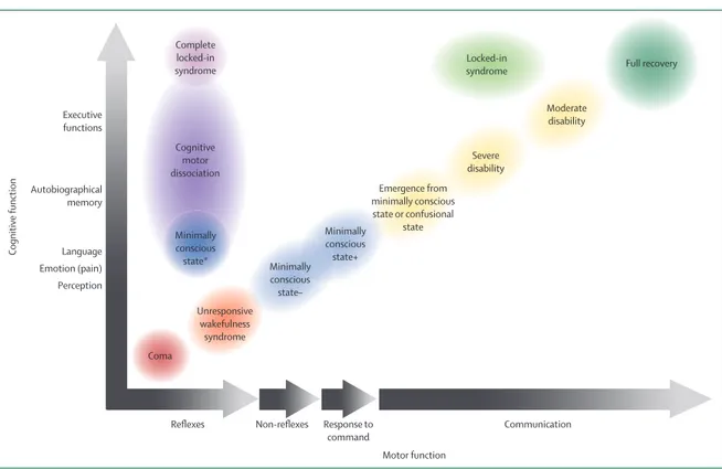
A lot of work has been done on the accurate diagnosis of patients with disorders of consciousness1,2 to establish prognostic indicators3,4 and to understand the neural corre lates of consciousness.5 This work is crucial because misdiagnosis can lead to important medical decisions, such as withdrawal of life-sustaining care.6 Disorders of consciousness include coma (unwakefulness, reflex behaviours only), unresponsive wakefulness syndrome (previ ously known as vegetative state; wakefulness but reflex behaviours only), and minimally conscious state (clini cal demonstration of signs of consciousness).7,8 Once patients recover functional communication or object use, they emerge from the minimally conscious state. Addi-tional entities have been proposed when dis sociation occurs between clinical diagnosis and neuro imaging results showing atypical brain activation, in clud ing mini-mally conscious state* and cognitive motor dissoci ation (panel 1; figure 1).14,18 Patients who have recovered from coma can remain severely disabled for several months, years, or even decades.
With regards to therapeutic options, only a few studies have investigated the treatment of patients with dis-orders of consciousness. Following a landmark paper on amantadine in 2012,22 this field has evolved rapidly, with new therapeutic approaches being tested and reported, but patients’ clinical management remains challeng ing, mostly because these patients cannot com municate and are dependent on others for care. The 2018 American practice guidelines for patients with dis orders of con-sciousness23 only recommend amantadine for patients with unresponsive wakefulness syndrome and minimally conscious state 4–16 weeks after a traumatic brain injury on the basis of one ran domised controlled trial.22 Given that the guidelines were developed on the basis of strict inclusion and exclusion criteria (eg, a minimum of 20 patients included, all at least 28 days after injury), many studies failed to meet their inclusion criteria and were not reported in these recommendations. In this Review, we critically evaluate the available therapeutic options
for patients with prolonged disorders of con scious-ness (ie, more than 28 days) that have been studied in the past 6 years. We discuss pharmacological and non-pharmacological interventions with the strongest evi dence and for which robust randomised controlled trials have been published. If no randomised controlled trials were available, we present open-label studies and anec dotal case reports with careful interpretation, be cause they might still provide insightful results to guide future research. We also report neuro imaging and neuro physiological results associated with positive treatment responses.
Pharmacological treatments
Amantadine (dopamine agonist and NMDA antagon-ist),22,24–26 intrathecal baclofen (GABA agonist),27 zolpi dem (non-benzo diazepine GABA agonist),28–32 midazolam (benzo diazepine GABA agonist),33 and ziconotide (calcium chan nel blocker)34 have been used to improve conscious-ness and functional recovery in patients with disorders of consciousness.
Amantadine and other neurostimulants
Only one large class II randomised controlled trial22 on amantadine has been published. 184 patients with prolonged disorders of consciousness (28–112 days after injury) after traumatic brain injury received either amantadine (up to 200 mg twice a day) or placebo for 4 weeks and were followed for a further 2 weeks.22The amantadine group recovered faster than the placebo group during the course of the treatment as measured by the Disability Rating Scale.35
In non-traumatic brain injury, one uncontrolled case report36 reported the positive behavioural effects of aman-tadine in patients in a minimally conscious state (16 months after injury). Another controlled case report24 showed an increased metabolism in the fronto-parietal cortex dur ing amantadine in an anoxic minimally con-scious state patient (figure 2A). These two case reports should encourage the development of a randomised controlled trial that evaluates the effect of amantadine in
patients with disorders of consciousness with causes other than traumatic brain injury.
Besides amantadine, the administration of one or more neurostimulants (ie, amantadine, bromocriptine, levo-dopa, methylphenidate, and modafinil) has also been explored in a retrospective study in a cohort of 115 patients with disorders of consciousness (<180 days after injury).25 The number of neurostimulants used was not associated with meaningful behavioural improvement in this study.
Zolpidem
Zolpidem is a hypnotic agent known to induce paradoxical transient effects in rare cases in patients with disorders of consciousness. A double-blind crossover randomised con-trolled trial28 in 84 patients with unresponsive wakeful ness syndrome or in a minimally conscious state (>4 months after injury) identified four (5%) respond ers following the intake of 10 mg of zolpidem. These four patients gained at least five points on the Coma Recovery Scale-Revised;46 one patient with unresponsive wakefulness syndrome and one in minimally conscious state minus became minimally conscious state plus, and two patients emerged from their mini mally conscious state for around 2 h.28 Another random ised controlled trial29 involving eight patients with un responsive wakefulness syndrome (1–114 months after injury) only reported slight clinical changes, such as yawns and hiccups, com bined with an EEG-recorded activity of lower amplitude after zolpidem intake. A two-phase study (ie, open-label and then a placebo-controlled trial if there was a change of Coma Recovery Scale-Revised diagnosis)31 included 60 patients with unresponsive wakefulness syndrome or in a minimally conscious state (1 month to 24 years after injury). 12 patients (20%; 11 in a minimally conscious state and one with unresponsive wakefulness syndrome) showed behav ioural improve ments, such as res ponse to command or object localisation, with out a change of diagnosis. One patient in a minimally conscious state could functionally use some objects after the open trial but did not demonstrate any improvement in the placebo-controlled phase. In one case report,30 recovery of con scious ness was reported in a patient with unresponsive wakefulness syndrome (>3 years after cardiac arrest) when using a higher dose of zolpidem (30 mg instead of 10 mg). The patient showed signs of consciousness when receiv-ing 20 mg and further improved after 30 mg of zolpi-dem, suggesting that higher doses might induce stronger effects.
With regard to zolpidem’s brain responses, studies using EEG,32 functional MRI,47 and PET37 have identified an in -crease in brain activity, mainly in prefrontal regions (fig-ure 2B), which supports the mesocircuit model (fig(fig-ure 3). This model could explain how zolpidem can modu late thalamo-cortical connectivity through disinhi bition of the thalamus by acting on the globus palli dus interna and, consequently, promote the recovery of consciousness.50
In summary, zolpidem shows improvement of con-sciousness and functional recovery (even if transient) in
Panel 1: Clinical entities of disorders of consciousness Coma
Coma is the result of a severe brain injury, in which patients are unarousable (ie, eye closure even when stimulated) and unaware of themselves and their environment.9 This state is temporary and after several days or weeks, patients might evolve to brain death (ie, irreversible coma with absence of brainstem reflexes and apnoea) or show some or full recovery.
Unresponsive wakefulness syndrome
When patients start opening their eyes but present only reflex movements, they are diagnosed with an unresponsive wakefulness syndrome (previously termed vegetative state).10 Patients in unresponsive wakefulness syndrome exhibit no signs of awareness, but they can present a variety of reflexive movements, such as grinding teeth, yawning, or groaning.10 This condition might be transitory, prolonged, or permanent.
Minimally conscious state
Once patients recover fluctuating but reproducible signs of consciousness, they enter the minimally conscious state.11 This entity is divided into minimally conscious state minus and minimally conscious state plus on the basis of language processing.12,13 Minimally conscious state minus describes patients showing visual pursuit and fixation, localisation to noxious stimulation, or automatic motor reactions (eg, grasping bed sheets). Patients in minimally conscious state plus follow simple commands, can make understandable verbalisations, or communicate intentionally but not functionally. Like unresponsive wakefulness syndrome, minimally conscious state can be temporary or permanent. The diagnostic label of minimally conscious state* has been suggested for unresponsive wakefulness syndrome patients who show no evidence of awareness at the bedside, while neuroimaging data show atypical brain patterns using active paradigm (eg, brain activity in motor area during a motor imagery task) or metabolic resting state (eg, preservation of the fronto-parietal network).14–16 This entity allows a more clinically accurate diagnosis when the bedside examination shows no evidence of consciousness. The term functional locked-in syndrome (as well as covert cognition) has also been proposed to indicate a dissociation between bedside behaviour and the results of neuroimaging assessments17 (like minimally conscious state*14 and cognitive motor dissociation18).
Cognitive motor dissociation
The syndrome of cognitive motor dissociation has been proposed to specifically refer to patients in coma, unresponsive wakefulness syndrome, or minimally conscious state minus who show consistent brain activation during mental imagery tasks using functional MRI or EEG, and hence show response to command using neuroimaging technologies.18 Cognitive motor dissociation indicates a wide range of uncertainty regarding the underlying cognitive capacity present in patients with no or little behavioural responses.
Emergence from minimally conscious state
When patients are able to functionally communicate or adequately use two different objects, they have emerged from the minimally conscious state. Most of these patients still have severe cognitive and motor impairments.11
Locked-in syndrome
Locked-in syndrome is defined by quadriplegia and anarthria due to a lesion in the corticospinal and corticobulbar pathways in the brainstem.19 These patients cannot move (some recover some distal movements, termed incomplete locked-in syndrome), but their sensations remain intact and they are fully conscious. The most common way for these patients to communicate is through vertical eye movements and blinks.20 In the case of complete locked-in syndrome, paralysis of the eyes prevents any communication and brain computer interfaces are needed.21
around 5% of patients with disorders of consciousness. Determining the behavioural and physio logical profile of zolpidem responders is crucial to better identify the patients that could benefit from this treatment.
Intrathecal baclofen and other drugs
Intrathecal baclofen is primarily used as a centrally act-ing treatment for spasticity but has been suggested as a potential drug to stimulate the recovery of consciousness in a few uncontrolled studies and case reports.27,51 The effects of midazolam (benzodiazepine receptor agonist)33 and ziconotide (atypical analgesic and selective blocker of N-type calcium channels)34 have also been reported in two single-case studies as stimulants for the recovery of consciousness of patients with prolonged disorders of consciousness (one in a minimally conscious state and one with unresponsive wakefulness syndrome, respect-ively).33,34 These anecdotal findings need to be confirmed with randomised controlled trials.
Non-pharmacological interventions
Non-pharmacological interventions have also been attemp-t ed attemp-to improve consciousness and funcattemp-tional re covery in
patients with disorders of consciousness. These include non-invasive brain stimulations (eg, transcranial direct cur rent stimulation, repeated transcranial magnetic stim-ula tion, transcutaneous auricular vagal nerve stimu la-tion, and low intensity focused ultrasound pulse), invasive brain stimula tion (ie, deep brain stimulation or vagal nerve stimulation), and sensory stimulation pro grammes (panel 2).
Non-invasive brain stimulation
Transcranial direct current stimulationA double-blind randomised controlled trial59 tested the effect of prefrontal transcranial direct current stimulation (ie, anode over the left dorsolateral prefrontal cortex for 20 min at 2 mA) on 55 patients, both in acute and prolonged disorders of consciousness (1 week to 26 years after injury). At the group level, behavioural improvements, as measured by the Coma Recovery Scale-Revised,46 were reported for patients in a minimally conscious state, but not for those with unresponsive wakefulness syndrome. At the individual level, 13 (43%) of 30 patients in a minimally conscious state showed an improvement (ie, recovery of a clinical sign of consciousness never
Figure 1: Motor and cognitive evolution following a severe brain injury
The different diagnoses after a severe brain injury can be best captured on a two-dimensional axis by comparing the degree of cognitive function against the degree of motor function. Red circles represent patients who are unconscious with only reflexive movements (coma and unresponsive wakefulness syndrome). Blue circles represent patients in a minimally conscious state (minimally conscious state plus and minimally conscious state minus depending on language preservation). When functional communication is detected (yellow circles), patients emerge from the minimally conscious state and can evolve to a confusional state or severe or moderate disability, before a full recovery (dark green circle). Dissociations between motor and cognitive functions exist in locked-in syndrome (light green circle), cognitive motor dissociation (dark purple circle), and minimally conscious state* (dark blue circle). In rare cases, the diagnosis of complete locked-in syndrome (light purple circle) can be done through neuroimaging examinations. The black-to-white gradient represents the evolution from absence (black) to the recovery of a behaviour (white; eg, from no response to command to consistent response).
Autobiographical memory Language Cognitive function Perception Emotion (pain) Executive functions
Reflexes Response to Communication
command Non-reflexes Motor function Full recovery Moderate disability Severe disability Locked-in syndrome Emergence from minimally conscious state or confusional state Minimally conscious state+ Minimally conscious state– Coma Unresponsive wakefulness syndrome Minimally conscious state* Complete locked-in syndrome Cognitive motor dissociation
observed before transcranial direct current stimulation, nor during sham session). No transcranial direct current stimulation related side-effects were reported in any patients. In a case report,60 one patient considered to have unresponsive wake fulness synd rome showed a response to command after one session of transcranial direct current stimulation over the dorsolateral prefrontal cort-ex. When looking at the neuroimaging assessments, a preservation of brain activity closer to what is usually observed in conscious individuals was identified, suggesting that the patient was in minimally conscious state*. In another randomised controlled trial,61 trans-cranial direct current stimulation was applied once a day for 5 consecutive days in 16 patients in a minimally conscious state (5 months to 30 years after injury) and the effects were assessed daily and at 1-week follow-up. A clinical improvement (eg, recovery of response to command, visual pursuit, or object localisation or manipulation) was observed after 5 days of transcranial direct current stimu lation and the effects remained up to a week in some patients, with a signficant treatment effect observed at the group level after 1-week follow-up.61 However, only four patients responded directly after
the first stimulation, indicating that a single session is insufficient to determine if a patient can benefit from the technique or not. A non-randomised controlled study62 evaluated the clinical effects of 5 days of sham then 5 days of active transcranial direct current stimulation applied either over the dorsolateral prefrontal cortex or the primary sensori motor cortex in ten patients with unresponsive wakefulness syndrome or minimally conscious state (6 months to 10 years after injury). The three patients in a minimally conscious state improved regardless of the site of stimulation (one patient in a minimally conscious state received prefrontal stimula tion and two received sensorimotor stimulation), whereas none of the seven patients in unresponsive wakefulness syndrome responded. Another double-blind randomised controlled trial63 showed that the observed behavioural improvement (Coma Recovery Scale-Revised total score) in five (38%) of 13 patients following five sessions of transcranial direct current stimulation were paralleled with EEG changes (enhancement of EEG background). Another double-blind randomised controlled trial64 in-cluded 26 patients with disorders of consciousness (1–17 months after injury) who received 20 sessions of
Figure 2: Pharmacological and non-pharmacological interventions to improve consciousness in patients with disorders of consciousness
(A) Amantadine has been shown to increase brain metabolism in the fronto-parietal network in one patient in a minimally conscious state. Reproduced from Schnakers and colleagues.24 (B) Zolpidem
induced an increase in brain metabolism in the prefrontal and mesiofrontal cortex in three minimally conscious state responders. Reproduced from Chatelle and colleagues.37 (C) Patients responding to
transcranial direct current stimulation (n=8) had more preservation of brain metabolism in the prefrontal cortex (stimulated area) compared with non-responders (n=13). Reproduced from Thibaut and colleagues.38 (D) Repeated transcranial magnetic stimulation of 20 Hz on the primary motor cortex induced EEG increases in beta (shown), alpha, and delta band power in one minimally conscious state
responder. Reproduced from Piccione and colleagues,39 by permission of SAGE Publishing. (E) Low intensity focused ultrasound pulsation is shown in a patient with unresponsive wakefulness syndrome who
became minimally conscious after stimulating the thalamic target (red circle). Reproduced from Monti and colleagues,40 by permission of Elsevier. (F) Transcutaneous auricular vagal nerve stimulation
induced increases in functional connectivity between posterior cingulate, precuneus, hypothalamus, thalamus, and prefrontal and temporal cortex (red), and decreases between the posterior cingulate, precuneus, and cerebellum gyrus(blue) in one patient with unresponsive wakefulness syndrome who transitioned to a minimally conscious state after stimulation. Reproduced from Yu and colleagues,41
by permission of Elsevier. (G) Deep brain stimulation electrode placement, as seen with MRI, in one patient in a minimally conscious state who subsequently recovered.42,43 Reproduced from Schiff and
colleagues.42(H) Brain connectivity patterns before (left) and after (right) invasive vagal nerve stimulation as measured with high-density EEG in one unresponsive wakefulness syndrome patient who
improved to a minimally conscious state after stimulation. Reproduced from Corazzol and colleagues,44 by permission of Elsevier.(I) Music stimulation induced an increase in functional connectivity in the
auditory network (and in the default mode network; not shown) in five patients with disorders of consciousness. Reproduced from Heine and colleagues.45
Amantadine Zolpidem
Transcranial direct current stimulation
Transcranial direct current stimulationRepeated transcranial magnetic stimulation Low intensity focused ultrasound pulsation
Deep brain stimulation Invasive vagal nerve stimulation Music stimulation
A B C
D E
G
H
H I
Transcranial direct current stimulation
Transcutaneous auricular vagal nerve stimulation
F
active or sham prefrontal stimulation over 10 days. Clinical improvement was observed in the minimally conscious state group but not in the un responsive wakefulness syndrome group, combined with an increase in P300 amplitude for the responders. A randomised controlled trial65 in 27 patients in a minimally conscious state (10 months to 33 years after injury) evaluated the effects of 20 sessions within 4 weeks of transcranial direct current stimulation over the dorsolateral prefrontal cortex applied by the patients’ relatives or caregivers at home or in nursing homes. Although the overall compliance was good (ie, 96% of sessions com pleted), the behavioural effect was not significant. How ever, when the five patients who did not receive at least 80% of the stimulation sessions were excluded, a signi ficant treatment effect was observed for the remaining 22 patients. Therefore,
patients with prolonged disorders of consciousness can show clinical improve ments after transcranial direct current stimula tion, such as the recovery of object manipulation or functional com munication, even years after the brain injury, but a continuous applica tion of transcranial direct current stimulation seems to be required. Beside trans cranial direct current stimulation, 101–640 Hz transcranial random noise stimulation was applied over the prefrontal cortex for 5 daily sessions of 20 min in a pilot random ised controlled trial66 on nine patients with unrespon sive wakefulness syndrome (30 days to 4 months after injury) which showed no clinical improvement. However, the small sample size prevents us from drawing any generalisable conclusions.
With regard to neuroimaging data of responders, a common pattern of metabolic and grey matter preserva-tions has been reported in eight responders com pared with 13 non-responder patients in a minimally con scious state.38 Clinical improvement following trans cranial direct current stimulation seems to require partial functional and structural preservation of the stimulated area (dorsolateral prefrontal cortex) and other brain regions crucial in consciousness recovery, such as the precuneus and the thalamus (figure 2C). A higher cortical connectivity within the theta band, known to be important for cons ciousness processes,67 was also reported in res-ponders compared with non-resres-ponders in a minimally conscious state.68 Additionally, EEG studies69,70 identified an increase in fronto-parietal coherence in the theta band after active transcranial direct current stimulation of the dorsolateral prefrontal cortex in patients in a minimally conscious state and an increase in glo-bal cortical excitability as measured with transcranial mag netic stimulation coupled with EEG, highlighting the possible neural effects of prefrontal transcranial direct current stimulation in patients with disorders of consciousness.
Compared with dorsolateral prefrontal cortex stimu-lation, transcranial direct current stimulation of the precuneus or the orbitofrontal cortex has shown less pro-mising results.71,72 In a double-blind randomised controlled trial,71 stimulation was applied over the precuneus once a day for 20 min for 5 days in 33 patients in a minimally conscious state (1–26 months after injury). A behavioural improvement at the group level was reported after the transcranial direct current stimu lation sessions, but the effect did not last when reassessed 5 days later. At the individual level, six (18%) of 33 patients were identified as responders with the recovery of visual pursuit, response to command, automatic motor reaction or objects manipula-tion, or localisation. In one prospective open-label study,72 no behavioural changes were observed after transcranial direct current stimulation applied over the orbitofrontal cortex in 22 patients with prolonged disorders of con-sciousness (4–33 months after injury). Cortical connectivity and excitability were increased after trans cranial direct current stimulation in all patients in a minimally conscious
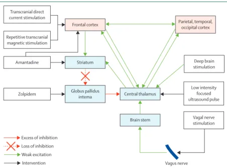
Figure 3: The mesocircuit fronto-parietal model for mechanisms underlying the effects of interventions in
severe brain injuries
This model provides a framework that explains the potential mechanisms of action of various therapeutic interventions and the neural mechanisms of impaired consciousness. This model supports the idea that, in normal cognitive processing, the central thalamus is regulated by both the dominant corticothalamic feedback provided by (pre)frontal regions (bidirectional green arrow)and through an inhibitory modulation by the internal globus pallidus, which itself is regulated by cortico-striatal and thalamo-striatal inputs. Activation of the central thalamus broadly drives activity of associative fronto-parietal cortical areas.48 However, in case of brain injury, a reduction of
corticothalamic and thalamo-striatal outflow following deafferentation and loss of neurons from the central thalamus withdraws important afferent drive to the medium spiny neurons of the striatum (green lines). Loss of active inhibition from the striatum (dashed red line) allows neurons of the globus pallidus interna to tonically fire and provide active inhibition (red line) to their synaptic targets, including relay neurons of the already strongly disfacilitatedcentral thalamus. This mesocircuit model might explain the potential mechanisms of several treatments that have shown promising results in the recovery of consciousness in patients with severe brain injuries. Partial preservation of the stimulated prefrontal cortex seems to be necessary to induce a clinical response to transcranial direct current stimulation,38 whereas repeated transcranial magnetic stimulation seems to induce a
global increase in cortical oscillations when applied over the primary motor cortex.39 The clinical improvement of a
patient who responded to amantadine correlated with increased fronto-parietal brain metabolism.24 Zolpidem
might produce broad excitation across the frontal cortices and striatum through direct excitation and through inhibition of the globus pallidus.32,49 Deep brain stimulation directly acts over the central thalamus, aiming to
stimulate the thalamo-cortical connectivity,42 whereas low intensity focused ultrasound pulse stimulates the
thalamus in a non-invasive way.40 Invasive and non-invasive vagal nerve stimulation directly stimulate the vagal
nerve.41,44 Blue rectangles represent subcortical regions, and pink rectangles represent cortical areas.Adapted from
Giacino and colleagues.7
Central thalamus
Brain stem Striatum
Globus pallidus intema
Frontal cortex Parietal, temporal,occipital cortex
Deep brain stimulation Amantadine Zolpidem Transcranial direct current stimulation Repetitive transcranial magnetic stimulation Vagal nerve stimulation Low intensity focused ultrasound pulse Vagus nerve Excess of inhibition Loss of inhibition Weak excitation Intervention
state and in some patients with unresponsive wakefulness syndrome, showing the poss ible neuroplasticity effects of transcranial direct current stimulation in patients with disorders of consciousness.
The prefrontal cortex seems to be a better target for stimulation compared with the precuneus and the motor cortex. Dorsolateral prefrontal cortex stimulation might induce a stronger connectivity between the prefrontal cortex and the thalamus because the prefrontal cortex has many connections with the striatum. By stimulating the striatum, a dis inhibition of the thalamus might occur, reinforcing thalamo-cortical connectivity (figure 3),49,73 and facilitating recovery from consciousness.
Repeated transcranial magnetic stimulation
In one double-blind randomised controlled trial74 of 11 patients with unresponsive wakefulness syndrome (9–85 months after injury), no behavioural improvements were reported following fiverepeated sessions at 20 Hz applied over the primary motor cortex (M1) for 10 min. Another random ised controlled trial75 also reported no behavioural im prove ment after one session of M1 20 Hz repeated transcranial magnetic stimulation for about 10 min in ten patients with disorders of consciousness (1–26 months after injury), but haemodynamic funct-ions (ie, cerebral blood flow velocity) were improved in the minimally conscious state group but not in the unresponsive wakefulness syndrome group. 5 Hz stimulation was applied on M1 for about 7 min in a third ran dom ised controlled trial76 in five patients with unresponsive wakefulness syndrome and five patients in a minimally conscious state (5–23 months after injury) evaluating its effects on sleep–wake cycles. Although no behavioural effect was reported, significant after-effects on slow wave activity power were reported in the mini-mally con scious state group but not in the un respon-sive wakefulness syndrome group. A small sample cross over randomised controlled trial77 eval u ated the effects of five sessions of M1 20 Hz repeated transcranial magnetic stimulation, lasting about 10 min, in three patients with unresponsive wakefulness synd rome, two in a minimally conscious state, and one emerging from a minimally conscious state (1–28 months after injury). At the group level, no treatment effect was found, but at the indivi dual level, one patient with unresponsive wakeful-ness syndrome recovered localisa tion to painful stimula-tion and maintained this behaviour at 1-week follow-up. This clinical improvement was paralleled with an increase in alpha and beta power, showing higher brain activity and supporting the recovery of a sign of con sciousness. Additionally, in a case report,39 an increased absolute and relative power in delta, alpha, and beta frequency bands was found with improved signs of consciousness in one patient in a minimally conscious state after stimulation over M1 (figure 2D).
The dorsolateral prefrontal cortex has also been targeted in a few uncontrolled studies. The effect of 20 sessions of
10 Hz dorsolateral prefrontal cortex repeated transcranial magnetic stimulation (each session lasting 11 min) was evaluated in 16 patients with disorders of consciousness (3–35 months after injury) in a single-blind uncontrolled study.78 Coma Recovery Scale-Revised46 total score in-creased in all five patients in a minimally conscious state and in four (36%) of 11 patients with unresponsive wakefulness syndrome, and the improve ments scored higher on the Coma Recovery Scale-Revised in patients in a minimally conscious state. In a small open-label study,79 ten anoxic patients with unresponsive wakefulness
Panel 2: Neuromodulation techniques Transcranial direct current stimulation
This neuromodulation technique modulates cortical excitability through the application of a weak (usually ≤2 mA) direct current through the brain between two electrodes. Physiologically, the establishment of the long-lasting after-effects depends on membrane potential changes as well as modulations of NMDA receptor efficacy, which can induce long-term potentiation and long-term depression-like effects.52–54 However, more mechanistic and in-vivo studies need to be done to better understand how transcranial direct current stimulation can influence cortical activity and act on neuroplasticity.
Transcranial magnetic stimulation
Transcranial magnetic stimulation uses an electromagnetic pulse to induce focalised neural depolarisation and firing. Repeated transcranial magnetic stimulation, compared with single pulse transcranial magnetic stimulation, can influence brain plasticity and cortical organisation through alterations of neuronal excitability. Repeated transcranial magnetic stimulation has been used to induce a sustained inhibition (about 1 Hz frequency) or activation (5–20 Hz frequency) of the neuronal population.
Low intensity focused ultrasound pulse
This technique uses low-energy sound waves to excite or inhibit brain activity. Compared with transcranial direct current stimulation and repeated transcranial magnetic stimulation, it is theoretically capable of directly targeting and stimulating subcortical and deep brain structures, such as the thalamus.
Vagal nerve stimulation
Vagal nerve stimulation can be invasive and surgically placed or non-invasive through transcutaneous auricular stimulation. Transcutaneous auricular vagal nerve stimulation consists of the injection of a thermal current to the external ear canal, which modifies the density of the endolymph in the internal ear and, consequently, alters the firing rate of the vestibular nerve. This technique is thought to induce compensatory responses, through basal forebrain or brainstem projections through the central thalamus and hypothalamus, in distal fronto-parietal and striatal networks.55 Invasive vagal nerve stimulation involves the surgical implantation of a vagus nerve stimulator, using a current of 1–2 mA. Mechanisms of stimulation are similar to transcutaneous auricular vagal nerve stimulation.
Deep brain stimulation
This neurosurgical procedure involves the implantation of a brain electrode that delivers a current to a targeted brain area. The underlying mechanisms of deep brain stimulation are not yet fully understood.56 In patients with severe brain injuries, the main target is the central thalamus to induce excitation of the projecting thalamo-cortical afference. The electrodes are usually implanted in the intralaminar nuclei, because this region seems to be particularly associated with recovery in patients with disorders of consciousness,57 and because of the pathophysiological mechanisms linked to the brain injury and cellular loss in the central thalamus.58
syn drome (4–15 months after injury) received a single ses sion at 10 Hz for 60 min. Although no clinical effects were observed at the group level, three (30%) patients showed behavioural improvements (ie, recovery of pain localisa tion) associ ated with an increase in brain connect-ivity (as measured with dual-coil transcranial magnetic stimula tion). The long-termsafetyof repeated transcranial magnetic stimu lation over the dorsolateral prefrontal cortex was reported in two patients with disorders of consciousness, 6 months and 9 years after injury, who received 30 sessions of stimulation (300 trains of paired stimuli; 100-ms interpulse interval, 5-s intertrain interval) over 6 weeksand who showed no serious adverse event related to repeated transcranial magnetic stimulation.80 The absence of severe adverse events linked to prolonged use of repeated stimulation is encouraging, but no con-clusion can be drawn on the basis of these two case reports alone.
As for transcranial direct current stimulation, the prefrontal cortex could be a better target than the prim-ary motor cortex, because all studies of repeated trans-cranial magnetic stimulation over M1 have not shown clini cal improvements. Preliminary results of uncon-trolled studies should encourage the design of repeated trans cranial magnetic stimulation randomised controlled trials targeting the prefrontal region.
Other novel non-invasive brain stimulation approaches
Novel non-invasive brain stimulation techniques, includ-ing low intensity focused ultrasound pulse, transcutan-eous auricular vagal nerve stimulation, and spinal cord stimulation, have been tested in a few case reports.40,41,81 The only published report40 of a patient in a mini mally conscious state (19 days after traumatic brain injury) who received one session of low intensity foc used ultrasound pulse targeting the central thalamus (figure 2E) showed a recovery of language comprehension and spatio-temporal orientation a few days later. The effects of transcutan-eous auricular vagal nerve stimula tion were presented in another case report of a patient with unresponsive wakefulness syndrome (50 days after anoxia; figure 2F).41 After 4 weeks of treatment (two daily stimulation sessions for 30 min each, with an intensity of 4–6 mA, at a frequency of 20 Hz), the patient regained some signs of con-sciousness. Caloric vestibular stimula tion is another technique that has been tested in two patients in a minimally conscious state (one caused by haemorrhagic stroke and one due to anoxia, about 6 months after injury).82 The protocol included two active or two sham daily sessions 4 or 5 days per week for 2 weeks. Both patients showed clinical improvement with the Coma Recovery Scale-Revised46 (ie, arousal and auditory scales) and the Wessex Head Injury Matrix83,84 (ie, gesture making and selective responses to relatives). Spinal cord stimula-tion has also been explored in some case reports or uncontrolled studies with mixed results.81,85 However, no randomised controlled trial eval u ating the effects of spinal
cord stimulationhas been done, and studies did not use standardised scales or well-defined outcomes to assess the effects of the intervention. As for all uncontrolled trials, the results of these case reports on novel non-invasive brain stimulation techniques could be linked to spontaneous recovery; however, these studies can be considered as feasibility studies.
Optimisation of non-invasive brain stimulation
Within the growing field of non-invasive brain stimula-tion techniques (ten out of the 14 randomised controlled trials reviewed investigated the effect of non-invasive brain stimulation; table), transcranial direct current stimu la tion is the only intervention that has shown a clinical effect in multiple randomised controlled trials, more specifically in patients in a minimally con scious state.59,63,64 However, not all pa tients respond, its effects are limited to the recovery of a few signs of consciousness (eg, recovery of visual pursuit, response to command, or object localisation or manipulation), and changes of diagnosis are transient and only observed in some cases. Therefore, the technique needs to be optimised to induce long-lasting clinically meaningful improvements, such as re covery of communication. Additionally, other brain areas could be stimulated according to patients’ remain-ing brain structures and function because patients’ clinical responsiveness is associated with the relative preservation of grey matter, brain metabolism, and cortical connect ivity.38,68 The emerging field of current modelling could also help the development of tailored stimulation montages based on individual structural brain changes.88 To this aim, neuroimaging should be done before brain stimulation to document the exact area to be stimulated and to tailor patients’ stimulation based on their brain lesions. Of note, no side-effects have been reported in all transcranial direct current stimulation or repeated transcranial magnetic stimulation studies (three studies63,64,77 did not mention if they collected possible adverse events).
With regard to the other non-invasive interventions, repeated transcranial magnetic stimulation did not have a significant effect at the group level in any random-ised controlled trials (all class III). Nonetheless, many parameters (eg, target area, frequency, or duration of stimulation) could be optimised to enhance its efficacy.
Invasive brain stimulation
A 7-year well-designed prospective open-label study89 on the effects of deep brain stimulation of the thalamic reticular nuclei in patients with disorders of conscious-ness (>6 months after injury) reported that only five (13%) of 40 patients met the inclus ion criteria (eg, EEG desynch-ronised activity <5% of the recorded time, somatosensory and auditory evoked potentials evoked on at least one side). Of the five eligible patients, two did not receive surgery owing to issues with the legal representative. The three patients who underwent the procedure showed small
Study design Class of evidence
N
Diagnosis
Time since injury
Procedure Results Effect sizes Study ca veats Pharmacological interv entions Zolpidem 28 Double-blind, crosso ver III 84
TBI and
non-TBI
patients
(no details
on
numbers of
each); 253 screened; 104 eligible 66 MCS, 18 UWS >4 months Single dose of 10 mg zolpidem, and if impro vement placebo-controlled
phase; evaluations at baseline and 1, 2, and 3 h after zolpidem
or placebo
intak
e
4 responders identified (ie, >5 points increase
on the
CRS-R
in the
placebo
controlled
double-blind phase); 2 patients
in MCS emerged, 1 patient
with
UWS, and
1 patient in MCS– regained response
to
command (ie, MCS+); side-effects: 1 sev
ere
with zolpidem, 24 mild (20 [83%]
of 24
with zolpidem)
Responders 1 h after zolpidem intak
e: d=2∙09;
2 h later: 1∙78*
Very low ratio
of
responders; heterogeneous population
Zolpidem 29 Double-blind, crosso ver III 8 (1 TBI, 7 non-TBI); no information on patients screened or eligible 8 UWS >1 month Single dose of 10 mg of zolpidem or placebo in
two sessions, separated
by 10–14
da
ys; evaluations at
baseline and 90 min after zolpidem or placebo intak
e
Signs
of arousal after zolpidem intak
e (yawns and hiccups), activation of EEG, and
a vagolytic chronotropic effect in all patients; no information
on side-effects
No beha
vioural
data
available to
calculate the effect size Small sample; heterogeneous population
Non-pharmacological interv
entions
Transcranial direct current stimulation
59 Double-blind, crosso ver II 55 (25 TBI, 30 non-TBI); 62 eligible 30 MCS, 25 UWS 1 w eek to 19 y ears Comparison of a single session (20 min) of activ e and sham stimulation o ver the left DLPFC with
CRS-R before and after stimulation
13 (43%) patients in MCS and 2 (1%) patients
with
UWS clinically impro
ved (reco very of visual pursuit or response to command); at
the group lev
el, clinical impro vement (2 points on the CRS-R) for patients in MCS; no side-effects observ ed For patients in MCS: d=0∙38* One session of
stimulation; no follow-up; heterogeneous population
Transcranial direct current stimulation
61 Double-blind, crosso ver III 16 (11 TBI, 5 non-TBI); 21 eligible 16 MCS >3 months Comparison of 5 sessions of activ e and sham transcranial direct current stimulation (20 minutes a da y) ov er the DLPFC, separated b y a w eek of washout; CRS-R performed before and after 5 da ys of stimulation, and at 1-w eek follow-up
9 (56%) responders; clinical impro
vement (2 points on the CRS-R) maintained up to 1 w eek after the end of the stimulation; no side-effects observ ed
After stimulation (before washout): d=0∙43; at 1-w
eek follow-up: d=0∙57 Short washout period; no long-term
follow-up; heterogeneous population
Transcranial direct current stimulation
63 Double-blind, crosso ver III 13 (1 TBI, 12 non-TBI); 15 eligible 7 UWS, 6 MCS >3 months Comparison of 5 da ys of activ e and sham stimulation ov er the DLPFC for 20 min a da y; EEG and CRS-R
done at baseline, after 5
da ys of stimulation, and up to 3-month follow-up At
the group lev
el, no statistical
difference
betw
een
the
two groups; at individual
lev
el, beha
vioural (CRS-R
total score) and
EEG changes in 5 (38%) patients (3 [50%] patients in MCS and 2 [29%]
with UWS); no information on side-effects No difference betw een groups Small sample; unequal group split (4 in one and 7 in the other);
parallel not crosso
ver
design; heterogeneous population
Transcranial direct current stimulation
64 Double-blind, parallel III 26 (12 TBI, 14 non-TBI); no information on patients screened or eligible 11 UWS, 15 MCS 1–18 months Comparison of 20 sessions of activ e or sham transcranial direct current stimulation ov er DLPFC for 20 min twice a da y for 10 da ys CRS-R impro vement in patients in MCS at
the group lev
el
in
the activ
e group
, coupled
with increase in P300 amplitude; no information on
side-effects
For patients in MCS: d=2∙22* Heterogeneous population; no follow-up
Transcranial direct current stimulation
65 Double-blind, crosso ver III 27 (12 TBI, 15 non-TBI); 86 screened 27 MCS 10 months to 14 y ears Comparison of 20 sessions of activ e and sham transcranial direct current
stimulation (20 min per
da y) ov er DLPFC, separated b y 8 w eeks; CRS-R
before and after 4
w
eeks
of
stimulation (20 sessions), and at 8-w
eek follow-up
No impro
vement at
the group lev
el; for
patient group
who receiv
ed at least 80%
of
the stimulation sessions, increase in
CRS-R
total scores; no
difference betw
een activ
e
and sham stimulation at 8-w
eek
follow-up; no stimulation-related side-effects observ
ed
Group lev
el:
d=0∙47; subgroup of patients who receiv
ed
>80% of
stimulation sessions: d=0∙53 Heterogeneous population; high rate of drop-out;
few
assessments done
(T
able continues
Study design Class of evidence
N
Diagnosis
Time since injury
Procedure
Results
Effect sizes
Study ca
veats
(Continued from previous page) Transcranial direct current stimulation
71 Double-blind, crosso ver III 33 (20 TBI, 13 non-TBI); 37 eligible 33 MCS >3 months Comparison of activ e or sham transcranial direct current
stimulation applied for 5
da
ys
ov
er
the posterior parietal cortex (20 min a da
y), separated by a w eek of washout; CRS-R performed before and after 5 da ys of stimulation, and at 5 da ys follow-up
9 responders; clinical impro
vement immediately after the 5 da ys of activ e stimulation (increase b y 1 point on the CRS-R); no effects at 1-w eek follow-up; no side-effects observ ed. After stimulation: d=0∙31 Short washout period;
heterogeneous population; no
long-term follow-up
(1 w
eek only)
Transcranial random noise stimulation
66 Double-blind, parallel III 9 (1 TBI, 8 non-TBI); no information on patients screened or eligible 9 UWS 30 da ys to 4 months Comparison of 5 sessions of
transcranial random noise stimulation (101–640 Hz)
ov
er
DLPFC for 20 min;
CRS-R performed
at baseline, after each session, and at 3-da
y follow-up; EEG at baseline,
the end of the 5 da ys protocol, and 3-da y follow-up No beha vioural or neurophysiological impro vement at
the group lev
el; no side-effects observ ed. No difference betw een groups Small sample; unequal group split (4 in one and 5 in the other);
parallel not crosso
ver
design; heterogeneous population (only 1 TBI)
Repetitiv
e
transcranial magnetic stimulation
74 Double-blind, crosso ver III 11 (2 TBI, 9 non-TBI); no information on patients screened or eligible 11 UWS 9–85 months Fiv e sessions of activ e or sham 20 Hz repetitiv e transcranial
magnetic stimulation for 10 min, for a
total of 1000 pulses deliv ered in 5 trains, ov
er left primary motor
cortex for 5 consecutiv
e da
ys; EEG
and
CRS-R performed before and
after stimulation No beha vioural or EEG impro vements; no side-effects observ ed No difference betw een groups
Small sample; heterogeneous population
Repetitiv
e
transcranial magnetic stimulation
75 Double-blind, crosso ver III 10 (4 TBI, 6 non-TBI); no information on patients screened or eligible 5 MCS, 5 UWS 1–28 months One session of activ e or sham 20 Hz repetitiv e transcranial magnetic stimulation ov
er M1 for 10 min, for
a total of 1000 pulses deliv ered in 20 trains,
CRS-R scores and cerebral
blood flow v
elocity
of
the middle
cerebral artery before and after stimulation
No beha
vioural (CRS-R) changes;
temporary increase in peak systolic v
elocity
and mean flow v
elocity
of
the left middle
cerebral artery for patients in MCS; no effects in patients
with UWS or in sham group; no side-effects observ ed No difference betw een groups One session of
stimulation; small sample; heterogeneous population
Repetitiv
e
transcranial magnetic stimulation
77 Double-blind, crosso ver III 6 (4 TBI, 2 non-TBI); no information on patients screened or eligible 3 UWS, 2 MCS, 1 EMCS 1–28 months Fiv e sessions of activ e or sham 20 Hz repetitiv e transcranial magnetic stimulation ov er primary
motor cortex for a
total of 1000 pulses deliv ered in 20 trains;
CRS-R scores and EEG reactivity collected before, after stimulation, and 1-w
eek follow-up No treatment effect on the CRS-R or on the
EEG metrics; clinical impro
vement in 1 patient maintained at 1-w eek follow-up (a patient with UWS impro ved to MCS–), which was paralleled b y EEG pow er spectra impro vement; no information on side-effects No difference betw een groups Crosso ver study in
acute patients but data analysesd as parallel
owing to
carry
ov
er effects;
small sample; heterogeneous population
(T
able continues
behavioural improvements (Coma Recovery Scale-Revised total scores improved 1–3 points compared with baseline). Additionally, the electrodes had to be removed for one patient due to a scalp infection. Given these results, the use of deep brain stimulation to improve patients’ recovery seems limited to a small proportion of patients with prolonged disorders of con sciousness and does not induce drastic clinical improve ments. In another prospective open-label study90 including 14 patients in unresponsive wakefulness syndrome or minimally conscious state (2 months to 11∙5 years after injury), positive effects of deep brain stimulation of the thalamic reticular nuclei on clinical recovery were reported in four patients (29%). Three of four patients in a minimally conscious state emerged and one of ten patients with unresponsive wakefulness synd rome regained response to command. However, dis entangling the effects of deep brain stimula-tion from spontaneous recovery is difficult, because these patients were enrolled 2–11 months after injury. Beside these two open-label studies, the only other study to employ a standardised and validated outcome measure (ie, the Coma Recovery Scale-Revised46) to evaluate the efficacy of deep brain stimulation in disorders of consciousness is the seminal paper published in 2007,42 in which a traumatic brain injured patient in mini-mally conscious state for 6 years was treated with deep brain stimulation of thalamic intra laminar nuclei in a double-blind alternating crossover study (figure 2G). Clinically, the patient recovered consistent responses to commands, oral feeding, and functional communica-tion. Improvements were seen immediately and over the course of 6 months. When deep brain stimulation was turned off, even if the clinical state of the patient decreased, it remained above baseline level suggesting some carryover effects.
To date, no sham-controlled trial has been published on deep brain stimulation in patients with disorders of consciousness. A treatment protocol still needs to be established that tests the generalisable effects of deep brain stimulation against a common set of criteria. Additionally, many clinical and ethical issues (eg, risk of infection and, consequently, clinical deterioration)should still be addressed.91
Invasive vagal nerve stimulation has been used in one uncontrolled case study44 of a patient with un responsive wakefulness syndrome for 15 years. The patient improved to a minimally conscious state and presented enhanced brain connectivity patterns (ie, activity increase in occipito-parieto-frontal and basal ganglia regions; figure 2H). This case report needs to be interpreted cautiously, but it illustrates the feasibility of using this approach in patients with disorders of consciousness.
Sensory stimulation programmes
Stimulation programmes include, among others, motor-based therapy, auditory-motor-based training, music therapy, and multi-sensory training programmes.
Study design
Class of evidence
N
Diagnosis
Time since injury
Procedure
Results
Effect sizes
Study ca
veats
(Continued from previous page) Tilt table
and
integrated stepping device
86 Single-blind, parallel III 50 (16 TBI, 34 non-TBI); 422 screened
UWS, MCS (no details on patient number)
4 w eeks to 6 months Comparison of
regular tilt table;
therap y with or without integrated stepping device on lev el of
consciousness; patients receiv
ed
the
interv
ention 10
times for 60 min
ov
er a 3-w
eek period
Both groups presented impro
vements (5 points on the CRS-R); no information on side-effects At 3-w eek follow-up: d=0∙34; at 6-w eek follow-up: d=0∙42
Single blind; no non-interv
ention
group because all had tilt table therap
y; heterogeneous population Sensory stimulation 87 Double-blind, parallel III 15 TBI; 55 screened 5 UWS, 10 MCS 1–6 months Comparison
of FAST and placebo
(silence) applied 10 min 4
times per da y for 6 w eeks Beha vioural impro
vements in both groups;
more impro
vement in
the FAST group
using Coma/Near Coma
Scale;
FAST
patients had more functional MRI activation in language regions and
whole brain in response to v ocal stimuli; no information on side-effects d=1∙88
Small sample; placebo condition was
silence;
unclear double-blind procedure; heterogeneous population
Only randomised controlled
trials published since 2013
that aimed
to impro
ve patients’ awareness and
used validated scale are listed.
TBI=traumatic brain injury
. UWS=unresponsiv
e wak
efulness syndrome. MCS=minimally conscious state.
DLPFC=dorsolateral prefrontal cortex.
CRS-R=Coma R
eco
very Scale-R
evised.
CBF=cerebral blood flow
. EMCS=Emergence from MCS. FAST=familiar auditory sensory
training. *Effect sizes
w ere tak en from the articles when a vailable or calculated (Cohen’ s
effect size small:
d=0∙2; medium: d=0∙5; large: d=0∙8) on the basis of data pro vided betw een activ
e and controlled condition
when a statistical
difference
was found.
Table
: Randomised controlled
trials assessing pharmacological and non-pharmacological inte
rv
entions in patients
with
disorders
In a single-blind randomised controlled trial,86 the effects of conventional tilt table and its combination with a stepping device were assessed in 50 patients with dis-orders of consciousness (1–6 months after injury). Be-havioural improvements were reported in both groups at the end of the 3-week intervention period and at the 3-week follow-up. No information was however provided regarding the type of behavioural recovery, and since the study did not include a group with no therapy, the improvement could also be related to spontaneous recovery.
Familiar auditory stimulation training (FAST)92 was used in a double-blind randomised controlled trial in 15 patients with prolonged disorders of consciousness (mean of 70 days after injury) after traumatic brain injury.87 FAST is composed of 5-min stories told by the patient’s relatives that involve autobiographical events (10 min, 4 times a day, with at least 2 h between, for 6 weeks), and the placebo protocol was silence. Both behavioural (using the Coma/Near Coma Scale35 and Coma Recovery Scale-Revised46) and neuroimaging data showed better results for the FAST group than for the control group (ie, more Coma/Near Coma Scale gains and higher MRI activation in language regions and whole brain). However, clinical improvements were within the boundaries of the Coma Recovery Scale-Revisedand the Coma/Near Coma Scale without changes of diagnosis. Additionally, baseline difference between groups and the small sample size might also be a source of bias in this study, reducing its interpretability.87
The effects of music therapy were evaluated in a controlled case series93 (two cycles of 15 sessions separated by 2 weeks) in ten patients with prolonged disorders of consciousness (time range not specified) showing some behavioural improvement (eg, more eye contact and smiles with fewer suffering expressions) and an improve-ment of haemodynamic parameters (ie, systolic and diastolic pressure) in patients in a minimally conscious state. Although no double-blind randomised controlled trial has been done to evaluate the clinical effects of music in patients with disorders of consciousness, neuroimaging has shown greater activation of the audit ory network (figure 2I) and stronger neurophysiological responses (ie, increase in P300 response), showing a possible enhancement of attent ional processes following music compared with other random sounds.45,94–96
An uncontrolled A-B-A-B design study97 including eight patients with unresponsive wakefulness syndrome and 18 in a minimally conscious statetested the effects of a multi-sensory stimulation programme including audit ory, visual, tactile, olfactory, and gustatory stimuli (20 min per session applied 3 days per week for 4 weeks). Higher Coma Recovery Scale-Revised total scores were reported during the treatments periods (B) compared with base line and treatment withdrawal periods (A) in the mini-mally conscious state group but not in unresponsive wakeful ness syndrome group. Double-blind randomised
controlled trials need to evaluate the possible superiority of a multi-sensory approach com pared with just one type of sensory stimulation.
Hyperbaric oxygen therapy98 and acupuncture99 have also been tested in uncontrolled studies. Some studies reported clinical improve ments following hyperbaric oxygen therapy, but the articles were either not available in English or they did not use validated scales to objectify the clinical improvements. Therefore, these articles do not meet our inclusion criteria.
Taken together, only one double-blind randomised cont-rolled trial92 has been done on sensory stimulations, show-ing that auditory stimulations (ie, FAST protocol) could speed up recovery in patients with prolonged disorders of consciousness.
Conclusions and future directions
Management of patients with disorders of consciousness is challenging because of the absence of communication, the scarcity of interaction with their environment, and their severe motor disability. Adapted thera peutic approaches that do not require patients’ active partici-pation need to be developed rapidly. Present findings suggest that some patients might benefit from rehabi-litative interventions,62,86,87 even years after the brain injury.59,63,65 As highlighted in the American practice guidelines for patients with disorders of consciousness,23 most studies are open-label studies and case reports, so results need to be interpreted with caution and cannot be translated directly into clinical practice. However, several randomised controlled trials have been published since 2013, even if not included in the guidelines (table), but more robust designs and larger samples are still needed.
Only a few randomised controlled trials on pharma-cological interventions have been done, and amantadine22 is the only drug tested that shows class II evidence for patients with traumatic brain injury during rehabilitation and is the only intervention recommended by the American practice guidelines for patients with disorders of consciousness.23 By contrast, many studies and ran-domised controlled trials have used neuromodulation techniques in this patient cohort, showing the growing interest in this field, which might be partly explained by the low cost and absence of severe side-effects. Tran-scranial direct current stimulation applied over the dorsolateral prefrontal cortex induced some clinical improvement in five randomised controlled trials (four class III61,63–65 and one class II59) in patients in minimally conscious states from traumatic brain injury and non-traumatic brain injury aetiolo gies. Although the sample sizes were relatively small (13–55 patients enrolled per study) and the field of non-invasive brain stimulation for patients with disorders of consciousness is still in its infancy, transcranial direct current stimulation seems a promising treatment ap proach for patients in a minimally conscious state. For patients in unresponsive wakefulness
syndrome, no treatment effects were found at the group level using this intervention.59,62,63 Repeated transcranial magnetic stimulation has also been investigated in three randomised controlled trials in patients with disorders of consciousness. However, at the group level, no behavi-oural enhancements were noticed in any of the random-ised controlled trials when applied over M1.74,75,77 Future randomised controlled trials should target the dorsolateral prefrontal cortex, similar to transcranial direct current stimulation, because two uncontrolled observational studies using repeated transcranial magnetic stimulation have shown some positive effects.78,79 Demographic and clinical characteristics of responders should also be investigated in larger randomised controlled trials or meta-analyses. Other brain areas could also be targeted according to patients’ brain lesions and neural residual function, because patients’ clinical responsiveness seems to depend on each patient’s brain damage or lesion.38
To advance the field of therapeutic options for patients with disorders of consciousness, large sample multi-centre randomised controlled trials, stratified for the level of consciousness, cause, and duration of the disease, should be done to confirm and validate the efficacy of a therapeutic intervention and to better target the clinical profile of patients who could benefit from specific interventions. All future randomised controlled trials also need to report how many patients were screened, enrolled, and lost to follow-up, especially when the sample size is small, because systematic reporting was not done in the randomised controlled trials described (table). Side-effects should also always be collected and reported.
Combining thera peutic interventions with neuro-imaging or neurophysio logical assessments would also help to improve our under standing of the neural correlates of a clinical res ponse and, therefore, of the possible neuroplastic mech anisms after an acquired brain injury. Additionally, bio markers of responsiveness are needed to provide a personalised intervention based on the patients’ clinical characteristics and their brain lesions.
In conclusion, several randomised controlled trials have been done, but only two show class II evidence (for amantadine22 and transcranial direct current stimulation59), and large double-blind randomised controlled trials are still needed to confirm possible therapeutic effects of other interventions. Because of the numerous challenges presented by this population (eg, high rate of drop-out due to medical complications and ethical issues), such randomised controlled trials are difficult to do. Given the promising effects of some treatments in patients with prolonged disorders of consciousness, we are convinced that the field of therapeutic interventions will make important progress in the years to come.
Contributors
AT reviewed the literature. AT and OG drafted the article. NS, JG, and SL revised it critically for important intellectual content. All authors gave final approval of the revised manuscript.
Declaration of interests
AT, SL, and OG have received grants from the Belgian National Funds for Scientific Research, the European Union’s Horizon 2020 Framework Program for Research and Innovation under the Specific Grant Agreement Number 785907 (Human Brain Project SGA2), the Luminous project (EU-H2020-fetopen-ga686764), the James McDonnell Foundation, Mind Science Foundation, IAP research network P7/06 of the Belgian Government (Belgian Science Policy), Fondazione Europea di Ricerca Biomedica, the Bial Foundation, the National Institute on Disability, Independent Living and Rehabilitation Research (Administration for Community Living Award numbers 90DP0039 and 90DP0060). SL has also received financial support from and serves as a member of the scientific advisory board of Fondazione European di Ricerca Biomedical Ferb onlus, Cephaly Technology, g.tec medical engineering, Neurosteer, Rinbeat, Neuroelectrics Barcelona, and Imagilys. NS serves on the scientific advisory board of EnspireDBS. JG reports grants from the National Institute on Disability, Independent Living and Rehabilitation Research, the National Institute on Neurological Disorders and Stroke, the US Department of Defense, and the James S McDonnell Foundation and philanthropic support from the Barbara Epstein Foundation.
Acknowledgments
We thank Zigfried Hampel for his help in proofreading the manuscript.
References
1 Wannez S, Heine L, Thonnard M, Gosseries O, Laureys S. The repetition of behavioral assessments in diagnosis of disorders of consciousness. Ann Neurol 2017; 81: 883–89.
2 Schnakers C, Vanhaudenhuyse A, Giacino J, et al. Diagnostic accuracy of the vegetative and minimally conscious state: clinical consensus versus standardized neurobehavioral assessment.
BMC Neurol 2009; 9: 35.
3 Faugeras F, Rohaut B, Valente M, et al. Survival and consciousness recovery are better in the minimally conscious state than in the vegetative state. Brain Inj 2018; 32: 72–77.
4 Estraneo A, Moretta P, Loreto V, Santoro L, Trojano L. Clinical and neuropsychological long-term outcomes after late recovery of responsiveness: a case series. Arch Phys Med Rehabil 2014; 95: 711–16. 5 Boly M, Massimini M, Tsuchiya N, Postle BR, Koch C, Tononi G.
Are the neural correlates of consciousness in the front or in the back of the cerebral cortex? Clinical and neuroimaging evidence.
J Neurosci 2017; 37: 9603–13.
Search strategy and selection criteria
We searched PubMed for articles published in English between Jan 1, 2013, and Oct 31, 2018, using the following search terms: “disorders of consciousness”, “vegetative state”, “unresponsive wakefulness syndrome” or “minimally conscious state”, and “therapy”, “treatment”, “therapeutics”, “revalidation”, or “drugs”. Of 558 papers, 45 matched our inclusion criteria: clinical trial, open label study, observational study, and case report using validated behavioural tools to evaluatetherapeutic interventions aiming at improving consciousness and functional recovery for patients with prolonged (>28 days after injury) disorders of consciousness. 16 of them were randomised controlled clinical trials. We did not include articles on rehabilitation methods not aimed at improving consciousness (eg, speech therapy or spasticity management). Additional references were collected and reviewed from the included articles’ bibliographies. From the 45 articles that matched our inclusion criteria, articles were selected on the basis of their originality and relevance to the topic. If no randomised controlled trial was found for a therapeutic option but open-label studies or case reports were available, we included them in this Review.