HAL Id: hal-02134921
https://hal.archives-ouvertes.fr/hal-02134921
Submitted on 20 May 2019HAL is a multi-disciplinary open access archive for the deposit and dissemination of sci-entific research documents, whether they are pub-lished or not. The documents may come from teaching and research institutions in France or
L’archive ouverte pluridisciplinaire HAL, est destinée au dépôt et à la diffusion de documents scientifiques de niveau recherche, publiés ou non, émanant des établissements d’enseignement et de recherche français ou étrangers, des laboratoires
HLA class I and KIR genes do not protect against
HIV-1 infection in highly exposed uninfected individuals
with hemophilia
Nicolas Vince, Arman Bashirova, Alexandra Lied, Xiaojiang Gao, Lucy
Dorrell, Paul Mclaren, Jacques Fellay, Mary Carrington
To cite this version:
Nicolas Vince, Arman Bashirova, Alexandra Lied, Xiaojiang Gao, Lucy Dorrell, et al.. HLA class I and KIR genes do not protect against HIV-1 infection in highly exposed uninfected individuals with hemophilia. Journal of Infectious Diseases, Oxford University Press (OUP), 2014, 210 (7), pp.1047-1051. �10.1093/infdis/jiu214�. �hal-02134921�
HLA class I and KIR genes do not protect against HIV-1 infection in highly exposed
uninfected individuals with hemophilia A
Running title: HLA/KIR do not prevent HIV-1 infection
Nicolas Vince, Arman A. Bashirova, Alexandra Lied, Xiaojiang Gao, Lucy Dorrell, Paul
J. McLaren, Jacques Fellay, Mary Carrington
Nicolas Vince. Cancer and Inflammation Program, Laboratory of Experimental Immunology, Leidos Biomedical Research, Inc., Frederick National Laboratory for Cancer Research,
Frederick, MD 21702, USA; Ragon Institute of MGH, MIT and Harvard, Cambridge, MA
02139, USA
Arman A. Bashirova. Cancer and Inflammation Program, Laboratory of Experimental Immunology, Leidos Biomedical Research, Inc., Frederick National Laboratory for Cancer
Research, Frederick, MD 21702, USA; Ragon Institute of MGH, MIT and Harvard, Cambridge,
MA 02139, USA
Alexandra Lied. Ragon Institute of MGH, MIT and Harvard, Cambridge, MA 02139, USA Xiaojiang Gao. Cancer and Inflammation Program, Laboratory of Experimental Immunology, Leidos Biomedical Research, Inc., Frederick National Laboratory for Cancer Research,
Frederick, MD 21702, USA
Lucy Dorrell. Nuffield Department of Medicine, University of Oxford, Oxford OX3 7FZ, UK Paul J. McLaren. School of Life Sciences, Ecole Polytechnique Fédérale de Lausanne & Swiss Institute of Bioinformatics, 1015 Lausanne , Switzerland
Jacques Fellay. School of Life Sciences, Ecole Polytechnique Fédérale de Lausanne & Swiss Institute of Bioinformatics, 1015 Lausanne , Switzerland
Mary Carrington. Cancer and Inflammation Program, Laboratory of Experimental
Immunology, Leidos Biomedical Research, Inc., Frederick National Laboratory for Cancer
Research, Frederick, MD 21702, USA; Ragon Institute of MGH, MIT and Harvard, Cambridge,
MA 02139, USA. Phone: +13018461390. Fax: +13018466771. Email: carringm@mail.nih.gov
Abstract word count: 100
Footnote page
Authors declare that they don’t have any conflict of interests.
This project was supported in part by funds from the Ragon Institute of MGH, MIT, and
Harvard. The project has been funded in part with Federal funds from the Frederick National
Laboratory for Cancer Research, National Institutes of Health, under contract
HHSN261200800001E. This research was supported in part by the Intramural Research Support
Program of NIH, Frederick National Lab, Center for Cancer Research.
Corresponding author: Mary Carrington. Cancer and Inflammation Program, Laboratory
of Experimental Immunology, Leidos Biomedical Research, Inc., Frederick National Laboratory
for Cancer Research, 1050 Boyles street, Frederick, MD 21702, USA. Phone: +13018461390.
Fax: +13018466771. Email: carringm@mail.nih.gov
Alternate Corresponding author: Nicolas Vince. Cancer and Inflammation Program,
Laboratory of Experimental Immunology, Leidos Biomedical Research, Inc., Frederick National
Laboratory for Cancer Research, 1050 Boyles street, Frederick, MD 21702, USA. Phone:
Abstract
A recent GWAS in patients with hemophilia A exposed to HIV-1, but uninfected, did not
reveal any genetic variants associated with resistance to HIV-1 infection beyond homozygosity
for CCR5-∆32. Since variation in HLA class I and KIR genes is not well interrogated by standard
GWAS, we tested whether these two loci were involved in protection from HIV-1 infection in
the same hemophilia cohort using controls from the general population. Our data indicate that
HLA class I alleles, KIR gene presence/absence, as well as functionally relevant combinations of
the HLA/KIR genotypes are not involved in resistance to parenterally transmitted HIV-1
infection.
Introduction
Hemophilia A is the most common inherited bleeding disorder and is caused by
deficiency of coagulation factor VIII (FVIII) [1]. The deficiency results from deleterious
mutations in the gene encoding FVIII, located on chromosome X. The incidence of the disease is
1 in 5000 male live births, occurring as mild, moderate, or severe forms depending on the
residual level of FVIII activity. Prevention of hemorrhagic episodes involves intravenous
infusions of FVIII, which have to be administered on a regular basis and at high frequency in
patients suffering from severe forms of the disease. Prior to the introduction of recombinant
FVIII in the 1990s, donor-derived pooled plasma concentrates were the only sources of
therapeutic FVIII. These concentrates were not treated for virus inactivation until 1984, and as a
result, more than 90% of the hemophilia A patients who received high and moderate dose
treatments between 1978 and 1984 were infected with HIV-1 [2]. The uninfected minority from
this patient group along with other cohorts of HIV-exposed seronegative individuals (HESN)
exposed to the virus by various routes represent a source of important genetic material for
studying natural resistance to HIV acquisition [3].
Attempts to identify the genetic basis for resistance to HIV-1 infection have only
demonstrated consistent results for the locus encoding the chemokine receptor CCR5, which also
serves as a co-receptor for R5-tropic viral isolates [4]. Homozygosity for a 32 bp deletion
(Δ32/Δ32) in the CCR5 gene, which occurs virtually only in Caucasians, results in abrogation of
the receptor’s cell surface expression. Up to 15% of hemophiliac HESN carry the Δ32/Δ32
genotype compared to ~1% in the general Caucasian population. A more rare mutation, m303,
(m303/m303) or compound heterozygous (m303/Δ32) carriers have also been observed among
HESN.
A recent study used a genome-wide approach to search for additional genes involved in
resistance to HIV-1 infection in hemophilia A patients [5]. No variants tested in 431 HESN and
765 HIV-infected controls reached genome-wide significance. Here we applied a candidate-gene
approach to the same cohort of hemophiliac HESN, and tested variation at two loci that are
known to be involved in HIV pathogenesis, the human leucocyte antigen (HLA) class I and the
killer cell immunoglobulin-like receptor (KIR) gene clusters [6]. Variation in the HLA class I
region shows the strongest and most consistent influence on the course of the HIV-1 disease,
including allelic associations with protection (B*57, B*27) and susceptibility (B*35 subtypes), as
well as the allele-defined level of HLA-C expression. These associations are thought to be due
primarily to effective anti-HIV CTL-responses. Interactions between KIR and HLA regulate
innate immune responses of natural killer (NK) cells and a subset of CD8+ T cells. A given KIR
gene recognizes a specific set of HLA allotypes, and certain combinations of KIR3DL1/S1 and
HLA-B alleles have been shown to delay disease progression [6].
Variation at the HLA and KIR loci is usually not extensively tested by standard GWAS,
because most HLA alleles are not efficiently tagged by any single nucleotide polymorphism
(SNP) present on the genotyping arrays [7] and because of the extreme insertion/deletion
polymorphism within the KIR locus. Given the importance of HLA and KIR for both innate and
acquired immunity, we tested whether variation within these loci may influence HIV-1
acquisition as they do for post-infection events. In contrast to the Lane et al. study [5], we used
individuals randomly drawn from the general population as controls in order to avoid the frailty
better HIV-1 control in cohorts of chronic patients, due to longer survival which confounds
association results [8]). Use of a random control population is essential when probing for an
effect of HLA on HIV infection, since the HLA class I is the only locus genome-wide to
consistently show an effect on control of HIV-1 after infection.
Methods
Study subjects
HESN with hemophilia A were included in the CHAVI014 protocol and described
previously [5]. Briefly, these individuals had moderate or severe hemophilia A, had been treated
with plasma-derived FVIII concentrates between 1979 and 1984, and were negative for HIV-1.
The control group included individuals within the 1958 Birth Cohort (1958BC,
http://www2.le.ac.uk/projects/birthcohort). The 1958BC provides a geographically representative
sample of British people primarily of European origin and has been used in a number of genetic
case-control studies. Local institutional review boards at each participating center approved the
study, and all participants provided informed consent for genetic testing.
Genotyping
The GWAS data (Illumina 1M) and the 2-digit HLA class I genotypes for the 1958BC
were obtained from The Wellcome Trust Case Control Consortium (http://www.wtccc.org.uk/).
The GWAS data (Illumina 1M/1Mduo) for the HESN were collected previously [5]. The HLA
class I loci were typed by the sequence-based typing method recommended by the 13th
the presence or absence of each gene was conducted by PCR with sequence-specific priming as
described previously [9], with some modifications. PCR was conducted using 5 ng genomic
DNA in a volume of 5 ul using SYBR Green Master Mix with Platinum Taq (Life technologies).
Presence/absence of specific PCR products was detected by melting curve analysis on the 7900
Real-Time PCR System (Applied Biosystems). Additional KIR2DS4 subtyping for the presence
of a 20 bp deletion resulting in a null allele was resolved by size discrimination using the
LabChip GX instrument (Perkin Elmer).
Statistical analyses
We applied quality control to the GWAS data as described earlier [5]. Logistic regression
tests were performed using the R software (www.r-project.org). To avoid spurious associations
due to population stratification, we used the EIGENSTRAT method [10]: after exclusion of
population outliers, genetic association tests were corrected for residual stratification using the
coordinates of the significant principal components axes as covariates. Bonferroni correction was
applied to calculate the significant p value threshold, p=0.0006. Since not all tests are
independent due to linkage disequilibrium, we also calculated an alternativesignificance
threshold based on permutation tests. For this, we randomly attributed case or control status to
each of the study subjects and repeated all logistic regression tests. With 1000 permutations, the
lowest 5% of p-values were below p=0.0009. Therefore, significance threshold based on
permutations (p=0.0009) was very close to that calculated by the Bonferroni method (p=0.0006).
To minimize the risk of false associations, we used the most stringent threshold, though it did not
Results and discussion
DNA samples were available for 442 HESN out of 483 included in the CHAVI014
protocol [5]. These were genotyped for HLA class I alleles and the presence/absence of the KIR
genes. A total of 117 individuals were excluded from further analyses based on the following
criteria: CCR5 mutation homozygosity (either Δ32 or m303), non-European ethnicity based on
self-report, GWAS quality control, relatedness, population outliers and genotyping failure. Thus,
325 white HESN with GWAS, KIR, and HLA class I data were used as cases in genetic tests. The
controls represent the general British population and genotypes available for these samples
included GWAS, HLA class I and KIR (GWAS/HLA-A, N=1916; GWAS/HLA-B, N=1882;
C, N=1602; GWAS/KIR, N=1305; A/KIR, N=1187;
GWAS/HLA-B/KIR, N=1176; GWAS/HLA-C/KIR, N=855).
No SNP reached genome-wide significance when the HESN were compared with the
general population controls, similar to the results obtained by Lane et al. where HIV-1 infected
individuals were used as controls [5]. Next, the frequencies of the HLA class I alleles were
compared between the HESN and the 1958BC individuals in dominant models using logistic
regression. Although some differences in allelic frequencies were observed between cases and
controls, none of them reached statistical significance after correction for population
stratification and multiple testing (Table 1). For example, HLA-B*08 was present at only about
half the frequency in the HESN as compared to the 1958BC individuals (15% vs. 27%,
respectively), but this difference is entirely attributed to differences in population structure
between cases and controls. The HESN group contains a mix of Europeans with substantial
compared to northern Europe, such that the difference between the cases and controls is
geographically based, as indicated by the statistical analysis (www.allelefrequencies.net).
Similarly, we tested the frequencies of the KIR genes (presence/absence) using logistic
regression. Again, no significant difference between cases and controls was observed (Table 2).
Given the known receptor-ligand interactions between HLA and KIR, we further performed tests
for KIR ligand groupings and genotypic combinations, some of which have been associated with
diseases previously (Tables 2 and S2). These included KIR3DL2-HLA-A*3/11, functional
KIR2DS4-HLA-A*3/11, KIR3DL1-HLA-Bw4, KIR3DS1-HLA-Bw4-80I, KIR2DL3-HLA-C1, KIR2DL1/S1-HLA-C2 [6]. None of the results were significant after correction for multiple
testing, but the lowest p-values were observed for the KIR2DL1-HLA-C2 compound genotype
(p=0.002, ORadj=1.68), which tends towards protection from HIV infection. This genotype
confers relatively high level of inhibitory KIR engagement, and may be indicative of efficient
NK cell licensing. However, the possibility of such a mechanism being involved in protection
from infection is inconclusive from our data, as the p value does not reach the threshold of
significance (p=0.0006).
Thus, we did not detect any significant influence of the HLA class I alleles and the
presence/absence of the KIR genes on HIV-1 acquisition within the hemophiliac HESN. Whereas
homozygosity for deleterious CCR5 variants is protective against mucosal and parenteral
transmission of HIV-1, there could be distinct genetic mechanisms of protection against HIV
infection depending on the route of exposure, and it is well-documented that level of viral
exposure impacts risk of infection [3]. These factors vary across groups who are at risk for HIV
infection, including sex workers, discordant couples, children of infected mothers, intravenous
quantifiable and therefore makes the genetic association tests for the risk of infection difficult to
interpret. In addition, the risk of infection is estimated to be less than 1% per exposure in sexual
and parenteral contacts, except for contaminated blood transfusion, in which the risk is about
90% (www.cdc.gov). The hemophiliac cohort that we analyzed herein is the most reliable as
compared to other HESN cohorts in terms of homogenously high risk of infection across
participants, detailed clinical data, and size of the cohort [5]. Several studies suggested a role for
HLA class I allele and/or KIR gene presence/absence in HIV-1 infection in sexual, parenteral,
and mother-to-child transmissions [11-13]. These results should be interpreted with caution due
to potential shortcomings in terms of samples sizes, exposure quantification, population
stratification and frailty bias which invariably leads to the enrichment of genotypes that protect
against disease progression in cohorts of chronically infected individuals. Nevertheless, the
negative data obtained here cannot be readily extrapolated to other types of HIV exposure due to
potentially distinct mechanisms of protection.
Conversely, HIV-1 transmission between sexual partners has been shown to associate
with certain HLA class I alleles in the infected partners [14, 15], which is likely due to the
influence of these HLA class I alleles on viral load. The same studies did not find HLA class I
allelic associations with resistance to the HIV-1 acquisition in uninfected partners, similar to our
findings.
In summary, HLA class I and KIR genes do not appear to impact HIV-1 acquisition
within hemophiliac patients exposed to contaminated blood products. Host genetic factors could
still be involved in the resistance phenotype observed in HESN individuals (e.g. low frequency
control groups with available genome-wide genotyping data, such as the 1958BC, allows a
thorough investigation of genetic associations in ethnically matched populations.
Funding
This project was supported in part by funds from the Ragon Institute of MGH, MIT, and
Harvard. The project has been funded in part with Federal funds from the Frederick National
Laboratory for Cancer Research, National Institutes of Health, under contract
HHSN261200800001E. This research was supported in part by the Intramural Research Support
Program of NIH, Frederick National Lab, Center for Cancer Research.
Acknowledgments
We would like to thank all the CHAVI014 sites that participated in collection of the
hemophiliac samples. We thank Joanna Roberts for administrative support in liaising with the
hemophilia centers. This study makes use of data generated by the Wellcome Trust Case Control
Consortium (WTCCC); a full list of investigators who contributed to the generation of this data
is available at www.wtccc.org.uk. The content of this publication does not necessarily reflect the
views or policies of the Department of Health and Human Services, nor does mention of trade
names, commercial products or organizations imply endorsement by the US Government.
Conflict of interests
References
1. Franchini M, Mannucci PM. Hemophilia A in the third millennium. Blood Rev 2013;
27:179-84.
2. Kroner BL, Rosenberg PS, Aledort LM, Alvord WG, Goedert JJ. HIV-1 infection incidence
among persons with hemophilia in the United States and western Europe, 1978-1990.
Multicenter Hemophilia Cohort Study. J Acquir Immune Defic Syndr 1994; 7:279-86.
3. Horton RE, McLaren PJ, Fowke K, Kimani J, Ball TB. Cohorts for the study of
HIV-1-exposed but uninfected individuals: benefits and limitations. J Infect Dis 2010; 202 Suppl
3:S377-81.
4. Arenzana-Seisdedos F, Parmentier M. Genetics of resistance to HIV infection: Role of
co-receptors and co-receptor ligands. Semin Immunol 2006; 18:387-403.
5. Lane J, McLaren PJ, Dorrell L, et al. A genome-wide association study of resistance to HIV
infection in highly exposed uninfected individuals with hemophilia A. Hum Mol Genet 2013;
22:1903-10.
6. Martin MP, Carrington M. Immunogenetics of HIV disease. Immunol Rev 2013; 254:245-64.
7. Carrington M, Bashirova AA, McLaren PJ. On Stand By: Host genetics of HIV control. Aids
2013; 27:2831-9.
8. McLaren PJ, Coulonges C, Ripke S, et al. Association study of common genetic variants and
HIV-1 acquisition in 6,300 infected cases and 7,200 controls. PLoS Pathog 2013; 9:e1003515.
9. Martin MP, Carrington M. KIR locus polymorphisms: genotyping and disease association
10. Price AL, Patterson NJ, Plenge RM, Weinblatt ME, Shadick NA, Reich D. Principal
components analysis corrects for stratification in genome-wide association studies. Nat Genet
2006; 38:904-9.
11. Boulet S, Sharafi S, Simic N, et al. Increased proportion of KIR3DS1 homozygotes in
HIV-exposed uninfected individuals. Aids 2008; 22:595-9.
12. Jennes W, Verheyden S, Demanet C, et al. Cutting edge: resistance to HIV-1 infection
among African female sex workers is associated with inhibitory KIR in the absence of their HLA
ligands. J Immunol 2006; 177:6588-92.
13. MacDonald KS, Embree JE, Nagelkerke NJ, et al. The HLA A2/6802 supertype is associated
with reduced risk of perinatal human immunodeficiency virus type 1 transmission. J Infect Dis
2001; 183:503-6.
14. Gao X, O'Brien TR, Welzel TM, et al. HLA-B alleles associate consistently with HIV
heterosexual transmission, viral load, and progression to AIDS, but not susceptibility to
infection. Aids 2010; 24:1835-40.
15. Tang J, Shao W, Yoo YJ, et al. Human leukocyte antigen class I genotypes in relation to
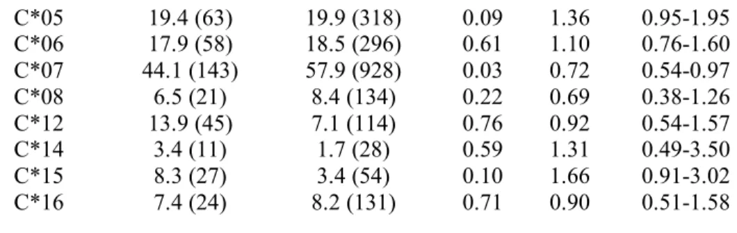
Table 1: Logistic regression with correction for population stratification comparing hemophiliac HESN cases (N=325) and the 1958BC controls (N=1916) for HLA class I alleles
with frequencies greater than 3%. The frequency is displayed in percentage. None of the
variables shown here reach the threshold of significance (p=0.0006). ORadj: Odd Ratio adjusted
for population stratification, CI: Confidence Interval. OR>1 reflect protection against HIV
infection and OR<1 reflect susceptibility to HIV infection.
HLA allele cases (N) controls (N) P ORadj 95% CI A*01 24.3 (79) 34.4 (660) 0.53 0.90 0.66-1.24 A*02 52.3 (170) 49.7 (953) 0.08 1.30 0.97-1.73 A*03 23.1 (75) 26 (499) 0.35 0.85 0.61-1.19 A*11 8.9 (29) 12.5 (239) 0.09 0.66 0.41-1.07 A*23 4.0 (13) 3.0 (57) 0.62 1.20 0.58-2.45 A*24 17.8 (58) 14.9 (286) 0.83 0.96 0.65-1.41 A*25 3.1 (10) 3.5 (67) 0.54 0.78 0.34-1.75 A*26 7.7 (25) 4.9 (93) 0.94 0.98 0.50-1.89 A*29 8.3 (27) 7.9 (152) 0.97 1.01 0.59-1.73 A*30 6.8 (22) 4.2 (81) 0.60 0.83 0.41-1.66 A*31 6.8 (22) 5.3 (102) 0.27 1.36 0.78-2.38 A*32 8.0 (26) 8.0 (153) 0.35 0.76 0.43-1.34 A*68 7.7 (25) 7.7 (147) 0.79 1.07 0.64-1.80 B*07 18.2 (58) 26.4 (497) 0.27 0.82 0.58-1.16 B*08 15.1 (48) 27.2 (511) 0.13 0.75 0.52-1.08 B*13 3.1 (10) 3.4 (64) 0.68 0.84 0.36-1.93 B*14 6.3 (20) 8.0 (151) 0.28 0.72 0.39-1.31 B*15 13.2 (42) 15.6 (293) 0.79 1.06 0.71-1.56 B*18 10.4 (33) 6.8 (128) 0.74 0.91 0.52-1.60 B*27 7.2 (23) 8.4 (159) 0.78 1.07 0.64-1.79 B*35 19.8 (63) 11.4 (214) 0.62 1.11 0.73-1.69 B*37 4.4 (14) 2.9 (55) 0.09 1.85 0.91-3.74 B*38 5.7 (18) 1.6 (31) 0.97 0.98 0.38-2.53 B*39 4.7 (15) 3.6 (67) 0.55 0.79 0.37-1.70 B*40 11.9 (38) 13.2 (249) 0.56 1.13 0.75-1.70 B*44 29.9 (95) 31.1 (586) 0.28 1.19 0.87-1.62 B*50 4.4 (14) 1.8 (34) 0.02 2.43 1.14-5.19 B*51 13.5 (43) 7.9 (148) 0.58 1.14 0.71-1.85 B*55 2.8 (9) 3.8 (71) 0.92 0.96 0.43-2.14 B*57 6.3 (20) 8.6 (162) 0.72 0.90 0.52-1.57 C*01 6.2 (20) 7.5 (120) 0.20 0.66 0.35-1.24 C*02 8.3 (27) 6.7 (107) 0.47 1.23 0.70-2.14
C*05 19.4 (63) 19.9 (318) 0.09 1.36 0.95-1.95 C*06 17.9 (58) 18.5 (296) 0.61 1.10 0.76-1.60 C*07 44.1 (143) 57.9 (928) 0.03 0.72 0.54-0.97 C*08 6.5 (21) 8.4 (134) 0.22 0.69 0.38-1.26 C*12 13.9 (45) 7.1 (114) 0.76 0.92 0.54-1.57 C*14 3.4 (11) 1.7 (28) 0.59 1.31 0.49-3.50 C*15 8.3 (27) 3.4 (54) 0.10 1.66 0.91-3.02 C*16 7.4 (24) 8.2 (131) 0.71 0.90 0.51-1.58
Table 2: Logistic regression with correction for population stratification comparing the HESN cases (N=325) and the 1958BC controls (N=1305) for KIR genes and genotypic
combination of KIR and HLA. The frequency is displayed in percentage. None of the variables
shown here reach the threshold of significance (p=0.0006). Only the functional KIR2DS4 allele
was included. ORadj: Odd Ratio adjusted for population stratification, CI: Confidence Interval,
hmz: homozygous. OR>1 reflect protection against HIV infection and OR<1 reflect
susceptibility to HIV infection.
Test cases (N) controls (N) P ORadj 95% CI KIR2DL1 96.3 (313) 96.9 (1264) 0.48 1.37 0.57-3.27 KIR2DL2 51.7 (168) 50.1 (654) 0.89 1.02 0.76-1.37 KIR2DL3 88.6 (288) 91.7 (1197) 0.97 1.01 0.59-1.72 KIR2DL5 53.2 (173) 46.2 (603) 0.27 1.18 0.88-1.59 KIR2DP1 96.3 (313) 96.9 (1264) 0.48 1.37 0.57-3.27 KIR2DS1 42.2 (137) 36.3 (474) 0.09 1.30 0.96-1.76 KIR2DS2 52.6 (171) 50.6 (660) 0.78 1.04 0.78-1.40 KIR2DS3 31.1 (101) 24.7 (322) 0.17 1.26 0.91-1.76 KIR2DS4 35.4 (115) 39.1 (511) 0.84 0.97 0.71-1.32 KIR2DS5 33.2 (108) 29.7 (388) 0.14 1.27 0.93-1.74 KIR3DL1 96.0 (312) 95.6 (1248) 0.49 1.31 0.60-2.85 KIR3DS1 41.8 (136) 36.2 (473) 0.07 1.32 0.98-1.79 KIR3DS1_hmz 4.0 (13) 4.4 (57) 0.49 0.76 0.35-1.66 HLA-A*3/11 30.8 (100) 35.6 (422) 0.19 0.81 0.59-1.11 HLA-A*3/11 + KIR2DS4 10.5 (34) 14.7 ((175) 0.08 0.65 0.41-1.06 HLA*Bw4 66.0 (210) 60.5 (711) 0.38 1.15 0.84-1.57 HLA*Bw4 80I 30.2 (96) 21.9 (258) 0.78 0.95 0.66-1.36
HLA*Bw4 80I + KIR3DS1 11.6 (37) 7.7 (91) 0.90 0.97 0.55-1.69 HLA group C1 81.8 (265) 88.7 (758) 0.13 0.71 0.46-1.10 HLA group C1 hmz + KIR2DL3 hmz 16.4 (53) 20.7 (177) 0.05 0.65 0.43-0.99 HLA group C2 66.0 (214) 56.0 (479) 0.006 1.58 1.14-2.20 HLA group C2 hmz + KIR2DL3 hmz 7.7 (25) 5.6 (48) 0.75 1.10 0.59-2.06 HLA group C2 + KIR2DL1 63.6 (206) 53.9 (461) 0.002 1.68 1.21-2.32 HLA group C2 + KIR2DS1 26.9 (87) 20.0 (171) 0.03 1.51 1.05-2.17