University of Montréal
Studies on the extra-neuronal cholinergic system in HIV-1
infection
Zainab AldbahDepartment of Microbiology, Infectiology and Immunology Faculty of Medicine
Master’s Thesis / Mémoire de maîtrise Thesis submitted for the Master Degree
In microbiologie immunologie January 2017 © Zainab Aldbah, 2017
Résumé
L’acétylcholine (ACh) est un important neurotransmetteur qui est produit dans le système nerveux. Cependant, cette molécule est aussi produite par d’autres cellules non-neuronales du corps humain. Cette dernière est produite en abondance par les lymphocytes T CD4+, qui sont la cible principale du virus de l’immunodéficience humaine (VIH). ACh exerce ses effets sur les cellules par l’intermédiaire de ses récepteurs nicotiniques (n) et muscariniques (m) qui sont exprimés à la fois sur les cellules immunitaires et non immunitaires dans le corps. Il est bien connu que l’ACh a des effets anti inflammatoires sur les cellules immunitaires, et c’est le récepteur nicotinique qui est un joueur indispensable de cet effet. SLURP-1 (Secreted Ly6/uPAR-related Protein-1), est une autre molécule secrétée par les cellules T activées et d’autres cellules. Elle agit comme un ligand allostérique pour le récepteur α7, et module les effets de l'ACh sur les lymphocytes T. Il est peu connu comment ce système cholinergique extra-neuronal (ENCS) est régulé chez les individus infectés par le VIH.
Nos résultats démontrent que le taux d'ACh et de SLURP-1 en circulation ne change pas significativement chez les sujets infectés par le VIH comparé aux témoins sains. Cependant, le niveau de ces médiateurs est plus élevé chez les sujets infectés à long termes non progresseur (LTNP), qui contrôlaient la réplication virale, depuis plus que sept ans, sans aucune thérapie. Il est tentant de spéculer que le niveau élevé de ces deux composantes de l'ENCS peut jouer un rôle dans leur capacité à contrôler la réplication du VIH. Les résultats de cette étude montrent que l’agoniste du récepteur α7 diminue, et que l’antagoniste de ce même récepteur augmente la réplication virale in vitro, dans les cellules activées par le phytohaemagglutinin (PHA). En outre, l'hémicholinium (HC-3), un composé qui inhibe la capacité des cellules à produire ACh en compétition avec leur absorption de choline, augmente la réplication virale. L'expression du récepteur α7 sur les lymphocytes T CD4 + provenant du sang périphérique, mais pas sur les monocytes, était significativement réduite (p <0,01) chez les individus infectés par le VIH, et elle n'a pas été entièrement restaurée par le traitement antirétrovirale (TAR). Tandis que l'expression du récepteur adrénergique β-2 a diminué significativement (p <0,01) sur les monocytes et les lymphocytes T CD4 + chez des individus
infectés par le VIH. Ces cellules répondent à la norépinephrine via ce récepteur et l’ACh secrété.
Dans l'ensemble, les résultats cette étude suggèrent que le VIH provoque une modulation significative des différentes composantes de l'ENCS chez les individus infectés par le virus. Ce système pourrait être manipulé pour réduire la réplication virale et l’inflammation chez ces patients.
Abstract
Acetylcholine (ACh) is an important neurotransmitter produced in the nervous system. However, the molecule is also produced by non-neuronal cells in the body. CD4+ T cells, the main targets of HIV-1, produce it abundantly. ACh exerts its effects on cells via its nicotinic (n) and muscarinic (m) receptors that are expressed on both immune and non-immune cells in the body. ACh is well known to exert anti-inflammatory effects on immune cells. The main receptor that is indispensable for the anti-inflammatory effects of ACh is the α7 nicotinic receptor. Another molecule, secreted by activated T cells and by other cells is SLURP-1 (Secreted Ly6/uPAR-related Protein-1), which acts as an allosteric ligand for α7 and fine tunes the effects of ACh on T cells. Little is known as to how this extra-neuronal cholinergic system (ENCS) is regulated in HIV-infected individuals.
Our results show that the circulating levels of ACh and SLURP-1 do not change significantly in HIV-infected individuals, as compared to the circulating levels in healthy controls. Interestingly, higher levels of these soluble mediators were detected in HIV-infected long-term non-progressors (LTNP) who control the viral replication for more than seven years without any chemotherapy. It is tempting to speculate that the increase in levels of these two soluble mediators of the ENCS present in HIV-infected LTNPs may play a role in their ability to control HIV replication. The results from this study show that an α7 agonist decreased HIV replication, whereas a receptor antagonist increased its replication in vitro in human PHA blasts. Furthermore, hemicholinium (HC-3), a compound that inhibits the ability of the cells to produce ACh, by competing with their uptake of choline, increases the viral replication. The expression of the α7 receptor on peripheral blood CD4+ T cells, but not on monocytes, was significantly reduced (p<0.01) in HIV-infected individuals, and it was not fully restored by antiretroviral therapy (ART). Interestingly, the expression of the β2 adrenergic receptor was decreased significantly (p<0.01) on both monocytes and CD4+ T cells in HIV-infected individuals. These cells respond to norepinephrine via this receptor and secrete ACh.
Overall, the results of this study suggest that HIV causes significant modulation of different components of the ENCS in virus-infected individuals. This system could be manipulated to reduce viral replication and inflammation in these patients.
Table of Contents
Résumé ... i
Abstract ... iii
Table of Contents ... iv
List of tables ... vi
List of figures ... vii
List of Abbreviations ... ix
Acknowledgments ... xiii
1 Introduction & Review of Literature ... 1
1.1 Extra-Neuronal Cholinergic System ... 1
1.1.1 Synthesis of Acetylcholine ... 2
1.1.2 ACh Receptors ... 4
1.1.3 Acetylcholine as a mediator of Anti-Inflammatory Reflex ... 7
1.1.4 The Autonomic Nervous System ... 7
1.1.5 Activation of the Cholinergic Anti-Inflammatory Pathway (CAP) ... 10
1.1.6 Extra-Neuronal Acetylcholine and its biological effects ... 13
1.1.7 ACh-mediated regulation of T cell activation: Role of SLURP-1 ... 18
1.2 Human Immunodeficiency Virus (HIV) ... 21
1.2.1 The virus and the disease ... 21
1.2.2 Structure of an HIV-1 virion ... 22
1.2.3 HIV life cycle ... 23
1.2.4 Stages of HIV infection ... 24
1.2.5 Immunopathogenesis of HIV infection ... 26
1.2.6 Antiretroviral drugs ... 27
1.2.7 Anti-retroviral drugs prolong survival but do not cure HIV infection ... 28
1.2.8 Regulation of CAP in HIV infection ... 29
2 Hypothesis & Objectives ... 31
2.1 Hypothesis ... 31
2.1.1 Rationale ... 31
2.2.1 Specific aims ... 31
3 Materials & Methods ... 32
3.1 Antibodies & reagents ... 32
3.2 Isolation of peripheral blood mononuclear Cells (PBMC) ... 32
3.3 Cell culture ... 33
3.4 Virus preparation ... 33
3.5 In vitro infection of cells with HIV-1 ... 33
3.6 Flow cytometry ... 34
3.7 Study participants ... 34
3.8 Measuring ACh and SLURP-1 concentrations ... 36
3.10 Statistical analysis ... 36
4 Results ... 37
4.1 Levels of ACh in HIV-infected individuals ... 37
4.2 Levels of SLURP-1 in HIV-infected individuals ... 38
4.3 Effects of α7nAChR agonist and antagonist on HIV replication ... 40
4.4 Effect of ACh depletion on HIV replication ... 41
4.5 Expression of α7nAChR on CD4+ T cells ... 43
4.6 Expression of α7nAChR on monocytes ... 46
4.7 Expression of β2 adrenergic receptor on peripheral blood CD4+ T Cells ... 48
4.8 Expression of β2 adrenergic receptor on CD3-CD14+ monocytes ... 50
5 Discussion ... 52
6 Conclusion ... 59
List of tables
Tableau I. Demographic and clinical parameters of the study participants ... 35
List of figures
Figure 1. Chemical structure of Acetylcholine (ACh) ... 1
Figure 2. Synthesis of Acetylcholine in neuronal cells ... 3
Figure 3. The Structure of different nicotinic Acetylcholine receptors ... 5
Figure 4. Structures of different muscarinic Acetylcholine receptors ... 6
Figure 5. Structural anatomy of the Autonomic Nervous System ... 8
Figure 6. Preganglionic and postganglionic nerve fibers and secreted neurotransmitters of the Autonomic Nervous System ... 9
Figure 7. Anatomy of the Inflammatory Reflex or Cholinergic Anti-Inflammatory Pathway (CAP) ... 11
Figure 8. A model of functioning of the Cholinergic Anti-Inflammatory Pathway .... 12
Figure 9. Synthesis and release of acetylcholine in lymphocytes ... 15
Figure 10. Signaling cascades activated by ligand-induced activation of α7nAChR .... 16
Figure 11. Expression of α7nAChR and its variant Dup-α7nAChR in human cells .... 17
Figure 12. Features of palmo-plantar keratosis in a patient suffering from Mal de Meleda (MDM) ... 19
Figure 13. The Structure of a mature HIV-1 virion... 23
Figure 14. Cycle of HIV-1 replication ... 24
Figure 15. Clinical stages of HIV infection ... 25
Figure 16. Concentration of ACh in the circulation of HIV-infected individuals ... 37
Figure 17. Comparison of SLURP-1 concentrations between ART+ and ART- HIV-infected individuals ... 39
Figure 18. Effect of the α7nAChR agonist and antagonist on HIV replication ... 40
Figure 19. Effect of reduction/inhibition of ACh synthesis on HIV replication in PHA blasts………..42
Figure 20. Expression of α7nAChR on CD4+ T cells in HIV-infected individuals ... 45
Figure 21. Expression of α7nAChR on CD3-CD14+ monocytes in HIV-infected individuals.... ... 47
Figure 23. Expression of β2 adrenergic receptor on CD3-CD14+ monocytes in HIV-infected individuals ... 51
List of Abbreviations
3TC: Lamivudine, an antiretroviral medication used to prevent and treat HIV/AIDS ABC: Abacavir
AcCo-A: Acetyl coenzyme A ACh: Acetylcholine
AChE: Acetylcholinesterase
AKT: AK (mouse) strain Thymoma (other name: Protein kinase B/PKB) ANS: Autonomic Nervous System
ART: Antiretroviral therapy
AZT: Azidothymidine (Zidovudine) BuChE: Butyrylcholinesterse Ca++/Ca2+: Calcium ions
CaM Kinase-II: Ca2+/calmodulin-dependent protein kinase II CAP: Cholinergic anti-inflammatory pathway
cART: Combination anti-retroviral therapy CCR5: C-C chemokine receptor type 5 cDNA: Complementary DNA
ChAT: Choline acetyltranferase
CHRFAM7A: CHRNA7-FAM7A fused gene CHRM1: Cholinergic receptor M1
CHRM5: Cholinergic receptor M5
CHRNA7: Cholinergic receptor nicotinic α-7 (α7) ChT: Choline transporter
CI: Chronically infected CNS: Central nervous system
CREB: cAMP response element-binding protein CTL: CD8+ T lymphocytes
CXCL: chemokine (C-X-C motif) ligand d4T: Stavudine
ddC: Zalcitabine (2′-3′-dideoxycytidine), also called dideoxycytidine, a nucleoside analog reverse transcriptase inhibitor (NRTI) sold under the trade name Hivid
ddI: Didanosine, marketed under trade name Videx, used to treat HIV/AIDS ddN: 2’,3’dideoxynucleoside
DMN: Dorsal motor nucleus
Dup-α7: Duplicated α7 (another name for CHRFAM-7α) ENCS: Extraneuronal cholinergic system
ENV: HIV envelope glycoprotein
FAM-7A: Family with sequence similarity-7α
Gag: Group-specific antigen, coding for structural proteins GALT: Gut-associated lymphoid tissue
GI tract: Gastro-intestinal tract GPCR: G-protein coupled receptor GPI: Glycophosphatidylinositol GSK-3: Glycogen synthase kinase-3
HAART: Highly aggressive anti-retroviral therapy HC: Healthy controls
HC-3: Hemicholinium 3
HIV-1: Human immunodeficiency virus type 1 HIV-2: Human immunodeficiency virus type 2 HMGB-1: High mobility group protein B-1
HMOX1: Heme oxygenase-1; also abbreviated as HO-1 HPA: Hypothalamus-pituitary-adrenal axis
IBD: Inflammatory bowel disease IgG1: Immunoglobulin G1
IL: Interleukin
INI: Integrate inhibitors IR: Inflammatory reflex JAK-2: Janus kinase-2 K+: Potassium ion
LAG-3: Lymphocyte-activation gene-3 LPS: Lipopolysaccharide
LTNP: Long-term non progressor
mAChRs: Muscarinic acetylcholine receptor MDM: Mal de Meleda
Na+: Sodium ion
nAChRs: nicotinic acetylcholine receptor ND: Non-detectable
NEF: Negative Regulatory Factor
NF-kB: Nuclear factor kappa-light-chain-enhancer of activated B cells NFE2L-2: Nuclear factor erythroid-derived-2-like-2
NLR: Nod-like receptors
NNTRI: Non-nucleotide reverse transcriptase inhibitors
NOD: nucleotide-binding oligomerization domain-like receptors NOR: Noradrenaline
Nrf2: Another abbreviation for NFE2L-2
NRTI: Nucleotide reverse transcriptase inhibitors NTS: Nucleus tractus solitaries
PBMC: Peripheral blood mononuclear cells PD-1: Programmed cell death-1
pDC: Plasmacytoid dendritic cells PHA: Phytohaemagglutinin PHI: Primary HIV infection PI: Protease inhibitor PKA: Protein kinase A PKC: Protein kinase C
PNS: Parasympathetic nervous system Pol: DNA polymerase
PR: Protease
REV: Regulator of expression of viral proteins RLH: RIG-like helicases
RT: Reverse-transcriptase
RTI: Reverse-transcriptase inhibitor SIV: Simian immunodeficiency virus SLC5A7: Solute carrier family 5 member 7
SLURP-1: Secreted mammalian Ly6/uPAR-related peptide-1 SNS: Sympathetic nervous system
STAT-3: Signal transducer and activator of transcription-3 SU: Surface unit
TAT: Trans-activator protein TH: T helper
TIM-3: T cell immunoglobulin and mucin-domain-3 TLR: Toll-like receptors
TM: Transmembrane
TNF-α: Tumor necrosis factor alpha ULK-4: Unc51-like kinase-4
uPAR: Urokinase type plasminogen activator receptor Vif: Viral infectivity factor
VIH: Virus de l’immunodéficience humaine Vpr: Viral protein R
Acknowledgments
First and foremost, praises and thanks to God, the Almighty, for His showers of blessings throughout my research work to complete the research successfully. I would like to express my deep and sincere gratitude to my academic research supervisor, Dr. Ali Ahmad, for giving me the opportunity to do research and for providing invaluable guidance throughout this research project. He contributed to a rewarding graduate lab experience by giving me intellectual freedom in my work, supporting my attendance at various conferences, engaging me in new ideas, and demanding a high quality of work in all my experiments. His dynamism, vision, sincerity and motivation have deeply inspired me. He has taught me the methodology to carry out and present the research as clearly as possible. It was a great privilege and honor to work and study under his guidance. I am extremely grateful for what he has offered me. I would also like to thank him for his friendship, empathy, and great sense of humor. Additionally, I would like to thank my committee members, Dr. Martin Guimond and Dr. John Stagg for taking time to serve as committee members.
Millions of thanks go to Suzanne Samarani, for her continuous support, and for all our helpful discussions. I also thank her for her excellent, valuable, and patient technical assistance with cell culture, FACS and analyses of results, etc., for believing in my potential, and for the nice moments we have had together. I would also like to thank her for showing so much enthusiasm while working under her supervision. Although she was very busy with her daily tasks, she has always been ready and available to answer my questions.
A special thanks to my family. Words cannot express how grateful I am to my mother for all of her sacrifices that were made on my behalf. Her prayers for me have sustained me thus far. I would like to thank very special persons of my personal life that contributed to making this work possible: my beloved husband, Ali Albiz. Thank you for supporting me for everything, and especially I can’t thank you enough for encouraging me throughout my studies. I also wish to thank my son Abdu Salam and my daughter Rassil for doing their best to understand a mother who had to take time for her studies for such a long time.
Last but certainly not least, thanks to the people and to the government of my country, Libya. I could not have gone through the Master’s program overseas without their financial
support. I would like to express my full appreciation to the staff of the Department of Scholarship, especially Francine Brisebois, the Academic manager for Students in Montreal, for her generous support and her advice, which were available whenever I needed her.
1 Introduction & Review of Literature
The project for my Masters’ thesis was aimed at investigating modulation of the extra neuronal cholinergic system (ENCS) in HIV-1 (hereafter referred to as HIV) infections.
Therefore, a brief introduction of the ENCS and HIV infection is provided.
1.1 Extra-Neuronal Cholinergic System
It is a part of the cholinergic system, which works within and outside the nervous system. The system is based upon the production and release of a neuro-endocrine transmitter called acetylcholine (ACh), a low molecular weight chemical (see Figure 1 for structure of ACh) and the first identified neurotransmitter in the brain (Gando et al 2001, Kawashima et al 2015). The transmitter is needed to conduct nerve impulses in the central and peripheral nervous tissues. The ACh released by the neuronal cholinergic system controls autonomic, cognitive, and motor functions in the body.
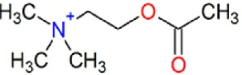
Figure 1. Chemical structure of Acetylcholine (ACh)
Acetylcholine (2-Acetoxy-N,N,N-trimethylethanaminium) is an organic compound that functions as a neurotransmitter, using chemicals released by nerve cells to send signals to other cells.
The extra-neuronal cholinergic system comprises ACh produced by non-neuronal cells and regulates a wide variety of cell functions outside the nervous system. The non-neuronal cholinergic system plays an important role in modulating inflammation and immune response in the body (Kawashima et al 2012). Large varieties of non-neuronal cell types in the body produce ACh and express ACh-specific receptors. ACh acts as a cytotransmitter and acts in both autocrine and paracrine manners. Interestingly, human and mouse CD4+ T cells abundantly produce ACh upon activation (Rinner et al 1998, Rosas-Ballina et al 2011; reviewed in Kawashima & Fujii 2004).
1.1.1 Synthesis of Acetylcholine
ACh is an evolutionarily conserved molecule and its existence predates the development of the nervous system in living organisms (Zoheir et al 2012). Almost every cell in the body of all living organisms (including plants) produces ACh to a variable extent. ACh is synthesized in body cells from choline and acetyl coenzyme A (AcCo-A). The enzyme choline acetyltransferase (ChAT) catalyzes this synthesis (Figure 2). Choline is mainly produced in the liver and is also absorbed by the gut from dietary constituents. Cells uptake choline free and phospholipid (phosphocholine) forms from the circulation, via a high-affinity choline-specific cell surface expressed transporter, Choline Transporter (ChT), which is also known as the solute carrier family 5 member 7 (SLC5A7; Kawashima et al 2004). Hemicholinium-3 (HC-3) is a competitive inhibitor of choline uptake by ChT. HC-3 is used in experiments to deplete ACh in cells and in tissues. Phosphocholine is hydrolysed inside cells before being used for synthesis of ACh. AcCo-A is produced in the cytosol of the mitochondria during the Kreb’s cycle and is transported across the mitochondrial membranes into the cytoplasm where it is used for production of ACh (Tucek 1990).
Figure 2. Synthesis of Acetylcholine in neuronal cells
This figure depicts how acetylcholine is synthesized within neuronal cells from choline and acetyl coenzyme A, and is degraded in the extracellular synaptic cleft by the enzyme Acetycholinesterase. (Accessed on 14 June 2016; Permission to use granted from: http://what-when-how.com/ neuroscience/ neurotransmitters-the-neuron-part-2/).
The bioavailability of choline and AcCo-A limits the synthesis of ACh. ACh, as a negative feedback mechanism, binds and inhibits ChAT. Once synthesized inside cells, ACh is packaged into vesicles. These vesicles are derived from the Golgi apparatus and store ACh. Each vesicle may store up to 10,000 molecules of ACh. The vesicles are stored in the axonal termini of neuronal cells (Tucek 1990). Upon a proper stimulus, ACh is rapidly released from the nerve termini into the synaptic clefts. The released ACh binds specific receptors on post-synaptic neuronal dendrites and propagates the nerve impulse across neuronal junctions. ACh also conducts impulses across neuro-muscular junctions. The release of ACh is rapidly metabolized into choline and acetate by the enzyme acetylcholinesterase (AcChE) in neuronal tissues and by butyrylcholinerase (BuChE) in non-neuronal tissues (Picciotto et al 2012). The
BuChE enzyme is mainly produced in the liver. Choline released from the action of these enzymes is transported back into the cells and is re-used. The activity of both of these enzymes is markedly increased during inflammation and these enzymes have often been used as markers for chronic inflammatory conditions such as Alzheimers’ disease, type II diabetes and obesity (Das 2007). The levels of acetylcholine in the circulation are normally decreased in chronic inflammatory diseases. Not surprisingly, reversible AChE inhibitors are used for the treatment of some brain diseases such as Alzheimer’s (Ohta et al 2017). Non-reversible AcChE inhibitors are fatal and are used as insecticides, pesticides and warfare agents (Colovic et al 2013).
1.1.2 ACh Receptors
ACh exerts its biological effects via two groups of receptors: nicotinic (n) and muscarinic (m). These receptors differ from each other with respect to their structures and functions (Kawashima et al 2012, Kruse et al 2014, Hurst et al 2013). The nicotinic acetylcholine
receptors (nAChRs) are ionotropic and act as ligand-gated ion channels and the nAChRs are designed to transmit impulses across synaptic clefts fast (in milliseconds). Upon binding of ACh or nicotine, the receptors cause an influx of calcium ions (Ca++) and sodium ions (Na+) and an efflux of potassium ions (K+), leading to activation of protein kinase C (PKC) and Ca2+/calmodulin-dependent protein kinase II (CaM Kinase-II). In parallel, they also activate Janus kinase II (JAK-2) independent of the Ca++ and Na+ influx. The two pathways induce activation of nuclear factor kappa-light-chain-enhancer of activated B cells (NF-kB) (Chernyavsky et al 2010). The nicotinic receptors in vertebrates could be of neuronal or of muscular type. The neuronal types are built from nine α (α2-10) and three β (β2-4) subunits (Figure 3). They could be homo- or hetero-oligomers. For example, α7 nicotinic AChR is made from five α7 subunits, and α4β2 is a hetero-pentamer made from three α4 and two β2 subunits (Gotti & Clementi 2004). The nAChRs are present in the central and peripheral nervous tissues and transmit motor signals from the presynaptic to the postsynaptic cells within autonomic nervous system. The muscle type nAChRs in adults are made from α1, β1, δ, and ε subunits in a 2:1:1:1 ratio. They are located in the neuromuscular junctions and cause contractions of skeletal muscles. Several drugs such as curare, hexamethonium, and α-bungarotoxin specifically block these receptors. Unlike nicotinic receptors, mAChRs are metabotropic and associate with
G-proteins, thus are G protein-coupled receptors (GPCR). The mAChRs mediate many of the effects of acetylcholine in the central and peripheral nervous systems.
Figure 3. The Structure of different nicotinic Acetylcholine receptors
(A) Schematic representation of a neuronal nAChR subunit in the plasma membrane. Each nAChR subunit comprises four transmembrane domains (designated as M1-M4) and extracellular amino- and carboxy-termini with M3-M4 intracellular loops of variable length. (B) Assembly of five subunits forming a functional receptor. (C) Functional homomeric receptors comprised of five α7, α9, or α10 (not shown) subunits. (D) Most high affinity nAChRs are heteromeric and are made up of a combination of α and β subunits and usually show high affinity for ACh. Heteromeric receptors may comprise more than two types of sub-units, e.g. the α4α6β3β2 receptor shown here. The red triangles represent ACh binding sites (Permission to use granted by Hendrickson et al 2013).
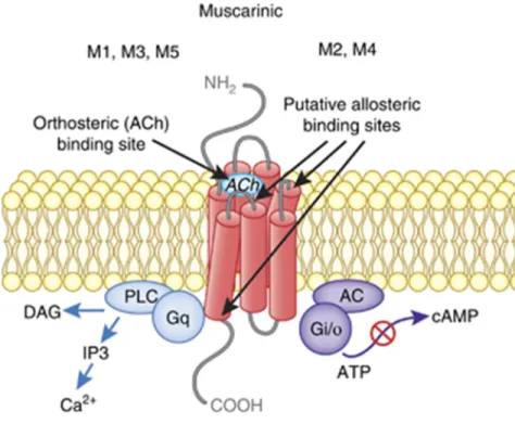
Muscarinic acetylcholine receptors (mAChRs) are mainly located in the sympathetic nervous system and at the neuromuscular junctions of cardiac and smooth muscles. Five different mAChR, named M1-M5, have been described. Although these receptors show significant sequence homologies, they differ in their preferences for associations with different G-proteins. M1, M3, and M5 associate with Gq/11, whereas M2 and M4 associate with Gi/o
proteins (Kawashima et al 2012, Kruse et al 2014). Atropine and scopolamine are used for blocking these receptors (Lochner & Thompson 2016). Several small chemically defined molecules have been discovered that act as specific agonists or antagonists for different n- or m-type AChRs. They have been used in a variety of human diseases such as Alzheimer’s, Sjogren’s syndrome, cancer, and psychosocial disorders, wherein they work by improving neurotransmission and by reducing inflammation (Mudo et al 2007, Greig et al 2013).
Figure 4. Structures of different muscarinic Acetylcholine receptors
This figure shows structures of M1-M5 muscarinic acetylcholine receptors, and their associated G-proteins. These receptors mediate the many actions of ACh in the central nervous system, as well as throughout non-nervous tissues. (Permission to use granted by Nature Publishing Group, Jones et al 2011).
1.1.3 Acetylcholine as a mediator of Anti-Inflammatory Reflex
The immune and the neuro-endocrine systems cross talk with each other and maintain homeostasis via the neuro-endocrine-immune axis. Several neural circuits function reflexively and achieve this homeostasis (Rosas-Ballina & Tracey 2009, Anderson et al 2012, Olofsson et al 2012). The best understood of these circuits is the anti-inflammatory reflex. It is also called the cholinergic anti-inflammatory pathway (CAP), named so as ACh plays the role of an essential effector molecule in the function of this pathway. The pathway works reflexively in response to increased concentrations of pro-inflammatory mediators (Interleukin (IL)-1β, Tumor Necrosis Factor (TNF)-α) in the circulation. The reflex works as a component of the autonomic nervous system.
1.1.4 The Autonomic Nervous System
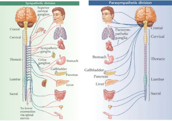
The CAP functions as a component of the Autonomic Nervous System (ANS), which maintains internal homeostasis of the body by regulating involuntary activities such as cardiovascular, urogenital, gastrointestinal, thermoregulatory and exocrine, systems (Vinik et al 2011, Schwartz & De Ferrari 2011, Vinik 2012). The ANS comprises two distinct functional and anatomical components: Sympathetic Nervous System (SNS) and Parasympathetic Nervous Systems (PNS; Figure 5). The two systems function synchronously, as well as independently from each other. The activities of the two systems often, but not always, oppose and cross regulate one another.
The SNS becomes active during fear, stress, and life threatening situations resulting in an increased heart rate, a rise in blood pressure, sweating, bronchodilation and piloerection. The PNS opposes these actions and tends to maintain the “rest and digest” functions. For example, it slows heart rate, increases peristaltic movements and gastric secretions, induces broncho-constriction and causes relaxation of the urinary bladder and GI tract sphincters.
Figure 5. Structural anatomy of the Autonomic Nervous System
This figure shows the sympathetic and parasympathetic components of the autonomic nervous system. The Sympathetic Nervous System is a short-acting system that originates from the thoracic and lumber spinal cord. It redirects blood flow to improve performance. The Parasympathetic Nervous System stems from the cranial nerves and the sacral spinal cord. It is responsible for maintaining the stability of normal body functions and handling energy acquisition and storage. (Permission to use granted by Bethopedia, Wikidot Inc. 2016)
The pre-ganglionic nerve fibers of the SNS originate from thoracic and lumbar regions of the spinal cord (Figure 5). They travel to para- and pre-vertebral ganglia, a collection of neurons outside the CNS (Elenkov et al 2000, Schwartz & De Ferrari 2011, Vinik 2011). They have relatively smaller pre-ganglionic fibers and longer post-ganglionic fibers, which supply the organs (Figure 6). They mainly secrete norepinephrine (NOR) and/or dopamine, with the exception of sweat glands and adrenal medulla, where they secrete ACh.
The PNS preganglionic fibers originate from cranial nuclei in the brain stem and in the sacral regions of the spinal cord (Figure 5). They are relatively long and travel within cranial and spinal nerves to the ganglia. The ganglia of the PNS are located close to or within the innervated organs. As a result, postganglionic fibers are relatively short compared with those of
the SNS (Figure 6; Vinik et al 2011, Olofsson et al 2012, Vinik 2012). The pre-ganglionic fibers of both the PNS and SNS secrete ACh at their termini, while the post-ganglionic fibers of the PNS and SNS secrete ACh and NOR respectively. Based upon the secreted neuro-transmitter, the nerve fibers are also called cholinergic (ACh) or adrenergic (NOR).
The main nerve of PNS is the vagus nerve, also known as the 10th cranial nerve (Figure 5). It supplies parasympathetic innervation to visceral organs present in the neck, thorax, and abdomen. The organs responding to the vagus nerve include the larynx, lungs, heart, esophagus, stomach, small intestine, colon, pancreas, gall bladder and blood vessels. The nerve provides structural and functional bases of the CAP (Van Der Zanden et al 2009, Pavlov et al 2012). For this reason, the words parasympathetic and vagal activities are used interchangeably.
Figure 6. Preganglionic and postganglionic nerve fibers and secreted neurotransmitters of the Autonomic Nervous System
This figure illustrates various secreted neurotransmitters in the parasympathetic, sympathetic, and adrenal pathways. Pre and Pst refer to preganglionic and postganglionic fibers, whereas E and NE refer to epinepharine and norepinepharine respectively. (Permission to use granted by Nature Publishing Group, Tracey 2002).
1.1.5 Activation of the Cholinergic Anti-Inflammatory Pathway (CAP)
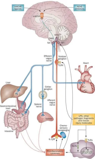
CAP reflex occurs in response to increased concentrations of pro-inflammatory mediators in the circulation (Huston 2012, Pavlov & Tracey 2015). These mediators include IL-1β, TNF-α, IL-6, High mobility group protein B (HMGB)-1, prostaglandins and microbial products (e.g. lipopolysaccharide (LPS)). The sensory (afferent) nerve fibers of the vagus nerve arise from neurons whose cell bodies lie in the nodose and jugular ganglia. The neurons are bipolar with one projection ending in the targeted organs and the other in the Nucleus Tractus Solitarius (NTS) in the brain stem (Valentin et al 2012; Figure 7). The sensory vagal fibers express receptors for proinflammatory cytokines, different metabolites and microbial products. Furthermore, vagal nerves endings in organs and tissues lie in close proximity to dendritic cells (DCs) and macrophages. When these cells become activated, they secrete proinflammatory cytokines and stimulate sensory fibers of the vagus nerve (Goehler et al 1999; Olofsson et al 2012). The sensory vagal nerves transmit signals to the nuclei present in the brain stem (e.g. the NTS), which is connected to the Dorsal Motor Nucleus (DMN) of the vagus. The motor vagal fibers originate from the DMN of the vagus. They innervate end organs and project to the hypothalamus and other nuclei in the CNS nuclei, and regulate activity of the Hypothalamus-Pituitary-Adrenal (HPA) axis. Thus, the activation of sensory vagal fibers results in activation of the vagal motor nerves as well as of the HPA axis reflexively.
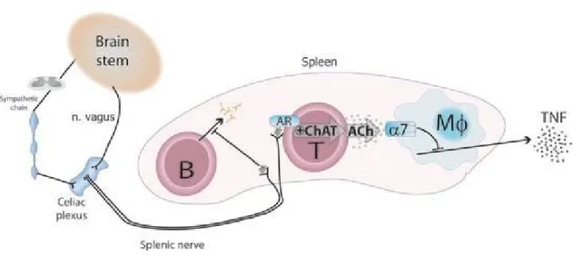
Activation of the HPA results in the release of glucocorticoids from the adrenal glands. The activation of vagal motor fibers activates CAP, which prevents the production of proinflammatory cytokines from macrophages and DCs. The mechanism of action of the CAP was discovered by in 2000 by Tracey’ group (Borovikova et al 2000). Studies have demonstrated that vagal stimulation attenuates systemic proinflammatory response to LPS in mice. In this work, the authors proposed the existence of an inflammatory reflex (IR), also known as CAP (Tracey 2002). Studies have revealed an essential role played by the spleen in the functioning of the inflammatory reflex, as vagal stimulation fails to inhibit the production of TNF-α in splenectomized mice (Huston et al 2006). This posed a conundrum, as the spleen is not directly innervated by any vagal motor nerve fibers. Rather, the spleen is innervated by the noradrenergic splenic nerve, which arises from the celiac plexus (Figure 8). Previous works showed that the activation of the vagus activates noradrenergic neurons present in the celiac ganglion and
Figure 7. Anatomy of the Inflammatory Reflex or Cholinergic Anti-Inflammatory Pathway (CAP)
This figure illustrates the neural and humoral pathways in which cells release cytokines that modulate the immune system. Immunomodulation of the central nervous system is achieved by the cholinergic anti-inflammatory pathway, HPA axis, and sympathetic nervous system. Activation of the sensory vagal fibres results in activation of the vagal motor nerves, as well as of the HPA axis. (Permission to use granted by Nature Publishing Group, Pavlov and Tracey 2012).
induces the release of NOR in the spleen (Rosas-Ballina et al 2011). In addition, the release of NOR in the spleen activates a population of β2 adrenergic receptor-expressing CD4+CD25-CD44 high and CD62L low T cells. These receptors lie in close proximity to
splenic nerve endings in the T cell-rich zones of the organ (Rosas-Ballina et al 2011). Upon activation by NOR, CD4+ T cells synthesize ACh, which inhibits the production of TNF-α and other proinflammatory mediators from neighboring monocytes, macrophages, B cells and DCs. The cells responding to ACh express a nicotinic AChR called α-7. These findings were supported by another group (Vida et al 2011). The ACh-producing CD4+CD25- T cells represent a population of regulatory T cells (Tregs) that are different from classical Tregs. Unlike ChAT+ T cells, classical Tregs express FoxP3 and CD25 (Vida et al 2011, Peña et al 2011). The ChAT+ CD4+ CD25- T cells, that function as effectors for CAP, also express high levels of choline transporter (ChT)-1 encoded by the solute carrier family 5 member 7 (SLC5A7) gene (Kawashima & Fujii, 2004). However, they do not store ACh. During vagal stimulation, ACh is released from postganglionic nerve endings (Figure 5 and 6), which are located within or very close to the innervated organs, e.g., intestines. The released ACh acts on neighboring immune and non-immune cells to regulate their functions.
Figure 8. A model of functioning of the Cholinergic Anti-Inflammatory Pathway
The model of the cholinergic anti-inflammatory pathway, proposed by the Tracey’s group, postulates that sensory or afferent signals from the brain stem travel through the vagus nerve to different nuclei in brain. The efferent vagus reflexively activates the splenic nerve arising in the celiac plexus and innervates the spleen. Splenic nerve endings release nor-adrenaline, which activates closely lying choline acetyltransferase (ChAT)-positive T and B cells. Upon activation, these cells produce and release ACh. Released ACh signals macrophages and other immune cells through the α7 nicotinic acetylcholine receptor and suppresses the production of TNF-α and other proinflammatory cytokines (Permission to use granted by PubMed Central, Olofsson et al 2012).
The model of CAP proposed by Tracey’s group (Pavlov & Tracey 2015) has been challenged, as no synaptic connections could be found between postganglionic parasympathetic (vagal) nerve fibers and preganglionic sympathetic neurons that project to spleen (Martelli et al 2016). A newly proposed model suggests that the efferent arm of the anti-inflammatory reflex travels in greater splanchnic sympathetic nerves, and not via vagal efferent nerves. The postganglionic sympathetic nerve fibers release NOR that inhibits the production of proinflammatory cytokines from macrophages via β2 adrenergic receptors. The released NOR also induces ACh from T cells. Furthermore, a direct stimulation of the vagus also has an anti-inflammatory effect. It does so by mobilizing ACh-producing T cells from intestinal lymphoid tissues. The mobilized ACh-producing T cells migrate to the spleen and probably to other lymphoid tissues to reduce inflammation. In this model, ACh released from T cells can also stimulate the sympathetic nerve terminals via the α7AChR (Martelli et al 2016).
1.1.6 Extra-Neuronal Acetylcholine and its biological effects
As mentioned above, ACh is an evolutionarily conserved molecule that is also produced by non-neuronal cells in the body. Morris was the first researcher to report the synthesis of ACh in the human placenta (Morris 1966). Since then, several studies have reported the production or the presence of ACh in various cell types, including vascular endothelial cells (Parnavelas et al 1985), keratinocytes (Zia et al 2000), epithelial cells (Nguyen et al 2000), T lymphocytes (Fujii et al 1996) and B cells (Arredondo et al 2009). There is overwhelming evidence suggesting that extraneuronal ACh is a ubiquitous cell signalling molecule that plays an important role in the homeostasis of a variety of cell functions, such as proliferation, differentiation, cell-to-cell contact, secretion and absorption in non-neuronal cells (Grando et al 2003).
The extraneuronal cholinergic system plays an important role in the regulation of immune responses, both innate and adaptive. ACh is produced by several types of immune cells including T cells, DCs, and B cells (Fujii & Kawashima 2001, Zdanowski et al 2015). However, the role of ACh has been investigated more extensively in lymphocytes, especially in CD4+ T cells. CD4+ T cells express all components of the cholinergic nervous system,
including ChAT, ACh, as well as various types of cholinergic receptors. The ACh synthesized in lymphocytes is not stored inside the cell but rather is continuously secreted via a special transporter called mediatophore (Fujii et al 2012; Figure 9). ACh acts as an immune modulator and regulates immune cell functions independently of the neuronal cholinergic system. It can function both in an autocrine and a paracrine fashion.
The biological effects of ACh depend on the expression of AChR on target cells. As mentioned above, there exist muscarinic and nicotinic AChR. Several studies have demonstrated the existence of both muscarinic and nicotinic type AChR on human and murine immune cells, including lymphocytes (Costa et al 1995, Fujino et al 1997 and Sato et al 1999).
Sato et al (1999) demonstrated the expression of mRNAs encoding α2, α5, and α7 subunits in human mononuclear cells, while there was no expression of α1, β1, and ε (the skeletal muscle type subunits) in these cells. On the other hand, the expression of α1 subunits was reported in human thymocytes (Wakkach et al 1999). Non-neuronal immune cells also express different types of muscarinic receptors. The stimulation of these receptors induces cell proliferation and the production of proinflammatory cytokines like IL-6 (Wessler & Kirkpatrick 2008). The production of proinflammatory cytokines and antigen-specific immunoglobulin G1 (IgG1) is markedly reduced in mice lacking the Cholinergic Receptor M-1 (CHRM1) and CHRM5 genes, which encode receptors M1 and M5 respectively (Fujii et al 2007b).
Figure 9. Synthesis and release of acetylcholine in lymphocytes
Lymphocytes express the enzyme ChAT, which catalyzes synthesis ACh from Acetyl-CoA and choline. Acetyl-CoA is produced from glucose and fatty acids through glycolysis and beta-oxidation respectively. Choline comes from different sources. It can be taken up by cells through ChT1 or is synthesized de novo inside cells. T cells, unlike neurons, do not store ACh in vesicles. It is continuously released from the cells upon their activation through mediatophore by as yet unknown mechanism. Black and blue arrows indicate known and unknown processes respectively. (Permission to use granted by PubMed Central, Olofsson et al 2012).
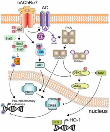
The most significant of the nicotinic acetylcholine receptors studied with regard to the immune system is the α7 nicotinic acetylcholine receptor (α7nAChR). It is a homo-oligomeric receptor made up of five α7 subunits. In addition to T cells, the receptor is also expressed by monocytes, macrophages, DCs, B cells, mast cells, endothelial cells, epithelial cells and keratinocytes. When this receptor is stimulated, it causes an influx of Ca++ and an efflux of K+ from the cells. The Ca++ influx leads to activation of CaM Kinase II and PKC. Recent studies have shown that the receptor activation also leads to metabotropic effects. This involves activation of JAK-2 and signal transducer and activator of transcription-3 (STAT-3); the inhibition of cAMP response element-binding protein (CREB) phosphorylation and glycogen synthase kinase-3 (GSK-3); and the nuclear translocation of nuclear factor (erythroid-derived 2)-like 2 (Nrf-2) induced by AK (mouse) strain thymoma (AKT) activation (Figure 10; Kalkman & Feuerbach 2016).
Figure 10. Signaling cascades activated by ligand-induced activation of α7nAChR
This image describes the anti-inflammatory signaling pathways activated by the α7nAChR. Upon binding ACh, the receptor activates Jak2, which leads to the inhibition of NF-kB and GSK3, and to the activation of CREB. (Permission to use granted by http://creativecommons.org/licenses/by/4.0/; originally published in Kalkman & Feuerbach 2016)
The biological effects of α7nAChR depend on the cell type: they are anti-inflammatory on B cells, macrophages, and mast cells; proinflammatory on lymphocytes and epithelial cells; and variable (pro- or antiinflammatory) for monocytes and neutrophils (Hallquist et al 2000, Aicher et al 2003; Arredondo et al 2009, Zdanowski et al 2015). The α7nACh receptor is required to mediate cytokine suppressive effects of the CAP and vagal stimulation. KO mice for the CHRNA7 gene, which encodes α7nAChR, produce abundant amounts of proinflammatory cytokines and IgG1 antibodies (Fujii et al 2007a). These effects are opposite to those observed
in CHRM1 and CHRM5 gene KO mice (Fujii et al 2007b). Another distinct CHRNA7-related gene, CHRFAM7A, has been described in humans and is located on the same chromosome (15q13.3) about 1.6 megabase upstream to CHRNA7 (Riley et al 2002; Costantini et al 2015a). The expression of the two genes is regulated independent from each other. The CHRFAM7A gene comprises exons 5-10 of the CHRNA7 gene with exons A-E from another partially duplicated gene, ULK-4 (Unc51-like kinase-4). The CHRFAM7A gene encodes the α7 subunit (called duplicated or du-α7) that does not bind ACh or nicotine. It acts as a negative regulator of α7. The dup-α7 is expressed relatively more (compared to α7) on non-neuronal cells. The two genes are expressed in different ratios in human leukocytes. The α-7 and dup-α7 subunits may combine in different ratios to form novel nicotinic receptors, which may dampen ACh-mediated anti-inflammatory effects (Costantini et al 2015b; Figure 11). Interestingly, an increase in the ratio between the expressions of CHRFAM7A and CHRNA7 genes occurs in the colons of patients with Inflammatory Bowel Disease (IBD; Baird et al 2016).
Figure 11. Expression of α7nAChR and its variant Dup-α7nAChR in human cells
A partially duplicated CHRNA7 gene has been discovered in humans. It is named CHRFAM7A and encodes duplicated (dup)-α7. It lacks ACh binding site. The α7 and dup-α7 can each form homopentameric receptors, which are responsive and unresponsive to ACh, respectively. They are shown as A and B in this Figure, respectively. The two subunits can also combine in different proportions to form AChR (C) with reduced responsiveness to ACh. The incorporation of dup-α7 units into the receptor decreases its responsiveness to ACh. The relative expression of CHRNA7 and CHRFAM7 in immune cells can regulate their responses to ACh and hence inflammation. (Permission to use granted by Copyright 2015, The Feinstein Institute for Medical Research; originally published in Costantini et al 2015b).
1.1.7 ACh-mediated regulation of T cell activation: Role of SLURP-1
The release of ACh upon T cell activation is an important aspect of the extra-neuronal cholinergic system. This activation may be polyclonal, T cell receptor (TCR)-mediated, or via another molecule such as CD44 (Kawashima & Fujii 2004, Zdanowski et al 2015). The activation of different T cell lines, as well as of peripheral blood mononuclear cells (PBMCs) with phytohaemagglutinin (PHA), results in the release of ACh from these cells and its detection in the blood (Fujii et al 1998, Fujii & Kawashima 2001). This circulating ACh not only inhibits the production of proinflammatory cytokines, it also modulates proliferation and differentiation of cells. Differentiation depends on the target cell’s ACh-specific expression profile. Upon activation, T cells increase the expression of the α7nAChR, ChAT, ACh, and AChE. In addition, activated T cells also release SLURP-1 (Kawashima et al 2012). SLURP-1 stands for the secreted mammalian Ly6/uPAR (the urokinase type plasminogen activator receptor)-related peptide-1. The SLURP1 gene is located on the long arm (q) of chromosome 8 at position 24.3.
SLURP-1 belongs to the Ly-6/uPAR superfamily of proteins. The family members play diverse roles in signaling, cell proliferation, differentiation and adhesion. There are sixty members of the family, including Ly6/uPAR and CD59. The members may be GPI-anchored to the plasma membrane or secreted. SLURP-1 has no GPI anchor and is secreted. It was discovered in 1999 in patients with the Mal de Meleda (MDM) disease, exhibiting a loss of function mutation (Adermann et al 1999). MDM is a genetic disease resulting from autosomal recessive mutations in the SLURP-1 gene. MDM patients show transgressive palmo-planter keratosis with erythematous borders, perioral erythema, brachydactyly, and nail abnormalities (Tjiu et al 2011; Figure 12). The keratosis in the disease may progress to dorsal surfaces of the hands and feet. The mutations deregulate epidermal cell homeostasis and leads to their enhanced proliferation and differentiation. The prevalence of the disease is 1 case per 100,000 in the population. It is so named, as it was first found and described in patients living in the Adriatic Island of Meleda (Neumann 1898; Perez & Khachemoune 2016).
SLURP-1 acts as a positive allosteric ligand for α7nAChR in T cells, keratinocytes and DCs (Chimenti et al 2003, Fuji et al 2014). SLURP-1 potentiates the effects of ACh on T
lymphocytes and other cells such as keratinocytes, epithelial cells and DC. During T cell activation, ACh and SLURP-1 binding to the α7nAChR fine-tunes T cell activation. Mutations arising in the gene encoding SLURP-1 have been shown to impair T cell activation (Tjiu et al 2011). Recombinant SLURP-1 affects the cholinergic pathway in three ways; first, it attenuates peripheral blood mononuclear cell proliferation; secondly, it increases ChAT gene expression in MOLT-3 cells, and lastly, it increases the synthesis and release of ACh by T cells. All these effects are abolished by the α7nAChR antagonists.
Figure 12. Features of palmo-plantar keratosis in a patient suffering from Mal de Meleda (MDM)
MDM patients show palmo-planter keratosis with erythematous borders, perioral erythema, brachydactyly and nail abnormalities. Similar lesions occur on the feet of the patients. (Permission to use granted by SciELO Brasil; originally published in Morais e Silva et al 2011).
SLURP-1 mRNA is expressed in the thymus and spleen (Moriwaki et al 2007). In addition, peripheral blood mononuclear leukocytes, DCs and macrophages have also been shown to express mRNAs encoding SLURP1. Barnes et al (2014) have shown that tonsillar CD205+ DCs also express SLURP-1 and are surrounded by CD4+ T cells, as well as other immune cells. The presence of SLURP-1 is essential for normal T cell activation. Other cell types that express SLURP-1 include keratinocytes, fibroblasts, neurons, and epithelial cells. Finally, SLURP-1 can be detected in saliva, tears, urine, and blood.
A SLURP-1 related molecule, SLURP-2, was discovered and found to be expressed in a tissue-specific manner. Unlike SLURP-1, SLURP-2 binds non-α7 (e.g. α3) nicotinic ACh receptors. It is expressed in keratinocytes, epithelial cells and immune cells (Moriwaki et al 2007). Mutation of the SLURP-2 gene in mice also causes plamo-plantar keratosis. SLURP-1 and -2 play a role in both keratinocyte turnover and wound healing in the skin and mucosae (Kong et al 2012). Interestingly, SLURP-2 is overexpressed in psoriasis (Tsuji et al 2003).
1.2 Human Immunodeficiency Virus (HIV)
1.2.1 The virus and the disease
The Human Immunodeficiency Virus type-1 (HIV-1) is a complex retrovirus, which is the causative agent of AIDS (Acquired Immune Deficiency Syndrome). HIV-1 caused an unprecedented global pandemic in the late 20th century. It is estimated that about 36.7 million people are infected by HIV-1 worldwide; with only 11 million people having access to anti-HIV medicines. In 2015, around 1.1 million individuals died from AIDS-associated illnesses, and about 2.1 million people became newly infected worldwide (World Health Organization 2016). These statistics show that the infection is still a global health problem.
The AIDS disease gained importance in the 1980s, when men in Los Angeles presented with multiple bacterial and fungal infections, as well as Kaposi’s sarcoma. The disease was officially named “AIDS” in 1981 by the US Centre for Disease Control and Prevention. The retrovirus responsible for the disease was first isolated at the Pasteur Institute in France in 1983. At first, it was known by several names, including Lymphadenopathy Associated Virus (LAV) and Human T Lymphotropic Virus (HTLV)-III, before formally being renamed by the International Committee on Taxonomy of Viruses as the Human Immunodeficiency Virus (Sabin & Lundgren 2013). It is believed that HIV-1 originated from the Simian Immunodeficiency Virus (SIV) in chimpanzees due to increased contact between humans and non-human primates, as chimpanzees are illegally hunted in Africa for “bush meat”. Some researchers have claimed that the oral Polio vaccine, which was grown in chimpanzee cells infected with SIV, may have resulted in the transmission to humans and the eventual development of HIV (Sabin & Lundgren 2013). Two types of HIV have been identified: HIV-1 and HIV-2. HIV-1 is more virulent and is responsible for the global pandemic. HIV-1 is composed of three subgroups: M, N and O; with M being the most prevalent. HIV-2 is not as virulent or as widespread as HIV-1 and the infection is confined to West Africa. HIV-2 is more similar to the Simian Immunodeficiency Virus (SIV).
1.2.2 Structure of an HIV-1 virion
HIV is a retrovirus of about 100 nm in diameter. The structure of a typical mature HIV virion is shown in Figure 13. The viral envelope is a lipid bilayer derived from the cell membrane during viral budding. The viral envelope proteins are studded in the envelope. Each envelope protein comprises a surface unit (SU) or glycoprotein (gp)-120, and a transmembrane part TM), gp-41. The SU is attached non-covalently to the TM. The gp120/41 complex is found as trimmers on the surface of the virion. Beneath the envelope lies the viral matrix comprising of the matrix (MA) protein, p17. The viral capsid comprises the capsid protein, p24. The viral nucleocapsid contains two copies of the single stranded viral RNA, viral reverse transcriptase (RT), integrase (IN), and protease (PR). In addition to the major structural proteins, group-specific antigen (gag), polymerase (pol), and envelope (env), the HIV-1 genome encodes two regulatory proteins, Regulator of expression of viral proteins (Rev) and Transactivator (Tat); and four accessory proteins, which include the Viral infectivity factor (Vif), Negative factor (Nef), Viral protein R (Vpr), and Viral protein U (Vpu). The proteins play a diverse role in ensuring the infection of non-dividing host cells, efficient replication, the budding of virions, and evasion from the host’s antiviral immune factors (Li et al 2005). The non-immune cellular factors such as the apolipoprotein B mRNA editing enzyme/catalytic polypeptide-like (APOBEC)-3G, SAM- and HD domain-containing protein (SAMHD)-1, and tetherin, inhibit HIV replication by different mechanisms. The virus has developed strategies to overcome and to evade the host’s antiviral activities (Malim & Bieniasz, 2012)
Figure 13. The Structure of a mature HIV-1 virion
The proteins making up the HIV-1 virion include the viral envelope studded with surface proteins, glycoprotein-120, and transmembrane gp-41; while the viral capsid houses the capsid protein p24, two copies of single stranded viral RNA, viral reverse transcriptase, integrase and protease. (Permission to use granted by National Institutes of Health; obtained from https://commons.wikimedia.org/wiki/File:HI-Virion-en.png).
1.2.3 HIV life cycle
CD4+ T cells are the main target of HIV infection. As the first step of infection, the viral envelope protein gp120 binds to the viral receptor, CD4 molecule, on target cells (Figure 14). This induces a conformational change in the receptor, exposing the fusogenic gp41 regions that bind the co-receptors, C-X-C chemokine receptor type 4 (CXCR4) for T cell (T) tropic or C-C chemokine receptor type 5 (CCR5) for macrophage (M) tropic HIV strains. After binding and penetration, the RNA genome is converted into DNA by the RT contained within the virus. The viral complementary DNA (cDNA) strand is then inserted by the viral integrase into the host genome. Upon cell activation, more viral proteins and viral RNA copies are made, which then assemble to form new virions. The newly formed virions hijack the multi-vesicular body-forming machinery for budding off the infected cells (Gomez & Hope 2005).
Figure 14. Cycle of HIV-1 replication
This figure illustrates the steps in the life cycle of HIV infection. The viral envelope protein gp120 binds to the CD4 receptor molecule and penetrates the target CD4+ cells. Viral RNA is then reverse transcribed into DNA by the RT contained within the virus, which is then integrated into the host genome. By hijacking the host’s replication system, the virus is able to replicate uncontrollably and infect the host. (Permission to use granted from Taylor & Francis online, originally published in Saayman et al 2015).
1.2.4 Stages of HIV infection
HIV infection could be arbitrarily divided into three different clinical stages (Figure 15) and are described below:
i) Acute or primary infection: This stage usually lasts six months after the initial infection. Patients exhibit mild flu-like symptoms. However, during this time, massive replication of the virus occurs, followed by dissemination in the host. With the induction of HIV-specific CD8+ T lymphocytes (CTL), the viral load decreases (An & Winkler 2010).
ii) Chronic infection: This stage can last for several years, typically 4-7 years. Normally, infected individuals show no clinical signs. However, during this phase, there is massive replication of the virus accompanied by destruction and regeneration of the CD4+ T cells. The virus-specific immune response (antibodies and CTL) keeps the virus under control. The viral load in this early stage, called the viral load set-point, determines the prognosis of the infection.
iii) AIDS: This is the final stage in which host’s immune response is no longer able to control viral replication. The host becomes immunodeficient; CD4+ T lymphocyte counts fall below 200 per μl of blood and the host becomes infected with opportunistic infections, develops neurocognitive defects, and AIDS-related cancer such as Kaposi’s sarcoma. If infection is not treated, death occurs after a variable period of illness.
The median time to develop AIDS in untreated patients from initial infection is 8-10 years. However, some infected individuals do not develop AIDS. It is worth noting that a small fraction (5-10%) of HIV-infected individuals are able to control HIV replication for more than seven years without receiving anti-retroviral treatments. These individuals are called slow progressors. Some of them may control the infection and show no signs of disease for more than 15 years. They are called elite controllers. Several factors, including infection with mutant viruses, strong immune responses, and/or anti-viral host genetic factors may underlie their ability to control the infection (Blankson 2010).
Figure 15. Clinical stages of HIV infection
The HIV infection is divided into three stages (primary/acute, chronic, and AIDS). The division is based upon the duration of the infection, CD4+ T cell counts, viral load in the blood and clinical symptoms. The chronic infection, when the infection is asymptomatic, is also called the period of clinical latency. (Permission to use granted by PubMed Central, originally published in An & Winkler 2010).
1.2.5 Immunopathogenesis of HIV infection
HIV induces a strong immune response in the host. Both HIV-specific antibodies and T cell (CD4+ and CD8+) responses can be readily demonstrated in infected individuals. However, the high mutation rate of the virus enables it to evade the host’s immune responses. Furthermore, depletion of CD4+ T cells deprives the generation of virus-specific antibodies and CTLs. As a result, T cells become weak and exhausted. They express a variety of co-inhibitory molecules such as programmed cell death protein 1, (PD-1), T-cell immunoglobulin and mucin-domain containing-3 (TIM-3), lymphocyte-activation gene 3 (LAG-3) and CD160 (Mohan et al 2014).
HIV-infected individuals exhibit chronic and aberrant immune activation of all essential cellular components of the innate and adaptive immune systems including T cells, B cells, Natural Killer (NK) cells, plasmacytoid dendritic cells (pDC) and the complement system (Imran et al 2016). Increased expression of CD38, a marker of T cell activation, is considered a predictor for a bad prognosis in HIV-infected individuals. Furthermore, infected patients also show signs of chronic inflammation. The systemic inflammatory response results largely from a defect in the intestinal barrier. CD4+ T helper 17 (TH17) cells residing in the gut are major targets of the virus because they express relatively high levels of CCR5 on their surface. The massive depletion of these cells in the gut occurs during all stages of the infection (Brenchley et al 2004; Brenchley & Douek 2012). These cells secrete IL-17 and IL-22, which are important for maintaining the intestinal barrier function. The loss of these cells explains defective intestinal integrity in HIV-infected individuals. HIV viral proteins also contribute to disrupt mucosal tight junctions by inducing proinflammatory cytokines from intestinal epithelial cells. The defective intestinal barriers lead to the translocation of bacterial products and fragments into the blood. This microbial translocation causes immune activation and inflammatory responses in the infected individuals. Further, viral nucleic acids and proteins also activate the immune system and induce inflammation by activating a variety of Pattern Recognition Receptors (PRRs) of which Toll-like Receptors (TLRs), RIG-like helicases (RLH), nucleotide-binding oligomerization domain (NOD)-like receptors (NLRs), and inflammasomes are the most important. The virus has also been shown to kill infected CD4+ T cells via pyroptosis; a type of cell death accompanied by the production of proinflammatory cytokines such as IL-1β
and IL-18. The virus-infected individuals show increased levels of proinflammatory cytokines, chemokines and pro-coagulation mediators in the blood such as IL-6, IL-18, IL-7, chemokine (C-X-C motif) ligand (CXCL)-10/IP-10, D-dimer and sCD14 (Catalfamo et al 2012). Paradoxically, immune over-activation and chronic inflammation further weaken the immune system, and due to its high rate of replication and mutability, the virus is capable to evade the immune system. This results in immunodeficiency and patients become highly susceptible to opportunistic infections, cancers and neurocognitive defects. The infection, if untreated, invariably results in death.
1.2.6 Antiretroviral drugs
Tremendous efforts have been made in developing very effective antiretroviral drugs (De Clerc 2009, Cao et al 2015). The drugs belong to the following classes:
Nucleoside/Nucleotide RT Inhibitors (NRTI): This was the first class of drugs developed against HIV. They inhibit the viral RT, interacting with the catalytic site of the enzyme. This class includes zidovudine (AZT), zalcitabine (ddC), didanosine (ddI), lamivudine (3TC), stavudine (d4T), abacavir (ABC), and tenofivir (a nucleotide RTI). All are 2’,3’dideoxynucleoside (ddN) analogues and compete with normal deoxynucleoside triphosphates inside cells. The drugs are incorporated during reverse transcription but terminate the reaction. AZT, in combination with 3TC, has been shown to effectively inhibit vertical transmission of the virus from infected mothers to their newborn babies (McIntyre et al 2009).
Non-Nucleoside RT Inhibitors (NNRTI): These drugs interact with an allosteric site located near the catalytic site of the RT. This category of drugs includes nevirapine, etravirine, delavirdine, and efavirenz.
Viral Protease Inhibitors: These drugs inhibit viral proteases and thus prevent the processing of precursor viral proteins into mature ones. This category of drugs includes ritonavir, indinavir, lopinavir, nelfinavir, saquinavir, amprenavir, atazanavir, and darunavir.
Integrase Inhibitors (INI): These drugs prevent the integration of the virus into the genome. This category of drugs includes Raltegravir and Elvitegravir.
Viral entry inhibitors: These inhibitors interfere with the entry of the virus into susceptible cells. Maraviroc, a CCR5 antagonist, is only effective against M-tropic viruses. An anti-CXCR4 (against T-tropic viruses), AMD3100, could not be used due to toxicity issues.
Fusion Inhibitors: The drug enfuvirtide is a polypeptide homologous to the heptad repeat region of gp41 and prevents the fusion of the viral lipid bilayer with that of susceptible cells. Consequently, the virus cannot enter and infect cells.
1.2.7 Anti-retroviral drugs prolong survival but do not cure HIV infection
The use of different anti-retroviral drugs in combination, often referred to as the highly aggressive anti-retroviral therapy (HAART) or simply as combination anti-retroviral therapy (cART), has saved the lives of millions of HIV-infected individuals. Today, if treated, patients no longer die from AIDS or from any AIDS-related illness. However, cART is not a cure. The treatment does not eliminate the virus and the virus can persist in a latent form in these individuals in immunologically and pharmacologically privileged sites called viral sanctuaries. Such sites include the gut, brain, testes, B cell follicles in lymph nodes, lungs and cornea. The residual viral loads at these sites constitute what are generally referred to as the viral reservoirs (Kimata et al 2016). These reservoirs are the main hurdle in curing HIV-infected individuals. The main cell type infected in these reservoirs is the long-lived memory CD4+ T cells. As a result, HIV-infected patients have to take cART to suppress viral replication and to reduce the level of proinflammatory mediators in the circulation, thereby partially restoring immune responses. However, the levels of the pro-inflammatory mediators rarely return to physiological levels and remain above normal values. Furthermore, the virus found in these reservoirs continues to undergo replication at very low undetectable levels. This low-level of viral replication and the continuous use of anti-retroviral drugs contribute to the chronic low levels of inflammation and immune activation found in these patients. Although HIV patients do not die from AIDS, they become more susceptible to AIDS-unrelated co-morbidities including enhanced aging, cardiovascular diseases, metabolic syndrome, liver and kidney diseases, osteopenia/osteoporosis, neurocognitive disease and cancers. Researchers are investigating strategies to deplete viral reservoirs. It has been found that an early initiation of anti-retroviral therapy after infection significantly reduces the size of the viral reservoirs. At the same time, avariety of anti-inflammatory drugs (e.g. statins and aspirin) can be used to reduce the adverse consequences of chronic inflammation in HIV-infected patients (Bandera et al 2016).
1.2.8 Regulation of CAP in HIV infection
Little is known about how CAP is regulated in HIV-infected individuals. The HIV envelope protein, gp120, was shown to bear significant sequence homology with snake curare-mimetic neurotoxins. Gp120 has also been shown to bind the nAChR α7 subunit and to increase the expression of α7nAChR. However, the protein does not reduce production of pro-inflammatory cytokines from macrophages and microglia (Neri et al 1990, Ballester et al 2012, Delgado-Vélez et al 2015). In the neurons obtained from post-mortem brains of HIV-infected individuals with HIV-associated neurocognitive defects (HAND), the expression of α7 was upregulated but dup-α7 was downregulated. In vitro, gp120 mimicked these changes in neuron receptor expression (Ramos et al 2016). Gp120 was also shown to induce increased production of mucus from bronchial epithelial cells, which express CXCR4. The signalling pathway implicated in mucus secretion comprised activation of the CXCR4-α7-nAChR-γ aminobutyric acid (GABA) receptor-the epidermal growth factor receptor (EGFR) cascade (Gundavarapu et al 2013). In vitro, as well as in vivo, ACh mimetics, AcChE inhibitors (Galantamine and Pyridostigmine), and α7 agonists inhibit PMA and ionomycin-induced T cell activation, proliferation and production of proinflammatory cytokines (Valdés-Ferrer et al 2009; Pohanka et al 2011).
In summary, chronic inflammation accompanying HIV infection is the main cause of AIDS immunopathogenesis and later, CD4+ T cell depletion. It causes aberrant immune activation. In cART-treated individuals, low grade inflammation persists even after successful suppression of viral replication (Ahmad & Rinaldo 2017). Unfortunately, cART does not cure HIV infection. As a result, HIV-infected individuals must take antiretroviral drugs for life. Once cART is stopped, latent virus residing inside the reservoirs start replicating within days. Lifetime use of cART not only puts a lot of burden on healthcare resources, it is also toxic for patients. The drugs, especially PIs and NRTIs, exert toxic side-effects (Hester 2012). The
upregulation of body’s natural homeostatic anti-inflammatory pathways could constitute an alternative strategy to reduce inflammation and immune activation. In this regard, we need to learn about the function of the extra-neuronal cholinergic system in these patients. This new knowledge may help identifying new molecular targets to reduce inflammation and strengthen the immune response in HIV-infected patients. Based on our actual understanding of the extra-neuronal cholinergic system, we hypothesize that the extra-extra-neuronal anti-inflammatory cholinergic pathway becomes hypo/non-functional in HIV-infected individuals. The main objective of this thesis was to understand how the extra-neuronal anti-inflammatory cholinergic pathway is affected during HIV infection and contributes to chronic inflammation and immune activation.
2 Hypothesis & Objectives
2.1 Hypothesis
We hypothesize that HIV infection dysregulates the extraneuronal cholinergic system, which promotes chronic inflammation and immune activation. New knowledge about this dysregulation may be exploited to attenuate inflammation and reduce immune activation in HIV-infected patients.
2.1.1 Rationale
It has been well documented that patients infected with HIV have chronic inflammation and show aberrant immune activation. Activated immune cells, especially CD4+ T cells, produce ACh and SLURP-1, which act in autocrine and paracrine manners to fine tune T cell activation and prevent over activation (Kawashima et al 2012, Fuji et al 2014). Furthermore, ACh and SLURP-1 may also attenuate inflammatory responses by signalling T cells and other immune cells. As HIV replication is more efficient in activated T cells, it is possible that a dysregulation of the cholinergic system may further promote HIV replication. This provides a rationale for investigating the regulation of the ENCS during HIV infection. Furthermore, we would like to investigate whether this system could be exploited to reduce HIV replication and benefit HIV-infected patients.
2.2 Main objective
The main objective of the study was to investigate how the ENCS is regulated in HIV-infected individuals and understand whether this system could be manipulated to reduce HIV replication and immune activation in HIV-infected individuals.
2.2.1 Specific aims
• To examine the production of ACh and soluble SLURP-1 in HIV-infected individuals • To investigate the effects of different cholinergic components on HIV replication
• To investigate the expression of α7nAChR and β2 adrenergic receptors on peripheral blood monocytes and CD4+ T cells of HIV-infected individuals