Université de Montréal
Structural and Biochemical Characterization of the Organomercurial Lyase MerB Par
Haytham Mohamed Gamaleldin Wahba Abdelgawwad
Département de Biochimie Faculté de Médecine
Thèse présentée à la Faculté de Médecine en vue de l’obtention du grade de PhD en biochimie
Université de Montréal Faculté des études supérieures
Cette thése intitulée:
Structural and Biochemical Characterization of Organomercuriallyase MerB
Présentée par:
Haytham Mohamed Gamaleldin Wahba Abdelgawwad
a été évaluée par un jury compose des personnes suivantes:
Résumé
Le mercure est présent dans l'environnement à cause de phénomènes naturels (volcans) ou des activités humaines (combustion de combustibles fossiles). Le mercure existe sous forme de mercure élémentaire (Hg0), ionique (HgII) ou organique tel le méthylmercure (MeHg). Ces diverses formes sont en flux constant les uns avec les autres dans le cycle biogéochimique naturel. De par leur grande hydrophobicité et leur capacité à pénétrer les membranes biologiques, les composés organomercuriels contituent la forme la plus toxique de mercure retrouvée dans l’environnement Des niveaux élevés de MeHg ont d’ailleurs été détectés dans la chaire de poissons de nombreuses régions du monde. Conséquemment, une consommation de produits de la mer contaminés représente un grave danger pour la santé humaine.
Certaines bactéries isolées à partir d'environnements contaminés par le mercure ont évolué vers un système qui leur permet de convertir efficacement les composés mercuriels présents autant sous forme ionique qu’organique en un mercure élémentaire moins toxique. Cette résistance au mercure s’explique par l'acquisition d'un élément génétique connu sous le nom d’opéron mer. L’opéron mer code entre autre pour deux enzymes importants : la lyase organomercurielle MerB et la réductase mercurielle MerA. MerA catalyse la réduction du HgII conduisant à la formation du mercure élémentaire Hg0 qui est un composé volatile et moins toxique. MerB, quant à elle, catalyse la protonolyse de la liaison carbone-mercure de composés organomercuriels pour produire un composé réduit de carbone et du mercure ionique (HgII). Au
vu des effets des organomercuriels et de la réduction de HgII, MerA et MerB sont considérés comme des enzymes clés pouvant servir à la biorestauration des cours d'eau contaminés par les organomercuriels. Une compréhension claire des détails mécanistiques de la façon dont MerA et MerB fonctionnent ensemble au niveau atomique est donc cruciale dans la mise en œuvre de biotechnologies implicant l’opéron mer dans les efforts de bioremédiation.
Dans cette étude, nous avons utilisé la résonance magnétique nucléaire (RMN)et la cristallographie aux rayons X pour caractériser la structure et le mécanisme enzymatique de MerB de E. coli. Sur la base d’études structurales précédentes de MerB de E. coli, trois résidus (Cys96, Asp99 et Cys159) ont été identifiés comme constituant la triade catalytique nécessaire au clivage de la liaison carbone-Hg. En guise de suivi aux études antérieures, mon projet consiste
d’abord à utiliser la cristallographie aux rayons X afin de définir les rôles de Cys96, Asp99 et Cys159 dans la liaison du substrat et dans le clivage.
Deux approches ont été mises en œuvre pour atteindre cet objectif. Tout d'abord, les mutants MerB ont été testés pour définir le rôle des résidus catalytiques. Deuxièmement, les inhibiteurs de MerB et d'autres substrats non organicomercuriels potentiels ont été utilisés pour explorer le site actif de MerB.
Une sérine se retrouve à la position de Asp99 dans quatre variants de MerB répertoriés chez les bactéries. Pour mieux comprendre le rôle de Asp99, nous avons comparé la sérine présente dans le variants MerB de Bacillus megaterium (MerB2) et introduit un variant D99S à la protéine MerB du type sauvage d’E. coli (MerB D99S). Nous avons pu constater que la forme purifiée de MerB D99S se caractérisait par une couleur rose après avoir visualisé sa structure cristalline aux rayons X, révélant la présence d'un métal lié au niveau de son site actif. Les analyses par spectrométrie de masse à plasma à couplage inductif (ICP-MS) et par fluorescence des rayons X indiquèrent que MerB D99S se liait au cuivre au niveau du site actif. En outre, les analyses par résonance paramagnétique électronique (EPR) et des études de RMN ont identifié la forme CuII du cuivre. L'addition de substrats organomercuriels a pu déplacer le CuII entrainant ainsi une diminution de l’activité catalytique de MerB D99S. En revanche, MerB2 n'a pu être co-purifié avec le cuivre, bien que la structure aux rayons X du complexe MerB2-Hg soit pratiquement identique à la structure du complexe MerB D99S-Hg. Ceci suggère que le résidu Asp99 est essentiel au clivage des liaisons carbone-Hg de composés organiques du mercure et dirige la spécificité de la liaison au métal. De plus, la liaison cuivre-MerB D99S propose un lien possible entre l'évolution de MerB et son homologue structural, la protéine NosL.
Dans la seconde approche, nous nous sommes intéressés au site actif de MerB en testant sa liaison à des composés organostanniques et à des composés organoplombiques avec un inhibiteur de MerB connu sous le nom de triéthylétain (TET) qui se lie au résidu Asp99 sans s’associer aux cystéines du site actif. Une liaison similaire a été observée avec un autre inhibiteur à savoir le triméthylplomb (TML). Quant au diméthylétain (DMT), il inhibe MerB à l'aide d'un mécanisme
PbIV qui se lient au site actif de manière similaire à HgII. DMT, DET et DEL présentent une affinité pour la liaison à MerB supérieure à celle de son substrat initial MeHg. Ces résultats suggèrent que les composés organomercuriels ne sont pas les seuls substrats pour MerB et Asp99 est le premier résidu à se lier aux composés organométalliques suivis de la liaison à Cys96 et Cys159.
Ces observations suggèrent un agrandissement de l’éventail d'applications possibles pour MerB dans la bioremédiation de certains sites contaminés par des composés organométalliques tels les organoplombiques et organostanniques.
Mot-clé: Organomercuriallyase, Merb, Organoplombiques. Organostanniques, Protéine de liaison cuivre, Carbone liaison métallique clivage, Méthylmercure, Organomercuriels, Biorestauration, Résonance magnétique nucléaire, La cristallographie aux rayons X.
Abstract
Mercury is introduced into the environment from either natural occurrences (volcanoes) or from human activities (combustion of fossil fuels). Mercury exists as elemental mercury (Hg0), ionic mercury (HgII) or organic mercury like methylmercury (MeHg) and these forms are in constant flux with each other as part of the natural biogeochemical cycle. Organomercurial compounds like MeHg are the most toxic form because of their hydrophobicity and their ability to efficiently permeate membranes and bioaccumulate in organisms. High levels of MeHg have been found in fish in many areas around the world, and therefore human consumption of contaminated seafood represents a serious danger for human health. Bacteria isolated from mercury-contaminated environments have evolved a system that allows them to efficiently convert both ionic and organic mercury compounds to the less toxic elemental mercury. The mercury resistance is due to the acquisition of a transferable genetic element known as the mer operon. The mer operon encodes for several proteins including two enzymes, the organomercurial lyase MerB and the mercuric ion reductase MerA. MerB catalyzes the protonolysis of the carbon-mercury bond of organomercurial compounds to produce a reduced-carbon compound and inorganic ionic mercury HgII. MerA catalyzes the reduction of HgII to elemental mercury Hg0, which is volatile and less toxic. Due to their ability to cleave MeHg and reduce the resulting HgII product, MerB and MerA are considered crucial to bioremediation efforts to clean up MeHg from contaminated waterways. A clear understanding of the mechanistic details of how MerB and MerA function together at the atomic level is crucial for appropriate utilization of the mer system in bioremediation efforts. We have been using nuclear magnetic resonance (NMR) spectroscopy and X-ray crystallography to structurally and mechanistically characterize E. coli MerB. Based on previous structural studies of E. coli MerB, three residues (Cys96, Asp99 and Cys159) have been identified as a catalytic triad which is required for carbon-Hg bond cleavage. As a follow up to the earlier studies, my project involves using X-ray crystallography to define the roles of Cys96, Asp99 and Cys159 in substrate binding and cleavage.
acid is replaced by a serine. To understand the role of Asp99, we compared a serine-containing MerB variant (Bacillus megaterium MerB2) and an E. coli MerB mutant (MerB D99S) to wild type E. coli MerB. Interestingly, the purified MerB D99S protein was found to contain a pink color. X-ray crystal structure indicated the presence of a bound metal in the active site of MerB D99S. Analysis by inductively coupled plasma mass spectrometry (ICP-MS) and X-ray fluorescence indicated that MerB D99S binds copper in the active site. Further, electron paramagnetic resonance (EPR) and NMR studies identified the copper as CuII. Addition of
organomercurial substrate displaces bound CuII but MerB D99S shows diminished catalytic activity. In contrast, MerB2 did not co-purify with copper although the X-ray structure of MerB2-Hg complex is virtually identical to the structure of the MerB D99S-Hg. This suggests that the aspartic acid residue is crucial for the cleavage of carbon-Hg bonds of organomercurials as well as metal-binding specificity. Furthermore, the binding of copper to the MerB D99S protein suggests a possible evolutionary link between MerB and its structural homolog, the copper-binding protein NosL. In the second approach, we probed the active site of MerB through testing its binding to organotin and organolead compounds. The known MerB inhibitor triethyltin (TET) binds to Asp99 without binding to any of the active site cysteines. A similar binding has been observed with trimethylead (TML). Dimethyltin (DMT) inhibits MerB using an alternative mechanism. It first binds to Asp99 then Cys96, which induces a dramatic change in the active site by disrupting a cation-π interaction between Try95 and Arg155. In contrast, diethyltin (DET) and diethylead (DEL) were found to be substrates for MerB, where both ethyl groups were cleaved and the SnIV and PbIV products bound to the active site in a similar manner
to HgII. DMT, DET and DEL show higher binding affinity to MerB than its initial substrate
MeHg. These results suggest that organomercurials may not be the only substrates for MerB and Asp99 is the first residue to bind to organometals followed by subsequent binding to Cys96 and Cys159. In addition, these observations suggest that there are other possible applications for employing MerB in bioremediation of organolead and organotin contaminated sites while other organometals may have implications when using MerB in bioremediation systems.
Keyword: Organomercuriallyase, MerB, Organolead. Organotin, Copper binding protein, Carbon metal bond cleavage, Methylmercury, Organomercuriels, Bioremédiation, Nuclear magnetic resonance, X ray crystallography.
Table des matiéres
RÉSUMÉ ………. i
ABSTRACT……….. iv
Table des matiéres………. vi
Liste des figures……… viii
Liste des abréviations……… ix
Remerciements……….. xii
Introduction………... 1
1.1 Different forms of mercury and their toxicity ………….………...…... 1
1.1.1 Metallic Mercury……….…… 1
1.1.2 Mercurous and Mercuric Mercury……….. 2
1.1.3 Organomercurial compounds……….. 3
1.2 Occurrences and sources of mercury in environment………....……... 4
1.2.1 Natural sources………..…….. 6
1.2.2 Human activities (Anthropogenic sources) ………..……….. 8
1.2.3 The chemistry of MeHg and organomercurials... 10
1.3 Remediation of mercury contaminated sites………..……... 11
1.3.1 Physical or chemical remediation………..………. 11
1.3.2 Biological remediation (Bioremediation) ………..………. 12
1.3.3 Phytoremediation………..……… 14
1.4 The Mer system………..………… 17
1.4.1 Regulation………..……….. 20
1.4.2 Transport………..……….... 21
1.4.3 Detoxification enzymes………..………... 23
1.4.3.1 MerA………..………... 23
1.4.3.2 MerB………..………... 24
1.4.3.2.6 Proposed catalytic mechanism of MerB based on X-ray structure... 38
1.4.3.2.7 MerB is structurally similar to NosL …..………... 40
1.4.3.2.8 Direct transfer of HgII product from MerB to MerA…... 43
1.5 Organotin compounds are substrates and inhibitors of MerB…... 44
1.6 Organolead compounds…... 47
1.7 Overall rational of the thesis…... 50
1.8 Experimental procedures used for studying metal ion binding to protein... 53
1.8.1 X-Ray crystallography... 53
1.8.2 X-ray Fluorescence spectroscopy... 54
1.8.3 NMR and EPR spectroscopy studies with paramagnetic metals... 54
1.8.3.1 Paramagnetic metals alter NMR specta in a characteristic manner... 55
1.8.3.2 EPR spectroscopy for identifying paramagnetic metals... 55
1.8.4 Inductively Coupled Plasma Mass Spectrometry... 56
1.8.5 Isothermal Titration Calorimetry... 56
Article 1…... 59
Article 2…... 104
Discussion ...…... 148
Liste des figures
Figure 1: Ice core record of atmospheric mercury deposited at Wyoming’s Upper Freemont
Glacier over the last 270 years 5
Figure 2: Biogeochemical cycle of mercury species in the environment 7 Figure 3: Structures of important organomercurial compounds 13
Figure 4: Reactions catalyzed by MerB and MerA 14
Figure 5:- Resistance of transgenic plant to methylmercury chloride 16
Figure 6. The mer operon 18
Figure 7:- The proteins of the Mer system 19
Figure 8:- Possible mechanistic routes for cleaving a carbon-Hg bond 27 Figure 9:- Walsh proposal for mechanism of cleaving carbon-Hg bond by MerB through SE2
mechanism 28
Figure 10:- NMR structure of MerB 31
Figure 11:- X-ray crystal structure of MerB and MerB-Hg complex 32 Figure 12. Comparison of the NMR and crystal structures of MerB 33
Figure 13. DTT-affected residues in MerB-Hg-DTT complex 35
Figure 14: The MerB active site 37
Figure 15: The mechanism of carbon-Hg bond cleavage by MerB 40 Figure 16: Folding similarity between core regions of MerB and NosL supports their
evolutionary link 41
Figure 17: Structures of some organotin compounds (RnSnIV) 46
Figure 18: Structures of some organolead compounds (RnPbIV) 48
Figure 19: Proposed mechanisms of carbon-Hg bond cleavage based on computational
studies 149
Figure 20: Schematic representation of proposed mechanisms of carbon-Hg bond cleavage
by MerB based on computational studies 150
Figure 21: Proposed mechanism of carbon-Hg bond cleavage through initial binding to D99 based on results with organolead and organotin compounds 152
Liste des abréviations
B. megaterium Bacillus megaterium B. subtillis Bacillus subtillis Bacillus sp. Bacillus species C. butyricum Clostridium butyricum
C159 Cysteine 159
C96 Cysteine 96
Cu Copper atom
D99 Aspartic acid 99
DEL Diethyl lead
DML Dimethyl lead
DMT Dimethyltin
DPTA Diethylenetriaminepenta-acetic acid
DTT Dithiothreitol
E. coli Escherichia coli
EDTA Ethylenediaminetetraacetic acid EPR Electron paramagnetic resonance
EtHg Ethylmercury
EXAFS Extended X ray absorption fine structure
FAD Flavin adenine dinucleotide
GSH Glutathione Hg Mercury atom Hg0 Elemental mercury HgCl2 Mercuric chloride HgI Mercury I HgII Mercury II
HgO Mercuric oxide
HgS Mercuric sulfide
HSQC Heteronuclear single quantum correlation ICP-MS Inductively coupled plasma mass spectrometry
ITC Isothermal titration calorimetry
LB Luria Bertani broth
MeHg Methylmercury
MerA Organomercurial reductase
merA MerA gene from mer operon
MerB E. coli Organomercurial lyase
merB MerB gene from mer operon
MerB2 Organomercurial lyase 2
MerP Periplasmic mercury transporter from mer operon
merP MerP gene from mer operon
MerR regulator for the mer operon
merR MerR gene from mer operon
MerT Mercury transporter from mer operon
merT MerT gene from mer operon
MR-SAD Molecular replacement - single anomalous dispersion NADPH Nicotinamide adenine dinucleotide phosphate
NMerA N-terminal MerA
NMR Nuclear magnetic resonance
NOS Nitrous oxide reductase gene cluster
NosL L protein from the nitrous oxide reductase (nos) operon
PCMB p-Chloromercuribenzoate
PDB Protein Data Bank
PHMSA p-hydroxymercuric sulphonic acid
PMA Phenylmercuric acetate
RMSD Root mean square deviation
RNAP RNA polymerase
TETA Triethyltin acetate
TEVT Triethylvinyltin
TML Trimethyl lead
TMTF Trimethyltin fluoride
TPT Triphenyltin
TTEL Tetraethyl lead
TTET Tetraethyltin
TTML Tetramethyl
TTMT Tetramethyltin
TVT Tetravinyltin
UNEP United Nation Environmental Program UV-Vis Ultraviolet–visible
Remerciements
First, I would like to express my sincere appreciation and thank to my supervisor Professor James Omichinski. I appreciate his contribution of time and ideas, which made my Ph.D. experience productive. He always has time to join his group frequently in the lab to give advice and share ideas. His priceless guidance helped me in all the time of research and writing of this thesis. I learned a lot from his advice on both research as well as on my career. I would like to thank Professor Omichinski also for his supportive funding in my final 2 years. I have been incredibly fortunate to have a chance to join his group. I would like to thank Professor Jurgen Sygusch for his guidance in X ray data collection strategy and processing and for giving me the opportunity for X ray data collection in National Synchrotron Light Source (NSLS-I). Without his precious support it would not be possible to conduct this research. I would like also to thank Professor Pawel Grochulski at University of Saskatchewan for allowing us to use the Macromolecular X ray crystallography facility at Canadian light source. Many thanks to Professor Pascal Legault and Professor Jacques Archambault; the members of my thesis committee, for their valuable and brilliant comments and insightful questions. Their advice, suggestions and contributions were essential for this study to be accomplished. I would like to thank each member of Omichinski group, specially Mathieu Lussier Price, Julien Lafrance-Vanasse, Laurent Cappadocia and Normand Cyr. They taught me about protein purification and several other biochemical tools during my first days in the lab. They shared their time and expertise unselfishly. The training I have received from them was essential to accomplish my study. I would like to thank also Ahmed Mansour for his contribution to this project during his intern in Omichinski lab. I gratefully acknowledge the funding sources that made my Ph.D. work possible. I was funded by Egyptian Ministry of Higher Education for my first 4 years. Special thanks to all members of Egyptian Bureau of cultural and educational Affairs in Montreal for all the support they provided during my travel and study. I am grateful to my parents and my brothers for all their love and encouragement to pursue my dreams. I have been so blessed to be joined with my wonderful wife who motivated and helped me throughout my PhD studies. Her
Chapter 1: Introduction
1.1 Different forms of mercury and their toxicity
Due to its unique and attractive properties, humans have extensively employed elemental mercury and mercury-containing compounds in industry and innovation for several centuries. One drawback to the extensive application of this metal in the environment is that exposure to relatively low levels of mercury compounds is often accompanied by a potentially high risk of toxicity to either humans or other animal species. In the environment, mercury exists in many different chemical forms and the toxic effects of mercury vary according to the type of mercury present. The four main forms of mercury are Hg0 (elemental or metallic mercury), HgI (mercurous), HgII (mercuric) and
organomercurial compounds. Although all four forms of mercury are toxic to some degree, organomercurial compounds are generally considered to be the most toxic form followed by the three ionic forms of mercury, in the order HgII > HgI > Hg0 (Bernhoft 2012, Syversen & Kaur 2012).
1.1.1 Metallic Mercury
Hg0 is the only heavy metal that is known to exist in the liquid state at room temperature, but it is highly volatile. Given the high reactivity of mercury with cellular components, exposure to Hg0 vapours through direct inhalation has been linked to several adverse effects since Hg0 is the form of mercury most commonly encountered in the environment. Following inhalation, Hg0 quickly enters the blood stream via the lungs and is distributed quickly throughout the entire body since it diffuses readily through cell membranes (Hursh et al. 1976). Acute exposure to high concentrations of Hg0 induces bronchitis leading to dyspnea (Garnier et al. 1981). In contrast, chronic exposure to lower levels of Hg0 vapours is known to produce neurological dysfunction accompanied by tremors and memory loss (Smith et al. 1983). Although Hg0 represents the least toxic form
of mercurial compounds, the risk to humans is the highest due to its common use in a number of industrial applications including gold mining as well as its continual persistence in the atmosphere from natural occurrences.
1.1.2 Mercurous and Mercuric Mercury
Following elemental mercury, ionic mercury salts are the next most common form of mercury compounds found in the environment and there are two oxidation states for ionic mercury salts, which are often referred to as mercurous (HgI) or mercuric (HgII). The most widely used form of HgI is as a chloride salt Hg2Cl2. Hg2Cl2 was used extensively in
numerous pharmaceutical preparations up until the 1950s, and is commonly known by its trade name calomel. Although it is poorly absorbed from the intestine, long-term exposure to calomel can lead to systemic pain and discoloration of the skin. This condition is known as pink disease (acrodynia) and the mortality rate from calomel poisoning in Britain reached 1 in 10 in 1940s. Once its toxic side effects were recognized, the use of calomel in medical preparations was discontinued (Warkany 1966). Similarly, the most commonly employed HgII salt is the chloride salt HgCl
2, which was used extensively as either a
preservative or as an antiseptic in numerous drug preparations. In particular, HgCl2 was the
treatment of choice for syphilis for decades before the discovery of antibiotics. As a medical treatment, HgCl2 was either applied topically to infected areas or ingested as an
oral medication, but its extensive use led to numerous health complications. The primary side effect following acute exposure to high levels of HgCl2 is necrosis of the gut mucosa,
which produces bloody diarrhea and eventually leads to either septic shock or even death in certain circumstances (Barnes et al. 1980). Following chronic exposure to HgCl2, HgII
accumulates in the kidney causing glomerulonephritis, renal tubular necrosis and ultimately renal failure (Taugner et al. 1966). After exposure to HgCl2, HgII is transported through
binding to the sulfhydryl group of glutathione. In a similar manner, HgII also targets the sulfhydryl group present in cysteine residues in cellular proteins and in many cases this can result in either a decrease or loss of cellular functions. For example, HgII accumulation in the renal tubule is the result of its ability to form a complex with the cysteine-rich protein metallothionein. In the case of metallothionein, each molecule of metallothionien has the
1.1.3 Organomercurial compounds
Despite the fact that organomercurials have not been as widely used as other mercury species, they are responsible for a considerable number of human toxicities with methylmercury (MeHg) and ethylmercury (EtHg) being the two most common forms linked directly to human toxicities. Before its toxic side effects were recognized, MeHg was used extensively as a pesticide. However, MeHg is also found naturally in the environment as many microorganisms have evolved specific enzymes that convert HgII into MeHg, including several different bacterial species (Ullrich et al. 2001, Clarkson 2002). Although the exact physiological reason for why these microorganisms convert HgII into MeHg is currently unknown, it serves as a continual source for introducing it into the environment. Due to its lipophilicity, MeHg has the capacity to bioaccumulate within the food chain and through this mechanism represents a constant concern to human health. The most common form of human exposure to MeHg comes from eating seafood (fish and shellfish) containing elevated levels of MeHg. Following the consumption of contaminated food, MeHg is efficiently absorbed from the intestinal tract and distributed to fat tissue throughout the body (Leaner & Mason 2002).
MeHg is the ultimate neurotoxic agent due to its ability to effectively target the neurological system. Unlike other forms of mercury, MeHg efficiently crosses the blood-brain barrier (BBB) and the blood-brain levels of MeHg are 3-6 times higher than circulating blood levels following acute exposure (Aschner & Clarkson 1989). MeHg has also been shown to target other organs, including the liver and kidney as well as being transported through the placenta to the brain of the fetus (Syversen & Kaur 2012). Once absorbed, MeHg has a high tendency to bind sulfhydryl groups present in cysteine residues in proteins in a similar manner to HgII. The resulting cysteine-Hg-Me complex mimics the neutral amino acid methionine and this facilitates MeHg entry into the cell through the large neutral amino acid carrier (Yin et al. 2008). On the other hand, MeHg can be transported from the cell either through complex formation with the sulfhdryl group of GSH or via passive diffusion (Ballatori & Clarkson 1982). Unlike the ionic forms of mercury where symptoms usually disappear when exposure ceases, exposure to MeHg
leads to persistent neurological symptoms and symptoms of neurotoxicity appear rather late after exposure to MeHg. It normally takes several weeks before the symptoms appear, but once they start they propagate very rapidly and the latency period appears to be independent of the level of exposure. The reason and mechanism for this latent period following exposure to MeHg is a challenging mystery that has yet to be answered by the scientific community (Clarkson & Magos 2006).
In addition to MeHg, a number of other organomercurials have been used as antimicrobial agents, but their use is also now limited due to their toxicity. Following MeHg, EtHg ranks second in terms of human exposure to organomercurial compounds, with the most common use of EtHg being as a preservative in vaccine preparations. In humans, the biodistribution of EtHg is very similar to MeHg, but EtHg has a much shorter biological half-life. Unlike MeHg, which mainly affects the central nervous system, exposure to high levels of EtHg causes mainly kidney toxicity (Dorea et al. 2013). In addition to EtHg, other organomercurial compounds have been used as antiseptics and antifungals, including merbromine and phenylmercury. Although these compounds have a significantly lower toxicity profile in comparison to either MeHg or EtHg, the usage of any mercury-containing compound encountered general scepticism from the public due to the stigma associated with other mercury containing compounds.
In general, human exposure to the different forms of mercurial compounds through ingestion, inhalation or skin contact leads to variations in the toxicity profile. These variable toxicity profiles following exposure to mercurial compounds is attributed to their different chemical properties that lead to variations in their absorption, distribution and metabolism in humans.
remaining one third is attributed to natural processes like volcanic eruptions, forest fires and rock weathering. To determine the difference between natural sources and anthropogenic contribution to environmental mercury levels over the last several hundred years, the deposition of mercury in the ice core of a Wyoming glacier was measured in 1991 and 1998. The results showed that naturally released mercury produces a constant background level in the atmosphere independent of human activities. However, with the beginning of the industrial revolution in the nineteenth century, the levels of mercury increased dramatically from human activities as depicted in Figure 1 (Schuster et al. 2002).
Figure 1: Ice core record of atmospheric mercury deposited at Wyoming’s Upper Freemont Glacier over the last 270 years. The ice core covering the period between 1720-1945 was collected in 1991 and it is represented by ◊, and the ice core collected in 1998 by covers the
years between 1945-1993. The green area represents the natural background of mercury deposition, where the preindustrial deposition rates until 1880 can be extrapolated to present time (4 ng/L) to illustrate the increases during the last 100 years (in red) and decreases in the past 20 years. The blue color shows the deposition rates corresponding to natural events like volcanic eruptions. The orange area reflects the elevated levels of mercury that are associated with the gold rush period in the United States (US). The pink area represents the increase in global environmental level of mercury after the beginning of the industrial period around 1880 (anthropogenic events). The figure is adapted from (Shuster et al. 2002).
1.2.1 Natural sources
Different forms of mercury exist naturally in the earth’s crust, atmosphere and oceans. Several natural processes are responsible for mobilizing mercury from the earth’s crust and introducing it to the atmosphere and oceans as mentioned above. The most abundant form of mercury in the earth’s crust is mercuric sulfide (HgS), which is commonly known as cinnabar ore. In nature, mercuric containing rocks are subjected to natural weathering factors that convert the naturally occurring HgII into the more volatile Hg0, which is then readily emitted to the atmosphere. Similarly, volcanic eruptions play an important role in mobilizing mercury from the earth’s crust into the atmosphere as Hg0 (Nriagu & Becker 2003). Following its release into the atmosphere, Hg0 is converted through an uncharacterized oxidative process to inorganic forms such as mercuric oxide (HgO). The resulting inorganic mercuric compounds are then deposited, and they return back through rainwater to the earth’s surface, where they accumulate in aquatic sediments. After deposition of HgII either in marine or fresh water sediments, select microorganisms convert HgII to MeHg through an enzymatically catalyzed biomethylation reaction (Mason & Sheu 2002). Following the biomethylation reaction, MeHg readily bioaccumulates in marine species and in particular fish. In the absence of mercury pollution associated with human activities, only low levels of MeHg will accumulate, but this has changed dramatically due
Figure 2: Biogeochemical cycle of mercury species in the environment. Mercury is introduced into environment as a result of natural (degassing from rock, soil and water surface and volcanic eruptions) and anthropogenic activities (gold mining, fossil fuel combustion). Hg0 is released into
the atmosphere, where it circulates for up to 1 year and becomes widely distributed. Hg0 undergoes
a slow photochemical oxidation, which converts it to inorganic mercury. The resultant inorganic mercury travels back to the earth’s surface in rain, which leads to it being deposited in aquatic systems and soil. The soil-deposited inorganic mercury can be released into the atmosphere as the results of forest fires, whereas aquatic-deposited inorganic mercury is converted to MeHg by select microorganisms. The MeHg is absorbed by plankton, and the plankton are consumed by higher organisms and this leads to bioaccumulation in fish, which represent an important food source for humans. The figure is adapted from University of Wisconsin-Eau Claire (2014) Mercury in the Environment and Water Supply.url: https://people.uwec.edu/piercech/Hg/mercury_water/ cycling.htm. (Last accessed on 30 March 2016)
1.2.2 Human activities (Anthropogenic sources)
Human activities have greatly amplified the rate of mobilization of mercury from the earth’s crust since the beginning of the industrial revolution. Humans have released tremendous amounts of mercury during the second half of nineteenth century when cinnabar ore was heavily mined to be used to prepare Hg0. The Hg0 prepared from the mining of cinnabar ore has been used for gold extraction around the world. Using Hg0 in gold mining can result in a significant release of Hg0 into the atmosphere as was seen following the gold rush period in the United States in the nineteenth century (Figure 1). Similarly, coal and fossil-fuel burning for power generation and oil refining are another major source of anthropogenic emission of mercury into the atmosphere because mercury exists naturally in both coal and fossil-fuels (Mason et al. 1994). There are numerous other sources of mercury that result from human associated activities and many products contain mercury like batteries, fluorescent lamps, thermometers and blood pressure gauges. The improper handling of mercury-contaminated waste from these consumer products represents another important mechanism for introducing mercury species into the environment and mercury emissions to the atmosphere from such human activities was estimated at 1960 tonnes in 2010 (UNEP 2013).
In addition to releasing inorganic mercury species into the atmosphere, human activities are also responsible for the introduction of several forms of synthetically generated organomercurial compounds. Up until the 1960s, the chloro-alkali industry was the main source for contaminating aquatic systems through the direct release of MeHg as waste. This industry used mercury sulfate cells as catalysts to produce caustic soda, and MeHg was produced as a side product; consequently thousands of tons of MeHg were dumped through waste-water into aquatic systems as a by-product. Since MeHg has the capacity to bioaccumulate in the marine food chain, its lethal effect in humans was readily manifested through the consumption of contaminated seafood (Morel et al. 1998). The first
MeHg contaminated fish from Minamata Bay and MeHg toxicity is now commonly referred to as Minamata disease (Dltri & Dltri 1978, Dltri 1991). A second major outbreak of MeHg poisoning occurred in Iraq in 1972 following the consumption of contaminated bread. The contaminated bread was prepared from wheat seeds treated with MeHg as a fungicidal agent that were originally intended for agricultural purposes and not for human consumption. Thousands of people developed symptoms highly similar to toxicity after consumption of MeHg contaminated fish (Bakir et al. 1973, Greenwood 1985).
After the Minamata Bay and Iraq incidents, several governmental regulations came into effect to prevent or at least minimize the risk of toxicity caused by organomercurial compounds. In Japan, a comprehensive mercury-control plan was implemented to reduce mercury production and usage. Mercury extraction from mines was completely stopped by 1974. The chloro-alkali industry developed mercury-free catalysts and mercury use in lamps, batteries and medical equipment were greatly reduced. On the other hand, the use of MeHg as an antifungal agent in agriculture was discontinued globally after the outbreak in Iraq. Although Europe and North America did not experience any significant mercury toxicity outbreaks, several safety measures were implemented to prevent the risk of mercury toxicity. In the USA, fishing is prevented in over three thousand mercury-contaminated lakes (Berlin et al. 2007). In Ontario, Canada, three first nation communities suffered from symptoms of Minamata disease in 1970 after eating local fish contaminated with mercury. Due to the increased concern over the possibility of mercury contamination, the government of Canada launched several projects to monitor and control mercury pollution throughout the country. By 1995, all chloro-alkali factories using mercury cells were closed, but this occurred only after large quantities of MeHg had already been discharged into waterways around cities like Sarnia, Hamilton and Cornwall (Mailman et al. 2006, Saint-Amour et al. 2006, Depew et al. 2013).
Despite considerable efforts to minimize anthropogenic mercury emissions through strict emission standards, there is still a high load of previously emitted mercury in the ecosystem, and it will take decades to diminish mercury levels to safe standards in several
China, are introducing and mobilizing considerable amounts of mercury into the environment, due to the absence of strict emission standards (UNEP 2013). In addition, it appears that levels of MeHg in arctic marine life has been increasing over the last several years, and it has been suggested that this is correlated with global warming and increased melting of the polar ice cap (Schartup et al. 2015). Taken together, mercury still represents an important global threat to human health and current efforts to reduce anthropogenic emission and release are not enough to reduce the risk of toxicity. Thus, remediation efforts for cleaning up existing areas of mercury contamination are required to control mercury pollution and avoid future outbreaks of mercury poisoning.
1.2.3 The chemistry of MeHg and organomercurials
In contrast to many other organometallic compounds, organomercurial compounds are typically stable in aqueous solutions. This stability is not due to the strength of the carbon-Hg (15-20 kcal/mole) since carbon-Cd and carbon-Zn bonds (the other members of the group 12 metals) are stronger and organozinc and organocadmium compounds are unstable in aqueous solutions (Mason & Benoit 2003). Rather, the stability of metal-carbon bonds in an aqueous solution is connected to bond polarity. Carbon-metal bonds are polarised (Mδ+ - C δ-) as a result of the difference in electronegativity between the carbon atom and the metal. For example, carbon-Cd and carbon-Zn bonds are more polarized than carbon-Hg bond, carbon-Pb and carbon-Sn bonds and this is consistent with their relative stability in aqueous solutions. Whereas organocadmium and organozinc compounds are rapidly degraded in an aqueous environment, organomercury, organotin and organlead compounds are often very stable (Mason & Benoit 2003).
In general, organomercurial ions (RHg+) and mercuric ions show similar reactivity patterns, and both have a strong preference for binding to thiols. Given their strong affinity for thiol-containing compounds, organmercurial compounds such as MeHg are generally
carbon-Hg bonds when more than one sulfur ligand is bound to the mercury atom. The binding of a second sulfur ligand increases the polarization of carbon-Hg bond (Hgδ+-Cδ-) and creates a low energy pathway leading to cleavage of the carbon-Hg bond (Mason & Benoit 2003).
1.3 Remediation of mercury contaminated sites
1.3.1 Physical or chemical remediationDue to the toxicities associated with all forms of mercury compounds, many efforts have been undertaken to remediate mercury-contaminated sites by either stabilizing the mercurial compounds in contaminated sites to prevent them from spreading into the environment or by completely removing them from contaminated soil. The current remediation technologies can be classified as being either non-biological or biological, with the two non-biological methods being either chemical or physical remediation (Wang et al. 2004, Wang et al. 2012). Within the chemical remediation methods, the most common approaches involve immobilization or stabilization techniques. In these cases, mercury-contaminated waste or soil is chemically treated with either sulphur-containing ligands or reducing agents to minimize mercury mobility. By forming thiol complexes that are both stable and insoluble, it prevents the mercury from leaching into the surrounding environment. Addition of sulphur-containing ligands such as colloidal sulfur to mercury-contaminated soil or waste precipitates the mercury as HgS, which has decreased solubility and lower volatility (Kot et al. 2007, Liu et al. 2008). In general, the materials employed in immobilization techniques are commercially available and inexpensive. Thus, they are often an attractive option to governmental organizations trying to minimize the potential toxic effects of mercury contamination at the lowest possible cost. However, the mercury is not removed from the contaminated site and this is a major disadvantage of these chemical remediation methods since it does not completely remove the potential risk. In addition, these methods increase the volume of waste and it is important to continually monitor the immobilized mercury in these contaminated sites. To overcome the disadvantage associated with chemical treatments, physical remediation methods have also been developed to
technique (Massacci et al. 2000). In the thermal desorption process, the soil is heated in a closed system to collect volatilized mercury and its compounds. The relative volatilization rates of mercury compounds are as follows: Hg0 ≈ (CH3)2Hg > Hg2Cl2 > HgCl2 > HgS >
HgO. Heating the soil to between 600-800 °C removes all mercury compounds from contaminated soil, which can then be subsequently condensed for safe disposal. Mercury is removed very efficiently using this approach. For example, heating contaminated soil from a chloro-alkali plant in Sweden at 500 °C for 20 min removed 99% of the total mercury content in the soil. However, the danger of using this technique lies in changing the soil properties due to the very high temperatures required. Raising the soil temperature will definitely alter the levels of other soil contents such as bacteria, trace elements and nutrients such as nitrogen, phosphorus and potassium. To counter the adverse effects of these elevated temperatures on the soil, a new approach using lower temperatures for longer periods of time has succeeded in removing mercury and maintaining the overall quality of the soil (Kucharski et al. 2005). Although this method is both safe and efficient, the high cost is a major disadvantage. In addition, it is often very difficult to access the soil under aquatic systems and this represents an important source contributing to the build up of mercury contamination.
1.3.2 Biological remediation (Bioremediation)
In an attempt to avoid using costly and inefficient chemical and physical remediation methods, considerable effort has been devoted to identifying efficient biological methods for remediating mercury-contaminated sites. The discovery of mercury-resistant bacteria isolated from mercury-contaminated sites represented a promising step in developing such bioremediation strategies (Summers 1986, Osborn et al. 1997). Select bacterial strains are resistant to mercury containing-compounds due to the presence of a set of genes known collectively as the mer operon. The genes present in the mer operon encode for a set of
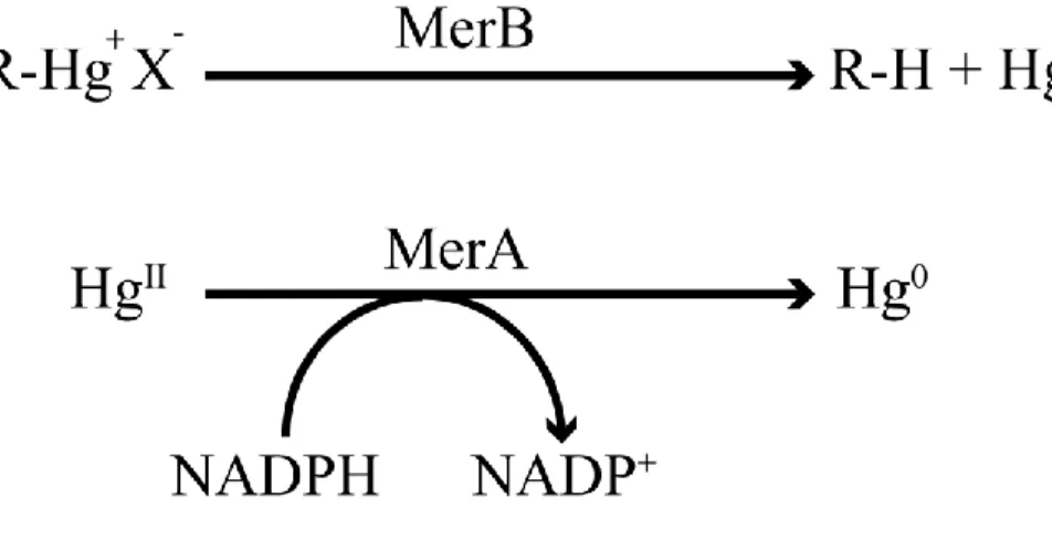
organomercurial compounds (Figure 3) and produces an organic moiety (CH4 in the case
of MeHg) and ionic HgII. The resultant HgII is then transferred directly to MerA, which reduces it to Hg0 (Figure 4) (Ji & Silver 1995). The resultant Hg0 is less toxic than either HgII or R-Hg, and the highly volatile Hg0 can be readily expired by the bacteria. Although the concept of using these mercury-resistant bacteria represents an attractive method for cleaning up mercury-contaminated sites, the major limitation is the small biomass associated with the bacteria. In efforts to use mercury-resistant bacteria on a larger scale, attempts have been made to develop bioreactors (Wagner-Dobler et al. 2000). In these bioreactors, the mercury-resistant bacteria are immobilized on a solid support. This allows mercury-contaminated wastewater to be inoculated in the bioreactor for sufficient time periods to allow the bacteria to detoxify either organomercurial compounds or HgII.
Bioreactors have been used for cleaning mercury-contaminated wastewater, but they are not suitable for remediating mercury-contaminated soil. In an attempt to address this limitation, MerA and MerB encoding genes were introduced into genetically modified plants to enable them to detoxify mercury-contaminated soil.
Figure 3: Structures of important organomercurial compounds. Structure of methylmercury; MeHg (A), Ethylmercury; EtHg (B), p-chloromercuric benzoic acid PCMB (C), p-hydroxymercuric sulphonic acid PHMSA (D) and phenylmercuric acetate PMA (E). X- is the counter ion typically
-Figure 4: Reactions catalyzed by MerB and MerA.
Bacteria isolated from mercury-contaminated sites have been shown to possess the MerA and MerB enzymes. MerB cleaves carbon-Hg bonds in organomercurial compounds to yield a hydrocarbon moiety and HgII products. The resultant HgII is subsequently reduced by MerA enzyme and this
produces the volatile and less toxic Hg0. R represents the alkyl or aryl group bound to the mercury
atom, whereas X represents the counter ion such as chloride.
1.3.3 Phytoremediation
In an attempt to develop a method that uses the two enzymes of the mer system to remediate contaminated soil, several groups have inserted MerA and MerB into different plant species (Meagher 2000, McGrath et al. 2006, Omichinski 2007). This technique is commonly known as phytoremediation and these systems have proven to be very successful in controlled trials. The advantage of phytoremediation systems is that the roots of the plant are able to extract subsurface mercury contamination and transport the mercury compounds to all sections of the plant. Inserting the MerA gene into the Arabidopsis thaliana genome allows for the efficient expression of MerA throughout the plant. The net result is that this significantly increases the plant’s capacity to grow in HgII-containing media in comparison with the wild-type plant. The volatilization of Hg0 from the plant was detected in parallel to
able to grow in organomercurial-containing media, but they showed a decrease in survival relative to plants expressing both MerA and MerB due to the accumulation of HgII (Figure 5) (Bizily et al. 2000, Bizily et al. 2003). Given the success in Arabidopsis thaliana, the MerA and MerB genes have now been successfully introduced into several additional plant species with larger biomass such as yellow poplar trees and tobacco plants (Merkle 2006, Hussein et al. 2007, Ruiz & Daniell 2009, Nagata et al. 2010). By creating a variety of mercury-resistant plant species that can grow in different environments and geographical locations, it will be possible in the future to select the most suitable plant species to fit the conditions present in a mercury-contaminated site. Moreover, employing mercury-resistant plants for cleaning up contaminated areas has several advantages since it presents a low cost and an environmentally friendly/green technology. In addition their large biomass and their ability to penetrate deep into the soil with their roots allows for the hyper-accumulation of mercury from contaminated soil. Thus, phytoremediation presents several advantages over physical and chemical remediation strategies for cleaning up mercury-contaminated areas. To optimize these phytoremediation technologies, there is a crucial need to obtain a comprehensive understanding of the catalytic mechanisms of MerA and MerB as well as how other components of the mer system function.
Figure 5: Resistance of transgenic plant to methylmercury chloride.
Growth of transgenic plants expressing the merB, merA/B or merA gene in comparison with wild-type Arabidopsis thaliana plants. The seeds for the various plants were germinated in growth media with MeHg concentrations of 0, 1, 5, 10 µM in A, B, C and D, respectively. A) In the absence of MeHg, all plants display regular growth behaviour indicating that merA and/or merB-containing plants grow normally. B) In 1 µM MeHg, both the merB and merA/B plants grow near normally whereas the merA and wild-type plants fail to germinate and grow. C) In 5 µM MeHg, the merA/B plants grow more efficiently than the merB plants, whereas both merA and WT seeds fail germinate. D) Although their growth is inhibited, merA/B plants still display resistance to 10 µM MeHg whereas the merB plants could germinate but they did not grow. Adapted from (Bizily et al.
1.4 The Mer system
The unique ability to grow in the presence of toxic concentrations of both inorganic mercury and organomercurial compounds has been observed for a wide range of gram-negative and gram-positive bacteria isolated from mercury-contaminated sites (Miller 1999). This resistance to mercurial compounds by these bacteria is attributed to the presence of a set of genes known as the mer operon, which is present on a transferable genetic element such as a plasmid or transposon. The genes of the mer operon encode for a set of proteins that function to detoxify and eliminate the mercurial compounds from the bacteria. The proteins of Mer system participate together to convert the highly reactive and highly toxic inorganic and organomercurial compounds to the less toxic and volatile Hg0,
which is readily expired by the cell. The gene contents of mer operon vary between the different strains of mercury-resistant bacteria, but the most common genes loaded on mer operons include merR which encodes for the transcriptional regulatory protein MerR, merP which encodes for the periplasmic HgII transporter protein MerP, merT which encodes for the membrane HgII transporter protein MerT, merA which encodes for the ionic mercuric reductase MerA and merB which encodes for the organomercurial lyase MerB (Figure 6). The proteins expressed from the mer operon represent the components of the Mer system (Figure 7) (Barkay et al. 2003). In the presence of ionic mercury, the transcriptional regulatory protein MerR induces the expression of the mer operon. When both MerA and MerB are expressed from the mer operon, the mercury resistance is classified as broad spectrum since the bacteria have the capacity to detoxify both ionic mercury and organomercurial compounds. However, when MerA is the only enzyme expressed by the mer operon, the resistance is classified as narrow spectrum because the bacteria are only able to detoxify ionic mercury and not organomercurial compounds (Nascimento & Chartone-Souza 2003).
All proteins of the Mer system contain critical cysteine residues that are essential for binding mercury and limiting damage to cellular proteins. Following exposure to HgII, the MerP protein traps HgII in the periplasmic space by coordinating it through the sulfhydryl groups of two cysteine residues. Next, MerP directly transfers the HgII to the sulfhydryl
groups of two cysteine residues on MerT, located on the periplasmic side of the inner membrane. MerT functions by transporting HgII from the two cysteine residues on the periplasmic side to two cysteine residues on the cytosolic side of the inner membrane. Once bound to the two cysteine residues on the cytosolic side of the inner membrane, the HgII is directly transferred from MerT to two cysteines located near the amino-terminal end of the mercuric reductase MerA, which reduces HgII to the volatile Hg0. The volatile Hg0 is subsequently expired by the bacteria with minimal damage to other cellular proteins. In the case of organomercurial compounds, they have the capacity to diffuse directly through the bacterial membrane into the cytosol. Once in the cytosol, the organomercurial compounds bind to key cysteine residues present in the active site of the organomercurial lyase MerB. MerB functions by cleaving the carbon-Hg bond to generate an organic moiety (methane in the case of MeHg) and HgII. The HgII product remains bound in the active site of MerB
until the resultant MerB-HgII complex directly transfers the HgII to two cysteine residues of
MerA without releasing it into the cytosol where it could damage other cellular proteins. As is the case following exposure to ionic mercury, MerA reduces the HgII to Hg0 as the final detoxification step. The direct transfer of mercury between proteins of the Mer system guarantees inaccessibility of the reactive mercury species to sulfhydryl groups of cellular protein (Barkay et al. 2003). Given the uniqueness of the system, each Mer protein has been biochemically characterized in attempts to define their exact role in mercury detoxification. The following section will describe the functional roles of the critical components of the Mer system, which carry out regulation, transport and catalysis.
Figure 7: The proteins of the Mer system.
MerP binds HgII in periplasmic space for transfer to MerT. MerT transports HgII from the periplasm
to the cytosolic side of the inner membrane. On the cytosolic side of the inner membrane, HgII is
transferred from MerT to MerA. In the cytosol, MerA reduces HgII to Hg0, which is volatile and is
released from the cell. The hydrophobic MeHg passes directly through the membrane without the need for a dedicated transport system. MeHg binds to MerB, which cleaves the carbon-Hg bond to generate methane and HgII. The HgII remains bound to MerB until it is transferred to MerA for the final detoxification step to the volatile and less toxic Hg0. All proteins and enzymes of the Mer
system possess thiol functional groups enabling them to bind mercury with high affinity. This figure was adapted from (Omichinski 2007).
1.4.1 Regulation
MerR regulates expression of the mer operon and the mechanism of regulation is unique in comparison with other prokaryotic transcriptional regulatory systems. The majority of prokaryotic transcriptional regulators function either as activators by recruiting RNA polymerase (RNAP) to the DNA promoter to initiate gene expression or as repressors by inhibiting recruitment of RNAP (Lee et al. 2012). In contrast, MerR functions as both a transcriptional activator and a transcriptional repressor (Shewchuk et al. 1989, Brown et al. 2003). In the case of the mer operon, RNAP forms a stable complex with the DNA promoter, but MerR is also bound to the promoter in the absence of ionic mercury and the mercury-free form of MerR blocks transcription. Binding to ionic mercury induces a significant structural change in the MerR protein. The conformational change in MerR also results in a conformational change in the associated DNA promoter, and this leads to the formation of an RNAP–promoter complex capable of expressing the downstream genes. This unique dual function of MerR as both a repressor and activator represents a novel transcriptional regulatory family (Ansari et al. 1992, Condee & Summers 1992, Parkhill et al. 1993, Ansari et al. 1995, Kulkarni & Summers 1999). MerR is a member of a family of regulators that function as repressors in their apo-form, but as activators in their metal-bound form. Other important members of this family include CueR, ZntR and PbrR, which regulate the expression of dedicated efflux pumps for Cu, Zn and Pb, respectively. There are 4 other metalloregulatory families and they are the ArsR, DtxR, Fur and NikR families. These proteins differ from the MerR family of regulators in terms of their mechanism of regulation. For more information about their mechanism of regulation see (Pennella & Giedroc 2005).
Structural and biochemical studies have provided a detailed description of the activation/repression steps of MerR regulation of the mer operon and this includes recent X-ray crystal structures of MerR in both its free and mercury-bound form (Chang et al.
by 20 bp so there are three additional bps in comparison with the typical bacterial promoter region. In the apo (metal-free) form, two identical MerR subunits arrange in an anti-parallel manner to form a functional homodimer. The two helix-turn-helix DNA-binding domains of the apo- MerR dimer bind to the mer promoter between the -10 and -35 RNAP recognition elements. The binding of the apo-MerR between -10 and -35 elements twists the promoter in a way that allows the RNAP to bind only to the 35 element and not to the -10 element.
This RNAP–mer promoter–apoMerR complex suppresses transcription of the mer operon. Mercury is chelated in a trigonal planar coordination state by two cysteine residues (Cys114 and Cys123) near the C-terminus of one subnunit of the MerR homodimer and one cysteine residue (Cys79) near the N-terminus of the second subunit of the homodimer. Thus, binding of two atoms of HgII induces significant structural rearrangements in the
regions around the two Hg-binding sites. Due to the near proximity of Hg-binding site to the DNA-binding domain in MerR, the structural rearrangement of the HgII-binding sites induces a dramatic conformational change in the DNA-binding domains of the MerR homodimer and this plays an essential role in modulating the conformation of the operator DNA. The conformational change induced by the HgII-MerR complex leads to an untwisting of the DNA promoter and a shortening of the distance between the -35 and -10 elements, which allows the pre-associated RNAP to now bind to both the -35 and the -10 elements and initiate transcription of the mer genes (Chang et al. 2015). The mechanism of allosteric HgII binding to MerR allows for an instantaneous response of the mer operon to the presence of HgII in the cell through the immediate transcription of the mer genes, which are required for the transport and detoxification of mercurial compounds.
1.4.2 Transport
The two most common HgII transport proteins expressed from the various mer operons are MerP and MerT. MerP is a 72 amino acid protein that is secreted in the periplasmic space following its synthesis. NMR solution and X-ray crystal structures of apo- and HgII -bound MerP reveal that MerP is a monomer that binds a single HgII ion (Steele & Opella 1997, Qian et al. 1998). MerP consists of a common βαββαβ structural fold with the two α
helices overlaying a four-strand antiparallel β sheet. Two critical cysteine residues located within a CxxC motif bind to HgII in a linear-coordination geometry. In general, MerP functions as a HgII scavenger in the periplasmic space and after binding Hg, it transfers it to MerT, a 116-residue protein located in the inner membrane of the bacteria cell, for transport into the cytosol (Serre et al. 2004). In contract to MerP, there are currently no high-resolution structures of MerT. However, based on biochemical and biophysical studies, the secondary structure of MerT is predicted to consist of 3 α-helices embedded in the inner membrane with two pairs of highly conserved cysteine residues located on either side of the inner membrane (Brown et al. 1991). The first pair is located in the helix near the N-terminus and they face the periplasmic side of the membrane. The second pair of cysteine residues is located between the second and third helix, and they are facing the cytosolic side of the inner membrane. The current working model is that HgII is transferred from the two
cysteine residues of MerP to the cysteine pair of MerT on the periplasmic side. Then, there is a second transfer to the cysteine pair of MerT on the cytosolic side (Morby et al. 1995, Brown et al. 2002). Once transferred to the cytolsolic side, the HgII is again directly transferred to MerA in the cytosol for reduction to Hg0 (Rossy et al. 2004). This final transfer involves two cysteines located in the N-terminal domain of MerA and this domain of MerA is structurally homologous to MerP (Ledwidge et al. 2010). The HgII transport system used by mercury-resistant bacteria is a unique system in comparison to other toxic metal transport systems found in prokaryotic organisms. The majority of bacterial metal detoxification systems function by promoting efflux of the metal ion from the periplasmic back to the extracellular environment, which prevents the reactive metals from entering the cell (Silver & Phung 2005, Hobman & Crossman 2015). In contrast, the Mer system imports the toxic HgII into the cell, where it is converted to the less toxic Hg0. MerP and MerT transport the toxic HgII inside the cell and enhance mercury resistance by delivering the toxic HgII to MerA for reduction to the less toxic Hg0.
1.4.3 Detoxification enzymes 1.4.3.1 MerA
All known mer operons encode for MerA, an enzyme that plays a key role in mercury detoxification by reducing HgII to Hg0, and several biochemical and structural studies have contributed to our understanding of the mechanistic details of HgII reduction (Fox & Walsh 1982, Miller et al. 1986, Walsh et al. 1988a). MerA is a homodimeric enzyme in its active form and it contains two functional sites at which the reduction reaction occurs. Each active site consists of a pair of redox-active cysteine residues (Cys207, Cys212; Tn501 transposon numbering), an NADPH-binding site and a bound FAD cofactor flanked between the two cysteines and a molecule of NADPH. This catalytic core represented by a combination of the cysteine pair, NADPH and FAD makes MerA similar in structure, and to some extent in function, to glutathione reductase and lipoamide dehydrogenase, which are both members of the flavin disulfide oxidoreductase family (Schiering et al. 1991). However, MerA is also characterized by the presence of several additional cysteine residues in comparison with other members of the flavin disulfide oxidoreductase family. MerA has a second critical pair of cysteine residues (Cys13, Cys16) located in its terminal domain. This N-terminal domain (residues 1-69) is a structurally and functionally homologous to MerP, which, as discussed above, plays a key role in sequestering HgII in the periplasmic space
(Ledwidge et al. 2005). In addition, there is a third crucial pair of cysteine residues (Cys628, Cys629) located near the C-terminus of the protein (Moore et al. 1992, Ledwidge et al. 2010). This third pair of cysteine residues is oriented so that they are facing the redox-active cysteine residues in the adjacent subunit of the MerA dimer. The close proximity of the C-terminal cysteine pair to the active site cysteine pair of the alternate subunit is essential for several functions in the mercury-resistant pathway including HgII trapping, transfer and binding to the active site. These additional structural features, which are absent in other members of flavin disulfide oxidoreductase family, allows HgII to be scavenged and reduced by MerA and this prevents the HgII from binding to other thiol-containing proteins in the organism. Furthermore, these additional cysteine residues enable MerA to reduce HgII and the subsequent Hg0 product does not inhibit its activity, whereas binding of
HgII to other flavin disulfide oxidoreductases inhibits their enzymatic activity (Picaud & Desbois 2006).
MerA is able to acquire HgII through two different mechanisms. In the presence of extraneous thiolate ligands in the cytosol, such as glutathione (GSH), the Hg-thiolate ligand complex functions as a substrate for MerA where the two C-terminal cysteine residues function to displace HgII from the Hg-thiolate ligand complex and acquire it (Ledwidge et al. 2005). The more dominant mechanism takes place in either the absence or depletion of cytosolic extraneous thiolate ligands, which occurs under oxidative stress or following exposure to electrophilic agents such as HgII (Lund et al. 1993). In this mechanism, it has been postulated that there is a direct transfer of HgII bound to MerT on the inner membrane
to the cysteine pair in the N-terminal domain of MerA (NMerA), which subsequently transfers HgII to the cysteine pair in catalytic core of MerA (Ledwidge et al. 2005). Both
NMerA and MerP adopt a βαββαβ structural fold with a conserved CXXC motif and this similarity suggested a role for NMerA in acquiring HgII from MerT on the cytosolic side of the inner membrane. This transfer would thus be similar to the transfer of HgII from MerP to MerT on the periplasmic side of the inner membrane and is also supported by the fact that the mercury bound form of NMerA was found to be structurally complementary to the active site cleft of MerA in molecular docking experiments (Ledwidge et al. 2005). In addition, biochemical studies demonstrated that Hg-NMerA complex was able to directly transfer HgII to the cysteine residues in the catalytic core of MerA (Johs et al. 2011). In summary, the mechanism of HgII reduction starts with the binding of HgII to the cysteine pair near the C-terminus through a direct transfer from either an extraneous thiolate ligand complex or from NMerA. Once bound to the cysteine pair near the C-terminus, the HgII is passed to the redox-active cysteine pair in the active site. Simultaneously, an electron pair is transferred from NADPH to FAD and these two electrons pass to the redox-active site to reduce HgII to Hg0, which is then released by the bacteria due to its volatility.