Université du Québec
Institut National de la Recherche Scientifique Centre Énergie, Matériaux et Télécommunications
UPCONVERTING NANOPARTICLES FOR INTEGRATION IN
BIOIMAGING AND THERAPEUTIC APPLICATIONS
Par Yue Huang
Thèse présentée pour l’obtention du grade de Philosophiæ doctor (Ph.D.)
en sciences de l’énergie et des matériaux
Jury d’évaluation
Président du jury et
examinateur interne Shuhui Sun
INRS-EMT
Examinateur externe Tomislav Friscic
McGill University
Examinateur externe Pat Forgione
Concordia University
Directeur de recherche Fiorenzo Vetrone
INRS-EMT
Codirecteur de recherche Federico Rosei
INRS-EMT
I
ABSTRACT
In recent years, lanthanide (Ln3+)-doped upconverting nanoparticles (UCNPs) have emerged as efficient and versatile bioimaging as well as therapeutic tools. In general, these nanoparticles can be excited with near-infrared (NIR) light and emit higher-energy photons spanning the ultraviolet (UV), visible and NIR ranges via a multiphoton process known as upconversion. The multiphoton excitation occurs through a plethora of 4f excited electronic energy states, which have long lifetimes (micro- to millisecond). Compared with conventional fluorophores, UCNPs possess several advantages including reduced autofluorescence background, remarkable tissue penetration, and low cytotoxicity. Driven by these factors, Ln3+-doped UCNPs could serve as excellent candidates for numerous biological applications. In this thesis, our work is mainly focused on the development of novel nanostructures combining UCNPs with other modalities for bioimaging and therapeutic applications.
In the first part, we develop novel hybrid nanomaterials that exploit the interesting optical properties of both UCNPs and gold nanorods (GNRs), and bring them together onto a single nanoplatform. It is well known that GNRs are good candidates for photothermal therapy (PTT) where cancer cells are destroyed by optical heating. In order to generate a temperature increase in diseased cells, GNRs absorb light causing electrons to undergo transitions from the ground state to the excited state. The electronic excitation energy subsequently results in an increase in the kinetic energy, which leads to overheating of the local environment around the light absorbing species. Therefore, local cells or tissues
II
could be destroyed by the heat produced. In addition, UCNPs can be applied as nanothermometers based on the temperature dependent luminescence where their luminescence intensity ratios (LIR) vary as a function of temperature. Thermal sensing with UCNPs could therefore be used for controlling the photothermal treatment, which would minimize collateral damage in healthy tissues surrounding the hyperthermia target.
In detail, Part I is divided into two sections based on two different nanostructures (Section I and Section II). In Section I, we developed a novel core/shell nanostructure using a multistep strategy consisting of a GNR core with an upconverting shell of NaYF4:Er3+, Yb3+ (GNR@UCNPs). The absorption of GNR was tuned to ∼660 nm, which was resonant with the upconverted red Er3+ emission emanating from the 4F9/2 excited state. Upon laser irradiation, UCNPs converted NIR light to UV/visible photons
via energy transfer, which could then be absorbed by GNRs and converted into heat.
Meanwhile, the intensity ratio of the upconverted green emission showed remarkable thermal sensitivity, which was used to calculate the temperature change due to rapid heat conversion from the GNR core. Doxorubicin (DOX), a model anticancer drug, was selected to load into the GNR@UCNPs. In terms of the drug release profile, it was shown that the release of DOX was significantly enhanced at lower pH and higher temperature caused by photothermal effect. This multifunctional nanocomposite, which is well suited for bioimaging and local heating, shows strong potential for use in cancer therapy.
In section II, we developed another novel multifunctional nanocomposite consisting of GNRs, silicon dioxide (SiO2), and NaGdF4:Er3+, Yb3+ UCNPs (GNR@SiO2@UCNPs), with highly integrated functionalities including luminescence imaging, PTT and
III
photodynamic therapy (PDT) capabilities. PDT is a light-activated clinical treatment, which causes the controlled death of diseased cells, such as tumor cells. It is based on a process in which a light sensitive drug called a photosensitizer is introduced in the cells, and is subsequently excited with light at an appropriate wavelength. The absorbed energy is transferred to the molecular oxygen present in the surroundings, generating reactive oxygen species (ROS) whose presence can trigger the death of the cells. Regarding this novel nanostructure, the surface plasmon resonance (SPR) of GNRs was tuned to 980 nm, which overlapped with the Yb3+ absorption. Under exposure of laser irradiation, UCNPs and GNRs could be excited simultaneously resulting in the generation of heat by the GNR with the ability to detect the temperature increment from the NaGdF4:Er3+, Yb3+ UCNPs as above. In addition, it is worth noting that luminescence enhancement was observed when compared with bare UCNPs due to the localized field created by the GNRs. Finally, a photosensitizer, zinc phthalocyanine (ZnPc), was loaded into the mesoporous silica. Under laser irradiation, UCNPs absorbed NIR light and converted it to visible light, subsequently activating the photosensitizer to release singlet oxygen for future applications in PDT. Therefore, such multifunctional nanocomposites, which are well suited for bioimaging, photothermal and photodynamic effects and show strong potential in cancer therapy.
Part II is focused on the hybrid nanocarrier consisting NaGdF4:Er3+, Yb3+ UCNPs that were encapsulated in the aqueous core of liposomes and the potential of the obtained nanocarriers for drug delivery was shown by co-loading DOX. Liposomes, which composed of a lamellar phase lipid bilayer, are considered as good candidates for drug delivery, since their structure is similar to that of cell membranes. They can be selectively
IV
trapped by tumor tissues due to the high permeability of tumor vasculature toward liposomes in combination with the lack of proper lymphatic drainage. Therefore, liposomes have been introduced as suitable nanocarriers for UCNPs. Under 980 nm excitation, a decrease of the green upconversion emission of the UCNPs was observed when DOX was co-loaded with the UCNPs in the liposome nanocarrier. This quenching effect was assigned to the energy transfer between the donor UCNP and the acceptor DOX, and most importantly, it allowed for the spectral monitoring of the DOX loading and release from the liposome nanocarriers. Thus, the drug loading, release, and spectral monitoring properties of the obtained liposome nanocarriers were thoroughly characterized allowing us to assess their potential as bioimaging and therapeutic nanocarriers.
V
ACKNOWLEDGEMENTS
Foremost I would like to thank my advisors, Prof. Fiorenzo Vetrone and Prof. Federico Rosei, for continual guidance and assistance throughout the duration of my Ph.D. I thank them for their enthusiasm, advice and effort, which make this thesis a reality. Their invaluable suggestions and inspirations make me find the right direction, which allow me explore a fascinating scientific field. Being able to study under their supervision is one of the most important achievements in my life. Their attitude towards academic research has greatly motivated me to be a good researcher. I learned lots from them.
I would be also grateful to all the group members from my lab for their help throughout the work. Thanks to have been part of such a supportive, hardworking, and inspiring group of graduate students and postdoctoral fellows. These people include: Rafik Naccache, Marta Quintanilla Morales, Antonio Benayas Hernandez, Eva Hemmer, Wagner Ferreira, Joe Gerald, Artiom Skripka, Riccardo Marin, Miao Wang as well as other collaborators including Jianming Zhang at Université du Québec à Montréal, Patricia Haro González, Lucía Labrador Páez at Universidad Autónoma de Madrid.
I would like to extend my gratitude to our various collaborators. I would like to acknowledge Prof. Jerome P. Claverie at Université du Québec à Montréal and Prof. Daniel Jaque at Universidad Autónoma de Madrid for their kind help and important comments on my project research.
VI
A massive thanks to all my friends who have made the last four years so enjoyable: Guozhu Chen, Shun Li, and Jalani Ghulam, etc., without you I cannot have such wonderful memories these years.
I also thank the departmental and technical staffs at INRS-EMT. They are very helpful.
I am eternally grateful to my dearest parents, who have been a constant source of love, encouragement for me. You have taught me everything I know about life and have not only been my parents but also my friends and my biggest supporters. I wish to thank to my wife, Nan Su, for your patience and love throughout this time. I thank my family and my friends for their continuing love and heartily support.
Finally, I wish to acknowledge the following organizations for their financial support to my research: the Natural Sciences and Engineering Research Council of Canada and Canada Foundation for Innovation. I also highly appreciate the Fonds de recherche du Québec-Nature et technologies (FRQNT) and the merit scholarship program for foreign students from the Ministere de l’Éducation , du Loisir et du Sport du Québec for my fellowship.
VII
CONTENTS
Chapter 1 Introduction ... 1
1.1 Basics ... 1
1.1.1 Lanthanide ion-based luminescent nanomaterials ... 1
1.1.2 Upconversion luminescence ... 4
1.1.2.1 Excited state absorption ... 4
1.1.2.2 Cross-relaxation upconversion and energy transfer upconversion ... 5
1.1.3 Composition of upconverting nanoparticles (UCNPs) ... 6
1.1.3.1 Host for UCNPs ... 6
1.1.3.2 Sensitizers for UCNPs ... 7
1.1.3.3 Activators for UCNPs ... 7
1.2 Synthesis of UCNPs ... 9
1.3 UCNPs for bioapplications... 10
1.3.1 UCNPs for in vitro and in vivo bioimaging ... 12
1.3.2 UCNPs for photodynamic therapy ... 14
1.3.3 UCNPs for photothermal therapy ... 16
1.3.4 UCNPs for drug delivery ... 19
1.4 UCNPs for other applications ... 22
1.4.1 UCNPs for thermal sensing ... 22
1.4.2 UCNPs for photocatalysis ... 24
Chapter 2 Research Objectives ... 28
2.1 Our objectives ... 28
Part I: Multifunctional nanocomposites combining UCNPs and gold nanorods (GNRs)... 28
VIII
Part II: Multifunctional liposomes encapsulated UCNPs and doxorubicin ... 30
2.2 Thesis organization ... 31
Chapter 3 Experiments and Characterizations ... 33
3.1 Materials ... 33
3.2 Methods ... 34
3.2.1 Experimental details relevant to GNRs and GNR@SiO2 ... 34
3.2.1.1 Synthesis of GNRs ... 34
3.2.1.2 Synthesis of porous silica coated GNRs (GNR@SiO2) ... 35
3.2.2 Experimental details relevant to UCNPs ... 35
3.2.2.1 Synthesis of UCNPs via hydrothermal method ... 35
3.2.2.1.1 Synthesis of Y(OH)CO3:Er3+, Yb3+ precursors ... 35
3.2.2.1.2 Synthesis of NaYF4:Er3+, Yb3+ UCNPs hollow nanoshells ... 36
3.2.2.2 Synthesis of UCNPs via the thermal decomposition method ... 36
3.2.2.2.1 Synthesis of (CF3COO)3Ln (Ln = Gd, Yb, Er) precursors ... 36
3.2.2.2.2 Synthesis of NaGdF4:Er3+, Yb3+ UCNPs... 36
3.2.2.2.3 Oleate-citrate ligand exchange ... 37
3.2.3 Preparation of liposome encapsulated UCNPs and doxorubicin ... 38
3.2.3.1 Preparation of liposome encapsulated UCNPs ... 38
3.2.3.2 Loading of the doxorubicin into the liposome ... 38
3.3 Characterization ... 39
3.3.1 Transmission electron microscopy (TEM), energy dispersive X-ray spectroscopy (EDX). ……….39
3.3.2 X-ray diffraction (XRD) ... 39
3.3.3 UV-Visible spectroscopy ... 40
IX
Chapter 4 ... 41
Chapter 5 ... 51
Chapter 6 ... 82
Chapter 7 Conclusion and Perspectives ... 102
7.1 Conclusions ... 102
7.2 Perspectives ... 105
7.2.1 Optimization of UCNPs properties and materials ... 105
7.2.2 Investigation of distance dependence of upconversion luminescence ... 106
7.2.3 Surface modification of UCNPs-based nanostructures ... 108
7.2.4 Biological experiments with a collaborator ... 108
References ... 110
X
LIST OF TABLES
XI
LIST OF FIGURES
Figure 1.1 Schematic representation of the excited state absorption process.
Figure 1.2 Schematic representation of the cross-relaxation upconversion process.
Figure 1.3 Schematic representation of the energy transfer upconversion process.
Figure 1.4 Energy level diagram for UCNPs in Yb3+/Er3+ and Yb3+/Tm3+ [19].
Figure 1.5 UCNPs-based multimode bioimaging [64].
Figure 1.6 Schematic diagram showing UCNPs-based targeted PDT in a mouse model of
melanoma intravenously injected with UCNPs [44].
Figure 1.7 Two main strategies to bring upconversion to photothermal therapy: an upconverting
nanoparticle is decorated with metal nanoparticles (left side of the particle) or a metal shell is grown around it (right side of the particle). When the UCNP is excited in the NIR, the subsequent emitted light can be absorbed by the metal and thus, be transformed into heat. On the right side of the figure a scheme of the effect of different temperatures in cells is added [62].
Figure 1.8 Schematic representations of the UCNPs-based drug delivery systems: (A)
mesoporous shells, (B) hollow spheres, and (C) PEG grafted amphiphilic polymer.
Figure 1.9 (A) Intracellular temperature as a function of the applied voltage, (B) luminescence
emission spectra under laser excitation as obtained at two different temperatures [90].
Figure 1.10 Energy transfer mechanism accounting for enhanced photocatalytic processes in
XII
LIST OF CHEMICAL COMPOUNDS, ABBREVIATIONS
AND SYMBOLS
Chemical compounds
ABDA 9,10-Anthracenediyl-bis(methylene)dimalonic acid AgNO3 silver nitrate
APTES 3-aminopropyltriethoxysilane BiFeO3 (BFO) bismuth ferrite
C6H8O6 ascorbic acid
C60MA monomalonic fullerene
CO(NH2)2 urea
CTAB cetyltrimethylammonium bromide
DOPC 1,2-Dioleoyl-sn-glycero-3-phosphocholine HCl hydrochloride acid
HNO3 nitric acid
HAuCl4• 3H2O hydrogen tetrachloroaurate (III) hydrate
LnCl3 (Ln= Y, Yb, Er) lanthanide chlorides
Ln2O3 (Ln=Gd, Yb, Er) lanthanide oxides
OA oleic acid ODE octadecene OLA oleylamine PEI polyethylenimine SiO2 silicon dioxide
NaBF4 sodium tetrafluoroborate
NaBH4 sodium borohydride
XIII Abbreviations
CR cross-relaxation CT computed tomography DOT diffuse optical tomography DOX doxorubicin hydrochloride
EDX energy dispersive X-ray spectroscopy EG ethylene glycol
ESA excited state absorption ETU energy transfer upconversion
EPR enhanced permeability and retention effect FBS fetal bovine serum
FCC face centered cubic
FRET Förster resonance energy transfer
GNR(s) gold nanorod(s)
GSA ground state absorption LIR luminescence intensity ratios
LRET luminescence resonance energy transfer LSPR localized surface plasmon resonance MRI magnetic resonance imaging
NIR near-infrared
PBS phosphate buffered saline PEG polyethylene glycol
PET positron emission tomography PL photoluminescence
XIV ROS reactive oxygen species SP(s) surface plasmon(s) SPR surface plasmon resonance TEM transmission electron microscopy TPL two-photo luminescence
UCNP(s) upconverting nanoparticle(s) UV ultraviolet
1
Chapter 1 Introduction
1.1 Basics
1.1.1 Lanthanide ion-based luminescent nanomaterials
Lanthanides refer to the series of metallic chemical elements with atomic numbers 57 through 71 located at the sixth period and IIIB group in the periodic table, ranging from lanthanum to lutetium. These lanthanide elements, along with the other chemical similar elements scandium and yttrium, are also known as the rare earth elements. Historically, the term “rare earth” was first named by the chemist Johan Gadolin in 1794 [1]. The rare earths were so named because of their low concentration in minerals, which were rare. However, some of the elements are neither “rare” nor “earths” [2, 3]. The lanthanides were first discovered in 1787 when a mineral was found in a town called Ytterby in Sweden [4]. The mineral was later separated into the various lanthanide elements. After that, Professor Gadolin obtained yttria (an impure form of yttrium oxide) in 1794 from the mineral [1]. Subsequently, with the development of lanthanide chemistry over two centuries, these elements have been applied in different fields including chemical industry, agriculture, and biomedicine [5-8].
The lanthanides have similarities in their electronic configuration, which presents most of their physical similarities. A summary of the electronic configurations of the lanthanides is shown in Table 1.1. These elements have electrons in the f orbital, which are different from the main group elements. After Lanthanum, the electron starts to fill the 4f sub-shell before the 5d sub-shell, due to the energy of the 4f sub-shell falls below that of the 5d
2
sub-shell. Some of the lanthanides, such as Yb2+, Tb4+ and Ce4+, may exist in the 2+, 4+ oxidation states, owing to the fact that the f orbital is fully, half occupied or empty, while the most common and stable lanthanide ions (Ln3+) are the 3+ oxidation state. This is because the 3+ oxidation state leaves the ions in the 4f sub-shell and the ionization energy of the f electrons is large, which could be considered core-like. This stability also indicates that modification through chemical methods is highly difficult. Since the 6s and 5d electrons are drawn closer towards the nucleus resulting in the well-known lanthanide contraction effect, the f electrons exhibit poor nuclear charge shielding behavior. The f electrons are the poorest for shielding, while the s electrons are the best. The 5s2 and 5p6 electrons penetrate the f sub-shell and as the nuclear charge increases, an increase in the contraction is also observed. The shielding of the f electrons by their s and p counterparts has a direct impact on the magnetic and spectroscopic properties especially in the fact that they are highly uninfluenced by the ligands coordinating the lanthanide atoms. In addition, the crystal field splitting is significantly less in comparison to that of the d block elements. As a result, the bands in the electronic spectra of the lanthanides are very sharp showing narrow emission profiles.
Z Name Symbol Electronic
outside the Ln Configuration [Xe] core Ln3+ Metallic radius pm Ionic radius M3+ pm Colour of Ln3+ 57 58 59 60 61 62 63 64 65 66 67 68 69 70 71 Lanthanum Cerium Praseodymium Neodymium Promethium Samarium Europium Gadolinium Terbium Dysprosium Holmium Erbium Thulium Ytterbium Lutetium La Ce Pr Nd Pm Sm Eu Gd Tb Dy Ho Er Tm Yb Lu 5d16s2 4f15d16s2 4f36s2 4f46s2 4f56s2 4f66s2 4f76s2 4f75d16s2 4f96s2 4f106s2 4f116s2 4f126s2 4f136s2 4f146s2 4f145d16s2 - 4f1 4f2 4f3 4f4 4f5 4f6 4f7 4f8 4f9 4f10 4f11 4f12 4f13 4f14 187 183 182 181 - 179 204 180 178 177 176 175 174 194 174 106 103 101 100 98 96 95 94 92 91 89 88 87 86 85 Colourless Colourless Green Lilac Yellow Yellow Pale pink Colourless Pale pink Yellow Yellow Rose pink Pale green Colourless Colourless
3
Lanthanides possess intrinsic luminescence that originates from f−f electron transitions in the 4f sub-shell and offer unique properties for optical imaging. Recently, much attention has been given to lanthanide luminescence when doped in inorganic hosts, in particular when the host has particle sizes in the nanometer regime. Typically, this occurs upon direct excitation into an excited state followed by emission and return to the ground state again. A suitable excitation source, which wavelength is resonant with the energy gap separating the ground and excited states, is required.
A promising method to achieve lanthanide emission is through a process known as upconversion. In brief, upconversion is a process where low energy near-infrared (NIR) excitation light is converted to higher energies covering a broad wavelength region from the ultraviolet (UV) to the visible to the NIR (with wavelengths shorter than the excitation source). In this process, upconversion is achieved by absorbing energy through two or more excitation photons to generate one emission photon, and it is characterized by the emission of light at shorter wavelengths than the excitation wavelength [11]. In particular, unlike two-photon luminescence (TPL), absorption of the photons is sequential and not simultaneous [12]. In the latter case, it requires excitation with costly ultrafast (femtosecond) pulsed lasers. The upconversion process benefits from real intermediate excited states possessing long lifetimes typically in the μs to ms range [13]. This allows for the sequential and step-wise absorption of NIR photons to achieve excitation of the final energy state followed by the generation of a higher energy photon.
4
Upconversion is well known to occur via three major mechanisms namely excited state absorption (ESA), cross-relaxation (CR) upconversion, and energy transfer upconversion (ETU). They are highlighted briefly below.
1.1.2 Upconversion luminescence 1.1.2.1 Excited state absorption (ESA)
ESA is an upconversion process, which involves only a single ion and is the successive absorption of two photons by a single Ln3+ ion. As shown in the Figure 1.1, if an incoming photon of a wavelength resonant with the energy gap separating ground state G and excited state E1, it will bring the ion to an intermediate excited level (E1) from the ground state (G). This phenomenon is referred to as ground state absorption (GSA). After that, due to the long lifetime of E1, a second photon promotes it to the upper emitting level (E2). The transition of electrons from E2 back to G results in the emission of a photon having higher energy than either of the photons absorbed. This is the least efficient upconversion mechanism, and this phenomenon occurs in materials having low lanthanide dopant concentrations since the distance separating them is too large for any effective interaction.
5
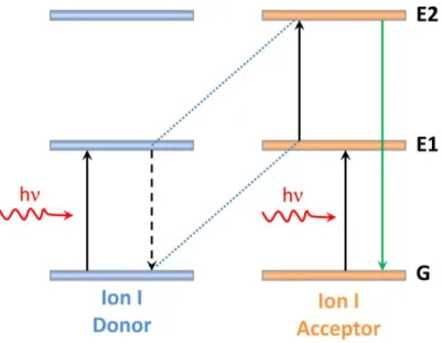
1.1.2.2 Cross-relaxation (CR) upconversion and energy transfer upconversion (ETU) There exist two upconversion mechanisms, which involve energy transfer between two neighbouring ions in close proximity where one ion acts as a donor of energy, while the second acts as an acceptor of energy. The first, CR upconversion, two identical ions in close proximity are both excited from the ground state (G) to intermediate excited state (E1) by GSA, then energy transfer occurs through a non-radiative process in which one ion returns to the ground state while another is promoted to the upper emitting level (E2) (Figure 1.2).
Figure 1.2 Schematic representation of the cross-relaxation upconversion process.
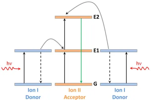
The second, ETU, occurs in co-doped materials, which involves the successive energy transfer from a donor ion to an acceptor ion. In this upconversion process, donor ions are excited to their intermediate states via GSA. Then, a non-radiative energy transfer from donor ions to the acceptor ions results in the promotion of the latter to its intermediate state. A second transfer subsequently occurs and promotes the acceptor ions to the emitting state (Figure 1.3).
6
Figure 1.3 Schematic representation of the energy transfer upconversion process.
1.1.3 Composition of UCNPs
UCNPs generally consist of an inorganic host doped with Ln3+ ions, in which the distance between Ln3+ ions should be below ~10 Å to allow energy transfer, which constitutes the most efficient mechanism of upconversion. The amount and type of dopants, size and phase of nanoparticles can be tuned to achieve multiple emissions over a wide spectral range [14].
1.1.3.1 Host for UCNPs
Host materials should optimally be highly transparent in the visible and NIR regions, as well as be characterized by low energy phonons to minimize host absorption losses and maximize the luminescence output. In that regard, the best materials known to date belong to the “fluorides family”, because fluorides usually exhibit low phonon energies (<500 cm−1), high chemical stability and thus are often used as host materials for upconversion processes [15]. Based on this, NaREF4 such as NaYF4 [16-19], NaGdF4 [20,
7
21], NaLuF4 [22] has been the most popular host for lanthanide-doped UCNPs due to their relative low phonon energy and excellent chemical stability [23]. To date, there are two phase structures of NaREF4 UCNPs, namely (α-) cubic phase and (β-) hexagonal phase. Generally, it is accepted that (β-) hexagonal phase is the most efficient phase due to their unique crystal structure [24].
1.1.3.2 Sensitizers for UCNPs
Regarding the ETU process, the sensitizer should possess a high absorption cross-section for the excitation wavelength and energy levels matching those of activator. In this case, Yb3+ is the best choice. This is because the energy level of Yb3+ is quite simple, with only one excited state (2F5/2) that is in resonance with 980 nm wavelength light. Hence, a 980 nm laser is applied as the excitation source to match the 2F7/22F5/2 transition of Yb3+. From Figure 1.4, we can find this energy gap between 2F7/2 and 2F5/2 is resonant with the energy gaps between several excited states of commonly used activator ions, such as Er3+
etc. Therefore, Yb3+ can be considered as an excellent sensitizer to donate energy to other ions.
1.1.3.3 Activators for UCNPs
According to the ETU process, the acceptors should be characterized by energy levels homogeneously distributed, with an energy separation between them equivalent to the sensitizer emission. Once sensitizers are excited, activators are likely to extract energy from nearby excited sensitizers to promote transitions to higher energy levels. Based on these considerations, Er3+, Tm3+ and Ho3+ (especially Er3+ and Tm3+) are ideal ETU activators owing to the unique ladder-like arrangement of their energy levels [25].
8
Among the commonly used activators, Er3+ shows the highest upconversion efficiency, due to similar energy gaps from 4I15/2 to 4I11/2 (first transition) and 4I11/2 to 4F7/2 (second transition) (Figure 1.4). In Yb3+-Er3+ co-doped UCNPs, the green and red emissions are most commonly observed under 980 nm laser irradiation. This mechanism is shown in Figure 1.4, where the green and red emissions emanate from the 2H11/2, 4S3/24I15/2 and 4
F9/24I15/2 transitions, respectively. Upon the laser irradiation, Yb3+ absorbs NIR photons with the generation of the 2F7/22F5/2 transition. Subsequently, the 4I11/2 energy level of Er3+ is resonant with the 2F5/2 level of Yb3+resulting in a very efficient energy transfer process, while Yb3+ drops back to its 2F7/2 ground state. Due to the energy level match, the Er3+ can be populated to the higher excited state (4F7/2, 4F9/2 state) through similar resonant energy transfer from the sensitizer. After multiphonon relaxation to the 2
H11/2 and 4S3/2, green emissions corresponding to 525 and 545 nm are observed to radiative decay from the 2H11/2 and 4S3/2 excited states to the 4I15/2 ground state. Alternatively, the red emission at 655 nm arises from the 4F9/2 state, which is attributed to either multiphonon relaxation from the higher 4S3/2 state or exciting Er3+ ions from the 4
9
Figure 1.4 Energy level diagram for UCNPs in Yb3+/Er3+ and Yb3+/Tm3+ [26].
1.2 Synthesis of UCNPs
The controlled synthesis of monodispersed UCNPs is necessary for extended bioimaging and therapeutic applications. To date, several methods including thermal decomposition (thermolysis) [27, 28], hydrothermal/sovothermal [29, 30], microwave [31, 32], etc., have been developed. Among all these methods, thermal decomposition with metal trifluoroacetate precursors has been recognized as a common route to synthesize high quality monodisperse UCNPs with small size and narrow size distribution. Thermal decomposition refers to a chemical decomposition caused by heat. This method was first used for synthesis of highly monodisperse LaF3 in 2005 [33]. LaF3 triangular nanoplates
2 F7/2 2
10
(2.0 × 16.0 nm) were synthesized by trifluoroacetate precursors (CF3COO)3La in octadecene/oleic acid solvent. The approach was later modified and first extended to the synthesis of NaYF4 nanoparticles in 2006 [27]. The NaYF4 nanoparticles vary in size from 10 to 50 nm in this work. Metal trifluoroacetates were used as precursors, octadecene used as high-boiling-point organic solvent and oleylamine was used as both solvent and surfactant to prevent the aggregation of nanoparticles. In this thesis, thermal decomposition method is adapted for the synthesis of UCNPs based on our group’s previous work [34].
In recent years, the synthesis of hollow UCNPs has also been developed. Based on the hollow structure, the interior cavities of UCNPs can be applied as effective tools for storage and delivery of therapeutic agents. The synthesis route of hollow UCNPs is generally divided into three categories, including hard templating synthesis, soft templating synthesis and self-templating synthesis [35]. In our group, we mainly focus on the formation of hollow structure of UCNPs through the self-templating method. Xu et al. utilized this method to synthesize GdPO4:Eu3+ hollow spheres [36], and the Gd element was also replaced by Yb in the following research [37]. This method was further optimized, and α-NaYF4 hollow spheres were fabricated [38, 39].
1.3 UCNPs for bioapplications
Optical imaging techniques have been developed for biological applications. However, conventional fluorophores such as organic dyes or semiconductor quantum dots (QDs) require excitation with high energy radiation in the UV or visible region and emission at longer wavelengths [40, 41]. Excitation at these wavelengths incurs a set of
11
drawbacks including low penetration depth into biological tissues due to diverse absorption and scattering processes, the risk of photon induced tissue damage due to the phototoxicity of UV light, photobleaching restricting their temporal use (in organic dyes) and autofluorescence of the background resulting in low contrast. Consequently, there is specific need to develop novel optical bioprobes that overcome these limitations and eventually allow for reliable and efficient optical deep tissue in vivo bioimaging. Thus, UCNPs are very promising as a luminescent probe for biological applications [42-44]. The main advantage of UCNPs is the ability to be excited by NIR photons and emit range from the UV to the visible to the NIR. Compared with normal UV excited bio-probes, for example QDs, conventional organic dyes, the development of UCNPs that can be both excited as well as emit in the NIR “biological window” (700-1000 nm) could potentially overcome the limitations of traditional fluorophores. In fact these nanoparticles possess several advantages, which include (1) significantly reduced background autofluorescence from the biological structures; (2) remarkable penetration depths in vivo; and (3) low cyto- and phototoxicity to the biological specimen under study. For these reasons, Ln3+ -doped UCNPs could serve as excellent candidates for numerous biological applications.
UCNPs have demonstrated significant potential in bio-imaging and cancer therapy applications. In bio-imaging, UCNPs have been used for in vitro imaging [45], in vivo imaging [46, 47] and more recently, combination with some other bio-imaging techniques such as positron emission tomography (PET) [48] and diffuse optical tomography (DOT) [49]. UCNPs have also been studied for cancer therapy where their luminescence has been harnessed for applications such as photodynamic therapy [50, 51], photothermal therapy [52, 53] and drug delivery [54-56].
12 1.3.1 UCNPs for in vitro and in vivo bioimaging
Since the first successful synthesis of UCNPs around 2001 by Dang and co-workers [57], UCNPs have been widely applied to cell and small-animal imaging. Regarding in vitro imaging, water dispersibility and cytotoxicity is a big issue for the effective use of UCNPs. Shan et al. attempted to prepare hydrophobically ligated NaYF4 UCNPs with amino and carboxyl groups, and their cellular cytotoxicity, cellular imaging were investigated [58]. Also, Vetrone et al. studied intracellular imaging of HeLa cells by using polyethylenimine (PEI) capped NaYF4:Er3+, Yb3+ UCNPs. The results showed redistribution of UCNPs inside the cell as the incubation time increased, which could have promising applications for real time imaging of cellular dynamics [59].
With regards to in vivo imaging, it was first reported that Caenorhabditis elegans were injected with UCNPs by Lim et al. [60]. In their studies, it was observed that there was strong luminescence in the intestines of the worms, and no cytotoxicity was detected over a 24 h period. However, the UCNPs used in this study were relatively large and the upconversion emission was weak. These drawbacks hinder the further applications of UCNPs for bioimaging. In 2008, Zhang et al. [61] selected rats as models and injected UCNPs subcutaneously. Upon laser irradiation, stronger upconversion luminescence was observed, when comparing with the control group with conventional inorganic QDs, which was also injected subcutaneously. It was further confirmed that NIR excitation provided a higher penetration depth and a higher signal-to-noise ratio than UV excitation because the excitation wavelength is located in the “biological window” of the biological tissues. In addition, it is also worth noting that these subcutaneously injected UCNPs
13
would be only trapped underneath the skin and did not participate in blood circulation. In fact, UCNPs intravenously injected would be more significant and clinically valuable.
Besides the bioimaging utilizing the upconversion emission, multimodal bioimaging is also studied through the combination of UCNPs and other optical image probes. For example, the computed tomography (CT), ultrasound imaging, magnetic resonance imaging (MRI) and PET could be integrated with UCNPs into one single nanoparticle for multimodal imaging [62-64] (Figure 1.5). These studies demonstrated that UCNPs could be potential candidate for multimodal bioimaging.
14 1.3.2 UCNPs for photodynamic therapy
Photodynamic therapy (PDT) is a type of light-activated clinical treatment in which a light sensitive drug, called a photosensitizer, is used to cause the controlled death of viruses or diseased cells, such as cancer cells. In this process, the photosensitizer is first introduced in the cells, and then excited with light at the appropriate wavelength (depending on the specific photosensitizer but will generally be in the visible range). The absorbed energy is transferred to the molecular oxygen present in the surroundings, generating reactive oxygen species (ROS) whose presence can trigger the death of the cells [65, 66]. Although PDT is a promising therapeutic modality, already being used for numerous clinical cancer treatments, some limitations still persist, such as the need of UV or visible light to excite the photosensitizers. UV and visible light exhibit a limited penetration depth into and subsequently propagation out of biological samples not allowing for deep-tissue imaging. Recently, however, the combination of PDT drugs with UCNPs has attracted increased attention. NIR-excited UCNPs can emit UV/visible photons that can be used to activate the photosensitizers in close proximity and subsequently generate ROS to kill the surrounding cancer cells. Thus, the application of UCNPs will allow the use of NIR excitation wavelengths, increasing the penetration depth in biological tissue from ~2 mm (in the case of standard PDT) to a few centimeters. This would, for instance, allow the treatment of deeper carcinoma and extend the versatility of this therapeutic modality [67, 68]. Moreover, the possibility of specific targeting cancer cells of PDT compounds could be monitored by UCNPs localizing the diseases though fluorescence imaging.
15
Up to now, several studies have successfully combined UCNPs and PDT molecules, most of them using NaYF4:Er3+, Yb3+ as the upconverting material and different surface modifications to create a PDT system, with either one photosensitizer [69], or more, in which case their absorption bands match different upconverted emissions of the UCNPs [51]. The first work combing UCNPs and PDT was published by Zhang et al. [65]. In their study, UCNPs were coated by a silica layer where PDT molecules were incorporated and their results demonstrated the concept works. However, depending on the photodynamic drug selected, other dopant ions could also be considered as good alternatives. Indeed, it has been demonstrated that an improvement of energy transfer between the UCNP and the PDT compound can be achieved using additional lanthanide ions as dopants. Following this idea, X.M. Liu et al. prepared core-shell NaYF4 nanoparticles with Tm3+/Yb3+ in the core (mainly emitting blue light) and Er3+/Yb3+ in the shell (mainly emitting green light). They used the emitted light to activate monomalonic fullerene (C60MA), a novel PDT drug that stands out because of its high quantum yield to form reactive species, and that has a broad absorption band. C60MA was covalently conjugated to the UCNPs by a crosslinking reaction between the amino group of the UCNPs and the carboxyl group of C60MA. In this work, the efficiency of the resulting structure in vitro as well as the increased effect obtained thanks to the addition of Tm3+/Yb3+ dopants to the UCNPs was demonstrated [70]. To date, the fast progress of UCNPs-based PDT and in vitro successful results also motivated the start of in vivo treatments and long-term toxicity experiments using these nanostructures [51] (Figure 1.6).
16
Figure 1.6 Schematic diagram showing UCNPs-based targeted PDT in a mouse model of melanoma intravenously injected with UCNPs [51].
1.3.3 UCNPs for photothermal therapy
Photothermal therapy (PTT) is a treatment strategy based on the damage that high temperatures can produce in cells (Figure 1.7), i.e. it uses heat to kill cancer cells [71]. Like PDT, PTT also has the advantage of allowing for the selectivity of cancer cells, so that surrounding healthy cells and tissue would not be subjected to harmful effects [72]. In order to generate a temperature increase in the cells, a highly efficient mechanism is to transfer it from a light source, more conveniently a laser (due to its high power, monochromaticity and easy position control), through optical absorption. Therefore, nanoparticles with a high optical absorption coefficient and thermal dissipation rate would increase the PTT efficiency.
17
Figure 1.7 Two main strategies to bring upconversion to photothermal therapy: an UCNP is decorated with metal nanoparticles (left side of the particle) or a metal shell is grown around it (right
side of the particle). When the UCNP is excited in the NIR, the subsequent emitted light can be absorbed by the metal and thus, be transformed into heat. On the right side of the figure, a scheme of
the effect of different temperatures in cells is added [73].
Noble metals, such as gold or silver, are considered as excellent candidates for PTT due to their strong surface plasmon resonance (SPR) absorption that can be followed by light emission and, more importantly here, heat release. SPR is the resonant oscillation of conduction electrons at the metal interface stimulated by incident light. These nanoparticles often have the SPR in the visible range, although for certain morphologies it can get down to the NIR. Particularly, gold nanospheres, that are especially convenient due to ease of synthesis, have the SPR absorption band located in the green/yellow spectral range, though the exact wavelength can be tuned by changing the particle size. Consequently, Er3+ doped UCNPs that emit green light after NIR excitation, can be used to excite gold nanoparticles and thus to bring the advantages of NIR excitation (higher
18
penetration depth and minimized heat transfer along the laser path) to PTT. Also, the PTT nanoparticle-based approach could be further improved with the addition of UCNPs allowing for imaging the location of the cancer cells or monitoring cellular events, which is not possible using only the metal nanostructures.
This option has been explored by several groups [74]. A particularly nice example was given by Qian et al. that prepared gold decorated UCNPs (NaYF4:Er3+, Yb3+/NaYF4/silica (core/shell/shell)) applying a reverse microemulsion method [75]. They observed that the total emission intensity of gold decorated UCNPs decreased compared with that of simple UCNPs. Absorption of the luminescence from gold nanoparticles, which triggered a temperature increase, was indeed one of the reasons for this emission decrease. However, a second effect was also pointed out to explain the intensity decrease: NIR light penetration into the particles could be smaller due to the presence of gold, so the excitation light reaching UCNPs would be weaker. Nevertheless,
in vitro data illustrated that BE(2)-C cancer cells were effectively killed after irradiation
with the laser at 980 nm, proving that UCNPs can be used as antennas for the radiation. The decrease of emission intensity can be avoided through, as when a gold nanostructure is excited to its plasmon resonance, it generates an electromagnetic field in its surroundings that can actually enhance the emission intensity of the particles in this area. However, the emission enhancement strongly depends on the design of the nanostructure [76]. Na Niu et al., for instance, developed mesoporous upconversion nanocomposites with 5 nm diameter gold nanoparticles integrated onto the surface. Meanwhile, an anticancer drug, doxorubicin (DOX) was selected and loaded in the mesoporous structure. In their case, upon laser irradiation, enhancement of upconversion intensity was observed
19
owing to the effect of the plasmonic field created by the gold nanoparticles. In addition, the green emission generated by the upconversion nanocomposite could be used to trigger photothermal effect through energy transfer to the gold nanoparticles, as well as rapid drug release [77].
1.3.4 UCNPs for drug delivery
Another area of recent application of upconversion in therapy is targeted drug delivery. Current drug delivery systems are based on different nanostructures (nanoparticles, liposomes, micelles, dendrimers, nanoemulsions, etc.) [78] with a size distribution in the range between 10 and 100 nm to prevent the clearance by the reticulo-endothelial system [79] and thus, keeping them in the bloodstream for a longer time [80]. The properties of these nanostructures can be improved, though, through optical tagging, to visualize their distribution in vivo, and thus, monitor the efficacy of the treatment. For this reason, UCNPs have emerged as an interesting element in drug delivery nanosystems, participating in monitoring drug location and studying the interaction with other cellular components.
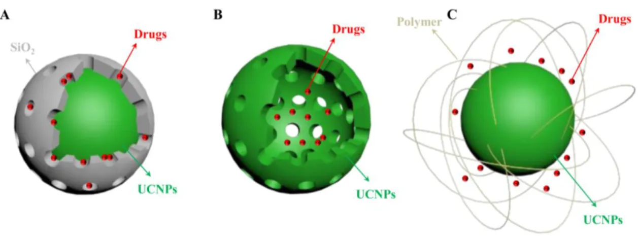
Figure 1.8 Schematic representations of the UCNPs-based drug delivery systems: (A) mesoporous shells, (B) hollow spheres, and (C) PEG grafted amphiphilic polymer.
20
Generally, drug delivery systems consist of a porous shell to load the drug and slowly release it in a rate normally regulated by diffusion or, alternatively, by the erosion of the shell. Because of the biocompatibility of silica materials, silanization is to date one of the most popular techniques for surface modification. In the case of UCNPs, a common strategy is to encapsulate them in a mesoporous silica shell, where the drug can be loaded through capillarity (Figure 1.8A). With the help of this shell, the potential application of UCNPs would be extended. The UCNP core can act as a luminescence imaging probe, and the porous shell can load drugs for localized therapy. An example is the work by Yang et al. using LaF3:Er3+/Yb3+ UCNPs with a silica shell and Ibuprofen as the model drug, which was observed to be totally released after 72h [81]. It is stated that this type of shell exhibits a high surface area, and the loading and drug diffusion capabilities are controlled by the size of the pores, which are advantageous properties to design a drug carrier. Moreover, it has been shown through intracellular analysis that this nanomaterial is non-toxic, which motivates further research following this path [82]. Parallel nanoplatforms have been developed using the NaYF4 host material instead. Also in this case, drug storage and in vitro release was demonstrated [83, 84].
An alternative strategy to the use of mesoporous silica shells is the use of hollow spheres made out of UCNPs encapsulating the drug (Figure 1.8B). The first challenge to follow such structures is the preparation of the hollow spheres, and thus, several techniques have been proposed. For example, the group of J. Lin followed a self-sacrificing method to prepare upconversion hollow spheres [37]. They synthesized core-shell structured Yb(OH)CO3@YbPO4:Er3+ hollow spheres ( ~ 380 nm size), and showed that DOX loaded spheres, that release the drug in 24 h, maintain the Er3+ luminescence and have
21
more cytotoxicity than free DOX on HeLa cells [37]. Applying a surface-protected “etching” and hydrothermal ion-exchange process, Yb(OH)CO3:Yb3+/Er3+ spheres can be used as templates to form hollow mesoporous NaYF4:Yb3+/Er3+ nanospheres, a more efficient upconversion material whose surface can be later modified with folic acid for targeted delivery [39]. A similar evolution of the structure was observed in a study by Ruichan et al., where GdF3 hollow shells were successfully prepared without any surfactant and catalyst [85].
In addition to UCNPs coated with mesoporous silica and to the use of upconverting hollow spheres, a third strategy has been applied for drug delivery with upconversion. This is, to coat the UCNPs with an amphiphilic polymer (Figure 1.8C), which allows to transfer hydrophobic UCNPs into the biological environment. The group of Z. Liu prepared polyethylene glycol (PEG) grafted amphiphilic polymer encapsulating UCNPs, modified the surface with folic acid to improve cancer targeting, and loaded DOX via physical adsorption [54]. In agreement with the results found working with hollow spheres, they found that drug release is affected by environmental pH, it being larger in acidic media. Following this path and aiming for controlled drug delivery, pH-sensitive polymers attracted the attention of the community and some nanoplatforms using them have been recently developed. Good examples are the use of TWEEN 80 [86] or poly(acrylicacid)-modified UCNPs [87], both showing optimal properties as carriers and pH dependent drug release. An alternative for controlled drug release is the use of a light-responsive copolymer. After excitation at 980 nm, the UCNP emitted UV light that triggered the light-responsive amphiphilic copolymer shell uncaging the upconversion nanocomposites, resulting in the release of anticancer drugs [88]. Recently, liposome,
22
which used to deliver cytotoxic drugs or genes, provides a multitude of innovative approaches for pharmaceutical and biomedical applications based on the encapsulation of nanostructures, such as QDs, metallic nanoparticles, or iron oxide [89-92]. In our group, we also developed the liposomes co-encapsulated NaGdF4:Er3+, Yb3+ UCNPs and the anticancer drug DOX for simultaneous bioimaging and therapeutic applications [93].
1.4 UCNPs for other applications 1.4.1 UCNPs for thermal sensing
In recent years, luminescence nanothermometry has emerged as a novel technique which is used to detect the local temperature of a living cell with sub-micrometric spatial resolution. Luminescence is the emission of light from a given substance, which depends on the properties of electronic states. These, in turn, depend on the local temperature and thus the relationship between temperature and luminescence properties can be exploited to achieve thermal sensing from the spatial and spectral analysis of the luminescence. Luminescence nanothermometry can be obtained based on the particular parameter of luminescence which is analyzed, the examples encompass temperature-dependent emission intensity and/or lifetime of organic dyes [94, 95], semiconducting QDs [96-101] and UCNPs [102-107], etc.
To date, there are several examples demonstrating that the intensity of the intra-4f transitions and of the lifetime of a particular 4f excited state are temperature dependent. Therefore, UCNPs can be considered as promising candidates used for nanothermometry. The upconversion luminescence mostly depends on the energy separation between the energy levels of the lanthanide ions. If this energy separation is small, then the electrons
23
will thermally populate and re-distribute among energy levels with similar energy, according to the Boltzmann distribution:
where R represents the luminescence intensity ratios between the emissions with proximity of energy levels, C is a constant, ΔE is the energy gap between the two excited states, k is the Boltzmann constant and T is the temperature. Based on the Boltzmann distribution, if the two energy states are very close to each other, small temperature changes can induce large population re-distributions. The most studied UCNP-based thermometer uses the Yb3+/Er3+ ion pair [108-112]. The Yb3+ ions were excited under laser irradiation at 980 nm, followed by energy transfer to nearby Er3+ ions [25]. Finally, green emission occurring from the 2H11/2 and 4S3/2 excited states to the 4I15/2 ground state as well as red emission from the 4F9/2 excited state to the 4I15/2 ground state is observed. Since the states of two green emissions are very close in energy (typically separated by only several hundred cm-1), the lower energy 4S3/2 state will thermally populate the higher 2
H11/2 state. As the temperature of the surroundings increase, the probability of the lower (4S3/2) state thermally populating the higher (2H11/2) state increases causing a change in the relative intensities of the two emission bands (2H11/24I15/2 and 4S3/24I15/2). Thus, by exploiting the intensity ratio of these two emissions, it is possible to use Er3+-doped materials as ratiometric thermal sensors. Single-cell thermal sensing based on NaYF4:Er3+, Yb3+ UCNPs was demonstrated by Vetrone et al. [59]. It was reported that UCNPs could homogeneously distribute in living cells without causing any remarkable toxicity [113]. Intracellular thermal sensing was achieved by incubating UCNPs in living
) exp( kT E C R
24
HeLa cells, which were exposed to laser irradiation. The optical transmission image of a single HeLa cell is shown in Figure 1.9A when different voltages were applied to a metallic plate in physical contact with the cell. Cell temperature was obtained from the changes of the relative intensities of luminescence bands (see Figure 1.9B). Based on the proper analysis of these changes, intracellular temperature in the 25–45°C range with a sub-degree resolution as a function of the applied voltage could be monitored.
Figure 1.9 (A) Intracellular temperature as a function of the applied voltage, (B) luminescence emission spectra under laser excitation as obtained at two different temperatures [106].
1.4.2 UCNPs for photocatalysis in emerging energy applications
Aside from the application of UCNPs in biology and nanomedicine mentioned above, they have also recently been studied for implementation for use in alternative energy applications such as photovoltaics [114, 115] or photocatalysis [116-118]. The latter is a chemical reaction between organic species and free radicals generated from photocatalysts under light irradiation from the UV to NIR. It is a significant technology which can meet goals of sustainable energy development via unlimited access to clean
25
solar energy. The basic mechanism of photocatalysis is shown in Figure 1.10 based on a semiconductor to generate free radicals (e.g. •OH and •O2-). To date, the energy conversion efficiency which is an ongoing research process is far from satisfactory since they only respond to a relatively small fraction of the solar photons with energy higher than the threshold bandgap (Eg) of the system. To boost the conversion efficiency, the outstanding optical properties of UCNPs have generated increasing interest for photocatalyst application. UCNPs have the ability to convert lower energy NIR excitation light to higher energy photons, covering a broad wavelength region from the UV to the visible to the NIR via the previously discussed upconversion process. The converted UV or visible emission can be recaptured and reused by photon acceptors, such as oxide semiconductors. In addition, they can transform two (or more) sub-bandgap NIR photons into one usable above-bandgap photon, hence minimizing non-absorption energy losses [115, 119].
Figure 1.10 Energy transfer mechanism accounting for enhanced photocatalytic processes in semiconductor catalysts modified with UCNPs [119].
26
The first report for upconversion materials used for photocatalysis can be dated back to 2005, when Wang et al. developed the first visible photocatalyst, based on a combination of Er2O3 upconversion materials with TiO2 [120]. From then on, significant progress has been made. To date, several studies have been demonstrated that by hybridizing with UCNPs (e.g., NaYF4:Tm3+, Yb3+), conventional oxides, e.g., TiO2, becomes NIR light active in photocatalysis, as the NIR illumination was converted into UV photons by UCNPs [121-123]. During this process, excellent photocatalytic activity was achieved via energy transfer between the UCNPs and TiO2, and thus allowing the efficient realization of upconversion effects in photocatalysis. Recently, bismuth ferrite (BiFeO3), which is one of the most important multiferroic materials, has been considered as a good candidate for photocatalysis [124-128]. With a bandgap of ~2.0-2.6 eV (vs 3.0-3.2 eV for TiO2), BiFeO3 (BFO) exhibits excellent response both to UV and visible light [129, 130]. Therefore, an appropriate combination of UCNPs with BFO is expected to yield efficient photon harvesting from the visible to NIR spectral ranges for application. In our group, we prepared UCNP@BFO core@shell hybrid nanostructures for visible-NIR photocatalysis and demonstrate that they are active for the photodegradation of organic compounds under visible and NIR irradiation [131]. The good response of the BFO shell is highly beneficial to capture visible light in a wide spectral range, and the intimate contact between the UCNP core and BFO shell results in the UCNPs and BFO acting as energy donors and energy acceptors, respectively, where the upconverted green emission is partially absorbed by the BFO. The efficient non-radiative energy transfer occurs between UCNP and BFO, thus opening the door to broad-spectrum highly active photocatalysis. While this work is not included in the thesis, this material can also have
27
applications in biomedicine as UCNPs@TiO2 hybrid nanomaterials have been used for applications in PDT [132, 133].
28
Chapter 2 Research Objectives
2.1 Our objectivesThis thesis is divided into two parts with two corresponding objectives:
Part I: Multifunctional nanocomposites combining UCNPs and GNRs
So far, UCNPs have been shown to be efficient and versatile tools for bioapplications. One of the most interesting applications is the use of UCNPs as optical nanothermometers. The majority of UCNP nanothermometers are based on the temperature dependent luminescence where their luminescence intensity ratios vary as a function of temperature. These types of nanothermometers open the door for therapeutic possibilities for many diseases, where a local temperature increases to thermally induced death. Based on this, these nanothermometers are envisioned to combine with local nanoheaters whose principle role is to cause contained temperature increases in order to induce cell death. During this process, GNRs are considered as excellent candidates for nanoheaters, owing to their strong, tunable SPR absorption between 650 and 1200 nm, especially when the plasmon is located in the NIR, making it compatible for in vivo applications. The GNRs absorb light causing electrons to undergo transitions from the ground state to the excited state. The electronic excitation energy subsequently results in an increase in the kinetic energy, which leads to the overheating of the local environment around the light absorbing species. Therefore, local cells or tissue could be destroyed by the heat produced. Recently, PTT using the localized SPR bands of gold nanoparticles and the UCNPs has been reported. However, there are still some considerable challenges and issues that need to be addressed. For example, the surrounding healthy cells could be
29
damaged due to the temperature increase along with the diseased cells. Therefore, care must be taken to obtain accurate control over the local temperature increment so as to not expose the surrounding healthy cells to this localized heating. To accomplish this, the nanoheaters could be coupled with a nanothermometer but must be located in the same cellular space. Hence, they should be transported together in the same vehicle. Thus, thermal sensing with UCNPs could be used for controlling the PTT treatment, which would minimize collateral damage in healthy tissues surrounding the hyperthermia target. Therefore, the nanocomposite could not only be well suited for bioimaging, but also can potentially be used for applications where a thermal gradient must be generated and monitored by a thermometer. In particular, a molecular drug could be also loaded in these nanocomposites, and the drug release behavior could be enhanced by the heat generated from the GNRs. This would result in a multi-pronged or multi-modal nanocomposite capable of delivering two therapeutic modalities.
Therefore, the objectives for this part are as follow:
1. Preparing and characterizing novel multifunctional nanocomposites combining UCNPs and GNRs.
2. Investigating the luminescence intensity ratio of UCNPs as a function of temperature and using it to detect temperature changes of nanocomposites under laser irradiation.
3. Comparing the UCNPs luminescence before and after combining GNRs and analyzing the GNRs influence on luminescence changing.
4. Model drugs were selected and loaded into the nanocomposites, drug release profiles were evaluated.
30
Part II: Multifunctional liposomes encapsulated UCNPs and doxorubicin (DOX) Liposomes are artificially prepared nanocarriers composed of a lamellar phase lipid bilayer. They are considered as good candidates for drug administration due to the fact that their structure is similar to that of cell membranes. Great potential for dramatically enhanced biocompatibility, reduced toxicity and improved therapeutic efficacy has been reported for liposome-based drug delivery. Recently, in the context of liposome-based nanocarriers, research reports have shown that the encapsulation of nanostructures, such as QDs, metallic, or iron oxide nanoparticles within liposomes provides a multitude of innovative approaches for pharmaceutical and biomedical applications. However, their use as delivery systems for various types of nanoparticles is still in the research stage. In particular for liposome encapsulated UCNPs, the data are still scarce, and further studies on the design, characterization, and application of liposome encapsulated UCNPs are required to develop their application in biomedicine. Therefore, we constructed a liposome co-encapsulated UCNPs and DOX, which combined optical imaging and therapeutic modalities. Both UCNPs and DOX were distributed in the internal aqueous phase of the liposomes because of their hydrophilic nature. Meanwhile, because there is a partial spectra overlap between the absorption spectrum of DOX and the upconverted green emission of the UCNPs in the wavelength region from 510 to 570 nm, the UCNPs and DOX molecules act as energy donors and energy acceptors, respectively. Luminescence resonance energy transfer (LRET) would occur based on the close distance between UCNPs and DOX, which could further used for highly sensitive, real-time drug release monitoring system. Therefore, this liposome nanocarrier encapsulated
31
with UCNPs and DOX could be used for multimodal biomedicine, involving the simultaneous detection and therapy of disease.
Therefore, the objectives for Part II are:
1. Preparing and characterizing liposome encapsulated UCNPs and DOX.
2. Investigating the UCNPs luminescence and analyzing the influence of UCNPs luminescence by DOX loading.
3. Using the luminescence intensity of UCNPs to monitor drug release by quantitatively investigating LRET from UCNPs to DOX.
2.2 Thesis organization
This thesis is divided into seven chapters and organized as follows:
Chapter 1 Introduction: briefly introducing the basic concepts of the background; Chapter 2 Research objectives: presenting the motivation and main goals of this thesis; Chapter 3 Experiments and characterization: describing experimental details of synthesis processes of GNRs, UCNPs and liposomes. The main characterization techniques are also described in this chapter;
Chapter 4 GNR@UCNPs multifunctional nanocomposites used for photothermal therapy and drug delivery, and the publication related to this chapter is
Y. Huang, F. Rosei, F. Vetrone. A single multifunctional nanoplatform based on upconversion luminescence and gold nanorods. Nanoscale, 2015; 7(12): 5178-85 [53]. Chapter 5 GNR@SiO2@UCNPs multifunctional nanocomposites used for photothermal and photodynamic therapy, and the publication related to this chapter is
32
Y. Huang, L. L. Páez, P. H. González, D. Jaque, F. Rosei, F. Vetrone. Upconverting nanoparticles used as multifunctional nanocomposites combining with photothermal and photodynamic effects. In preparation.
Chapter 6 Liposome encapsulated UCNPs and DOX used for bioimaging and drug delivery, and the publication related to this chapter is
Y. Huang, E. Hemmer, F. Rosei, F. Vetrone. Multifunctional liposome nanocarriers combining upconverting nanoparticles and anticancer drugs. J. Phys. Chem. B. 2016; 120(22): 4992-5001 [93].
Chapter 7 Conclusions and perspectives: concluding the main results and discussing some prospective potential investigations in future work.
Most of work in this thesis was done by Yue Huang, however, some parts were conducted through collaboration. More specifically, cellular assays were performed by Lucía Labrador Páez, Dr. Patricia Haro González working with our collaborator Prof. Daniel Jaque’s group at Universidad Autónoma de Madrid. UCNPs used in chapter 6 were provided by Dr. Eva Hemmer in our group, who also assisted with the PL measurements.
Following the main body of this thesis is an appendix containing a summary of this thesis in French according to INRS policy.
33
Chapter 3 Experiments and Characterizations
In this chapter, experimental details for the synthesis, characterization of GNRs, UCNPs and liposomes are described. The first section mainly introduces the synthesis of GNRs at different SPR absorption (660 nm and 980 nm), and then GNRs were coated with a layer of silica. The second section focuses on the synthesis of UCNPs using different methods. In order to obtain the UCNPs nanoshell, we first used the hydrothermal method to prepare UCNPs and further coated the GNRs in the core to form the GNR@UCNPs core@shell structure nanocomposites (chapter 4). We also synthesized the UCNPs via the thermal decomposition method. Moreover, the oleate-capped UCNPs were further transferred to water through ligand exchange, and the oleate ligand was replaced by the sodium citrate molecule. The last section focuses on the liposome preparation. We prepare the liposomes using the thin-film hydration method and encapsulate the water dispersible UCNPs in the aqueous core of the liposomes, DOX was further loaded into the liposomes via an ammonium sulfate gradient method.
3.1 Materials
The lanthanide chlorides (LnCl3 (99.99%), Ln = Y, Yb, Er), lanthanide oxides (Ln2O3 (99.99%), Ln = Gd, Yb, Er), trifluoroacetic acid (99%), sodium trifluoroacetate (98%), oleic acid (90%), 1-octadecene (90%), hydrogen tetrachloroaurate (III) hydrate (HAuCl4•3H2O), silver nitrate (AgNO3, >99%), L-ascorbic acid (C6H8O6, >99%), 5-bromosalicylic acid (>98.0%), zinc phthalocyanine (ZnPc, 95%), doxorubicin hydrochloride (DOX) were purchased from Alfa Aesar. Cetyltrimethylammonium bromide (CTAB), sodium tetrafluoroborate (NaBF4), sodium salicylate (99%),

![Figure 1.4 Energy level diagram for UCNPs in Yb 3+ /Er 3+ and Yb 3+ /Tm 3+ [26].](https://thumb-eu.123doks.com/thumbv2/123doknet/5006261.125018/24.918.155.752.128.591/figure-energy-level-diagram-ucnps-yb-and-yb.webp)
![Figure 1.5 UCNPs-based multimode bioimaging [64].](https://thumb-eu.123doks.com/thumbv2/123doknet/5006261.125018/28.918.211.716.467.882/figure-ucnps-based-multimode-bioimaging.webp)
![Figure 1.6 Schematic diagram showing UCNPs-based targeted PDT in a mouse model of melanoma intravenously injected with UCNPs [51].](https://thumb-eu.123doks.com/thumbv2/123doknet/5006261.125018/31.918.145.770.109.298/figure-schematic-diagram-showing-targeted-melanoma-intravenously-injected.webp)

![Figure 1.10 Energy transfer mechanism accounting for enhanced photocatalytic processes in semiconductor catalysts modified with UCNPs [119]](https://thumb-eu.123doks.com/thumbv2/123doknet/5006261.125018/40.918.189.736.669.963/transfer-mechanism-accounting-enhanced-photocatalytic-processes-semiconductor-catalysts.webp)

