Université de Montréal
Utilisation de lymphocytes T en thérapie cellulaire pour le
traitement de la néphropathie au polyomavirus BK chez les
greffés rénaux
par Caroline Lamarche, MD
Programme des Sciences Biomédicales Faculté de Médecine
Mémoire présenté à la Faculté des études supérieures en vue de l’obtention du grade de Maîtrise en Sciences (M.Sc)
en Sciences Biomédicales option Générale
Août 2016
RÉSUMÉ
Le polyomavirus BK est un virus très prévalent qui demeure normalement en phase de latence dans l’uroépithélium sans entrainer de complications. Chez les greffés rénaux, il peut cependant se réactiver et mener à une néphropathie pouvant nuire à la survie du greffon. L’immunité du receveur est la pierre angulaire de la prévention et du traitement de cette néphropathie, puisque le seul traitement démontré efficace est une diminution de l’immunosuppression. Cependant, une augmentation non spécifique de l’immunité augmente également le risque de rejet. Notre objectif était donc d’adapter et de valider un protocole transférable en clinique d’immunothérapie adoptive antivirale nous permettant de produire des lignées de lymphocytes T BK-virus spécifiques à partir du sang de patients greffés virémiques, afin de prévenir et traiter ces néphropathies. Nous avons tout d’abord comparé les lignées cellulaires produites à partir de donneurs sains à celles de patients immunosupprimés soumis à une immunosuppression chronique. Par la suite, nous avons adapté le protocole en ajoutant une stimulation à l’aide de cellules dendritiques afin de maximiser l’expansion cellulaire, le statut de différentiation et la spécificité. Bien que les lignées étaient polyclonales, elles n’ont pas démontré de potentiel alloréactif in vivo et in vitro, et ce, malgré une persistance et une prolifération in vivo. Nous avons donc élaboré un protocole qui est prêt à être transféré en étude clinique de phase I/II et qui pourrait nous permettre de prévenir et traiter la néphropathie associée au polyomavirus BK, sans augmenter le risque de rejet.
Mots clés: BK polyomavirus, transplantation rénale, immunothérapie, thérapie cellulaire, néphropathie associée au polyomavirus
ABSTRACT
More than 75% of the population has been exposed to BK polyomavirus and carries latent virus in the uroepithelium without any complications. However, it can reactivates in kidney transplant recipients (KTR) and lead to a nephropathy affecting graft survival. Recipient anti-viral immunity is the cornerstone of BK-virus associated nephropathy prevention and treatment and thus, reduction of immunosuppression is the only well-accepted treatment. Adoptive immunotherapy is a promising solution to this problem, allowing a specific T cell mediated response against this virus without the alloreactive risk. It was demonstrated efficacious for other viral infections in immunocompromised hosts but it has not been used in this specific context. Our objective was to adapt and validate a clinical-compliant protocol to obtain BK-specific T cell lines from viremic KTR and to compare their expansion, differentiation and specificity to ones obtained from healthy donors. Although comparable specificity and differentiation status, cell expansions form KTR were not systematically sufficient for a therapeutic dose. The addition of a stimulation with dendritic cells improved cell expansion in addition to favors a central memory phenotype and refined BK-specificity. Despite polyclonality, T cell lines didn’t demonstrated alloreactivity in a chromium release assay and in vivo. Furthermore, T cell lines could persist and proliferates in vivo. This protocol is ready for a phase I/II clinical trial. This opens the possibility to solve the current conundrum and treat PVAN without increasing rejection risk.
Keywords: BK polyomavirus, kidney transplant, cellular therapy, immunotherapy, polyomavirus-associated nephropathy
RÉSUMÉ DE VULGARISATION
La greffe rénale permet d’améliorer la survie des patients souffrant d’insuffisance rénale terminale. Dans les dernières décennies, la survie du greffon s’est beaucoup améliorée, notamment grâce à des immunosuppresseurs (anti-rejets) plus puissants. Cependant, l’affaiblissement du système immunitaire entraine une augmentation du risque de certaines infections, notamment le polyomavirus BK.
Une grande proportion de la population a contracté ce virus durant l’enfance et il demeure latent (inactif) dans les reins et les uretères (conduits entre les reins et la vessie), sans entrainer de complications. Par contre, chez les greffés rénaux, l’affaiblissement du système immunitaire fait en sorte que le virus peut se réactiver et entrainer des dommages pouvant évoluer jusqu’à la perte du greffon. Pour l’instant, le seul traitement démontré efficace consiste à diminuer les immunosuppresseurs, mais cela augmente le risque de rejet. Une solution consisterait à prélever les lymphocytes (globules blancs), de les éduquer à combattre le virus en laboratoire et ensuite de les ré-injecter au patient. Il s’agit donc de redonner au patient son propre système immunitaire, mais apte à contrôler le virus. Nous avons donc développé un protocole nous permettant de produire des lignées de lymphocytes T éduqués contre le polyomavirus BK (spécifiques), de bonne qualité et en quantité suffisante pour traiter un patient, à partir du sang de patient greffé ayant un virus actif. De plus, elles sont sécuritaires, donc n’entraineraient pas de rejet de greffon. Nous sommes maintenant prêts à en faire une étude clinique pour le tester chez des patients.
TABLE DES MATIÈRES
RÉSUMÉ II
ABSTRACT III
RÉSUMÉ DE VULGARISATION IV
LISTE DES FIGURES VII
LISTE DES ABRÉVIATIONS VIII
REMERCIEMENTS XI
INTRODUCTION
1
1 - La transplantation rénale
2
1.1 Mise en contexte 2 1.2 Alloréactivité 3 1.3 Immunosuppression 6 1.4 Infections 82 - Le polyomavirus BK
10
2.1 BK polyomavirus and the transplanted kidney; immunopathology and
therapeutic approaches 10
Abstract 13
Introduction 14
1. Epidemiology and diagnosis of PVAN 16
2. Virology and pathogenesis of BK 18
3.1 Environmental factors and the inception of PVAN 22
3.2 Immunology of PVAN 23
4. Current Therapeutic approaches 31
5. Perspectives 36 Conclusion 38 References 39 Table 1. 62 Figures 63
3- Immunothérapie adoptive
67
3.1 Mise en contexte 673.2 Efficacité / Innocuité 69
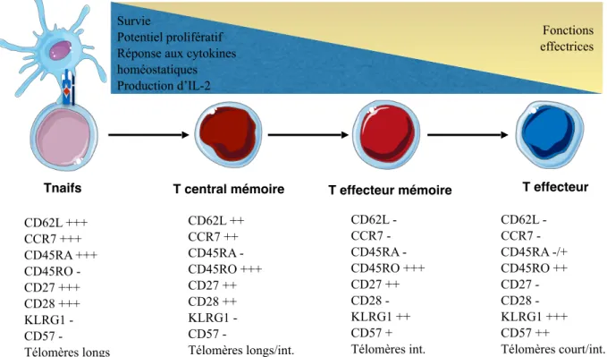
3.3 Différenciation des lymphocytes T 70
3.4 Résumé 73
RÉSULTATS
74
4.1 Clinical-scale Rapid Autologous BK-virus Specific T cell Line generation from Kidney Transplant Recipients with Active Viremia for Adoptive Immunotherapy 75
Abstract 77
Introduction 79
Materials and Methods 81
Results 85
Discussion 90
Acknowledgments 93
Disclosure 94
Figures 95
Description of Supporting Information 99
References 100 Supplementary Table 1. 111 Supplementary Figure 1. 112
DISCUSSION ET PERSPECTIVES
113
CONCLUSION
121
Références
123
LISTE DES FIGURES
Figure 1. Médicaments immunosuppresseurs utilisés en greffe rénale et leur site d’action selon le modèle des trois-signaux
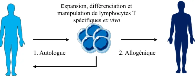
Figure 2. Immunothérapie adoptive
LISTE DES ABRÉVIATIONS
BrdU, 5-Bromo-2′-deoxyuridine
CMH, Complexe majeur d’histocompatibilité CMV, Cytomégalovirus
DC, Cellule dendritique EBV, Epstein-Barr virus
ELISpot, Essai d'immunospot enzymatique ERE, Élément de réponse oestrogénique
GRE/PRE, Élément de réponse glucocorticoide et/ou progestatif GVHD, Maladie du greffon contre l’hôte
HC, Control sain
HCV, Virus de l’hépatite C HSV, Virus de l’herpes simplex
HLA, Antigènes des leucocytes humains HPV, Papillomavirus humain
HSCT, Transplantation de cellules souches hématopoïétiques ICAM, Molécule d’adhésion intracellulaire
IFN-γ, Interferon-γ
IRT, Insuffisance rénale terminale IVIG, Immunoglobuline intraveneuse
KDIGO, Kidney Disease | Improving Global Outcomes KIR, Récepteur killer cell immunoglobulin-like
KTR, Receveur d’une transplantation rénale LT, Large tumoral
LTA, Antigène large tumoral
MCP, Protéine chimiotactique monocytaire MIP, Protéines inflammatoires de macrophages MPA, Acide mycophénolique
mRNA, ARM messager
NCCR, Région de contrôle non-codante NK, Natural killer
NSG, NOD/SCID/IL2Rγnull
PBMC, Cellules mononucléaires du sang périphérique PCR, Réaction de polymerase en chaine
PTLD, Syndrome lymphoprolifératif post-transplantation PVAN, Néphropathie associée au polyomavirus
RCITO, Registre canadien des insuffisances et des transplantations d’organes SV40, Simian virus 40
TCR, Récepteurs des lymphocytes T Tcm, Lymphocyte T central mémoire Tem, Lymphocyte T effecteur mémoire TGF, Transforming growth factor
Tn, Lymphocyte T naif
TNF-α, Tumor necrosis factor-α
VIH, Virus d’immunodéficience humaine VP, Protéine encodant le virus
REMERCIEMENTS
Je voudrais remercier Dr Jean-Sébastien Delisle, mon directeur de recherche pour ses précieux conseils, son écoute et son appui ainsi que tous les membres du laboratoire qui m’ont accompagné dans ce processus.
Je voudrais également remercier Dr Vincent Pichette et Dre Marie-Josée Hébert pour leur mentorat dans ma jeune carrière de recherche et pour être des modèles de rôles positifs comme cliniciens-chercheurs.
Finalement, je voudrais remercier les membres du Conseil des Médecins, Dentistes et Pharmaciens pour avoir cru en mon potentiel en recherche et pour leur soutien financier.
1 - La transplantation rénale
1.1 Mise en contexte
L’insuffisance rénale chronique est définie comme la présence d’anomalies dans la structure ou la fonction des reins, présents pour une période de plus de 3 mois et ayant des implications pour la santé (1). L’insuffisance rénale terminale (IRT), quant à elle, se définit comme une fonction rénale insuffisante pour maintenir un individu en vie. Afin de survivre, une thérapie de remplacement rénal sera nécessaire, soit sous la forme de dialyse ou de transplantation rénale (2). En 2013, au Canada, selon le dernier rapport du registre canadien des insuffisances et des transplantations d’organes (RCITO), plus de 40 000 Canadiens souffraient d’IRT. De ces derniers, 57,5% recevaient un traitement de dialyse et 42,5% avaient un greffon rénal (3). Les causes principales d’IRT au Canada sont le diabète, les glomérulonéphrites et les maladies vasculaires rénales (3).
Malgré ces thérapies de remplacement de la fonction rénale, l’IRT demeure une maladie chronique morbide et mortelle. En effet, la survie à 5 ans en dialyse n’est que de 44,8% selon les dernières données canadiennes (3), ce qui est inférieur à la survie avec un cancer du sein pour les stades non métastatiques (0-III) (4). Après une transplantation rénale, cette survie augmente à 89,2% pour les patients ayant reçu un rein provenant d’un donneur vivant et 82,6% pour un donneur décédé (3). Bien que les patients admissibles à une transplantation sont généralement en meilleure santé et ont une espérance de vie en dialyse supérieure aux patients qui ne sont pas
admissibles, leur risque relatif de mortalité est diminué de 32% à 18 mois seulement après une transplantation (5).
Au Québec seulement, en 2015, il y a eu 297 reins transplantés. Cependant, encore 613 patients étaient en attente de transplantation rénale (6). La survie de la greffe n’est pas éternelle. En effet, en 2005 aux États-Unis, la demi-vie d’un greffon était de 8.8 ans pour les greffes de donneurs décédés et 11.9 ans pour celles de donneurs vivants (7). De plus, environ 13% des patients inscrits sur la liste d’attente de transplantation aux États-Unis le sont suite à la perte de leur greffon (8). Considérant l’avantage de la transplantation rénale, l’énorme besoin au sein de notre population et la rareté de la ressource, il est d’autant plus important de combiner nos efforts afin de prolonger la longévité d’un greffon.
1.2 Alloréactivité
Après une transplantation allogénique, donc entre deux membres non identiques d’une même espèce, le principal risque à la survie de l’organe est son rejet. De façon précoce après la transplantation, il y aura d’abord une atteinte par le système immunitaire inné et plusieurs médiateurs inflammatoires, suivi par une réponse immunitaire adaptative, antigène spécifique.
Le risque de rejet est accru dans les premiers jours suivant la transplantation puisque le prélèvement de l’organe et la transplantation entrainent inévitablement des lésions d’ischémie-reperfusion menant à une augmentation de l’expression des antigènes
des leucocytes humains (HLA) et la relâche de chimiokines telles que GRO/CXCL1, MCP1, MIP-1 et IP-10, de cytokines telles que TNF-α, IFN-γ, IL-2 et IL-6, et des molécules d’adhésions dans la greffe telles que le CD11/CD18 et ICAM-1 (9, 10). L’activation des cellules immunitaires innées causera alors du dommage soit directement ou indirectement via l’activation et le recrutement de lymphocytes T.
Les cellules dendritiques (DC) seront les principales cellules présentatrices d’antigènes pour activer les lymphocytes T. Lorsque matures, les DC du donneur (voie directe) ou du receveur (voie indirecte) migrent dans les organes lymphoïdes secondaires et présentent un allo-antigène aux récepteurs des lymphocytes T (TCR) via leur complexe majeur d’histocompatibilité (CMH). Le contact entre le TCR, le peptide et le CMH correspond au premier signal d’activation des lymphocytes T. Pour compléter l’activation, ils fourniront également des signaux de costimulation (signal 2). La production de cytokines est également importante (signal 3).
Les lymphocytes T CD4+ ayant été activées contribueront également à l’activation d’autres cellules telles que les macrophages, les lymphocytes B et les lymphocytes T CD8+, perpétuant ainsi l’inflammation et le dommage cellulaire à la greffe.
Normalement, un lymphocyte T ne reconnait pas un antigène directement, mais seulement lorsqu’elle est présentée dans un CMH du soi. Cependant, puisque les variations alléliques entre les différents CMH sont petites, les TCR du receveur peuvent avoir une forte affinité pour un CMH intact du donneur et peuvent le
reconnaitre directement. Normalement, seulement 1 lymphocyte T sur 10 000 ou sur 1 000 000 va répondre à un certain antigène. Cependant, en transplantation, 5-10% des lymphocytes T vont répondre à une molécule CMH étrangère (11). Ce phénomène est particulièrement important en greffe rénale puisque les HLA ne sont que rarement appareillés entre le donneur et le receveur. Voilà pourquoi la plupart des traitements immunosuppresseurs en transplantation ciblent les lymphocytes T.
Le rejet d’un organe après la transplantation ne semble pas être la seule conséquence d’une reconnaissance du système immunitaire de l’organe comme le ¨non-soi¨. En effet, notre système immunitaire tolère plusieurs protéines étrangères inertes (par exemple dans l’alimentation) ainsi que plusieurs protéines du soi qui n’étaient pas présentes lors de la sélection négative qui permet d’éliminer les lymphocytes réagissant contre des antigènes du soi dans le thymus (tel que les hormones lors de la puberté). En effet, selon la théorie du danger élaborée par Polly Matzinger, le système immunitaire est d’abord activé par la perception de signaux de dangers (12). Ces signaux d’alarme sont multiples et peuvent être relâchés suite à un stress, un dommage ou lors de la nécrose cellulaire. Il peut s’agir de protéines de choc thermique, de nucléotides, de produits de dégradation de la matrice extracellulaire, de cytokines, etc (13). Lors d’une transplantation, les cellules présentatrices d’antigènes sont donc activées par ces signaux d’alarmes endogènes libérés par les cellules en détresse ou détruites, inévitablement présents suite au dommage d’ischémie-reperfusion. Cette théorie permet d’expliquer pourquoi une transplantation utilisant un organe d’un donneur vivant a un meilleur pronostic qu’une
transplantation utilisant un organe d’un donneur cadavérique pour un même degré de concordance HLA (14).
1.3 Immunosuppression
Une grande partie du succès de la transplantation est secondaire au succès de l’immunosuppression utilisée en induction et en maintenance. En effet, au cours des dernières décennies, la survie moyenne des greffons s’est nettement améliorée, avec une probabilité de perte de greffon à 5 ans qui est passée de 36,2% en 1996 à 26,9% en 2008 (8) et le taux de rejet aigu a diminué à environ 15-20% (15). En général, les différents immunosuppresseurs diminuent ou bien le nombre absolu de lymphocytes, tel que les immunoglobulines anti-thymocytes humains, ou leur fonction, en altérant un des 3 signaux précédemment décrits. La figure 1 démontre les différents immunosuppresseurs utilisés en greffe rénale, selon ce modèle.
De façon courante, les anticorps monoclonaux dirigés contre la sous-unité alpha du récepteur de l’IL-2 (CD25) (basiliximab) ou les immunoglobulines anti-thymocytes sont utilisés en association avec les corticostéroïdes comme agents d’induction selon le risque immunologique du patient. Par la suite, le patient est mis sous une triple thérapie consistant en de la prednisone, un inhibiteur de la calcineurine (préférentiellement le tacrolimus) et un anti-métabolite, souvent l’acide mycophénolique (MPA).
Cependant, en plus de leur action thérapeutique (diminution du risque de rejet), l’utilisation d’immunosuppresseurs plus puissants entraine des conséquences liées à l’immunodéficience tel que les infections et les cancers ainsi que de la toxicité cellulaire non liés à leur effet immun (hypertension artérielle, diabète…).
Figure 1. Médicaments immunosuppresseurs utilisés en greffe rénale et leur site d’action selon le modèle des trois-signaux, adapté de Halloran PF, NEJM 2004 (16). MPA, Acide mycophénolique.
1.4 Infections
Les infections sont une cause importante de mortalité et morbidité après une transplantation. En effet, ils sont la deuxième cause de mortalité chez les patients avec un greffon fonctionnel (17). Le taux d’infection par année durant les trois premières années après la greffe est d’environ 45% (18). L’utilisation d’immunosuppresseurs augmente le risque de toutes sortes d’infections, soit virale, bactérienne, fongique et parasitaire, de façon dépendante du temps après la greffe. Dans le premier mois après la greffe, il y a surtout le risque d’infection nosocomiale, pneumonie d’aspiration, infection de cathéter ou de plaie, colite à C. difficile, le risque d’infections provenant du donneur tel que le VIH ou l’HSV ou encore les infections reliées au receveur s’il était colonisé avant la greffe avec de l’aspergillus ou du pseudomonas. Normalement, les donneurs infectés par le VIH ou l’hépatite C ne sont pas utilisés en transplantation. Malheureusement, si l’infection est très récente, elle peut être manquée au moment du dépistage et être transmise au receveur. Entre 1 et 6 mois, les infections les plus fréquentes sont virales, telles que de la famille des herpesvirus (HSV, VZV, cytomégalovirus (CMV), Epstein-Barr virus (EBV)), l’HPV, le BK, l’adénovirus et l’influenza. Il peut s’agir de la réactivation d’un virus latent, d’un virus transmis par le donneur ou plus rarement d’une infection acquise dans la communauté. Également, il y a le risque de Pneumocystis, C difficile, Cryptococcus,
Mycobacterium tuberculosis, Listeria, Nocardia, toxoplasmose, strongyloïdes,
leishmaniose ou Toxoplasma cruzi. Afin de minimiser la transmission de virus par le donneurs, la présence de plusieurs virus est évaluée avant la transplantation et les donneurs peuvent être rejetés sur la base de la présence de certains virus tels le VIH
et l’hépatite C. Cependant, il arrive parfois que la présence de ces virus ne soit pas démasquée, principalement si l’infection est très récente. Finalement, après 6 mois il y a les pneumonies acquises en communauté, les infections urinaires, l’aspergillus, les moisissures atypiques, le mucor, la Nocardia, le Rhodococcus et toujours plusieurs infections virales telles que le CMV, HBV, HCV, HSV, JC et EBV (19).
L’utilisation d’immunosuppresseurs plus puissants a altéré la prévalence et la cinétique de plusieurs infections. De plus, cela a amené l’émergence de nouveaux types d’infections, telle la néphropathie au polyomavirus BK, qui peut être un réel danger pour le greffon (20).
Puisque le rejet représente le risque le plus important pour la survie du greffon et que la dose minimale d’immunosuppresseurs permettant de prévenir cette complication pour un patient donné n’est pas connue, la pratique actuelle est souvent de sur-immunosupprimer les patients aux dépens du risque infectieux et néoplasique (21).
2 - Le polyomavirus BK
2.1 BK polyomavirus and the transplanted kidney; immunopathology and
therapeutic approaches
Le polyomavirus BK est un virus acquis durant l’enfance demeurant latent dans l’uroépithélium. Un système immunitaire compétent permet de contrôler sa réactivation. Par contre, chez les greffés rénaux, ce virus peut se réactiver et entrainer une néphropathie affectant jusqu’à 10% d’entre eux (22). Un ensemble de facteurs agissent simultanément dans cette population afin de créer les conditions parfaites à sa réactivation. Tout d’abord, nous croyons que l’infection chez les greffés provient des donneurs. Il s’agit donc d’une souche qui peut être différente de celle contre laquelle le receveur a développé une immunité. Ensuite, nous savons maintenant que l’environnement inflammatoire présent tôt après la greffe peut favoriser la transcription virale. De plus, les lésions d’ischémie-reperfusion peuvent libérer le virus. Finalement, les immunosuppresseurs utilisés après la transplantation ciblent préférentiellement les lymphocytes T, qui s’avèrent primordiaux pour le contrôle de la réactivation virale. Lorsque le virus se réactive, on le retrouve dans l’urine puis dans le sang de façon séquentielle avant de causer une néphropathie. À ce jour, le seul traitement démontré efficace est une diminution de l’immunosuppression, ce qui peut cependant augmenter le risque de rejet. Il est donc important de mieux comprendre les interrelations le virus, l’environnement et le système immunitaire afin d’améliorer notre approche face à cette problématique.
Statut: Publié
Journal: Transplantation, 2016 Jul 7, [Epub ahead of print] Contribution des auteurs:
CL: Principale auteure, revue de littérature et écriture du manuscrit JO et JSD: Aide à la revue de la littérature et l’écriture du manuscrit SC, LS, MJH, ER, LAT: Révision et édition du manuscrit
BK polyomavirus and the transplanted kidney; immunopathology and therapeutic approaches
Caroline Lamarche, MD1,2,7, Julie Orio, MSc1, Suzon Collette, MD2,7, Lynne Senécal, MD2,7, Marie-Josée Hébert, MD3,4,7, Édith Renoult, MD4,7, Lee Anne Tibbles, MD5,7, Jean-Sébastien Delisle, MD, PhD1,6,7.
1 Centre de Recherche de l’Hôpital Maisonneuve-Rosemont (CRHMR), Montreal, Canada
2 Division of Nephrology, Department of Medicine, Hôpital Maisonneuve-Rosemont (HMR), Montreal,
Canada
3 Centre de recherche du Centre Hospitalier de l’Université de Montréal (CRCHUM), Montreal, Canada 4 Division of nephrology, Centre Hospitalier de l’Université de Montréal (CHUM), Montreal, Canada 5 Faculty of Medicine, Institute of Infection, Immunity and Inflammation, University of Calgary, Calgary,
AB, Canada
6 Division of Hematology, Department of Medicine, Hôpital Maisonneuve-Rosemont (HMR), Montreal,
Canada
7 Members, Canadian National Transplant Research Program (CNTRP)
Disclosures: The authors declare no conflicts of interest.
Abbreviations: DC, dendritic cells
ELISpot, Enzyme-linked immunospot assay ERE, estrogen response element
GRE/PRE, glucocorticoid and/or progesterone response element HLA, human leucocyte antigen
HSCT, hematopoietic stem cell transplantation IFN-γ, interferon-γ
IVIG, intravenous immunoglobulin
KDIGO, Kidney Disease | Improving Global Outcomes KIR, killer cell immunoglobulin-like receptors
LT, large tumor
mRNA, messenger RNA
NCCR, non-coding control region NK, natural killer
PCR, polymerase chain reaction
PVAN, polyomavirus-associated nephropathy SV40, simian virus 40
TGF, transforming growth factor TNF-α, tumor necrosis factor-α VP, virus-encoded protein
Abstract
BK polyomavirus is ubiquitous, with a seropositivity rate of over 75% in the adult population. Primary infection is thought to occur in the respiratory tract, but asymptomatic BK virus latency is established in the urothelium. In immunocompromised host, the virus can reactivate but rarely compromises kidney function except in renal grafts, where it causes a tubulo-interstitial inflammatory response similar to acute rejection. Restoring host immunity against the virus is the cornerstone of treatment. This review covers the virus-intrinsic features, the post-transplant micro-environment as well as the host immune factors that underlie the pathophysiology of PVAN. Current and promising therapeutic approaches to treat or prevent this complication are discussed in relation to the complex immunopathology of this condition.
Introduction
Polyomaviruses were first discovered by Ludwig Gross in 1953 as murine leukemia viruses. Notably, newborn mice injected with cell-free extracts of murine leukemia tissues developed adenocarcinomas of the parotid gland in addition to leukemia, suggesting that an infectious agent was the cause of the malignancies1. The infectious agent was named using the Greek words for many (poly) and cancer (oma)2. So far, about 30 species of polyoma viruses have been identified in birds and mammals, including thirteen in humans: BK, JC, KI, WU, Merkel cell polyomavirus, H6, H7, H9, H10, H12, STL, trichodysplasia spinulosa-associated polyomavirus and NJ3. BK polyomavirus was first isolated by Gardner and al. in 1971 from the urine of a renal allograft recipient and was named after the patient’s name4.Whether BK virus is oncogenic is controversial, but a role in the development of urothelial cancers has been proposed in immunocompromised patients5. In immunocompetent patients, the presence of BK virus DNA was found in numerous cases of bladder, urothelial and other tumors6,7. However, given the high prevalence of BK virus infection and latency in those tissues, the detection of BK in tumors does not imply a causal relationship8.
It is estimated that at least 75% of the adult population is latently infected with BK v i r u s9. I m m u n o c o m p e t e n t s u b j e c t s a r e u s u a l l y a s y m p t o m a t i c , b u t immunocompromised hosts can suffer BK-related complications. In kidney transplant recipients, BK and possibly other polyoma viruses can cause nephropathy and ureteral stenosis10, while hemorrhagic cystitis is prevalent in hematopoietic stem cell transplantation (HSCT) patients11. Rare cases of BK disseminated disease
(tubulointerstital nephritis, desquamative pneumonitis, meningoencephalitis and retinitis) have also been described12, especially in patients with acquired immune deficiency syndrome13.
Such wide range of presentations associated with BK virus suggests that condition-specific interactions between host and virus factors ultimately dictate the clinical complications related to BK-virus reactivations. As an illustration, polyomavirus-associated nephropathy (PVAN) in kidney transplantation patients and hemorrhagic cystitis occurring after HSCT are both associated with risk factors that are specific for these conditions. In HSCT, risk factors notably include myeloablative conditioning 14, CMV viremia14, recipients of cord blood units15 and acute graft-versus-host disease15, which may relate to two main factors; urothelial damage (myeloablative conditioning) and profound immunosuppression. Immunosuppression following kidney transplantation is likewise necessary for the development of PVAN as well as specific features uniquely associated with renal transplants. As opposed to HSCT where BK reactivation occurs in host tissues, in kidney transplantation the virus reactivates in the graft and the infection is mostly donor-derived16. Bohl and colleagues have shown a concordance in BK virus infection in receiving pairs from the same donor, with a match in sequences of segments of the two genes (non-coding control region (NCCR) and virus-encoded protein (VP1)) in these patients compared to recipients from different donors17, strongly supporting a donor origin of the virus in this case. Moreover, there is also a higher rate of reactivation in recipients from BK virus seropositive donors16.
This review focuses on the virology and BK-specific immunity in the context of renal transplantation, highlighting the interplay between three major variables; the virus, the kidney graft environment and the immune system. The rationale and merit of actual as well as plausible future prevention and treatment approaches for PVAN occurring following BK-virus reactivation are discussed in relation to BK-associated immunopathology.
1. Epidemiology and diagnosis of PVAN
Reactivation of BK virus in the transplant kidney can lead to PVAN in up to 10% of kidney transplant recipients18. BK virus reactivation is first observed with the appearance of decoy cells or BK virus DNA in the urine preceding viremia by a median of 4 weeks18. Decoy cells are virally infected uroepithelial cells that can be observed with standard light microscopy. They can be used as a screening method for PVAN, but their positive predictive value is weak (11,7%)19. These early findings are followed by BK viremia, which precedes nephropathy by a median of 8 weeks. As such, viremia has a better positive predictive value for nephropathy than viruria, especially if viral load is more than 10000 copies/mL20,21, with the caveat that BK virus polymerase chain reaction (PCR) assays are not standardized across centers22. Fifty percent of all detectable viremia occurs in the first 2 months and 95% in the first two years after transplant18. This timing for reactivation may be related to several factors including intense immunosuppression, tubular injury and ensuing inflammation that characterize the early post-transplantation period.
The intensity of immunosuppressive regimens is a risk factor for the development of PVAN23. The occurrence of PVAN correlates with the use and dosage of tacrolimus23,24 and/or mycophenolate mofetil23,25, anti-thymocyte globulin induction25 and anti-rejection treatment20. Other risks factors are less consistently reported in the literature, but include various recipient-related factors (older age, male sex26), donor factors (degree of human leucocyte antigen - HLA mismatches and BK virus seropositive status27), and factors associated with renal injury (cold ischemia time, delayed graft function and ureteral stent placement)27. The diagnosis of PVAN is highly suggested by the detection of viral inclusion bodies on kidney biopsy but is confirmed with immunohistochemical staining for simian virus 40 (SV40) large T antigen and/or in-situ hybridization for BK virus genetic sequences28. According to the Banff classification, the histopathological findings further categorize PVAN into three stages. Grade A refers to inflammatory changes without acute tubular necrosis, while grade B is defined by tubular epithelial cell lysis and acute tubular necrosis. Finally, the presence of interstitial fibrosis characterize grade C PVAN29,30. Graft prognosis correlates with PVAN severity as 2-year graft survival is 90% for grade A, but only 70% and 50% for grade B and C respectively31. Several biomarkers have been developed to assess intrarenal viral disease. The urinary polyomavirus-haufen test which relies on the detection of urinary casts composed of uromodulin, lysed tubular cell and virions is reported to predict PVAN onset, intrarenal viral activity and resolution 31. Urinary VP1 mRNA is also proposed as a new biomarker to identify PVAN32 which may be used with granzyme B mRNA, proteinase inhibitor-9 mRNA and plasminogen activator inhibitor-1 mRNA to predict graft failure risk33,34. Although
promising, these tests require further validation before widespread clinical use31. Furthermore, granzyme B and proteinase inhibitor-9 mRNAs are not specific for PVAN and they were used as biomarkers in the CTOT-O4 study to predict acute cellular rejection35. Interestingly, the diagnostic signature elaborated in this study was also associated with BK virus infection35 indicating that non-specific surrogate markers of immune activation can only be used in conjunction with other diagnostic information.
2. Virology and pathogenesis of BK
BK polyomavirus genome shares about 72% nucleotide homology with JC virus and 70% with SV40. It consists of a single molecule of circular viral DNA of 5300 base-pairs, complexed with cellular histones (H2A, H2B, H3, H4) and surrounded by an icosahedral capsid containing three virus-encoded proteins, VP1, VP2 and VP336. BK virus genome contains three functional regions; NCCR which regulates viral replication and transcription, the early and the late regions37. The early region contains large tumor (LT) and small tumor antigen proteins, which are derived by alternative splicing of a common precursor36 and are believed to be the first proteins expressed. The late region contains genetic information for the three virus-encoded proteins (VP) and agnoproteins37.
Role of the viral proteins
BK polyomavirus binds to the target cells through interaction with two ganglioside receptors, GT1b and GD1b and then uses caveolae-mediated endocytosis to reach the endoplasmic reticulum38 (Figure 1). Following partial uncoating of the virus by
reduction and isomerization of the disulfide bonds that link VP1 proteins, BK virus retrotranslocates to the cytosol for a second rearrangement of the capsid, thereby enablinga liaison to the nuclear pore and passage of viral DNA into the cell nucleus39, possibly facilitated by nuclear localization signals on the minor capsid proteins, VP2 and VP340. Following infection of human kidney epithelial cells in vitro, LT expression is observed at 36 hours before VP1 expression and viral DNA replication41 and is required for viral DNA replication and expression of the late genes42. It can also induce an oncogenic effect by specifically binding and inactivating tumour suppressor proteins, including retinoblastoma family genes and p53 (Figure 1). Thus, it can promote the transition of the cell into the S phase43. In an elegant study, Seemayer and colleagues demonstrated with indirect immunofluorescence that the enlargement of nucleus seen in polyomavirus-infected tubular cells is associated with viral replication, large T antigen expression and p53 accumulation. This observation correlates with an activation of the cycle cell, as seen by the expression of Ki-67. Viral replication can also lead to cellular demise. The absence of caspase 3, bcl-2 as well as a regular distribution of nuclear DNA indicates that cells die mostly by necrosis and not apoptosis44, in accordance with the previous observation made in PVAN patients by light and electronic microscopy45.
Viral strains
We can classify BK virus as “archetype” or “rearranged” types, based on the genotype of the NCCR. The NCCR regulates viral replication and transcription46. The rearranged variant implies numerous mutations in the NCCR region which can
amplify the replication potential47, a phenomenon that has been validated in vivo48. In their meta-analysis, Sharma and colleagues concluded that there is a correlation between the rearranged variant and the development of nephritis49. Two hypotheses have been suggested to explain this relationship; 1) the rearranged variant is more virulent and leads easily to nephritis, or 2) nephritis is associated with a more rapid viral turnover which favors the development of mutations49. Chatterjee and al found both virus types can be found in peripheral blood cells of healthy individuals. Hence, they proposed that leukocytes may play a role in the NCCR rearrangement process and transport of BK virus46, 50.
Transmission,
The exact route of transmission from human to human is unknown51. Oral transmission has been proposed 52, but the most accepted hypothesis is that BK is spread through the respiratory tract53. Primary infection is indeed associated with upper respiratory symptoms in one third of children54. Moreover, Goudsmith et al. have demonstrated that BK seroconversion is present in 8% of children admitted to the hospital for any upper respiratory tract illness, compared with 15% for adenovirus, influenza A, parainfluenza, respiratory syncytial virus and mycoplasma pneumoniae combined53 further strengthening the hypothesis that primary BK infection occurs through the respiratory tract.
Latency
After primary infection, BK virus persists mostly in the renal tubular epithelial cells and the uroepithelium, in a latent form. BK virus DNA was found in 33% of kidneys by
DNA-DNA hybridization in normal subjects 55 and in 25% of fresh frozen prostate specimens of patients with prostate adenocarcinoma, using nested PCR56. It was also found in 2/67 autopsy brain specimens with southern blot57 and in 17/18 healthy donors peripheral blood leucocytes by PCR amplification with in situ hybridization58. Interestingly, Dolei and colleagues detected BK virus NCCR DNA by nested PCR in 22% of healthy donors, but the presence of VP1 DNA in only 7% of subjects, a prevalence that was declining with age. NCCR positive prevalence in peripheral blood monocytes cells was 37,5% in the less than 20 years old group, to 12,5% in the 21 to 40 years old and 0% in the more than forty years old59. Therefore, they hypothesized that blood cells do not host biologically active BK virus for a long time after acute infection or reactivation59.
Reactivation
Seroprevalence in general population is about 50% in children of four years old and more than 75% in adults9. Newborns have maternal antibodies that decline with a nadir at 6 months9. Viruria, which may represent the first evidence of reactivation, can be detected in both healthy and immunocompromised subjects. Pregnant women can have asymptomatic viruria60, a possible consequence of hormones (mostly glucocorticoid and the combination of oestrogen and progesterone) on viral replication61. Viruria has also been noted in 7% of asymptomatic healthy blood donors62 and in more than 60% in immunocompromised patients63. Indeed, in addition to kidney transplant and hematopoietic stem cell transplant recipients, viral reactivation has been described in patients with HIV13, lupus erythematosus
patients64, Wiskott-Aldrich syndrome7, hyperimmunoglobulin M immunodeficiency65, cartilage-hair hypoplasia and Hodgkin's disease66, in non-renal solid transplant67 and in multiple sclerosis patients receiving Natalizumab therapy68.
In kidney transplant recipients the virus initially replicates in the distal tubular epithelial cells, leading to necrosis and initiation of local damage and inflammation. The spread of virus in the adjacent environment will result in viruria and the infection of adjacent cells. Following this initial insult, denudation and dissolution of the tubular basement membrane occurs, allowing infection to spread in the intertubular space and by peritubular capillaries resulting in viremia45 (Figure 2). This is followed by recruitment of inflammatory cells in the tubulo-interstitial space and viral spreading to proximal cells. Infection control will normally occur with the reestablishment of immune competence. A two-hit phenomenon is usually required for BK virus associated nephropathy development: environmental factors promoting viral replication and immunodeficiency.
3.1 Environmental factors and the inception of PVAN
PVAN occurs early after transplant, likely in the context of a “perfect storm” where immunosuppression is at its peak and active tubular lesions from ischemia-reperfusion or surgical trauma coincide. Indeed, electron microscopic data of kidney biopsies from patients with BK nephropathy demonstrated extensive tubular necrosis, even of the non-infected cells. Therefore, a question is whether tubular injury can
trigger BK-mediated nephritis, or does PVAN requires an environment conducive to tubular injury45.
Mouse model of polyomavirus infection demonstrated that kidney damage, either chemical or ischemic, can promote viral replication69. Viral replication is controlled by the NCCR and can be regulated by numerous cellular transcription factors, including nuclear factor I, Sp1, NFAT, AP1, Smad3, estrogen response element (ERE), glucocorticoid and/or progesterone response element (GRE/PRE), p53, NF-κB, C/ EBP and maybe PEA3, AP-2, CREB and GM-CSF (as reviewed by Liang and colleagues)70. Several of these molecules articulate numerous pro or anti-inflammatory pathways that are active following kidney injury and that could link ischemia/reperfusion, as well as inflammatory responses, to BK virus replication. As examples, factors such as TGF-β and tumor necrosis factor alpha (TNF-α) that can directly enhance transcriptional activity and promote viral replication70. Microenvironmental factors can therefore explain the particular vulnerability of the transplanted kidney. In addition, immunosuppression strategies could also directly activate viral replication. Hence, glucocorticoid pulses often used for treatment of acute rejection are well known risk factors for BK virus reactivation20. Independently of their effects on the immune system, steroid hormones can increase virus transcription by their action on GRE/PRE and ERE transcription factors on NCCR71. Once the virus has started to replicate, it must be held in check by a proficient immune system.
3.2.1 Cellular
T cells, especially CD8+, are pivotal to the anti-BK response and surveillance as they can detect and kill infected cells. The presence of BK virus specific T cells in the blood of seropositive healthy patients was demonstrated by T cell production of interferon-γ (IFN-γ), TNF-α, granzyme A and B and CD107 expression following stimulation with BK’s VP1 and LT antigens72. This was also demonstrated in patients with BK viremia and nephropathy by assessing IFN-γ producing cells by flow cytometry and multiplex analysis of the supernatant of peripheral blood mononuclear cells stimulated with BK VP1 peptide mix73. Cellular immune responses against LT and VP1 antigens are also higher in patients with decreasing or past viremia, compared to those with increasing or persisting viremia74, or BK nephropathy75, suggesting again that they play a role in the control and resolution of BK virus reactivation. Additional evidence of T cell activation during viremia or PVAN include the expression of messenger RNA (mRNA) associated with a cytotoxic program in T cells76.
In a study by Comoli and colleagues, transplant recipients with or without BK viruria had lower BK specific T cells evaluated by enzyme-linked immunospot assay (ELISpot) compared to healthy patients, which may suggest an impact of immunosuppression on BK immunosurveillance77. However, Chakera et al. failed to demonstrate a correlation between BK-specific T cells against any of BK peptides by ELISpot assays and tacrolimus trough levels or the total burden of immunosuppression, suggesting other factors must contribute to the lack of specific immunity post transplant78. In the study by Comoli et al., viremic patients had
undetectable CD4 and CD8 for BK virus. Appearance of BK reactive T cells coincided with graft function improvement and resolution of viremia77 results that had been confirmed by at least two other groups78,79. Compared to viremic patients without BK nephropathy, patients recovering from an episode of viremia had improved T cell response, as evaluated by ELISpot79. With the inherent limitations associated with testing peripheral blood and not lymphoid organs or the kidney, these data nonetheless suggest that the restoration of immune competence is central to viral control.
Mueller and colleagues have found that the five BK virus specific proteins (VP1, VP2, VP3, LT, sT) were able to elicit memory T cell response, demonstrated by specific production of IFN-γ, IL2 and TNF-α by flow cytometry analysis. All patients with a history of PVAN had a response to at least VP3 and 74% had a response to all five80. Also, these patients had a greater CD4 response than patients with asymptomatic viremia, as seen by a greater production of IL-2 and INF-γ80. However, T cells producing three cytokines (IFN-γ, IL2 and TNF-α), were more frequent in patients with asymptomatic viremia or no BK virus reactivation compared to PVAN patients, suggesting a possible protective role, or that strong T cell activation in PVAN leads to exhaustion and loss of polyfunctional responses80. In a study by Schmidt et al., transplant recipients with BK virus complications had more BK-specific T cells but less polyfunctional compared to transplant recipients without BK complications, suggesting also exhaustion of those T cells81.
T cells recognize peptide antigens presented by HLA molecules. HLA matching could therefore be important to elicit an optimal response. Whether HLA mismatching has an impact on PVAN is controversial. While some studies found an association between BK virus nephropathy and HLA mismatch82, others did not83. With the significant caveat that patients with many HLA mismatches are more aggressively immunosuppressed, thereby impeding anti-viral T cell responses, HLA mismatching could further limit viral antigen recognition on mismatched HLA molecules. Matching of HLA-A2, B44 and DR15 may be protective against BK viremia84, and the absence of C7 in either the donor or the recipient may be a risk factor for BK infection17, a result that was not confirmed in another cohort85.
Little is known about the resolution process of PVAN. There is an inflammatory response resembling histologically and genetically to acute rejection76,86. Whether this response is appropriate or is overwhelming, as an immune reconstitution syndrome, is not known. Two questions remain; does this process trigger fibrosis87 and/or allospecific damage? In the study by Menter and colleagues, PVAN resolution was not associated with fibrosis, but all biopsies were obtained relatively early after PVAN resolution (within one year)86. Despite the risk of alloreactive damage, the central tenet of PVAN prevention and treatment is a reduction in iatrogenic T cell immunosuppression.
Many studies used serological testing as a surrogate maker for B cells activity in BK virus infection. However, two critical elements must be considered; i: no current serological assay is standardized88 and ii; seropositivity indicates that a patient has been in contact with the virus and seroconverted, but this does not imply the development of effective anti-BK T cell memory responses which are principally needed to control BK reactivation77.
Qualitative and quantitative serostatus
Pediatric studies demonstrated a correlation between seronegative status and an increase risk of viruria89 and PVAN90. However, this correlation is controversial in adults. Two hypotheses have been proposed to explain the difference between these two patient populations. First, seropositivity may decline with time91. Shah and colleagues reported 100% seropositivity at 10-11 years old and 67% after 35 years old92. Hence, antibodies may be present, but under the threshold of detection. Second, adults have been exposed to many different viruses and may have acquired a cross-reactive protection89. Bohl demonstrated that a seropositive status in adults pre-transplant does not prevent viremia93 and Hirsch showed that seronegativity in patients before transplantation is not a risk factor for PVAN20. However, another group found a higher risk for BK viremia in seronegative recipients who received a kidney from a seropositive donor94. These discrepancies may be accounted by variability in the assays used to detect BK-specific antibodies and quantitative differences in anti-BK antibody titers. It was previously shown that viremic patients had a lower antibody level pre-transplant than those who never developed BK viremia93. Moreover, kidney
recipients from a seropositive donor will have a larger increase in antibody titers than those receiving a graft form a seronegative donor16, regardless of their own status. This suggests that BK virus transferred through the transplanted kidney can elicit a host primary or recall humoral response. Finally, there is an increase in IgG titer with PVAN resolution, suggesting humoral immunity could play a role in viral control95.
3.2.3 Innate immune response
Natural killers
Natural killers (NK) cells play an important role in the innate immune response against viral infections, and probably in polyoma infection/reactivation as well76. NK cell activity is controlled by opposing signals that come from a balance between activating and inhibitory receptors and can contribute to the orchestration of the adaptive immune response as well as mediating direct killing of infected cells. Many strategies are developed by viruses to avoid recognition by NK cells96. For example, BK virus miRNA can mediate down regulation of the NKG2D ligand ULBP397. Trydzenskaya and colleagues found a relation between activating killer cell immunoglobulin-like receptors (KIR) genotype and the control of BK virus infection as well as nephropathy in kidney transplant recipients98. NK from PVAN patients had lower activating receptors compared to the control group. However, they did not find any correlations between KIR, HLA compatibilities and BK virus infection98. Although less studied than in T cells, the impact of immunosuppressive therapy on NK function reveals that NK cells are inhibited by currently used medications. Cyclosporin A affects NK-cell function, phenotype99 and proliferation100, while prednisolone inhibits
their proliferation when exposed to allogenic tubular epithelial cells and tacrolimus may counter their capacity to degranulate in the same context101. Also, mycophenolate mofetil possibly inhibits proliferation induced by IL-2100. However, the relative importance of NK cells relative to other immune effectors remains to be defined and whether NK cells could be mobilized for prevention or therapy of BK-related diseases is unclear.
Dendritic cells
Dendritic cells (DC) are central to the adaptive cell response, as they are efficacious antigen-presenting cells. Kidney transplantation and chronic immunosuppression lead to an absolute decrease in DC counts in the peripheral blood102,103. Transplant surgery in itself induces a strong decline in the number of DC (and possibly with a greater reduction for plasmatoid DC103), in kidney transplant recipients as well as in kidney donors. This decline can last up to 3 months after surgery102. As opposed to donors, patients on chronic immunosuppression fail to recover normal counts102. Hackstein and colleagues demonstrated that all DC subtypes were lower in patients treated with long term immunosuppression (more than a year) in kidney transplant recipients compared to age and sex matched controls, independently of total leucocyte count104. Despite this possible DC deficiency, Yapici and colleagues found significant amount of myeloid DC in PVAN biopsies and those cells were found closely to BK virus infected tubules105, suggesting a role in PVAN physiopathology.
Pre-transplant DC deficiency, both absolute and functional, is associated with an increased BK viremia risk after transplant, even after adjustment for ureteral stent, tacrolimus and cyclosporine use106. Functional DC deficiency was evaluated by the production of IL-12 of a pool of peripheral blood mononuclear cells after lipopolysaccharide (LPS) stimulation. Furthermore, the absolute DC number in PVAN patients is reduced compared to other kidney recipients, despite the presence of ureteral stent and the use (not trough level) of tacrolimus103. Whether these findings reflect a direct impact of DC deficiency on BK reactivation is unclear. Nonetheless, DC levels and function could be further studied as biomarkers for the prediction of BK reactivation and disease.
Monocytes/macrophages
Little is known about monocytes’ role in BK nephropathy. Patients with BK viruria (not PVAN) have increased soluble interleukin-1 receptor antagonist levels in their urine, a counter regulator of monocyte activation which can be produced by monocytes (as well as other cell types, as endothelial and epithelial cells upon inflammatory stress)107. More research is needed to decipher the role of inflammatory macrophages (M1) and anti-inflammatory (M2) macrophages in PVAN, as they could respectively propagate the initial immune response and orchestrate the resolution of inflammation as well as the development of fibrosis.
4. Current Therapeutic approaches
A first strategy to prevent BK reactivation would be to tailor immunosuppressive regimen according to BK virus reactivation risk. Although promising, there is currently not enough evidence to recommend such approach currently108,109. Hence, a pre-emptive strategy is used. To date, the best preventive PVAN strategy is to routinely monitor BK virus reactivation and to reduce immunosuppression pre-emptively if needed. According to the KDIGO (Kidney Disease | Improving Global Outcomes) recommendations, BK screening should be performed monthly early after transplant (first 3-6 months), then every 3 months until the end of the first year post-transplant110. Testing should be repeated and performed at increased frequency if there is an unexplained rise in serum creatinine and after treatment for acute rejection. PCR quantification of BK viremia is recommended as the screening method as it has the best sensibility and specificity19. If not accessible, urinary cells or urinary PCR are acceptable surrogate markers of BK reactivation19,111. Kidney biopsy of patients with viral load of 10000 copies/mL should be performed as it is highly associated with PVAN 20,21. Absence of histological changes associated with PVAN, associated with viremia over 10000 copies/mL may be called ¨presumptive PVAN¨. The conventional approach is to treat those patients as definitive PVAN. However, to minimize the risk of acute rejection associated with a reduction in immunosuppression in patients who might not develop definitive PVAN, Nickeleit and Singh recently proposed to better stratify these patients using the urinary polyomavirus-haufen test and urinary mRNA in order to personalize therapeutic interventions and avoid under
treating BK reactivation in the kidney 31. These complementary analyses are not available to all centres and have not made their way into the KDIGO recommendations.
When there is viral reactivation, the only recommended treatment is a reduction in immunosuppression (KDIGO), but it comes with the risk of acute rejection112. These approches include to first reduce the calcineurin inhibitor 83,113,114,115, or reduce/ discontinue the anti-metabolite116,117, to reduce them both simultaneously118,119,120 or to switch to less potent drugs, such as cyclosporin A (if tacrolimus is used as first line) 83,113,121,122, azathioprine, sirolimus123 or leflunomide. However, these protocol have never been compared head to head, thereby leaving clinicians rely on their experience and the clinical context. There are only four randomized-controlled trials on PVAN prevention or treatment (Table 1). Despite the lack of clinical evidence supporting a particular approach, many treatments are proposed for PVAN notably based on the demonstration of anti-viral activity in vitro. At our center, we first revised downwards the calcineurin inhibitor target and halved the anti-metabolite. If possible, we randomized PVAN patients in clinical trials.
Sirolimus
The Mammalian Target of Rapamycin (mTOR) complex-1 inhibitor Sirolimus is used as an immunosuppressive drug owing mostly to its capacity to inhibit IL-2 dependent T cell proliferation. It also has an impact on effector T cell metabolic programming and TReg generation and maintenance124. In addition, Sirolimus was shown in vitro to
reduce LT antigen replication but not BK virus DNA replication125. This could also occur in vivo and provide direct anti-viral effects126. However, Sirolimus is likely less potent as an immunosuppressive agent than calcineurin inhibitors127,128. Hence, it might be difficult to dissect the relative contribution of immunomodulation and anti-viral effects in human studies.
Leflunomide and cidofovir
Leflunomide has been increasingly used in PVAN patients. In its’ active form, A771726, Leflunomide inhibits protein kinase activity and the synthesis of pyrimidines129. In vitro, it reduces LT antigen expression and BK DNA replication130. Cidofovir is a cytosine nucleoside analog which inhibits viral DNA polymerase in cytomegalovirus infections, but its antiviral effect in BK nephropathy is not known131. Although proposed as a potential therapeutic agent in PVAN, concerns remain related to Cidofovir’s nephrotoxicity in patients with precarious renal function. Also, a pharmacology study concluded that Leflunomide and Cidofovir activity against BK virus is modest and that the selectivity index is low132. Finally, a systematic review on the treatment of PVAN concluded that there is no benefits of adding Cidofovir or Leflunomide to reduction of immunosuppression112. However, and as pointed by the authors, this conclusion is made from small cohorts and has not been addressed in a large randomized study.
Fluoroquinolones could also have an in vitro activity against polyomaviruses133. They inhibit the helicase activity of SV40 LT antigen134, as well as DNA topoisomerase135. However, one month of levofloxacin wasn’t superior to standard treatment in the treatment of BK viremia136, and a 3-months course after transplant failed to prevent viruria and was associated with bacterial resistance in a randomized-control trial137.
Immunoglobulin
Intravenous immunoglobulin (IVIG) were also proposed to treat BK nephropathy. As for other viral infections, the main effect of such treatment would be from neutralizing antibodies preventing cellular infection138. There is evidence supporting that this treatment might be useful in some refractory cases139. In vitro, co-incubation of BK virus with IVIG for 2 hours before WI-38 cells infection led to more than 90% diminution of viral DNA after 7 days in culture138. However, this effect was significantly diminished if IVIG treatment was given directly to cells before or 2 hours after the infection, suggesting direct neutralization of BK virus by BK-specific antibodies.
Cyclosporine A
The widely used calcineurin inhibitor Cyclosporine A was also shown to inhibit LT antigen and VP1 in vitro. However, its inhibitory effect on BK-specific T cells may override its benefits24. A randomized controlled trial comparing Cyclosporine A to Tacrolimus demonstrated a lower incidence of viruria in the Cyclosporine A group, but no decrease in viremia116. Whether these effects can be related to the anti-viral
effects or the relative reduced potency of Cyclosporine A140 as an immunosuppressive drug is unknown.
In summary, very little evidence would support any strategy over the others. As such, clinical trials are required to define the best pharmacological approach to BK virus reactivation and PVAN. However, based on the available information, current clinical practices and existing recommendations, we can outline an algorithm (Figure 3) to guide clinical practice and summarize the areas of uncertainty.
5. Perspectives
To this day, reduction in immunosuppression remains the cornerstone of PVAN treatment, highlighting the role of the host’s immune system in controlling viral reactivation and infection of the transplanted kidney. Unfortunately, reducing immunosuppression puts the patient at risk of rejection. Hence, providing specific anti-viral immunity without risking organ-threatening alloreactivity remains an unachieved goal. To overcome this hurdle, several approaches using immunosuppressive drugs with anti-viral properties are under evaluation, including the use of Everolimus (ClinicalTrials.gov NCT01624948, NCT01289301 and NCT01911546) and the association of Sirolimus and Leflunomide (controlled-trials.com ISRCTN40228609).
As cellular immunity is the key to control BK virus reactivation, measures to augment BK-specific T cell may become a form of next-generation PVAN treatment. There are
two ongoing studies evaluating the presence of BK-specific T cells to predict risk of BK reactivation and nephropathy (ClinicalTrials.gov NCT02049827 and NCT01109186). These studies may provide important information about the degree of T cell immunity required to protect against the development of PVAN. Several approaches may be considered to boost BK-specific immunity, among them adoptive immunotherapy which seems particularly promising.
Adoptive T cell immunotherapy refers to the transfer of ex vivo manipulated T cells. The use of ex vivo “educated” T cells to prevent or treat viral reactivation in multiple
settings has been shown to be safe and efficacious. This approach was developed in the early 1990s to treat hematopoietic stem cell transplant patients suffering from EBV-related complications141. There is now evidence that several infectious agents can be treated with this approach in both HSCT and solid organ transplant patients. Although requiring expert cell-processing capabilities and clinical cell-therapy infrastructure, anti-viral adoptive immunotherapy has been shown to be cost-effective for the treatment of CMV and EBV-related complications142,143. The feasibility of producing autologous BK-specific T cells lines from viremic renal transplant patient was initially demonstrated by Comoli and colleagues144. Peripheral blood mononuclear cells were stimulated using autologous DC pulsed with BK virus antigen and exogenous IL-12, IL-7 and IL-2. In addition to the production of BK-reactive conventional T cells, the culture generated up to 66% ɣδ T cells which were found to be active against BK infected cells in vitro. A role for γδ T cells in the control of BK infection in vivo remains to be demonstrated, but innate lymphoid cells are increasingly recognized a key actors in viral infections145. A second group successfully expanded 15 BK-specific T cell lines, including one from a viremic kidney transplant recipient. However, cell expansion was limited and up to 20% NK cells were present in the final product146. Finally, the first demonstration that BK-specific T cell lines could be used clinically came from the Baylor College of Medicine group who treated HSCT patients with donor-derived multi-virus specific T cell lines147. The treatment cleared BK viremia in 5 out 7 patients and was not associated with significant side effects.
Conclusion
The occurrence of BK virus nephropathy almost exclusively in kidney transplant recipients but not in similarly immunosuppressed patients or in other settings of kidney injury indicates that a convergence of factors hinging around local injury and immunosuppression lead to PVAN. Additional factors may be the virulence of the donor-derived virus and HLA-mismatching. Despite these limitations, the central aspect of PVAN prevention and treatment remains a proficient host T cell immunity. In order to better prevent or treat BK-associated nephropathy, several variables will have to be defined, notably the relative contribution of virus-related and inflammation-related damage to renal dysfunction. Intervention trials designed to target the virus and/or fine tune BK-specific immunity will be required to ultimately define the best approaches to protect renal transplant recipients against PVAN.