T
T
H
H
È
È
S
S
E
E
En vue de l'obtention du
D
D
O
O
C
C
T
T
O
O
R
R
A
A
T
T
D
D
E
E
L
L
’
’
U
U
N
N
I
I
V
V
E
E
R
R
S
S
I
I
T
T
É
É
D
D
E
E
T
T
O
O
U
U
L
L
O
O
U
U
S
S
E
E
Délivré par Institut National Polytechnique De Toulouse
Discipline ou spécialité : Microbiologie & Biocatalyse Industrielles
Présentée et soutenue par BACHA NAFEES Le 15/09/2009
Titre :
CARACTERISATION DES POLYCETONES SYNTHASES INTERVENANT DANS LA BIOSYNTHESE D’OCHRATOXINE A, D’ACIDE PENICILLIQUE, D’ASPERLACTONE ET
D’ISOASPERLACTONE CHEZ ASPERGILLUS WESTERDIJKIAE
JURY
MATHIEU Florence Professeur, Université de Toulouse Président
FORGET Florence Directeur de recherche,INRA, Bordeaux Rapporteur RATOMAHENINA Robert Ingénieur de recherche,INRA, Montpellier Rapporteur BARREAU Christian Chargé de Recherche CNRS, Bordeaux Examinateur PUEL Olivier Ingénieur de recherche,INRA, Toulouse Examinateur LEBRIHI Ahmed Professeur, Université de Toulouse Directeur de these
Ecole doctorale : École doctorale: Sciences Ecologiques, Vétérinaires, Agronomiques et
Bioingénieries
Unité de recherche : Laboratoire de Génie Chimique Directeur de Thèse : M. LEBRIHI Ahmed (Pr. ENSAT-INPT) Co-Directeur de Thèse : Mme MATHIEU Florence (Pr. ENSAT-INPT)
First of all I am thankful to the alm ighty ALLAH, whose blessi ng and guidance let me windup m y thesis with good health. Second, m y sincere apprec iation goes to m y supervisor Professor Ahmed LEBRIHI and co-supervisor Professor Florence MATHIEU, whose scientific approach, careful reading and constructiv e comm ents were valuable. Their timely and efficient contributions helped me shape my research work into its final form and I express m y sincerest ap preciation for their assistance in an y way that I m ay have asked.
I also wish to thank the “Higher Educa tion Comm ission (HEC)” of Pakistan, its leadership and the staf f who in their limited resources supported me financially for my studies in F rance. I m ust also acknowledge the way HEC rem ained in contac t with me and addressed what so ever problem I have faced during my stay. The placement of new doctorate degree holders was not an easy task. I appreciate th e efforts of HEC to provide me an opportunity to serve m y country. I m ust also m ention the services provided by SFERE (Société française d'exportation des ressources éducatives) to facilitate m y living in France.
I am also indebted to my colleag ues at “Laboratoire de Genie Chem ique (LGC)”, particularly Patricia NOUVET, Thierry LIBOZ, Morie Cormen MONJE, Alaoui Lamrani HAFSA, Rafik ERRAKHI and Samia AFOULOUSE who al ways welcomed, helped and appreciated me. I am also thankful to Ali ATOUI, Anne-Claire Chorin Gwenaelle JARD, Muhammad SHAHID a nd Muhammad ARSHAD for their guidance and support. I a m grateful to a ll my Pakistani f riends specially “Muhammad Ali Nizam ani” for rem inding me that I am not away from my fam ily. Special thanks, tribute a nd appreciation to all those their names do not appear here who ha ve contributed to the successful com pletion of this study.
Finally, I’m forever indebted to m y family, who prioritized my studies over all f amily related duties. My humble thanks to my mother, late father, wife Asma, brother Hafiz-ul-Asad, Cousin Syed Tamiz-ud-din, sisters Af sheena, Neelam, Nighat and Farkhanda and last but not the least my daughter (Aiza) eyes bab.
i
Introduction Générale ...1
1-
Literature Review ... 4
1.1 Fungi and secondary metabolites ... 4
1.2 Aspergillus ochraceus / Aspergillus westerdijkiae NRRL 3174 ... 11
1.2.1 Ochratoxin A... 13
1.2.2 Penicillic acid... 16
1.2.3 Methylsalicylic acid, asperlactone and isoasperlactone... 18
1.2.4 Mellein and Hydroxymellein ... 21
1.3 Fungal polyketides synthases ... 23
1.3.1 PKS producing reduced polyketides... 27
1.3.2 PKS producing partially reduced polyketides... 27
1.3.3 PKS producing non-reduced polyketides... 28
1.4 Architectur base classification of PKSs ... 29
1.5 Nonribosomal Peptide Synthetases ... 31
1.6 Hybrid between PKS and NRPS ... 36
1.7 Biosynthetic pathways of different fungal polyketide metabolites... 37
1.7.1 Biosynthetic pathway of OTA ... 37
1.7.2 Biosynthetic pathway of aflatoxin B1... 39
1.7.3 Biosynthetic pathway of Lovastatin... 42
1.7.4 Biosynthetic pathway of Patulin ... 45
1.8 Organization of polyketide gene clusters... 47
2-
Materials and Methods ...53
2.1 Materials ... 53
2.1.1 Liste of kits utilized ... 53
ii
2.1.3 Apparatus used... 56
2.1.4 Culture medium ... 57
a. Czapek Yeast Extract Agar (CYA)... 57
b. Czapek Dox Agar (CZ)... 58
c. Synthetic Medium (SM) ... 58
d. Malt Extract Agar (MEA)... 59
e. Yeast Extract Saccharose (YES)... 59
f. LB Broth ... 59
2.1.5 Oligonucleotide primers used ... 60
2.2 Methodology ... 61
2.2.1 Inoculums preparation and conservation ... 61
2.2.2 Extraction of ochratoxin A and other fungal secondary metabolites from solid culture medium... 62
2.2.3 Extraction of ochratoxin A and other fungal secondary metabolites from liquid culture medium ... 62
2.2.4 Fluorescence and UV based HPLC Analyse ... 62
2.2.5 Fungal nucleic acid extraction ... 63
a. Preparation of fungal material ... 63
b. Genomic DNA extraction ... 63
i. High molecular weight DNA extraction ... 64
ii. CTAB method... 64
iii. Quick method of DNA extraction... 65
c. Total RNA extraction... 65
d. Quantification of DNA and RNA ... 66
2.2.6 Horizental agarose gel electrophoresis ... 66
a. Gel preparation and migration ... 67
b. Vizualisation of the gel after electrophoresis... 67
2.2.7 PCR (polymerase chain reaction) ... 67
2.2.8 Reverse transcription –PCR... 68
2.2.9 Cloning... 69
a. Principal of cloning... 69
b. Preparation of Petri dishes containing LB agar ... 69
c. Cloning protocol ... 70
d. Ligation ... 70
e. Transformation... 70
f. Analysis and conservation of clones... 70
g. Mini preparation of plasmid DNA... 71
iii
a. Formation of transformation vector by classical method ... 71
b. Formation of transformation vector by modified double joint PCR... 72
c. Complementation vector formation ... 74
d. Formation of protoplasts ... 74
e. Purification of protoplasts... 75
f. Transformation... 75
2.2.11 Confirmation of gene disruption and analyses of the mutants through PCR and Southern hybridization ... 76
Southern hybridization... 77
a. Restriction digestion of genomic DNA... 77
b. Southern transfer to Nylon membrane ... 77
c. Prehybridization... 79
d. Radioactive labeling of probe ... 79
e. Hybridization ... 80
f. Wash ... 81
g. Detection ... 81
3-
Results and Discussion ...82
3.1 Aspergillus westerdijkiae polyketide synthase gene “aoks1” is involved in the biosynthesis of ochratoxin A. ... 85
Abstract ... 86
1. Introduction... 87
2. Materials and methods ... 89
3. Results and Discussion ... 94
References ... 98
3.2 A polyketide synthase gene “aolc35-12” controls the differential expression of ochratoxin A gene “aoks1” in Aspergillus westerdijkiae ... 113
Abstract ... 114
1. Introduction... 115
2. Materials and Methods... 117
3. Results and discussion ... 122
iv
3.3 Cloning and characterization of novel methylsalicylic acid synthase gene involved in the biosynthesis of isoasperlactone and asperlactone in Aspergillus
westerdijkiae... 136
Abstract ... 137
1. Introduction... 138
2. Materials and Methods... 140
3. Results ... 145
4. Discussion ... 148
References ... 151
3.4 A polyketide synthase – non ribosomal peptide synthase gene deferentially controls penicillic acid, isoasperlactone and asperlactone genes in Aspergillus westerdijkiae... 162
Abstract ... 163
Introduction ... 164
Materials and Methods... 166
Results ... 171
Discussion ... 174
References ... 177
4-
General discussion and Perspectives ... 191
4.1 General discussion ... 191
4.2 Perspectives ... 195
List of abrevations
ACP: Acyl Carrier Protein AT: Acyl transferase
Blast: Basic Local Alignment Search Tool bp: Base pair
cDNA: Complementary desoxyribonucleic acid CoA: Coenzyme-A
DNA: Desoxyribonucleic acid DH: Dehydratase
ER: Enoyl reductase Etc: et cetera
FAS: Fatty Acid Synthase
HPLC: High performance liquid chromatography KR: Ketoreductase
KS: Ketosynthase MSA: Methylsalicylic acid
MSAS: Methylsalicylic acid synthase MT: Methyltransferase
NRPS: Non-ribosomal peptide synthase OD: Optical Density
OTA: Ochratoxine A
PCR: Polymerase chain reaction PKS: Polyketide synthase Q-PCR: Quantitative PCR
RT-PCR: Reverse Transcription PCR RNA: Ribonucleic acid
TE: Thioesterase UV: Ultraviolet
List of publications and communications
PUBLICATIONS________________ ______________________________________
1. Bacha, N., Atoui, A., Mathieu, F., Liboz, T., Lebrihi A., 2009. Aspergillus westerdijkiae
polyketide synthase gene "aoks1" is involved in the biosynthesis of ochratoxin A. Fungal Genetics and Biology 46, 77-84.
2. Bacha, N., Dao, H.P., Atoui, A., Mathieu, F., O’Callaghan, J, Puel, O., Liboz, T., Dobson, D.W., Lebrihi A., 2009. Cloning and characterization of novel methylsalicylic acid synthase
gene involved in the biosynthesis of isoasperlactone and asperlactone in Aspergillus westerdijkiae. Fungal Genetics and Biology 46, 742-749.
3. Bacha, N., Mathieu, F., Liboz, T., Lebrihi A., A polyketide synthase – non ribosomal
peptide synthase gene differentially controls penicillic acid, isoasperlactone and asperlactone genes in Aspergillus westerdijkiae (under consideration in Microbial biotechnology).
4. Bacha, N., Mathieu, F., Liboz, T., Lebrihi A., A polyketide synthase gene “aolc35-12”
controls the differential expression of ochratoxin A gene “aoks1” in Aspergillus westerdijkiae (under preparation)
INTERNATIONAL CONFERENCES PROCEEDINGS__ _________________
The AMMI Canada - CACMID Annual Conference, Vancouver (February 27 - March 1, 2008)
Bacha, N., Mathieu, F., Lebrihi A., Characterization of two polyketide synthase genes
involved in the biosynthesis of penicillic acid, Asperlactone and Isoasperlactone in Aspergillus ochraceus NRRL 3174.
7th Congrès National de la Societe Francaise de Microbiologie Nantes France (30 mai – June 1, 2007)
Atoui, A., Bacha, N., Mathieu, F., Lebrihi A., Characterization of a polyketide synthase gene
involved in the biosynthesis of lactone metabolites in Aspergillus ochraceus NRRL 3174.
POSTER PRESENTATION ___________ ____________
International Society for Mycotoxicology Annual Conference 2009 Vienna Austria Bacha, N., Mathieu, F., Liboz, T., Lebrihi A. Characterization of various PKS genes
included in secondary metabolites biosynthesis pathway in Aspergillus westerdijkiae NRRL 3174.
7th Congrès National de la Societe Francaise de Microbiologie Nantes France (30 mai – 1 June 2007)
Bacha, N., Mathieu, F., Lebrihi A., Characterization of the second polyketide synthase gene
1
Introduction Générale
Dans les changements mondiaux actuels, les mesures préventives et sécuritaires pour la santé humaine sont un besoin fondamental. La sécurité sanitaire des aliments est devenue un souci international majeur ces dernières années, la contamination microbiologique et chimique est extrêmement préoccupante, les risques chimiques résident dans la contamination de l’alimentation humaine et animale par les mycotoxines (les métabolites toxiques de champignons). Ces molécules représentent actuellement un important problème de sécurité sanitaire des aliments.
La plupart des mycotoxines appartiennent au groupe polycétones de metabolites secondaires (Doull et al., 1993; Kawaguchi et al., 1988). Aspergillus et Penicillium sont les espèces le plus abondamment étudiées pour la biosynthèse de plusieurs polyketides toxiques et pharmacologiques (Nancy et al., 1994) incluant l’aflatoxine, l’ochratoxine A (OTA), la patuline, l’acide methylesalicylique, l'acide penicillique, la melleine, l’aspyrone, l’asperlactone, l’isoasperlactone et la penicillin. La diversité chimique et les utilisations potentielles de ces composés sont pratiquement illimitées (Panagiotou et al., 2009) .
Dans le 11ième siècle une pathologie provoquée par l’ergot de seigle (Claviceps purpurea) a été découverte en France, c’est seulement en 1960 que l’aflatoxine a été mise évidence en Grande-Bretagne ouvrant la voie à la découverte d’autres mycotoxines. Certaines mycotoxines comme les aflatoxines, l’OTA et la patuline ont été largement étudiées et réglementées tandis que d’autres comme acide penicillique et sterigmatocystin restent moins bien connues. La mondialisation des échanges a compliqué la façon dont nous traitons les mycotoxines dans les normes que les organismes de réglementation deviennent souvent monnaie d'échange dans les négociations commerciales. Si les pays développés ont bien développé les infrastructures pour le suivi et le respect des normes de qualité des aliments, les pays en développement ne sont pas encore protégés contre les aliments contaminés. Selon la FAO au moins 25% des cultures alimentaires sont contaminées par les mycotoxines au moment où la production agricole des produits de base soutient à peine la croissance de la population mondiale (Boutrif & Canet, 1998).
2 Parmi les polycétones certaines sont toxiques, alors que d’autres jouent un rôle pharmacologique très important (Doull et al., 1993; Kawaguchi et al., 1988). La lovastatine est un métabolite de type polycétone avec de puissant effets hypocholestérolémiant (Alberts et al., 1980) et c’est un précurseur pour la synthèse de médicaments largement prescrits, la simvastatine (W. F. Hoffman et al., 1986). Il existe d'autres polycétones comme la mellein et l’hydroxumellein qui ont été utilisées avec succès comme phytotoxine contre les plantes indésirables (mauvaises herbes) (Webb, 1991). L'acide Penicillique possède des activités antibactériennes contre les bactéries Gram (+) et Gram (-) et présente une haute activité herbicide (Madhyastha et al., 1994; Michito et al., 1996). C'est aussi un agent antifongique efficace, surtout contre Phytophthra spp (Kang & Kim, 2004). Deux autres metabolites secondaires fongiques l’isoasperlactone et l’asperlactone, sont des agents antimicrobiens efficaces (Rosenbrook & Carney, 1970; Torres et al., 1998) et ont egalement des activités ovicidales contre Nezara viridula (Balcells et al., 1995; Balcells et al., 1998).
La biosynthèse des polycétones est catlysée par des groupes d’enzymes « polycétone synthase (PKS) » et « les hybrides entre la polycétone synthéase et les peptides synthetases non ribosomaux (PKS-NRPS)». Il existe plusieurs techniques comme les complementation chimiques, l’étude de RMN, l’incorporation des précurseurs marqués et les techniques de biologie moleculaire, qui ont été utilisés pour comprendre le mécanisme de biosynthèse de plusieurs polycétones (Harris & Mantle, 2001; Staunton & Sutkowski, 1991). Mais entre ces differentes techniques les techniques de biologie moleculaires ont permis l’identification et la caractérisation de plusieurs gènes de polycétones dans différents organismes (Atoui et al., 2006; Kao et al., 1994; McDaniel et al., 1993; Puel et al., 2007).
Aspergillus westerdijkiae est un champignon filamenteux qui est capable de produire plusieurs métabolites de type polycétone d'importance économique, y compris l'ochratoxine A, la mellein, l’hydroxymellein, l’acide penicilliaue, l’asperlactone et l’isoasperlactone. Récemment dans notre laboratoire, avec les techniques de biologie moleculaire plusieurs gènes de polycétones ont été identifié chez Aspergillus westerdijkiae et Aspergillus carbonarius (Atoui et al., 2006) .
3 Les principaux objectifs de cette thèse sont ;
1. Cloner et caractériser les différents gènes des PKSs et de PKS-NRPS (polycétone synthase- peptide synthetases non ribosomaux) chez le champignon A. westerdijkiae NRRL 3174.
2. Identifier les différentes étapes dans les voies de la biosynthèse de l’ochratoxine A, de l’acide penicillique, de l’asperlactone et de l’isoasperlactone.
3. Chercher les différents intermédiaires et leur implication pour les productions de différentes polycétones.
4. Utiliser les gènes caractérisés pour la construction d’une Banque d’ADN génomique et identifier les différents clusters des gènes.
Cette étude permettrait dans le future l'expression des gènes hétérologues et l’obtention de souches non toxigéniques à partir des souches toxigéniques.
4
1- Literature Review
1.1
Fungi and secondary metabolites
Fungi are a monophyletic group of microorganisms, also called the Eumycota (true fungi or Eumycetes). They live anywhere i.e. in the air, in water, on land, in soil, and in and on plants and animals. Within their varied natural habitats fungi usually are the primary decomposer organisms present. Many species are free-living saprobes (users of carbon fixed by other organisms) in woody substrates, soils, leaf litter, dead animals, and animal exudates. However, many other fungi are biotrophs, and in this role a number of successful groups form symbiotic associations with plants (including algae), animals (especially arthropods), and prokaryotes.
Fungi are important not only because they are equally distributed in our environment but they also offer enormous biodiversity, with around 70,000 known species, and an estimated 1.5 million species in total (Geoffrey, 2000). Most of these are filamentous fungi, which differ from the yeasts not only in their more complex morphology and development (e.g. asexual and sexual structures), but also in their greater metabolic complexity. In particular, they are known for production of secreted enzymes and secondary metabolites. Most of the secondary metabolites produced by fungi appear unnecessary for their primary functions. However, many metabolites appear indirectly important, influencing fungal growth and survival. The chemical characteristics of many secondary metabolites have been successfully exploited by man. At the beginning of the 21st century, fungi were involved in the industrial processing of more than 10 of the 20 most profitable products used in medicine. Two anti-cholesterol statins, the antibiotic penicillin and the immunosuppressant cyclosporin A produced by Penicillium chrysogenum and Cephalosporium acremonium respectively are among the top 10. Each of these has a turn over in excess of $1 billion annually (Anke & Thines, 2007; Wainwright, 1995). Some of the fungi are utilized in food industry e.g. Penicillium roquefortii and P. camembertii, for the production of cheese, several others are exploited for the production of different enzymes (40% of the enzymes are produced industrially), and organic acids (e.g. citric acid and gluconic acid produced by Aspergillus and
5 Penicillium species) (Carlile & Watkinson, 1997; Kiffer & Morelet, 1997; Perry et al., 2004). About 22 % of the antibiotics identified are produced by filamentous fungi (Berdy, 1974; Strohl, 1997). Further several fungal species are considered to be effective as biocontrol agents. The fungal antagonists restrict the growth of plant pathogens by three suggested mechanisms: antibiosis, competition and parasitism. Besides, they also induce the defense responses in host plants, termed “induced systemic resistance” (van Loon et al., 1998). Among the abovementioned mechanisms, antibiosis is considered the most important, in which the antagonists produce an array of secondary metabolites such as antibiotics and toxin, which contribute to the antagonistic activity of fungal biocontrol agents against plant pathogens (Mathivanan et al., 2008).
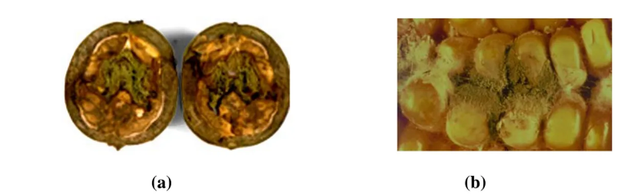
The vast majority of the known fungal species are strict saprophytes, however, there are several fungal genera containing species that cause disease (e.g., infections, allergies, toxicity) in plants, animals and man. The undesirable presence of these fungi can change different aspects of food commodities, principally due to the production of different pigments e.g. dark pigment “melanin” produced by many aspergillus species, undesirable color change can occur (Figure 1a) (Butler & Day, 1998). Development of certain other fungi on food commodities can produce undesirable moldy structures and smells (Figure 1b). These changes can both affect the quality and quantity of food products and hence can cause reduction in food productions. At present crop loss due to invasive plant pathogens, especially fungi, is estimated at $21 billion per year only in the United States, greater than the loss caused by non-indigenous insects (Rossman, 2009).
Metabolites produced by fungi during their growth are also major cause of the impairment of foodstuff quality. These metabolites can not only changes the organoleptic properties (taste) of food, but can also produce serious threats to human and animal health e.g. risk of intoxication due the presence of mycotoxins (Table 1) (Steve & Gary, 1999). The appearance of human mycosis and allergies can be the result of fungal contaminated food consumptions. In some eastern countries of Europe, it has been shown that the endemic nephropathie of the Balkans (NEB) (renal cancer and cancer of urinary tract) is linked to the consumption of cereals contaminated with ochratoxin A (Krogh, 1987).
6
(a) (b)
Figure 1: Contamination of food by fungi: (a) Aspergillus-infected walnuts, (b) The fungus Aspergillus sporulating on corn.
Table 1. Some mycotoxins, fungi that produce them and symptoms in animals
consuming feed contaminated with them (Steve & Gary, 1999).
Mycotoxin Fungi associated Symptoms/toxicology
Aflatoxin Aspergillus flavus, A. parasiticus
liver necrosis, liver tumors, reduced growth, depressed immune response, carcinogen
Fumonisin Fusarium verticillioides, F. proliferatum
Equine leukoencephalomalacia, porcine pulmonary edema Trichothecenes and Deoxynivalenol F. graminearum, F. culmorum, F. poae F. sporotrichioides
alimentary toxic aleukia, necrosis, hemorrhages, oral lesion in chickens, feed refusal, reduced weight gain Ochratoxins P.verrucosum,
A. ochraceus
Porcine nephropathy; various symptoms in poultry Citrinin Penicillium sp., Aspergillus sp. kidney damage Cyclopiazonic acid Penicillium sp., Aspergillus sp. Neurotoxin
7 Although reasons for why fungi produce secondary metabolites are not clear but it has been observed that these metabolites are commonly associated with sporulationprocesses in microorganisms (Hopwood, 1988; Mapleston et al., 1992; Stone & Williams, 1992). Secondary metabolites associated with sporulation can be placed into three broad categories: (i) metabolites thatactivate sporulation (for example, the linoleic acid-derived compounds produced by A. nidulans (Calvo et al., 2001; Calvo et al., 2002; Champe & El-Zayat, 1989; Mazur et al., 1991)), (ii) pigmentsrequired for sporulation structures, for example melanins required for the formation or integrity of both sexual and asexual sporesand overwintering bodies (Alspaugh et al., 1997; Kawamura et al., 1999), and (iii) toxic metabolitessecreted by growing colonies at the approximate time of sporulation,for example the biosynthesis of some deleterious natural products, such as mycotoxins (Hicks et al., 1997; Trail et al., 1995). These and other examples of secondary metabolites that fit into these categories are shown in table 2 (Bennett & Papa, 1988; Calvo et al., 2001; Champe et al., 1987; Champe & El-Zayat, 1989; Guzman-de-Pen˜a et al., 1998; Mazur et al., 1991; Takano et al., 2000).
Fungal secondary metabolites are classified into several groups including polyketides (e.g. aflatoxin and fumonisins), non-ribosomal peptides (e.g. sirodesmin, peramine and siderophores such as ferricrocin), terpenes (e.g. T-2 toxin, deoxynivalenol (DON)), indole terpenes (e.g. paxilline and lolitrems) (Ellen & Barbara, 2008) (Figure 2). Among these groups, polyketides which are natural products biosynthesized by the polymerisation of acetyl and propionyl subunits in a similar process to fatty acid synthesis (James, 2008) are the largest group of metabolites occurring in their greatest number and variety. They all are originating from a polyketone, the polyketide chain, giving these substances their name.
Fungal polyketide metabolites are a major potential source of novel bioactive compounds. They are the building blocks for a broad range of natural products or are further derivatized. The wide spectrum of activity of polyketides makes them economically, clinically and industrially the most sought after molecules. Ascomycetes and the related “imperfect” fungi commonly known such as Penicillium and Aspergillus
8 are considered to be the large producers of polyketide secondary metabolites (Nancy et al., 1994).
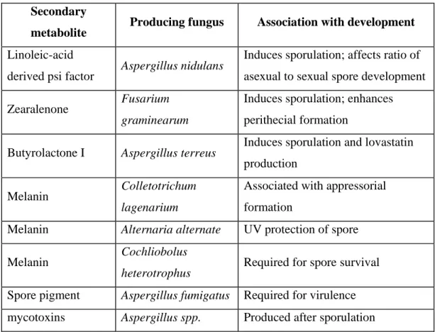
Table 2. Fungal secondary metabolites associated with sporulation processes and
development (Calvo et al., 2002).
Secondary
metabolite Producing fungus Association with development
Linoleic-acid
derived psi factor Aspergillus nidulans
Induces sporulation; affects ratio of asexual to sexual spore development Zearalenone Fusarium
graminearum
Induces sporulation; enhances perithecial formation
Butyrolactone I Aspergillus terreus Induces sporulation and lovastatin production
Melanin Colletotrichum lagenarium
Associated with appressorial formation
Melanin Alternaria alternate UV protection of spore Melanin Cochliobolus
heterotrophus Required for spore survival Spore pigment Aspergillus fumigatus Required for virulence mycotoxins Aspergillus spp. Produced after sporulation
9
Figure 2: Different classes of fungal secondary metabolites: (a) polyketides, (b)
10 Polyketide metabolites present a remarkable diversity in their structure (Figure.3). Varying from the simplest tetraketides such as 6-methylsalicilic acid (1, 6-MSA) and orsellinic acid (2) to highly modified aflatoxins (5) (Isao et al., 1998). Fungal polyketides are mostly aromatic compounds, though reduced complex type polyketides patuolide (6), brefeldin (7), lovastatin (8) and related compounds are not uncommon in fungi.
6-MSA (1) orsellinic acid (2) T4HN (3) (+) geodin (4)
aflatoxin B1(5) patuolide A (6) brefeldin A (7) lovastatin (8)
Figure 3. Diversity in the chemical structure of polyketides.
Fungal polyketides are a very diverse family of natural products with diverse biological activities and pharmacological properties. Several are known as mycotoxins like aflatoxin, ochratoxin, zearalenon, citrinin, patuline fumonisines, penicillic acid etc (Reboux, 2006). Several others are pharmacologically important like the antihypercholestemic drug lovastatin and its derivatives, the immunosuppressant cyclosporin, and ergotamine (Mathias & Dirk, 2007), the anti-fungal drugs echinocandins (Nyfelder & Keller, 1974) and antivirals i.e. HIV-integrase inhibitors, integrasone and integric acid (Marchand et al., 2003). The polyketide metabolites asperlactone and isoasperlactone have both insecticidal and weedicidal effects (Balcells et al., 1995; Balcells et al., 1998).
11
1.2
Aspergillus ochraceus / Aspergillus westerdijkiae NRRL 3174
Aspergillus ochraceus NRRL 3174 also known as Wilhelm strain (Wilhelm, 1887) belongs to the Aspergillus section Circumdati (also known as A. ochraceus group of Raper & Fennell (Raper & Fennell, 1965)), class Deuteromycetes (imperfect fungi, reproducing asexually), Ordre Hypomycetes, and family Moniliaceae. This group of organisms is especially well known for its production of ochratoxin A, named after the producer A. ochraceus CBS 263.67 / NRRL 3174 (Van der Merwe et al., 1965) and has attracted much interest by its role in the ochratoxin A contamination of coffee (Frisvad et al., 2004b; Sua´rez-Quiroz et al., 2004). Interestingly A. ochraceus NRRL 3174 or Wilhelm strain is now re-identified as Aspergillus westerdijkiae1 (Frisvad et al., 2004a). Hence several species previously identified as A. ochraceus could now be effectively named A. westerdijkiae. This fungus has got its name in honour of a former director of the CBS, Prof. Dr Johanna Westerdijk.
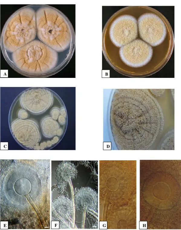
After 7 days of incubation on CYA (Czapek Yeast Agar) medium at 25 °C colony of A. westerdijkiae NRRL 3174 attains a diameter of 49-57 mm, while in MEA (Malt agar extract) its colony could be 42-47 mm in diameter, but never grows in CYA at 37°C. Cultural characteristics of this fungus include: production of good conidia on CYA at 25°C, coloring pale to light to dull yellow (color code:3B3–3A3); mycelium white, inconspicuous; sclerotia sparsely produced; pale yellow after 7 day, becoming dull orange at age. Reverse crème brown, no soluble pigment present. Good sporulation on MEA, velvety, pale to light or dull yellow (3A3–3B3) after 7 day; mycelium white, sclerotia sparsely formed, overgrown by conidial state and in shades of orange, reverse brown centre with yellow to medium–colored edge (Figure 4) (Frisvad et al., 2004a) (http://www.aspergillus.org.uk/indexhome.htm?secure/speciesdatabase/westerdijkiae.php ~main) . A. westerdijkiae is morphologically similar to Aspergillus ochraceus, though it is unable to grow at 37 °C. The white to cream sclerotia produced by A. westerdijkiae differ from the pink to vinaceous purple sclerotia of A. ochraceus.
1 Aspergilus westerdijkiae and Aspergillus ochraceus NRRL 3174 are same (Frisvad et al., 2004a). Hence
12
Figure 4. Aspergillus westerdijkiae after 7 days of incubation at 25 °C in CYA (A) and
MAE (B). Conidiophores (E, F). (Frisvad et al., 2004a).
Aspergillus ochraceus after 7 days of incubation at 25 °C in MAE (C and D). Conidiophores (G, H).
A B
C D
13 A. westerdijkiae is xerophile filamentous fungi hugely found in our environment. This fungus is frequently isolated from decomposed vegetables, soil, and desert. A. westerdijkiae principally causes seed and grain rot especially in maize. It contaminates many agriculture products including cereals, coffee, grapes (Christensen & Tuthill, 1985; Kozakiewicz, 1989). As this fungus best grows in temperature range of 15-31°C with water activity of 0.79, so most of the word regions favour the fungus to grow.
Aspergillus westerdijkiae has double economic interest. This fungus is used in food industry for the bioconversion of steroids and alcaloides (Chen, 1994; Richard et al., 1983) and also in the production of various enzymes (Chadha & Garcha, 1992). A. westerdijkiae is capable of producing many secondary metabolites including ochratoxin A & B, penicillic acid, mellein, 4-hydroxymellein, xanthomegnin, viomellein, vioxanthin, circumdatins (A–G), asperlactone, isoasperlactone, asperloxins and aspergamides (=avrainvillamides = stephacidins) etc (Ciegler et al., 1972; Robbers et al., 1978; Stack & Mislivec, 1978; Van der Merwe et al., 1965). The partially characterised extrolite NB1 is produced by all isolates of A.westerdijkiae, but not produced by any strain of A. ochraceus.
Details of some of the economically important polyketide metabolites produced by A. westerdijkiae are given bellow;
1.2.1
Ochratoxin A
The family of ochratoxin (Chemical formula C2OH18ClNO6 and Molecular weight 403.8
g/mol) contains a dozen of known molecules, but its most important representative is ochratoxin A (OTA) ((5-Chloro- 3,4-dihydro- 8-hydroxy- 3-methyl- 1-oxo- 1H-2-benzopyran-7-yl) –carbonyl)- L- phenylalanine) (Figure 5). OTA is consisting of a dihydroisocomurin moiety (the pentaketide-derived ochratoxine α) linked through thr carboxyl group to phenylalanine. Corresponding des-chloro analogues are ochratoxine B (OTB) and β. OTA is a toxic metabolite produced primarily by Aspergillus, but also by Penicillium and other molds in a divers array of environmental conditions. It is a white crystalline powder. Recrystallized from xylene, it forms crystals that emit green (acid
14 solution) and blue (alkaline solution) fluorescence in ultraviolet light; the melting point of these crystals is 169ºC (IARC, 1993). Infact the optimal temperature for the production of OTA by A. westerdijkiae is 28 °C, bellow 15 °C and above 37 °C its production is highly reduced. In contrary Penicillium viridicatum can produce OTA in a divers range of temperatures between 4 to 30 °C. Hence in cold regions OTA is mostly produced by Penicillium sp., while in hot regions it is mostly Aspergillus sp. producing it (Pohland et al., 1992; Varga et al., 1996).
Figure 5. Chemical structure of ochratoxine A.
Ochratoxine A is principally found in cereals (wheat, corn, rye, barley, oats), but also can be found in rice, soya, coffee, cocoa, beans, peas, peanuts fresh grapes and dry fruits (figs, grapes). It has also been detected in various cereal based product like flour, bread and pasta (Majerus et al., 1993), in beer (El-Dessouki, 1992) and even in wine and in grape juices (Zimmerli & Dick, 1996). Grains stored under high moisture/humidity (> 14 %) at warm temperatures (> 20 °C) and / or inadequately dried potentially can become contaminated by OTA (Ominski et al., 1994). Damage to the grain by mechanical means, physical means or insects can provide a portal of entry for the OTA producing fungus. Initial growth of fungi in grains can form sufficient moisture from metabolism to allow for further growth and mycotoxin formation.
Ochratoxin A is primarily a kidney toxin but in sufficiently high concentrations it can damage the liver as well. OTA is reasonably anticipated to be a class B animal and human carcinogen based on sufficient evidence of carcinogenicity in experimental animals. When ochratoxin A was administered in the diet, hepatocellular tumors (designated as well-differentiated trabecular adenomas), renalcell tumors (renal cystadenomas and solid renal-cell tumors), hepatomas (some exhibiting the trabecular
15 structure), and hyperplastic hepatic nodules were observed in male mice. In another study, administration of ochratoxin A in the diet induced hepatocellular carcinomas and adenomas in female mice. Gavage administration of ochratoxin A to male and female rats resulted in a dose-related increase in the incidence of renal-cell adenomas and adenocarcinomas; further, metastasis of the renal-cell tumors was also observed in male and female rats. When administered by gavage, ochratoxin A increased the incidence and multiplicity of fibroadenomas of the mammary gland in female rats (IARC, 1993; Ribelin et al., 1978).
The occurance of OTA in various foodstuffs is shown in table 3 (Magan & Olsen, 2004). These data confirm that OTA is found in a wide range of food stuffs from many different countries and in varying concentration. In a total diet study in the UK, low concentration of OTA was detected in all 50 samples analysed. In this study, OTA levels have been
Table 3. Occurance of OTA in selected foodstuff (Magan & Olsen, 2004)
Food stuff Origin Concentration (µg/kg) Contaminated samples
wheat Germany 1997 0.6-0.7 2/14
wheat Germany 1998 0.6-0.8 7/29
wheat Hungary 0.12-0.5 3/36
Wheat flour Hungary 0.11-0.15 2/16
Maiz meal Hungary 0.4 1/1
Oats Norway 0.065-0.47 3/22
Beer South Africa 3-3340 10/32
Beer Europ 0.025-0.121 40/40
Pure roasted coffee Hungary 0.17-1.3 33/50
Coffee products Europe 0.9 299/933
Chocolate UK 0.16 30/40
16
Table 4. European Union regulation for ochratoxin (μg/kg) (FAO Food and Nutrition
Paper No 81, 2004)
Product Concentration
Raw cereal grains 5
Products derived from cereals intended for direct human consumption 3 Dried vine fruit (currants, raisin and sultanas) 10
shown to varry between different commodities and withen different batches of commodities. Regulations for ochratoxin A are present in the European Community (FAO Food and Nutrition Paper No 81, 2004) (Table 4).
1.2.2
Penicillic acid
Penicillic acid (ϒ-keto-β-methoxy-∂-methylene-Aα-hexenoic acid) is a α, β-unsaturated, five-member lactones and a typical mycotoxin produced by Aspergillus sp. and Penicillium sp. (Figure 6) (Korzybski et al., 1978; Smith & Moss, 1985). This compound was first isolated in 1913 from Penicillium puberulum and subsequently from P. cyclopium by Raistrick who for the first time established its structure (James, 2008).
(a) (b)
Figure 6. Chemical structure of penicillic acid: (a) Ket-penicillic acid and (b)
17 The potential carcinogenic nature of penicillic acid possibly makes it a cause for concern, however work that has reported elsewhere with P. roqueforticell M-247 has shown that these toxins may not be produced in proteinaceous substrates, such as cheese (Dickens & Jones, 1961). This mycotoxin is toxic in experimental animals and has also been reported to be carcinogenic. The cytotoxicity of penicillic acid was studied in rat alveolar macrophages (AM) in vitro. The effects of penicillic acid on membrane integrity were studied by measuring cell volume changes and 51Cr release (Sorenson & Simpson, 1986). The carcinogenic effect of PA was studied in combination with other toxins. A direct interaction of patulin and penicillic acid with cellular DNA has been consistently observed in appropriate bacterial and mammalian cell systems. Single and double strand breaks were induced by both toxins in HeLa and mammary carcinoma cell DNA. An approximately fourfold larger total dose of penicillic acid was used to achieve carcinogenic effects in rats and mice, similar to those obtained with patulin (Umeda et al., 1977). PA also inhibit the Na± /K± ATPase (Na+, K+ activated ATPase) (Hayes, 1980), which catalyses the active transport of Na+ and K+ across the cell membrane. Penicillic acid is mostly known for its toxicity to animals but some previous work showed the phytotoxic effect of this molecule. P. cyclopium and P. canescens are the main contaminant fungi detected in seeds harvested in the northwest of France. Such contaminants are fearsome, since they affect the seeds before harvest time and may find optimal developing conditions when the seeds are stored, leading to alteration of the germination quality of these seeds. It has been found that the altered germinative qualities of the seeds were solely due to the growth of contaminating fungi or to the related action of phytotoxic metabolites penicillic acid (Jacqueline & Daniel, 1985). This metabolite was further reported to inhibit the growth of young plant roots of rice (Sassa et al., 1971) and of oat seedlings by lessening their respiration (Bastin & Roey, 1954). It was also reported to inhibit urease (Reiss, 1979) and RNase (Tashiro et al., 1979) activities in experimental animals.
Hence penicillic acid should now be considered as a phytotoxin as are aflatoxin B1, rubratoxin B, and zearalenone (Brodnik, 1975). The toxicity of penicillic acid is about 5 % that of aflatoxin B1, which gave 50 % inhibition of seed germination at the dose of 25
18 µg / ml, whereas 500 µg of penicillic acid per ml were necessary for a 50 % decrease of the main root length. Nevertheless, penicillic acid occurs in such high quantities (up to 2% dry weight) in infected corn (Kurtzman & Ciegler, 1970) that it must be considered seriously. Several other factors increased the seriousness of penicillic acid as phytotoxin. About 50 % of P. cyclopium strains produce penicillic acid (Le Bars, 1979), and its biosynthesis is higher on broken kernels (Le Bars, 1980). Penicillic acid is stable in substrates with low water content (Le Bars, 1982) and accumulates at the low temperatures (Ciegler & Kurtzman, 1970) of typical storage conditions. There is no additional evidence for natural occurance of PA in foodstuffs since the earlier report of its occurance (up to 230 µg/kg) in commercial maize and beans (Magan & Olsen, 2004). An Indian survey of over 1000 samples of food grains was negative. This paucity of occurrence of data may be due to its instability e.g. it is reactive with glutathione.
1.2.3
Methylsalicylic acid, asperlactone and isoasperlactone
Methylsalicylic acid (C8H8O3), is the archetypical tetraketide. It is a colourless or yellow
to red oily liquid with characteristic smell (Forrester & Gaucher, 1972). Esters of methylsalicylic acid are largely used in the production of perfumes and food aroma under the name oil Wintergreen (http://fr.wikipedia.org/wiki/Salicylate_de_méthyle). Its smell is very strong and attractive. It is also known for its analgesic and antipyretic properties. Several esters of methylsalicylic acid can provoke the anti-insect-herbivores defens system in plants. If the plant is infested with herbivorous insects, the release such compound may function as an aid in the recruitment of beneficial insects to kill the herbivorous insects. Several other esters of methylsalicylic acid can be toxic when absorbed and can be even lethal when inters into the body system in large quantity. A dose of 5 mL (a coffee spoon) is possible to be fetal for a child (Tintinalli et al., 2004). Apart from the toxic effect, this pheromon molecule has also been used to provoke successfully the salicylic acid based defence system in tobacco against tobacco mosaic virus (Vladimir et al., 1997).
19
Figure 7. Chemical Structure of 6-methylsalicylic acid
Methylsalicylic acid is biosynthesized by many Penicillium and Aspergillus species including A. westerdijkiae. This substance was intimately involved in establishing the polyketide biosynthetic pathway (Bu'Lock et al., 1968); it is one of the few fungal secondary metabolites whose enzymology has been examined successfully in detail (Dinroth et al., 1976); it was the model system that Bu'Lock and his colleagues (1968) used to develop the trophophase (A period in culture production characterized by active microbial cell growth and the formation primary metabolites) / idiophase (A period in culture growth during which secondary metabolites are produced) description of secondary metabolism; and it is one of the limited number of fungal systems wherein it has been shown convincingly that induction of the metabolic enzymes in submerged liquid culture correlates with a medium nutrient depletion (Grootwassink & Gaucher, 1980). In view of the pivotal role that 6- methylsalicylic acid has played in the development of our knowledge of fungal secondary metabolism, it is clear that any general explanation of why this process exists and what function it serves must be validated in 6-methylsalicylic acid-producing systems.
Beside the direct role of 6-methylsalicylic acid, it is the key precursor / intermediate of several other important polyketides like mycotoxin patulin (James, 2008) and antibiotic avilamycin (Gaisser et al., 1997). The importance of 6-methylsalicylic acid as precursor of patulin has been studied in various fungi. The first step in the production of patulin resides in the formation of 6-methylsalicylic acid, by the condensation of one molecule of acetyl-coA and 3 molecules of malonyl-coA. It has been shown that 6-methylsalicylic
20 acid specifically marked has been converted to patulin in Penicillium patulum (Bu’Lock, 1961; Tanenbaum & Bassett, 1958).
Asperlactone and isoasperlactone (9-chloro-8-hydroxy-8,9-deoxy) (Figure 8), along with patulin, isopatulin and orsellinic acid constitute a class of chemically related simples 5-membered cyclic lactones. The chemical structures of the two fungal metabolite i.e. asperlactone and isoasperlactone, have been deduced for the first time by the comparison of their spectroscopic data with that of aspyron and other known co-metabolites (Mary et al., 1984). Both asperlactone and isoasperlactone have the same chemical structure but they are stereochemically different. Not much is known about the biosynthetic pathways of isoasperlactone and asperlactone, except for a hypothetical scheme proposed by James & Andrew (1991). According to this scheme, the isomeric metabolites aspyrone, isoasperlactone and asperlactone are derived from a common biosynthetic precursor, the diepoxide. James & Andrew (1991) further stated that asperlactone is formed directly from the diepoxide, while isoasperlactone is formed from diepoxide via aspyrone pathway. Asperlactone is biosynthesized from aspyrone by the decarboxylation and arrangements of the polyketide intermediates followed by stereospecific opening of epoxy circle (Brereton et al., 1984).
(a)
(a) (b)
21 Asperlactone and isoasperlactone have been isolated for the first time from Aspergillus sp. in 1970 (Rosenbrook & Carney, 1970). It has been found that A. melleus is the key producer of both asperlactone and isoasperlactone (Garson et al., 1984). It is only recently found that A. ochraceus (re-identified as A. westerdijkiae) is also an active producer of these metabolites (Awad et al., 2005).
The activities of asperlactone and isoasperlactone are extensively studied in combination with other active polyketides metabolites like aspyrone and its synthetic derivatives. It has been found that asperlactone is more active than aspyrone (Balcells et al., 1995). Asperlactone equally contains antibacterial and antifungal activities (Torres et al., 1998). The bactericidal and fungicidal activities of asperlactone, isoasperlactone, aspyrone and some of its derivatives have been studied, with asperlactone and isoasperlactone in particular exhibiting strong activity against both bacteria and fungi; when compared to aspyrone derivatives (Balcells et al., 1998). In addition asperlactone isolated from an Aspergillus ochraceus Wilhelm strain displayed insect growth regulator (IGR) activity against Tribolium castaneum (Herbst). When the synthetic derivatives of aspyrone were tested in combination with asperlactone and isoasperlactone for IGR and ovicidal activities an improved impact of the combination was observed against T. castaneum (Balcells et al., 1998).
1.2.4
Mellein and Hydroxymellein
Recently, the isocoumarin compounds, mellein (3-methyl-8-hydroxy-3, 4-dihydroisocoumarin) and 4-hydroxymellein (3-methyl- 4, 8-dihydroxy-3, 4-dihydroisoco- umarin) (Figure 9) were isolated from the culture medium of several Aspergillus species including the ochratoxin producing strain A. westerdijkiae (Cole et al., 1971; Sasaki et al., 1970).
Mellein and hydroxymellein are structurally similar to the dihydroisocoumarin moiety of OTA and they possibly have similar biological properties (Van der Merwe et al., 1965). Because of this structural similarities with OTA, these metabolites were considered as intermediates of OTA (Huff & Hamilton, 1979). But later experiments with labelled
22 precursors of OTA ruled out this possibility and suggested that mellein and hydroxyl mellein are not intermediates in the biosynthetic pathway of OTA (Harris & Mantle, 2001).
Figure 9. Chemical structures of mellein and 4-hydroxymellein
The optimal biosynthetic conditions for mellein and hydroxyl-mellein have remained the topic of several studies in its principal producer Aspergillus ochraceus Wilhelm strain (Moor et al., 1972). Moor et al., (1972) demonstrated that mellein production showed a linear relationship with increasing sucrose concentration over the range studied, but production efficiency was best at 4, 8, and 16 %. 4-Hydroxymellein production efficiency was the same at sucrose concentrations of 2, 4, 8, and 16 %, similar to ochratoxin A and B production reported (Davis et al., 1969). For both mellein and 4-hydroxymellein production a sucrose concentration of 8 % was optimal (based on total amount produced per flask). Evaluation of production on medium, with equimolar concentrations of 15 carbon source, revealed that these compounds which are oxidized normally through both the hexose monophosphate and glycolytic pathways supported both growth and metabolites production. The organism grew poorly on the tricarboxylic acid cycle intermediates and metabolite production was poor. L-Glutamic acid at 6 g / liter produced the maximal amounts of mellein and hydroxymellein. Production of mellein and 4-hydroxymellein coincided with rapid mycelial growth at 20, 25, 30, and 35˚C. Incubation for longer than 8 to 10 days resulted in decreased yields of mellein but not 4-hydroxymellein. Incubation for 10 to 14 days at 25 to 30˚C was selected as optimal for production of both metabolites in the same medium (Moor et al., 1972).
23 Mellein and hydroxymellein has been shown to posses an impressive array of biological activities (Sun & Toia, 1993). For example, mellein is one of the constituents of the mandibular secretion of Carpenter ants and the defensive secretion of termites. It occurs in the male hair pencil of the oriental fruit moth and has been shown to have pheromonal properties in the castes of Camponotus pennsylvanicus. Mellein, hydroxumellein or combination of these two has been successfully used as phytotoxine against several unwanted plants (weeds) (Webb, 1991). Sasaki et al. (1970) reported the LD50 of mellein and 4-hydroxymellein in intraperitoneally injected mice as 250 to 500 and 1,000 to 1,500 mg/kg, respectively.
1.3
Fungal polyketides synthases
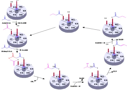
In spite of the fact that polyketides are greatly diverse both in their structures and functions, they are biosynthesized through the same mechanism analogous to the biosynthesis of fatty acid (through the action of fatty acid synthase (FAS)). The key chain-building step of polyketide biosyhnthesis is a decarboxylative condensation analogous to the chain elongation step of classical fatty acid biosynthesis which occurs in a well-understood way described in Figure 10. Unlike fatty acid biosynthesis, however, in
24 which each successive chain elongation step is followed by a fixed sequence of ketoreduction, dehydration and enoyl reduction, the individual chain elongation intermediates of polyketide biosynthesis undergo all, some, or none of these functional group modifications, resulting in a striking level of chemical complexity in the products (Figure 11). Additional degrees of complexity arise from the use of different starter units and chain elongation units as well as the generation of new stereo-isomers (Cox, 2000; Khosla et al., 1999).
Figure 11. Comparison of the biosynthetic schemes of fatty acid and polyketide
Polyketides are synthesized by sequential reactions catalyzed by a collection of modular enzymes activities called polyketide synthases (PKSs), nonribosomal peptide synthetase (NRPS) and hybrid between polyketide synthase and nonribosomal peptide synthetase (PKS-NRPS) (Bentley & Bennett, 1999; Hopwood & Sherman, 1990; Sebastian et al., 2007). These enzymes govern complex enzymology with fascinating mechanisms and great commercial appeal, because these enzymes create a multitude of secondary metabolites, many of which have become important drugs. These enzymes represent
25 some of the largest proteins known, and, as a consequence of their multifunctional character, a single protein can catalyze dozens of discrete biochemical reactions. For example, the rapamycin PKS (Schwecke et al., 1995) and cyclosporin NRPS (Weber et al., 1994) catalyze 51 and 40 steps in assembly of their respective products.
Polyketide synthases are large multi-enzyme protein complexes that contain a coordinated group of active sites or domains i.e. β-keto-acyl synthase (KS), acyl transferase (AT), ketoreductase (KR), dehydratase (DH), enoyle reductase (ER), methyl transferase (MT); acyle carrier protein (ACP) and thioesterase (TE). However AT, KS and ACP are the three principal domains while the remaing domains are optional (Figure 12).
Figure 12. Structure of a typical polyketide synthase, showing different functional
domains.
The PKS act in a step-wise manner to biosynthesize the corresponding polyketide from simple 2-, 3-, 4-carbon building blocks such as acetyl-CoA, propionyl CoA, butyryl-CoA and their activated derivatives, malonyl-, methylmalonyl- and ethylmalonyl-CoA (Figure 13).
Different functional domains on a PKS play different functions during the biosynthesis of a polyketide (Gokhale & Tuteja, 2001). AT domain serves for the loading of starter, extender and intermediate acyl units; ACP domain hold the growing macrolide as a thiol ester; KS domain, which is the most conserved domain among different PKSs, catalyses chain extension; KR domain is responsible for the first reduction to an alochol functionality; DH domain eliminates water to give an unsaturated thiolester; ER domain catalyses the final reduction to full saturation; MT domain catalyses the incarporation of
26
Figure 13. Different starter and extender units of polyketides.
Figure 14. Chemistry of polyketide synthases: role of different functional domains
27 methyle group; and finally a thiolesterase (TE) domain catalyses macrolide release and cyclisation (Figure 14)
.
In case of fatty acid biosynthesis, the ß-cetone group generated is completely reduced by a single sequential cycle of reduction (KR, DH and ER) before the following condensation until the skeleton attains the desired length. While this is not the case with PKSs, where the level of reduction varies by the partial or total omission of reduction reactions, which is the key to the diversity of fungal PKS (Fujii et al., 2001).
Based on the level of reduction of polyketides, fungal PKSs are divided into three main classes (Figure 15), as described bellow;
1.3.1
PKS producing reduced polyketides
These are the PKSs which contains along with principal domains, all of the optional domains responsible for reduction reactions (KR, DH and ER). The polyketides generated by this group of PKS undergo several stages of reductions in their biosynthesis, so that all ß-keto grouping are almost reduced. Example of these PKSs are the nonaceton and diceton synthases of lovastatine (A. terreus) and compactine (P. citrinum), PKS of citrinine (M. ruber), PKS of fumonisine (F. moniliform) and PKS of T-toxine (F. tricinctum) etc.
1.3.2
PKS producing partially reduced polyketides
These PKSs contain KS, AT, DH, KR and ACP functional domains or where there is only one cycle of reduction leading to the partial reduction of polyketides. Several PKSs of this class have been characterized as methylsalicylic acid synthase (MSAS) important for the biosynthesis of patulin in P. patulum (Beck et al., 1990), and MSAS in A. terreus (Feng & Leonard, 1998).
28
1.3.3
PKS producing non-reduced polyketides
The PKSs in this class do not contain any domain of reduction (ER, DH ou KR), which explains the absence of any reduction activity. They can have certain additional domains of ACP and a domaine Claisen-type cyclase or C-terminus (Kroken et al., 2003). This class includes the PKS of norsolinic acid, first intermediate in the biosynthetic pathway of aflatoxins in A. flavus, A. parasiticus and sterigmatocystine in A. nidulans (Feng & Leonard, 1998; Yu & Leonard, 1995), The PKS of wA type implied in the biosynthesis of spore pigments, YwA1 in A. nidulans and tetrahydroxynaphtalène synthase in the biosynthesis of melanin in C. lagenarium (Cox, 2000).
Figure 15: Organisation of functional domains diversity of fungal PKSs (NR : non
29
1.4
Architectur base classification of PKSs
At least three architecturally different PKS systems have been discovered in the microbial world (http://linux1.nii.res.in/~pksdb/polyketide.html). These systems are;
Type I systems consist of very large multifuctional proteins which can be either modular
or processive (for example the unique modular systems responsible for synthesis of macrolides in bacteria like erythromycin, rapamycin, rifamycin etc. (Figure 16a)) or iterative (Figure 16b)(for example the lovastatin nonaketide synthase) (Gokhale & Tuteja, 2001). Iterative Type I synthases are analogous to vertebrate fatty acid synthases. These are typically involved in the biosynthesis of fungal polyketides such as 6-methylsalicylic acid and aflatoxin. These PKSs are large multidomain proteins carrying all the active sites required for polyketide biosynthesis.
(a)
(b)
30 The iterative Type II systems consist of complexes of mono-functional proteins exemplified by the actinorhodin PKS from Streptomyces coelicolor. In these synthases, active sites are distributed among several smaller, typically monofunctional polypeptides. Type II synthases catalyse the formation of compounds that require aromatization and cyclization, but not extensive reduction or reduction / dehydration cycles. These PKSs are analogous to bacterial Fatty Acid Synthases and are involved in the biosynthesis of bacterial aromatic natural products such as actinorhodin, tetracenomycin and doxorubicin. A hypothetical "dynamic" model of this system is presented in Figure 17.
Figure 17. Iterative Type II system of PKSs.
Type III polyketide synthases are responsible for the synthesis of chalcones and
stilbenes in plants and polyhydroxy phenols in bacteria. Chalcone synthase like proteins are comparatively small proteins with a single polypeptide chain and are involved in the biosynthesis of precursors for flavonoids (Figure 18). Unlike all other PKSs, these proteins do not have a phosphopantetheinyl (P-Pant) arm on which the growing polyketide chains are tethered.
31
Figure 18. Representative model of type III polyketide synthases.
Most of the fungal PKSs are iterative type I. Recently, several chalcone synthase (CHS)-like genesPKS (belong to the type III PKS) have been discovered in some fungi like A. oryzae, Neurospora crassa et F. graminearum (Seshime et al., 2005).
1.5
Nonribosomal Peptide Synthetases
Nonribosomal peptide synthetases (NRPS) are multimodular enzymes that make nonribosomal peptides through a thiotemplate mechanismindependent of ribosomes. A unique feature of NRPS system is the ability to synthesize peptides containing proteinogenic as well as non-proteinogenic amino acids. In many cases these enzymes work in conjunction with polyketides synthases giving hybrid products. The products of these multifunctional enzymes have a broad spectrum of biological activities, some of which have been useful in medicine, agriculture, andbiological research (Kleinkauf & Doehren, 1996; Schwarzer & Marahiel, 2001; Smith et al., 1990).
NRPSs are organized into coordinated groups of active sites termed modules, in which each module is required for catalyzing one single cycle of product length elongation and modification of that functional group. The number and order of module and the type of domains present within a module on each NRPS determines the structural variation of the
32 resulting peptide product by dictating the number, order, choice of the amino acid to be incorporated and the modification associated with a particular type of elongation.
The minimum sets of domains required for an elongation cycle consist of a module. The modules are itself subdivided into different domains including Adenylation (A), Thiolation (T) or Peptidyl Carrier Protein (PCP), and Condensation (C) (Figure 19) (Schwarzer & Marahiel, 2001).
Figure 19. Minimum set of domains required in an NRPS
Each domain serves a specific function during the monomer incorporation. The A domain 500 to 600 amino acid residues is required for amino acid substrate recognition and activation. The 80 to 100 amino-acid-residue T domain, located downstream of the A domain, is the site for 4'-phosphopantetheine co-factor binding; the holoenzyme then activates aminoacyl substrates to form a thioester bond. A condensationdomain ( 450 amino acids) is typically found after each A-T module and functions in peptide bond formation and elongation of the nascent peptide. Generally, the number and order of modulespresent in a nonribosomal peptide synthetase determine the lengthand structure of the resulting nonribosomal peptide. In additionto A, T, and C domains, an N-methyl transferase (M) domain thatmethylates the amino acid specified by the A domain may be insertedbetween the A and T domains of any given module, and an epimerase(E) domain that changes an amino acid from the L- to the D-formmay be inserted between the T and C
33 domains. In some nonribosomalpeptide synthetases, a thioesterase domain is found at the C-terminalend of the protein and is thought to release the nonribosomalpeptide from the nonribosomal peptide synthetase (Cosmina et al., 1993)
There exist three major peptide biosynthesis types through the involvement of NRPS. The first one is linear biosynthesis (Figure 20a). In this type of biosynthesis there is collinearity between the synthetase and the peptide product. This means that the modular sequences of synthetase corresponds to that of the monomers incorporated in the product. This type of biosynthesis is the one most frequently occured. The second type is iterative biosynthesis (Figure 20b). In this case, some modules of the synthetase are used more than once during the synthesis of a peptide. The last type is the non-linear biosynthesis (Figure 20c). In this type of biosynthesis, the arrangement of modules of synthetase is different from that of the monomer peptide.
All the multi-modular enzymatic assemblies are organized with a common molecular logic for natural product: acyl chain initiation, chain elongation, and chain termination, catalyzed by protein modules that pass the growing chain from N to C termini of the assembly lines (Figure 21). Monomer units, such as amino or carboxy acids for NRPS and acetate or propionate for PKS, are selected and installed as acyl / aminoacyl-S-enzyme intermediates on a carrier protein domain in each module. The reactive thiol group in each acyl carrier/peptidyl carrier protein domain is introduced as a 4′-phosphopantetheinyl arm by posttranslational priming by dedicated 4′-phosphopantetheinyl transferases (Walsh et al., 1997). Specialized domains located within the corresponding NRPS/PKS modules can further modify the acylated substrates. Chain growth occurs as a series of translocating, elongating acyl/peptidyl-S-pantetheinyl carrier protein intermediates. The chain elongation chemistry in each module is by C-C bond formation in PKS assembly lines and by C-N amide bond formation in NRPS assembly lines, in upstream to downstream direction. When the full-length polyketide / polypeptide chain reaches the ultimate downstream carrier protein domain, the complete acyl chain is disconnected from its covalent thioester ether by a C-terminal thioesterase (TE) domain (Figure 21) (Bruner et al., 2002).
34 a)
b)
c)
Figure 20. Les trois types de biosynthèse a) Biosynthèse linéaire : synthèse du tripeptide
ACV, précurseur de la pénicilline et de la céphalosporine. b) Biosynthèse itérative : synthèse de l’entérobactine composée d’un dipeptide répété trois fois. c) Biosynthèse non linéaire : synthèse de la vibriobactine, peptide mixte NRPS/PKS
35
Figure 21. Surfactin Biosynthesis by the Modular Peptide Synthetase (A) The srf operon
(top) with the three genes srfA-A, srfA-B, and srfA-C coding for the surfactin synthetase subunits shown below the genes. Bars indicate the positions of modules within the protein, whereas the individual domains are shown as colored balls: A, adenylation domain; PCP, peptidyl carrier protein domain; C, condensation domain; E, epimerization domain; TE, thioesterase domain. The 4-phosphopantetheinyl cofactors with their active thiol groups are shown with the corresponding peptides attached at their current synthesis states. The growing peptide chain is passed from left to right, until the linear product at the last PCP domain is cyclized to the lipopeptide by the TE domain. (B) The SNAC (S-N-acetyl cysteamine) acyl peptide mimic can be cyclized by the genetically excised SrfTE domain. The native peptide (R =DLeu) and the soluble substrate (R = DOrn) are illustrated. The β-hydroxy fatty acid (FA) is likely to be attached by the N-terminal C domain.
To date, the majority of characterized nonribosomal peptide synthetases and their nonribosomal peptide products have beenfrom bacteria, especially spore-forming gram-positive soil bacteriasuch as Bacillus and Streptomyces species (Schwarzer & Marahiel, 2001; von Dohren et al., 1997). Relatively fewer fungal genes encoding nonribosomal
36 peptide synthetaseshave been fully sequenced and their corresponding peptide structures determined. These include Tolypocladium inflatum (niveum) simA, which controls production of the immunosuppressive drug cyclosporine(Weber et al., 1994), Penicillium chrysogenum acvA, which controls productionof the antibiotic penicillin (Baldwin et al., 1991), Ustilago maydis sid2 and Aspergillus nidulans sidC, each of which controls production of a siderophore (Eisendle et al., 2003; Yuan et al., 2001), Claviceps purpurea cpps1 and cpps2, both of whichare involved in the synthesis of ergot alkaloids (Correia et al., 2003; Tudzynski et al., 1999), Hypocrea virens tex1, which controls peptaibol synthesis (Wiest et al., 2002), Leptosphaeria maculans SirP, which controls sirodesmin biosynthesis (Eisendle et al., 2003),and three genes that control production of peptide virulence effectors: HTS1, which controls production of HC toxin by C. carbonum, a maize pathogen (Scott-Craig et al., 1992), AMT1, which controls production of AM toxin by Alternaria alternata, an apple tree pathogen(Johnson et al., 2000), and Esyn1, which controls production of enniatin by Fusarium scirpi and other Fusarium spp., potato and tomato pathogens(Haese et al., 1993; Herrmann et al., 1996).
1.6
Hybrid between PKS and NRPS
The structural and catalytic similarities between NRPSs and PKSs support the idea of combining individual NRPS and PKS modules for combinatorial biosynthesis. Recent advances in cloning and characterization of biosynthetic gene clusters for naturally occurring hybrid polyketide-peptide metabolites have provided direct evidence for the existence of hybrid NRPS-PKS systems (Metz, et al., 2001, Niermanm, et al., 2005, Sebastian, et al., 2007). Sequencing genome of several fungi including A. fumigatus and other filamentous fungi have revealed the presence of at least one gene that encodes a “hybrid” enzyme possessing domains typical of both PKS and NRPS. While such hybrid genes were described some time ago for bacteria (Du and Shen, 2001), only recently have the products of a few of the predicted fungal hybrid PKS-NRPS genes been determined. These include fusA, which is required for synthesis of a precursor of fusarin C in Fusarium moniliforme and F. venenatum (Song, et al., 2004) a gene required for equisetin biosynthesis in F. heterosporum (Sims, et al., 2005) and a gene that encode stenellin synthetase from Beauveria bassiana (Eley, et al., 2007).