UNIVERSITE DE LILLE
FACULTE DE MEDECINE HENRI WAREMBOURG
Année :2019
T H E S E P O U R L E D I P L O M E D ' E T A T
D E D O C T E U R E N M E D E C I N E
Facteurs prédictifs de cancer de prostate cliniquement significatif
chez les patients ayant une IRM négative : revue systématique de la
littérature et méta-analyse
Présentée et soutenue publiquement le 04 juin 2019 à 18h
Au Pôle Formation
Par Marc-Alexandre PAGNIEZ
_______________
JURY
Président :
Monsieur le Professeur VILLERS Arnauld
Assesseurs :
Monsieur le Professeur PUECH Philippe
Monsieur le Professeur LEROY Xavier
Directeur de Thèse :
Monsieur le Docteur OLIVIER Jonathan
Avertissement
La Faculté n'entend donner aucune approbation aux opinions
émises dans les thèses : celles-ci sont propres à leurs auteurs.
Anglais Français Abbreviation Signification Abréviation Signification ADC Apparent diffusion
coefficient
BN Biopsy naive
CCL Cancer core lengh csPCa clinically significant
prostate cancer csPCa Cancer de prostate cliniquement significatif DCE Dynamic Contrast
Enhanced
DWI diffusion weighted imaging GGG Gleason grade group
GS Gleason score
HR Hazard-ratio
isPCa insignificant prostate
cancer isPCa Cancer de prostate non significatif MRI multiparametric magnetic
resonance imaging IRM Imagerie par résonance magnétique multiparamétrique
NPV Negative predictive value VPN Valeur prédictive négative
OR Odd-ratio
PCa prostate cancer PCa Cancer de prostate
PBN Previous negative biopsy PPB Previous positive biopsy
Résumé ... 1
1. Introduction... 3
2. Materials and methods ... 5
2.1. Objective ... 5
2.2. Data acquisition and search strategy ... 5
2.3. Inclusion and exclusion criteria ... 5
2.4. Study selection and data collection ... 6
2.5. Quality assessment of included studies ... 6
2.6. Data synthesis and analysis ... 7
3. Results ... 9
3.1. Quantity of evidence identified ... 9
3.2. Risk of bias within studies ... 11
3.3. Characteristics of studies ... 12
3.4. Predictive factors ... 18
3.5. Meta-analysis ... 25
4. Discussion ... 28
4.1. Principaux résultats ... 28
4.2. Test index : IRM multiparamétrique ... 28
4.3. Examen de référence ... 28
4.4. Impact sur la pratique clinique et la recherche ... 29
4.5. Place de l’étude dans la littérature ... 31
4.6. Forces de l’étude et limites ... 31
5. Conclusion ... 34 6. Références ... 35 7. Suppléments ... 40 Supplément 1: Abstract... 40 Supplément 2: Introduction ... 42 Supplément 3: Discussion ... 43 Supplément 4: Conclusion ... 46 8. Annexes ... 47
Annexe 1: Research terms ... 47
Résumé
Contexte : Certaines sociétés savantes suggèrent que les patients ayant une
suspicion de cancer de prostate (PCa) et une IRM de prostate négative (IRMn) pourraient éviter des biopsies de prostate. Cette stratégie entraine le sous-diagnostic de 5 à 15% de cancers de prostate cliniquement significatifs (csPCa). Dans le but de réduire le risque de sous-diagnostiquer des csPCa chez ces patients avec IRMn, des facteurs cliniques et des marqueurs biologiques ont été utilisés en addition à l’IRM pour augmenter la valeur prédictive négative (VPN) de l’IRM.
Objectif : Réaliser une revue systématique de la littérature chez les patients naïfs de
tout cancer de prostate ayant une IRMn, dans le but d’identifier des facteurs prédictifs de csPCa dans cette population.
Méthode : Nous avons réalisé une revue de la littérature dans les bases Medline et
Scopus jusqu’en mars 2019. Les facteurs cliniques et marqueurs biologiques étudiés étaient l’âge, le taux de PSA, les isoformes du PSA, la densité de PSA (PSAD), le PCA3, le volume prostatique, les antécédents familiaux de cancer de prostate, les origines ethniques, et les calculateurs de risque. L’objectif principal était d’évaluer la valeur ajoutée de ces facteurs à la VPN de l’IRM pour réduire le risque de sous-diagnostiquer des csPCa. Les objectifs secondaires incluaient la réalisation d’une méta-analyse sur les performances de l’IRM combinée à ces facteurs prédictifs.
Résultats : Seize études ont été éligibles à l’inclusion. La PSAD<0.15ng/ml/ml était
le facteur prédictif le plus étudié pour le diagnostic de csPCa chez les patients avec
IRMn. Huit études (1015 patients) étaient éligibles pour évaluer la valeur ajoutée de la PSAD<0.15ng/ml/ml à la VPN de l’IRM pour réduire le risque de sous-diagnostiquer des csPCa. Lorsqu’elle était combinée à la PSAD, la VPN globale de l’IRM passait de 83.7% à 90.4% chez les patients naïfs de PCa. Cette augmentation était de 82.7% à 88.7% chez les patients naïfs de biopsies, et de 88.2% à 94.1% chez les patients ayant déjà eu des biopsies négatives. La PSAD (HR = 7.57 (2.73-21) p<0.001) et la PSAD<0.15ng/ml/ml (OR = 7.7 (2.8-21.3) p<0.001) présentaient les meilleures performances pour le diagnostic de csPCa.
Conclusion : La PSAD<0.15ng/ml/ml était le facteur prédictif le plus décrit pour
exclure les csPCa en cas d’IRMn. La PSAD pourrait être ajoutée aux résultats de l’IRMn pour mieux sélectionner les patients ne nécessitant pas de biopsies.
1. Introduction
Le cancer de prostate (PCa) est le cancer le plus fréquent chez l’homme en France avec une incidence de 48 427 nouveaux cas en 2013 (1). Le cancer de prostate est au cinquième rang des décès par cancer tous sexes confondus avec 8625 décès en 2013 (1). La mortalité spécifique au PCa est en baisse grâce à une détection plus précoce du PCa, dans la fenêtre de curabilité, et au perfectionnement de la prise en charge du PCa métastatique (2).
Une des principales préoccupations en lien avec le dépistage du PCa et son diagnostic précoce est le sur-diagnostic et le sur-traitement des cancers indolents. L’IRM de prostate multiparamétrique modifie le parcours diagnostique du PCa. De nombreuses études (3–6) ont démontré que l’utilisation de l’IRM améliorait le diagnostic de cancer de prostate cliniquement significatif (csPCa). L’IRM a aussi une excellente valeur prédictive négative (NPV) pour le diagnostic de csPCa, de l’ordre de 85 à 95% (7). Enfin l’IRM permet une réduction du diagnostic de cancer de prostate cliniquement non significatif (isPCa) (3,4).
L’IRM est maintenant recommandée avant toute série de biopsies de prostate (2,8,9). L’IRM de prostate est négative dans 20 à 30 % des cas (3,4) dans les populations de patients naïfs de PCa, et le dépistage précoce du cancer de prostate a tendance à augmenter ce taux (10). L’IRM pourrait être utilisée comme un test de tri (« triage test ») pour l’indication des biopsies de prostate chez les patients suspects de PCa. Cette hypothèse est basée sur les résultats de l’étude PRECISION (4) dans laquelle des patients naïfs de PCa et sans antécédent de biopsies ayant
une IRM de prostate négative (IRMn) ne bénéficiaient pas de biopsies de prostate. Ces résultats sont en partie nuancés par ceux de l’étude MRI-FIRST (5) dans laquelle 14% de csPCa n’étaient pas diagnostiqués par les biopsies ciblées seules, fournissant un argument pour la réalisation de biopsies systématisées. L’indication de biopsies de prostate chez les patients ayant une IRMn (PI-RADS 1-2) est donc controversée. Les patients ayant une IRMn pourraient éviter des biopsies de prostate, mais 5 à 15% de csPCa ne seraient alors pas diagnostiqués (3,4,11).
Des recommandations récentes soutiennent que les patients ayant une IRMn pourraient éviter des biopsies, surtout « si la densité de PSA (PSAD) est faible, et que la surveillance par PSA est appropriée » (8), ou « si la suspicion clinique de cancer de prostate est faible, et que le patient est informé et donne son accord pour cette prise en charge » (9).
A l’heure actuelle, nous ne sommes pas en mesure d’éviter des biopsies de prostate aux patients ayant une IRMn sans risquer de sous-diagnostiquer les csPCa.
L’association de facteurs prédictifs de csPCa à l’IRM pourrait augmenter la VPN de l’IRM et réduire la pratique de biopsies de prostate chez les patients dont le risque de csPCa est faible.
L’objectif de ce travail était de réaliser une revue systématique de la littérature dans le but de trouver les facteurs prédictifs de csPCa chez les patients naïfs de PCa ayant une IRMn.
2. Materials and methods
2.1. Objective
The primary objective was to systematically review the NPV of patient factors and biochemical markers as adjuncts to MRI in ruling out csPCa. Secondary outcomes were to systematically evaluate the predictive factors of csPCa in cancer naive patients, biopsy naive patients (BN) and in men with previous negative biopsies (PNB) with a nMRI, and to perform a meta-analysis of the NPV of MRI combined with these predictive factors.
2.2. Data acquisition and search strategy
The review was performed according to the Preferred Reporting Items for Systematic Reviews and Meta-Analysis (PRISMA) statement (12). Methods of the analysis and inclusion criteria were specified in advance and documented in a protocol. The
review protocol was published in the PROSPERO database
(http://www.crd.york.ac.uk/ PROSPERO; registration number CRD42019125549). A literature search using the PubMed and Scopus databases was performed, covering from January 01, 2010 to March 29, 2019, to identify eligible studies evaluating predictive factors of PCa/csPCa in patients with nMRI. The detailed search strategy is presented in Supplement 1.
2.3. Inclusion and exclusion criteria
No restriction on study type was imposed. Participants were male, adults, human patients. Included studies concentrated on men who were assessed for suspected PCa by MRI before undergoing prostate biopsy. Systematic prostate biopsies were
used as reference standards, with positive or negative cases of csPCa being determined by histopathological examination. Studies enrolling BN and PNB patients were included. Prebiopsy prostate MRI was considered the index test and comprised T2-weighted imaging (T2WI), and at least two functional imaging techniques (diffusion-weighted imaging [DWI], dynamic contrast-enhanced imaging [DCE], or apparent diffusion coefficient [ADC]). No results with biparametric MRI were included. Language restrictions were applied, excluding articles not written in English.
2.4. Study selection and data collection
Abstract and full-text screenings were performed by two reviewers independently (MAP and JO). Disagreement was solved by consensus. A standardized form was used to extract data on study methodology, patient characteristics, imaging protocols and main results (supplementary table S2). Any discrepancy concerning data extraction was solved by consensus. References from the included studies were manually retrieved to identify additional studies of interest.
2.5. Quality assessment of included studies
To assess the risk of bias (RoB), all included reports were independently reviewed by two reviewers using the Quality Assessment of Diagnostic Accuracy Studies (QUADAS-2) tool for diagnostic accuracy studies (13). Any discrepancy concerning the RoB assessment was solved by consensus.
2.6. Data synthesis and analysis
Clinical factors studied were age, prostate specific antigen (PSA), PSA isoforms, PSAD, prostate cancer gene 3 (PCA3), prostate volume, family history, ethnicity, and risk calculators. Predictive factors of all PCa, csPCa for patients with nMRI were extracted as notified in the studies. Outcome data regarding false negative and true negative values of MRI before prostate biopsy were recorded as reported by authors. NPV of each study was calculated if not available. NPV of MRI combined with any marker were recorded as reported by authors, or calculated if not available. Authors of the included papers were asked for any missing data.
A meta-analysis was undertaken to calculate the pooled NPV of MRI coupled with PSAD across the available studies. To ensure appropriate clinical homogeneity of the studies included in this meta-analysis, we selected only the studies enrolling BN patients and/or patients with a PNB, and fulfilling the following criteria that were defined a priori:
- reference standard consisting of prostate biopsy with at least 12 samples on all patients;
- MRI protocol comprising at least T2WI, DWI and DCE;
- MRI results presented as a five-level score, using a subjective Likert scale or the Prostate Imaging Reporting Data System (PI-RADS) score ;
- definition of positive MRI as a score ≥3/5;
- only studies defining csPCa as GGG≥2 were selected for the meta-analysis assessing the MRI NPV for csPCa coupled with PSAD.
To combine the pooled NPVs, we firstly transformed individual NPVs to a quantity using the Freeman-Tukey variance stabilizing arcsine transformation (14), and secondly we used fixed or DerSimonian-Laird random effects models (15) to obtain
the combined estimates. Random effects model were used in case of significant heterogeneity between studies. Between-studies heterogeneity in NPV was quantified by the I² statistic and tested by the Cochran Q test. Forest plots were created using the DistillerSR Forest Plot Generator from Evidence Partners (https://www.evidencepartners.com/resources/forest-plot-generator/). For other studies not included in the meta-analysis based on the criteria described above, a narrative synthesis of the data was performed.
3. Results
3.1. Quantity of evidence identified
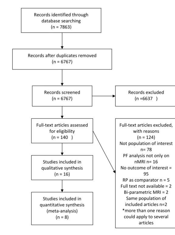
The study selection process is depicted in the PRISMA flow diagram (figure 1). A total of 7863 abstracts were retrieved. After abstract screening and removal of duplicates, 140 articles were eligible for full text screening, of which 16 studies were eligible for inclusion in this systematic review.
Figure 1: flow chart of included studies
Records identified through database searching (n = 7863) Sc reen in g In clu ded
El
ig
ib
ili
ty
Id en tif icat io nRecords after duplicates removed (n = 6767)
Records screened
(n = 6767) Records excluded (n =6637 )
Full-text articles assessed for eligibility
(n = 140 )
Full-text articles excluded, with reasons
(n = 124)
Not population of interest n= 78
PF analysis not only on nMRI n= 16 No outcome of interest =
95
RP as comparator n = 5 Full text not available = 2
Bi-parametric MRI = 2 Same population of included articles n=2 *more than one reason
could apply to several articles Studies included in qualitative synthesis (n = 16) Studies included in quantitative synthesis (meta-analysis) (n = 8) 10
3.2. Risk of bias within studies
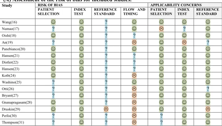
Out of the 16 included studies, 12 were single-center, 3 were multicenter studies, and 1 did not specify. Eight studies were prospective and 8 were retrospective. RoB assessment using QUADAS-2 was performed for each of the individual studies. Overall, the RoB was heterogeneous across studies for patient selection and flow and timing, homogeneous in index test and reference, in which RoB was low and unclear in all studies, respectively. RoB assessment is reported in figure 2.
Figure 2: risk of bias QUADAS-2
(A) Assessment of the risk of bias for included studies.
Study RISK OF BIAS APPLICABILITY CONCERNS
PATIENT
SELECTION INDEX TEST REFERENCE STANDARD FLOW AND TIMING PATIENT SELECTION INDEX TEST REFERENCE STANDARD
Wang(16) ? Numao(17) ? ? ? Oishi(18) ? ? An(19) ? ? ? Panebianco(20) ? Hansen(21) ? ? Distler(22) ? ? Hansen(23) ? ? Kotb(24) ? Washino(25) ? ? Otti(26) ? ? ? Bryant(27) ? ? Gnanapragasam(28) ? Druskin(29) Perlis(30) ? ? ? Thompson(31) ? ? ?
(B) Risk of bias summary graph.
3.3. Characteristics of studies
3.3.1. Main characteristics
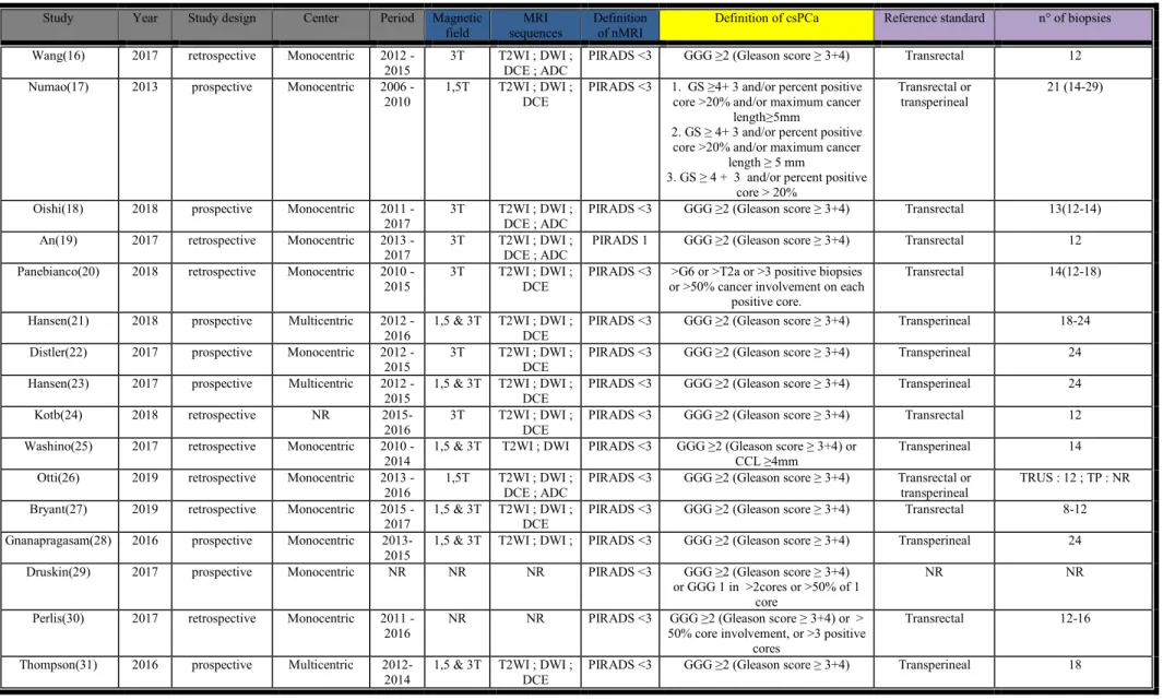
The study and patient baseline characteristics are presented in Table 1.
Table 1: Baseline characteristics of included studies.
Study Year Study design Center Period Magnetic
field sequences MRI Definition of nMRI Definition of csPCa Reference standard n° of biopsies
Wang(16) 2017 retrospective Monocentric 2012 -
2015 3T T2WI ; DWI ; DCE ; ADC PIRADS <3 GGG ≥2 (Gleason score ≥ 3+4) Transrectal 12
Numao(17) 2013 prospective Monocentric 2006 -
2010 1,5T T2WI ; DWI ; DCE PIRADS <3 1. GS ≥4+ 3 and/or percent positive core >20% and/or maximum cancer
length≥5mm
2. GS ≥ 4+ 3 and/or percent positive core >20% and/or maximum cancer
length ≥ 5 mm
3. GS ≥ 4 + 3 and/or percent positive core > 20%
Transrectal or
transperineal 21 (14-29)
Oishi(18) 2018 prospective Monocentric 2011 -
2017 3T T2WI ; DWI ; DCE ; ADC PIRADS <3 GGG ≥2 (Gleason score ≥ 3+4) Transrectal 13(12-14)
An(19) 2017 retrospective Monocentric 2013 -
2017 3T T2WI ; DWI ; DCE ; ADC PIRADS 1 GGG ≥2 (Gleason score ≥ 3+4) Transrectal 12
Panebianco(20) 2018 retrospective Monocentric 2010 -
2015 3T T2WI ; DWI ; DCE PIRADS <3 >G6 or >T2a or >3 positive biopsies or >50% cancer involvement on each
positive core.
Transrectal 14(12-18)
Hansen(21) 2018 prospective Multicentric 2012 -
2016 1,5 & 3T T2WI ; DWI ; DCE PIRADS <3 GGG ≥2 (Gleason score ≥ 3+4) Transperineal 18-24
Distler(22) 2017 prospective Monocentric 2012 -
2015 3T T2WI ; DWI ; DCE PIRADS <3 GGG ≥2 (Gleason score ≥ 3+4) Transperineal 24
Hansen(23) 2017 prospective Multicentric 2012 -
2015 1,5 & 3T T2WI ; DWI ; DCE PIRADS <3 GGG ≥2 (Gleason score ≥ 3+4) Transperineal 24
Kotb(24) 2018 retrospective NR
2015-2016 3T T2WI ; DWI ; DCE PIRADS <3 GGG ≥2 (Gleason score ≥ 3+4) Transrectal 12
Washino(25) 2017 retrospective Monocentric 2010 -
2014 1,5 & 3T T2WI ; DWI PIRADS <3 GGG ≥2 (Gleason score ≥ 3+4) or CCL ≥4mm Transperineal 14
Otti(26) 2019 retrospective Monocentric 2013 -
2016 1,5T T2WI ; DWI ; DCE ; ADC PIRADS <3 GGG ≥2 (Gleason score ≥ 3+4) Transrectal or transperineal TRUS : 12 ; TP : NR
Bryant(27) 2019 retrospective Monocentric 2015 -
2017 1,5 & 3T T2WI ; DWI ; DCE PIRADS <3 GGG ≥2 (Gleason score ≥ 3+4) Transrectal 8-12
Gnanapragasam(28) 2016 prospective Monocentric
2013-2015 1,5 & 3T T2WI ; DWI ; PIRADS <3 GGG ≥2 (Gleason score ≥ 3+4) Transperineal 24
Druskin(29) 2017 prospective Monocentric NR NR NR PIRADS <3 GGG ≥2 (Gleason score ≥ 3+4)
or GGG 1 in >2cores or >50% of 1 core
NR NR
Perlis(30) 2017 retrospective Monocentric 2011 -
2016 NR NR PIRADS <3 GGG ≥2 (Gleason score ≥ 3+4) or > 50% core involvement, or >3 positive
cores
Transrectal 12-16
Thompson(31) 2016 prospective Multicentric
2012-2014 1,5 & 3T T2WI ; DWI ; DCE PIRADS <3 GGG ≥2 (Gleason score ≥ 3+4) Transperineal 18
* mean results and not median; ˟ results on all patients (and not only nMRI) NR: Non Reported
AA: Afro-American ; BN : biopsy naive ; PNB : previous negative biopsy ; PPB : previous positive biopsy ; DRE : digital rectal examination ; PSAD : PSA density
patients (>T1c) (median) history of
PCa Wang(16)* 84 84 39 30 15 Afro-American BN 61.9(+-7.2) 5.9(+-6.3) 18% 0.17(0.15) 2(5%) 18% PNB 64(+-7.6 9.4(+-6.9) 10% 0.19(0.21) 1(3%) 20% Numao(17) 351 194 194 0 0 65 (59–70) 6.3 (4.9–9.1) 19% 32 (24–42) 0.19 (0.14–0.30) Japanese 5.9% Oishi(18) 135 135 48 60 27 64(58-69) 5.9(4.1-8.0) 8.1% 55(38-79) 0.1 (0.073-0.15) 24% An(19) 114 114 20 53 41 61 (57-67) 5,5(3,6-8,7) 4.4% 57(42-80) Caucasian 93(81.6%); AA 12(10.5%); other 9(7.9%) Panebianco(20) 1255 1255 659 596 0 BN 66(62-69) 5.9(3.9-7.6) 10% 50(42-68) 0.11(0.08-0.14) 11% PNB 68(60-72) 5.6(3.2-7.8) 9% 60(38-73) 0.11(0.08-0.15) 7% Hansen(21) ˟ 807 236 236 0 0 65(59-70) 6.5(4.9-8.8) 23% 42(30-58) 0,15(0,10-0,22) Distler(22) ˟ 1040 344 NR NR 0 65 7,2 45 0,16 Hansen(23) ˟ 487 144 0 144 0 66(60-71) 9,0(6,7-13,4) NR 56(40-80) 0,15(0,10-0,24) Kotb(24) ˟ 228 81 0 81 0 64.6 ± 6.6 8.1 ± 7.8 0.14 ± 0.16 Washino(25) ˟ 288 131 131 0 0 69(64-74) 7,5(5,5-11,0) 16% 28,7(23,3-39,4) 0,26(0,17-0,38) Otti(26) ˟ 792 348 348 0 0 66 +- 10 6.75 (4.18) 19.5% 49.50 (36) 0.13 (0.12) Bryant(27) ˟ 1789 278 278 0 0 68 (37-88 ; 63-73) 7.6 (0.4-2668 ; 5.7-11.5) 48% 56.1 ( 10.4-244 ; 40.1-79) 0.13 (<0.1-42.4 ; 0.09 - 0.23) 17% Gnanapragasam(28) ˟ 279 94 0 NR NR 66 (45-80) 52 (11-230) Druskin(29) ˟ 104 20 NR NR 0 65.0(59.3-70.8) 7.0(4.9-.10,2) 0% 50 (37.32-70,0) 0.14(0.096-0.21) AA 11,6% Perlis(30) ˟ 286 58 0 NR NR 62.5(58-68) 6.35(4,6-8,8) 5,6% 47(35-63) 18,8% Thompson(31) ˟ 344 79 79 0 0 62.9 5.2 44.40% 40 26,70% 14
® results after median follow-up of 38 months in BN and 60 months in PNB
csPCa : clinically significant prostate cancer ; BN : biopsy naive ; PNB : previous negative biopsy ; AS/PPB : active surveillance/ previous positive biopsy ; NPV : negative predictive value ; nMRI : negative MRI
csPCa csPCa csPCa in
BN csPCa in BN csPCa in PNB in PNB csPCa in AS/PPB csPCa in AS/PPB MRI for csPCa csPCa in MRI for BN MRI for csPCa in PNB nMRI Wang(16) 13.1% 11 10.30% 4 16.70% 5 13.3 2 86.90% 89.7% 83.3% 18.1% Numao(17) 20% 38 20% 38 (i). 90.7% ; (ii). 87.4% ; (iii). 88.1% (i). 90.7% ; (ii). 87.4% ; (iii). 88.1% 55% Oishi(18) 18% 24 21% 10 8% 5 33% 9 82% 79% 92% 12.90% An(19) 3.60% 4 0% 0 0% 0 9.80% 4 96.5% 100% 100% NA Panebianco(20)® 4.80% 60 5.50% 36 4.00% 24 NA NA NA 31% Hansen(21) 20,3% 48 20.30% 48 80% 80% 29.2% Distler(22) 20.60% 71 NR NR NR NR 79.4% 76.8% 83.0 % 33.10% Hansen(23) 7,6% 11 7.60% 11 92.4% 92.40% 29.7% Kotb(24) 12.40% 10 12.40% 10 88% 88% 24.10% Washino(25) 13.70% 18 13.70% 18 86.30% 86.30% 44.40% Otti(26) 14.40% 50 14.40% 50 85.60% 85.60% 44% Bryant(27) 15.10% 42 15.1% 42 84.90% 84.90% 35.10% Gnanapragasam(28) 22.3% 21 NR NR NR NR 78% 78% 33.7% Druskin(29) 10% 2 90% 19.20% Perlis(30) NR NR NR NR NR NR NR 55.2% Thompson(31) 8% 6 8% 6 92% 92% 23% 15
3.3.2. Biopsy status
The patient population consisted of BN in 6 studies, PNB in 2 studies, BN and PNB in 3 studies, BN, PNB and previous positive biopsy (PPB) in 3 studies. 2 studies included PNB and PPB.
3.3.3. MRI characteristics
The magnetic field strength was 1.5 and 3 Tesla in 2 and 6 studies, respectively. Six studies used both 1.5 and 3 Tesla MRI systems. In 2 studies the magnetic field strength was not specified. DWI, DCE and ADC were used in 14, 12 and 4 studies, respectively.
3.3.4. Definition of negative MRI
The definition of nMRI varied little across studies. Fifteen studies used PI-RADS
score <3 and one study PI-RADS score <2.
3.3.5. Biopsy methods
Regarding the reference standard, TRUS-guided biopsies were used in 7 studies, trans-perineal biopsies in 6 studies, and mixed TRUS-guided and trans-perineal biopsies in 2 studies.
In one study, the biopsy approach was unclear. The number of cores per biopsy procedure was <18 in 9 studies, >18 in 4 studies, and variable among patients in 2 studies. In one study, the number of biopsy cores taken was unclear.
3.3.6. Definition of clinically significant prostate cancer
The definition of csPCa varied little across studies. 12 studies used Gleason Grade Group (GGG) ≥2 (Gleason score ≥ 3+4). Perlis et al. (30) used two definitions: (i) Gleason 7 or a cancer core length of 6 mm or greater or (ii) any Gleason 7. Druskin et al. (29) used GGG ≥2 or GGG =1 in >2 cores or >50% of any core). Panebianco et al. (20) used the EAU-ESTRO-SIOG guidelines selection criteria for isPCa, eligible for active surveillance (32). Numao et al. (17) used 3 definitions: (i) GS ≥4+3 and/or percent positive core>20% and/or maximum cancer length ≥5mm; (ii) GS ≥3+4 and/or percent positive core>20% and/or maximum cancer length ≥5mm; (iii) GS ≥ 3+4 and/or percent positive core > 20%.
3.4. Predictive factors
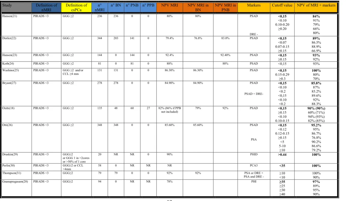
Four studies reported predictive factors for PCa in patients with nMRI at biopsy (clinical predictors of biopsy outcome) and one study reported predictive factors for csPCa in patients with nMRI during follow-up. Twelve studies reported NPV of MRI combined with a marker or a clinical factor.
Table 2: NPV of MRI combined with markers for the diagnostic of csPCa Study Definition of
nMRI Definition of csPCa nMRI n° n° BN n° PNB n° PPB NPV MRI NPV MRI in BN NPV MRI in PNB Markers Cutoff value NPV of MRI + markers
Hansen(21) PIRADS <3 GGG ≥2 236 236 0 0 80% 80% PSAD
DRE -<0,15 <0.10 0.10-0.20 ≥0.20 84% 91% 79% 66% 80%
Distler(22) PIRADS <3 GGG ≥2 344 203 141 0 79.4% 76.8% 83.0% PSAD <0,15
<0.07 0.07-0.15 ≥0.15 89% 86.5% 88.9% 66.9%
Hansen(23) PIRADS <3 GGG ≥2 144 0 144 0 92.4% 92.40% PSAD <0,15
≥0.15 93% 92%
Kotb(24) PIRADS <3 GGG ≥2 81 0 81 0 88% 88% PSAD <0,15 93%
Washino(25) PIRADS <3 GGG ≥2 and/or
CCL ≥4 mm 131 131 0 0 86.30% 86.30% PSAD 0.15-0.29 <0,15
≥0.3
100% 80% 70%
Bryant(27) PIRADS <3 GGG ≥2 278 278 0 0 84.90% 84.90% PSAD
PSAD + DRE-<0,15 <0.10 <0.2 <0,15 <0.10 <0.2 85.8% 87% 85.2% 89.6% 92% 88.3% Oishi(18) PIRADS <3 GGG ≥2 135 48 60 27 82% (86% if PPB
not included) 79% 92% PSAD <0,15 ≥0.15
<0.10 0.10-0.15 90% (90%) 60% (71%) 94% (93%) 82% (85%)
Otti(26) PIRADS <3 GGG ≥2 348 348 0 0 85.60% 85.60% PSAD
PSA <0,15 <0.12 0.12-0.15 ≥0.15 <5 5-10 ≥10 95.2% 95% 86.7% 76.8% 90.2% 86.6% 79.2% Druskin(29) PIRADS <3 GGG≥2 or GGG 1 in >2cores or >50% of 1 core 20 NR NR 0 90% PHID >0.44 100% Perlis(30) PIRADS <3 GGG≥2 or CCL >4mm 58 0 NR NR NR PCA3 <35 100%
Thompson(31) PIRADS <3 GGG≥2 79 79 0 0 92% 92% PSA or DRE +
PSA and DRE - ≥10 <10 100% 90%
Gnanapragasam(28) PIRADS <3 GGG≥2 94 0 NR NR 78% PHI ≥35
≥25 ≥30 ≥40 97% 89% 95% 90% 19
Table 3: studies including predictive factors of all prostate cancer / csPCa.
*controlling for time to biopsy, PV and MRI suspicion (no MSR vs PI-RADS I vs II), the PCPTRC estimated risk for csPCa (as a continuous variable) was a significant independent predictor for csPCa on biopsy (OR 1.01, P < .01).
● Results were told as significant predictors of subsequent csPCa, but no Odd-ratios were available
Study n°
nMRI Definition of nMRI Definition of csPCa Age PSA Prostate Univariate analysis Multivariate analysis
volume PSAD PSAD < 0.15 Previous negative
biopsy > T1c Ethnicity (AA vs non AA) Family history of PCa
Other Age PSA Prostate
volume PSAD PSAD <0.15 Previous negative
biopsy
> T1c Ethnicity other
Predictive factors of csPCa at biopsy Wang(16) 84 BN 39 PNB 30 PPB 15 PIRADS <3 GGG ≥2 6.1 +4.6 vs 14.4 +10.5 p<0.01 0.14 + 0.12 vs 0.63 + 0.28 p<0.01 PCPTRC 8.4 +6.5% vs 23.1 + 22.7% p<0.01 PCPTRC* OR = 1.01 p< 0.01). Numao(17) 151 BN 151 PIRADS <3 1. GS ≥4+3 and/or percent positive core >20% and/or maximum cancer length ≥5 mm OR = 1.1 (0.37–3.5) p=0.81 OR = 1.4 (0.46–14) p= 0.56 OR = 8.1 (2.1–54) p<0.01 OR = 11 (2.1–200) p<0.01 OR = 1.4 (0.07–9.0) p =0.76 % fPSA (cutoff 15%) OR=3.3 (1.1–11) p=0.03 OR =8.1 (2.1–54) p< 0.01 2. GS ≥3+4 and/or percent positive core >20% and/or maximum cancer length ≥5 mm ; OR =1.3 (0.49–3.5) p = 0.60 OR =1.5 (0.56–4.0) p= 0.44 OR =5.2 (1.8–19) p < 0.01 OR =3.0 (1.0–11) p<0.04 0.99 (0.05– 6.1) p=0.99 % fPSA (cutoff 15%) OR = 2.6 (0.97–7.1) p=0.06 OR =5.2 (1.8–19) p< 0.01 3. GS ≥ 3+4 and/or percent positive core >20% OR =1.5 (0.55–4.1) p = 0.44 OR =1.7 (0.63–4.9) p= 0.30 OR =4.8 (1.6–18) p < 0.01 OR =2.8 (0.94–10) p<0.06 1.1 (0.05– 6.5) p=0.96 % fPSA (cutoff 15%) OR =2.3 (0.85–6.4) p=0.10 OR =4.8 (1.6–18) p< 0.01 Predictive factors of no csPCa at biopsy
Oishi(18) 135 BN 48 PNB 60 PPB 27 PIRADS <3 GGG ≥2 OR = 0.99 (0.94-1.06) p = 0.97 OR = 1.02 (0.93-1.11) p = 0.7 OR=1.03 (1.01-1.06) p=0.001 OR = 0.01 (0.0002-0.55) p = 0.02 OR = 5.93 (2.32-15.2) p <0.001 OR =3.73 (1.3-10.7) p=0.01 OR = 0 (0-1.81) p= 0.2 OR = 0.96 (0.35-2.68) p = 0.9 BN (Y/N) OR = 0.73 (0.30-1.79) p=0.5 PPB (Y/N) OR = 0.32 (0.12-0.85) p=0.02 PIRADS v.1 vs v.2 OR = 0.62 (0.24-1.56) p=0.31 OR = 7.7 (2.8-21.3) p <0.001 OR = 5.2 (1.6-16.5) p = 0.005
Predictive factors of all PCa at biopsy An(19) 114 BN 20 PNB 53 PPB 41 PIRADS <2 GGG ≥2 OR = 0.99 (0.93-1.05) p = 0.79 OR = 0.90 (0.80-1.01) p = 0.19 OR = 0.99 (0.97-1.00) p=0.06 OR = 2.36 (0.37-14.95) p=0.36 OR = 1.14 (0.29-4.58) p = 0.85 OR = 1.01 (0.95-1.07) p = 0.82 OR = 0.99 (0.81-1.21) p = 0.93 OR = 0.98 (0.96-1.01) p=0.19 OR = 1.41 (0.21-9.55) p = 0.73 OR = 0,98 (0,23-4,19) p=0,98 Predictive factors of csPCa with follow-up
Panebianco(20) 1255 BN 659 PNB 596 PIRADS <3 >G6, >T2a NR● NR● NR● NR● HR = 0.93 (0.89-0.98) p = 0.005 HR =1.21 (1.1-1.32) p<0.001 HR = 7.57 (2.73-21) p<0.001 HR = 1.01 (0.53-1.93) p = 0.97 20
3.4.1. PSA
Two studies reported NPV of MRI combined with PSA. Thompson et al. (31) reported results on BN patients. The NPV of MRI for csPCa among men with a pre-biopsy PSA≥10.0ng/ml or an abnormal DRE was 100%. Among all men with a normal DRE and a PSA<10.0 ng/ml, the NPV was 90% for csPCa.
Otti et al.(26) reported a NPV of 85.1% in 348 BN patients with nMRI. Patients were stratified in three groups: PSA <5ng/ml, PSA between 5 and 10ng/ml and PSA ≥10 ng/ml. In those patients, NPV of csPCa was 90.2%, 84.8% and 79.2%, respectively. PSA was reported in 5 studies (16–20), and was a significant predictive factor of csPCa in 2 studies (16,20). In multivariate analysis, PSA was reported in 2 studies (19,20) and was a significant predictive factor of csPCa in one study (20) (HR=1.21 (1.1-1.32) p<0.001).
3.4.2. PSA density
Eight studies (18,21–27) reported NPV of MRI combined with a PSAD<0.15ng/ml/ml. NPV of MRI coupled with PSAD<0.15ng/ml/ml varied between 84 and 100%, independently of biopsy status. PSAD was reported in 4 studies (16–18,20), and was a significant predictive factor of csPCa in all 4 studies, except for definition 3 in reference (17). In multivariate analysis, PSAD was reported in one study (20) and was a significant predictive factor of csPCa (HR = 7.57 (2.73-21) p<0.001). PSAD<0.15 ng/ml/ml was reported in one studies (18) and was a significant predictive factor of no csPCa (OR = 7.7 (2.8-21.3) p<0.001).
3.4.3. PCA3
Perlis et al.(30) reported results on patients with PNB and some with previous positive biopsy (PPB). 154 patients with PCA3 score and MRI had repeat biopsy. No patient (0/26) with nMRI and a normal PCA3 score had csPCa on biopsy (NPV=100%).
3.4.4. PHI and PHI density
Druskin et al. (29) reported results on Prostate Health Index (PHI) density (PHID) combined with MRI in PNB for the diagnosis of csPCa. 241 cancer naive patients were included, 104 had MRI. MRI NPV for csPCa was 90% (18/20 patients). NPV of MRI coupled with PHID ≥0.44 was 100%.
Gnanapragasam et al.(28) reported results of PHI test and MRI in 279 patients (PNB and PPB), including 94 with nMRI. In those patients, and with a PHI score ≥35, the NPV for csPCa was 97% (84–100%) whereas NPV of MRI alone was 75.5% (71/94 patients).
3.4.5. Age
Age was reported in 4 studies (17–20) and was a significant predictive factor of csPCa in one study (20). In multivariate analysis, age was reported in 2 studies (19,20) and was a significant predictive factor of csPCa in one study (HR=0.93 (0.89-0.98) p=0.005).
3.4.6. Prostate volume
Prostate volume was reported in 3 studies (17–19), and was a significant predictive factor of csPCa in 2 studies (17,18). In multivariate analysis, prostate volume was reported in one study (17) and was a significant predictive factor of csPCa, according to all 3 definitions of csPCa (OR =8.1 (2.1–54) p < 0.01 ; OR =5.2 (1.8–19) p<0.01 ; OR=4.8 (1.6–18) p<0.01).
3.4.7. Family history
Family history was reported in 2 studies (17,18), and was never a significant predictive factor of csPCa.
3.4.8. Clinically palpable tumor (>T1c)
Clinically palpable tumor was reported in 2 studies (19,20) and was never a significant predictive factor of csPCa or PCa. (HR = 1.01 (0.53-1.93) p = 0.97; OR=1.41 (0.21-9.55) p=0.73).
3.4.9. Previous negative biopsy status
Previous negative biopsy status was reported in 2 studies (18,20). Surprisingly, it was a significant predictive factor in both studies of csPCa (20) and of no csPCa (18). In multivariate analysis, PNB status was reported in two studies (18,20), and was a significant predictive factor of no csPCa (OR = 5.2 (1.6-16.5) p = 0.005) in reference (18), but was not a significant predictive factor of csPCa (HR = 1.01 (0.53-1.93) p=0.97) in reference (20).
3.4.10. Other factors
Ethnicity (19), %free PSA (17), risk calculator (Prostate Cancer Prevention Trial PCPT) (16), biopsy naive status (yes or no) (18), PIRADS v.1 vs PIRADS v.2 (18) were each reported in one study. None of these factors except PCPTRC were significant predictive factors of csPCa. In multivariate analysis, ethnicity was not a predictive factor of PCa in one study (19) (OR=0,98 (0,23-4,19) p=0,98).
PCPTRC was a significant predictive factor of csPCa in one study (16) (OR=1.01, p<0.01).
3.5. Meta-analysis
Eight studies reported NPV of MRI coupled with PSAD< 0.15ng/ml/ml for csPCa and fulfilled the inclusion criteria for meta-analysis. Six studies included BN patients, and 4 studies included PNB. All studies used GGG ≥ 2 for defining csPCa. All 8 studies used a score of PIRADS < 3 for defining nMRI.
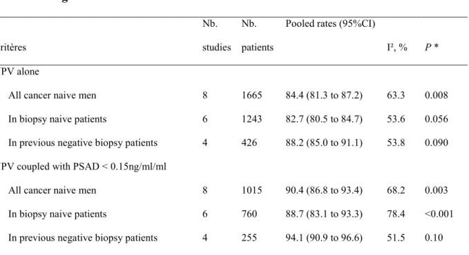
Table 4: meta-analysis of studies reporting results of MRI coupled with PSAD<0.15ng/ml/ml Critères Nb. studies Nb. patients
Pooled rates (95%CI)
I², % P * NPV alone
All cancer naive men 8 1665 84.4 (81.3 to 87.2) 63.3 0.008
In biopsy naive patients 6 1243 82.7 (80.5 to 84.7) 53.6 0.056
In previous negative biopsy patients 4 426 88.2 (85.0 to 91.1) 53.8 0.090
NPV coupled with PSAD < 0.15ng/ml/ml
All cancer naive men 8 1015 90.4 (86.8 to 93.4) 68.2 0.003
In biopsy naive patients 6 760 88.7 (83.1 to 93.3) 78.4 <0.001
In previous negative biopsy patients 4 255 94.1 (90.9 to 96.6) 51.5 0.10
* P-value associated with Chi-squared test for heterogeneity.
When coupled with PSAD, the NPV of MRI increased from 83.7%, 82.7% and 88.2% to 90.4%, 88.7% and 94.1% in all cancer naive, biopsy naive and previous negative biopsy patients respectively (table 4 and figure 3).
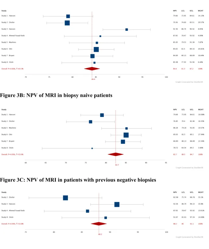
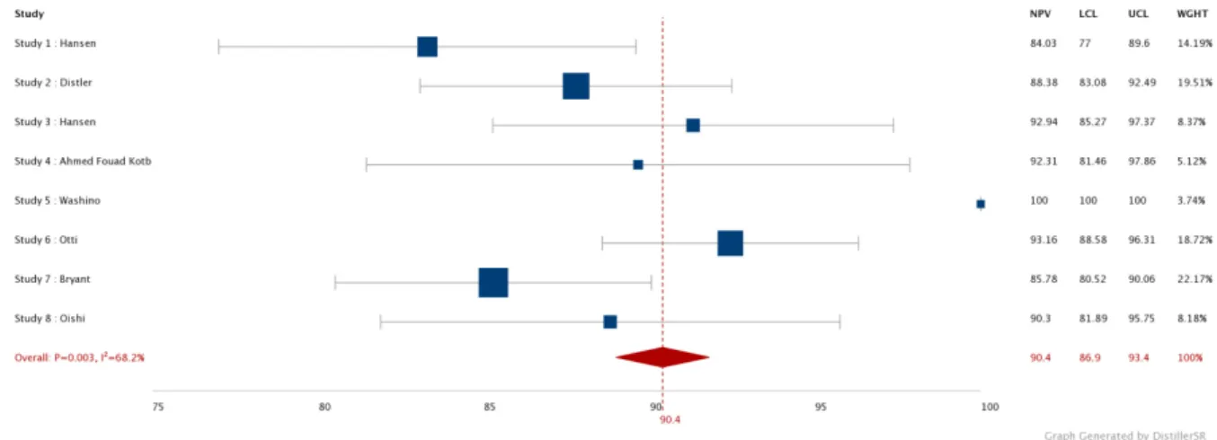
Figure 3: Forest plot showing the NPV for csPCa of MRI alone and combined with PSAD<0.15ng/ml/ml, in cancer naive patients, biopsy naive patients and patients with previous negative biopsies
Figure 3A: NPV of MRI in cancer naive patients
Figure 3B: NPV of MRI in biopsy naive patients
Figure 3C: NPV of MRI in patients with previous negative biopsies
Figure 3D: NPV of MRI combined with PSAD<0.15ng/ml/ml in PCa naive patients
Figure 3E: NPV of MRI combined with PSAD<0.15ng/ml/ml in biopsy naive patients
Figure 3F: NPV of MRI combined with PSAD<0.15ng/ml/ml in patients with previous negative biopsies
4. Discussion
4.1. Principaux résultats
D’après notre revue de la littérature, la PSAD était le marqueur le plus fréquemment couplé à l’IRM pour le diagnostic de csPCa. Lorsqu’elle était couplée à la PSAD<0.15ng/ml/ml, la NPV de l’IRM passait respectivement de 83.7% à 90.4% chez les patients naïfs de PCa, de 82.7% à 88.7% chez les patients naïfs de biopsie, et de 88.2% à 94.1% chez les patients ayant au moins une série de biopsies négatives.
4.2. Test index : IRM multiparamétrique
Parmi les études inclues dans notre revue, la définition de l’IRMn variait peu (15 études considéraient PI-RADS <3 et une étude PI-RADS<2), à l’origine d’un faible biais de sélection.
4.3. Examen de référence
Nous avons inclus uniquement des études qui rapportaient les résultats de biopsies systématisées comme examen de référence chez des patients ayant une IRMn. Les études incluant des biopsies ciblées (pour des lésions PI-RADS 2) n’ont pas été inclues pour ne pas modifier la VPN de l’IRM. Le risque de biais était « douteux » dans la plupart des études car aucune ne rapportait si l’examen anatomopathologique des biopsies était réalisé en aveugle des résultats de l’IRM. Deux études (17,26) incluaient des patients qui n’ont pas reçu le même examen de référence (nombre de biopsies et/ou voie différente). Dans une étude (26), tous les
patients qui ont eu une IRM n’ont pas eu de biopsies (231 patients), parmi lesquels 84.8% avaient une IRM négative, amenant un biais de sélection.
4.4. Impact sur la pratique clinique et la recherche
D’après notre revue de la littérature, il n’existe à l’heure actuelle que peu de données sur les facteurs prédictifs de csPCa chez les patients ayant une IRMn. La PSAD était le facteur prédictif le plus rapporté de csPCa chez les patients ayant une IRMn.
La NPV de l’IRM augmentait lorsqu’elle était couplée à la PSAD, quel que soit le statut biopsique des patients (cancer naïfs, naïfs de biopsies, ou biopsies négatives antérieures). Les patients ayant une IRMn et PSAD<0.15ng/ml/ml pourraient éviter les biopsies de prostate en diminuant le risque de sous-diagnostic de csPCa.
Ces données sont en concordance avec celles du parcours diagnostique suggéré par Padhani et al. (33) dans lequel les patients avec une IRMn et une PSAD<0.1-0.15ng/ml/ml pourraient éviter les biopsies, mais seulement dans le cas où une surveillance du PSA est une stratégie possible. L’utilisation de la PSAD soutient donc l’utilisation de l’IRM avant les biopsies, car cela permet à plus d’hommes ayant une IRMn d’éviter des biopsies de prostate. Par ailleurs le rapport coût-efficacité de l’IRM pré-biopsie est dépendant du fait de ne pas réaliser de biopsies en cas d’IRMn (34,35).
Les marqueurs tels que PHI, PHID et PCA3, lorsqu’ils étaient couplés aux résultats de l’IRM, permettaient une augmentation de la VPN de l’IRM pour le diagnostic de csPCa. Mais d’après notre revue de la littérature, ces données sont très limitées pour le diagnostic de csPCa. La grande majorité des études en rapport avec ces marqueurs rapportent des résultats sur tous les PCa, sur de faibles nombres de
patients (moins de 50), avec des définitions peu claires d’IRMn et de csPCa, rendant l’extrapolation de ces résultats difficile.
La NPV de l’IRM combinée au taux de PSA n’augmentait que faiblement par rapport à la VPN de l’IRM seule. Le PSA est un marqueur produit à la fois par les cellules épithéliales prostatiques et par le PCa. Le taux de PSA augmente lors des PCa, mais bien que les hauts taux de PSA soient prédictifs de PCa avancés, une grande partie des PCa confinés à la prostate présentent un taux de PSA plus bas, qui se confond avec celui des prostates de haut volume sans PCa. Le taux de PSA étant lié au volume prostatique, il semblerait donc que la PSAD (rapport du taux de PSA sur le volume prostatique) pourrait être un marqueur plus précis de diagnostic de csPCa que le PSA, qui pourrait être utilisé en tant que marqueur de surveillance chez les patients ayant une IRMn qui n’ont pas eu de biopsies (33).
Les calculateurs de risque (Risk calculators) semblaient eux aussi intéressants pour déterminer le risque de PCa d’un patient, et réduire le nombre de biopsies. Plusieurs outils (36) sont disponibles sur internet, comme celui de la cohorte PCPT (Prostate Cancer Prevention Trial) : PCPTRC 2.0 (qui n’inclut pas les données de l’IRM mais inclut des données sur les marqueurs urinaires comme le PCA3), ou ceux de la cohorte ERSPC (European Randomized Study of Screening for Prostate Cancer). Plusieurs outils sont issus de cette cohorte, et récemment un nouvel outil de l’ERSPC a été publié (37), incluant les résultats de l’IRM (et l’âge), qui pourrait aider à la sélection des patients pour l’indication des biopsies de prostate (38). En l’état actuel des connaissances, aucune recommandation n’est validée sur la meilleure façon de stratifier le risque de csPCa des patients avant d’envisager de les référer à
l’IRM et aux biopsies. Les nomogrammes utilisés pour le diagnostic de PCa et csPCa semblent pleins de promesses, mais aucun n’a été utilisé dans une population de patients ayant une IRM négative, et aucun n’a été validé pour le dépistage de csPCa (39).
4.5. Place de l’étude dans la littérature
Cette revue de la littérature est la première à rapporter la valeur ajoutée de la PSAD à l’IRM pour le diagnostic de csPCa. Des études existent rapportant les performances diagnostiques isolées de la PSAD (40,41), de la PSAD rapportée au taux de PSA (42), ou la VPN de l’IRM seule pour le diagnostic de csPCA (7).
C’est aussi la première revue à essayer d’identifier les facteurs prédictifs de csPCa chez des patients ayant une IRMn.
4.6. Forces de l’étude et limites
La PSAD était le seul facteur prédictif de csPCa suffisamment décrit pour réaliser une méta-analyse, et seules 8 études ont pu être inclues dans cette méta-analyse. Les études inclues avaient les mêmes définitions pour l’IRMn (PI-RADS <3) et pour le csPCa (GGG≥2), et incluaient des patients BN, PNB ou les deux.
Notre étude est limitée par le faible nombre d’études rapportant des facteurs prédictifs de csPCa chez les patients ayant une IRMn. Plusieurs études (16,18,19) ont rapporté des résultats sur des populations incluant des patients en surveillance active (patients non cancer naïfs), ce qui peut rendre difficile l’extrapolation de leurs résultats sur les facteurs prédictifs de csPCa sur tous les patients naïfs de PCa.
Une seule étude (17) a détaillé la méthode de calcul de la PSAD, en utilisant le volume prostatique calculé sur l’IRM. Les quinze autres études n’ont pas précisé quel volume de prostate était pris en compte dans le calcul de la PSAD (volume échographique ou IRM). Dans l’hypothèse où les patients ayant une IRMn et une PSAD<0.15ng/ml/ml éviteraient les biopsies de prostate, une mesure du volume prostatique la plus fiable possible serait nécessaire. Christie et al. (43) ont réalisé une revue de la littérature sur les performances de l’échographie, du scanner et de l’IRM sur la mesure du volume prostatique. L’IRM était légèrement plus précise pour la mesure du volume prostatique que l’échographie ou le scanner, données corrélées par Tai et al. (44) qui rapportent que l’échographie sous-estime le volume prostatique par rapport à l’IRM, et ce surtout pour les volumes supérieurs à 40cc.
L’expérience et l’expertise des radiologues analysant les IRM est un facteur important pour le diagnostic de csPCa. Branger et al. (45) ont comparé les résultats IRM des csPCa non diagnostiqués chez des patients IRMn entre des IRM faites en centre expert et faites par des radiologues généraux. Les csPCa non diagnostiqués par les radiologues de centres experts par rapport aux radiologues généraux étaient plus fréquemment confinés à la prostate, avaient un contingent moins important de Gleason 4, un plus petit diamètre tumoral et un plus petit volume tumoral. Ce qui sous-entend la nécessité de radiologues spécialisés en urologie pour tirer bénéfice de l’IRM pour le diagnostic de csPCa. Besoin qui est comblé dans les centres experts, mais qui ne l’est pas forcément en dehors de ceux-ci, ce qui pourrait gêner l’extrapolation des résultats et du management des patients ayant une IRMn dans la « vraie vie ». Par ailleurs, 2 études (46,47) rapportent les résultats d’une relecture des IRMn des patients ayant un csPCa. Les principales raisons de non diagnostic de
csPCa sur l’IRM sont la présence d’anomalies diffuses sans lésion focale (post-prostatite en général), ou de mauvaise classification des lésions. Itatani et al. (48) rapportant les raisons des faux négatifs de l’IRM, retrouvait principalement les petits volumes tumoraux, les lésions diffuses liées aux antécédents de prostatite, et l’hypertrophie bénigne de prostate. D’après notre revue, l’expérience des radiologues était souvent rapportée, mais aucune étude ne rapporte de double lecture de l’IRM.
Comme l’usage systématique de l’IRM avant tout série de biopsies de prostate est un fait nouveau dans les recommandations, et que le management des patients ayant une IRMn est une préoccupation récente, les données de la littérature sur ce sujet sont maigres. Des études larges, prospectives rapportant ces facteurs sur des patients ayant une IRMn sont nécessaires pour permettre d’identifier avec certitudes ces facteurs, et ainsi mieux sélectionner les patients avec IRMn devant avoir une biopsie de prostate.
5. Conclusion
La densité de PSA<0.15ng/ml/ml est le facteur prédictif le plus décrit pour exclure les cancers de prostate cliniquement significatifs en cas d’IRM négative. La densité de PSA pourrait être ajoutée aux résultats de l’IRM pour mieux sélectionner les patients naïfs de cancer ne nécessitant pas de biopsies.
6. Références
1. Jéhannin-Ligier K. Projection de l’incidence et de la mortalité par cancer en France métropolitaine en 2017 / 2018 / Maladies chroniques et traumatismes / Rapports et synthèses / Publications et outils / Accueil [Internet]. [cité 8 mai 2019]. Disponible sur: http://invs.santepubliquefrance.fr/Publications-et-outils/Rapports-et-syntheses/Maladies- chroniques-et-traumatismes/2018/Projection-de-l-incidence-et-de-la-mortalite-par-cancer-en-France-metropolitaine-en-2017
2. Rozet F, Hennequin C, Beauval J-B, Beuzeboc P, Cormier L, Fromont-Hankard G, et al. Recommandations françaises du Comité de Cancérologie de l’AFU – Actualisation 2018– 2020 : cancer de la prostate. Prog En Urol. nov 2018;28(12):S79‑130.
3. Ahmed HU, El-Shater Bosaily A, Brown LC, Gabe R, Kaplan R, Parmar MK, et al. Diagnostic accuracy of multi-parametric MRI and TRUS biopsy in prostate cancer (PROMIS): a paired validating confirmatory study. Lancet Lond Engl. 25
2017;389(10071):815‑22.
4. Kasivisvanathan V, Rannikko AS, Borghi M, Panebianco V, Mynderse LA, Vaarala MH, et al. MRI-Targeted or Standard Biopsy for Prostate-Cancer Diagnosis. N Engl J Med [Internet]. 18 mars 2018 [cité 13 avr 2019]; Disponible sur:
https://www.nejm.org/doi/10.1056/NEJMoa1801993?url_ver=Z39.88-2003&rfr_id=ori%3Arid%3Acrossref.org&rfr_dat=cr_pub%3Dwww.ncbi.nlm.nih.gov 5. Rouvière O, Puech P, Renard-Penna R, Claudon M, Roy C, Mège-Lechevallier F, et al. Use of prostate systematic and targeted biopsy on the basis of multiparametric MRI in biopsy-naive patients (MRI-FIRST): a prospective, multicentre, paired diagnostic study. Lancet Oncol. 1 janv 2019;20(1):100‑9.
6. Simmons LAM, Kanthabalan A, Arya M, Briggs T, Barratt D, Charman SC, et al. The PICTURE study: diagnostic accuracy of multiparametric MRI in men requiring a repeat prostate biopsy. Br J Cancer. 25 avr 2017;116(9):1159‑65.
7. Moldovan PC, Broeck TV den, Sylvester R, Marconi L, Bellmunt J, Bergh RCN van den, et al. What Is the Negative Predictive Value of Multiparametric Magnetic Resonance Imaging in Excluding Prostate Cancer at Biopsy? A Systematic Review and
Meta-analysis from the European Association of Urology Prostate Cancer Guidelines Panel. Eur Urol. 1 août 2017;72(2):250‑66.
8. Appayya MB, Adshead J, Ahmed HU, Allen C, Bainbridge A, Barrett T, et al. National implementation of multi-parametric magnetic resonance imaging for prostate cancer detection – recommendations from a UK consensus meeting. BJU Int. 2018;122(1):13‑25. 9. EAU Guidelines on Prostate Cancer | Uroweb [Internet]. [cité 28 avr 2019]. Disponible
sur: https://uroweb.org/course/eau-guidelines-on-prostate-cancer/
10. Leest M van der, Cornel E, Israël B, Hendriks R, Padhani AR, Hoogenboom M, et al. Head-to-head Comparison of Transrectal Ultrasound-guided Prostate Biopsy Versus Multiparametric Prostate Resonance Imaging with Subsequent Magnetic
guided Biopsy in Biopsy-naïve Men with Elevated Prostate-specific Antigen: A Large Prospective Multicenter Clinical Study. Eur Urol. 1 avr 2019;75(4):570‑8.
11. Haffner J, Lemaitre L, Puech P, Haber G-P, Leroy X, Jones JS, et al. Role of magnetic resonance imaging before initial biopsy: comparison of magnetic resonance imaging-targeted and systematic biopsy for significant prostate cancer detection. BJU Int. 2011;108(8b):E171‑8.
12. Zorzela L, Loke YK, Ioannidis JP, Golder S, Santaguida P, Altman DG, et al. PRISMA harms checklist: improving harms reporting in systematic reviews. BMJ. 1 févr
2016;352:i157.
13. Whiting PF. QUADAS-2: A Revised Tool for the Quality Assessment of Diagnostic Accuracy Studies. Ann Intern Med. 18 oct 2011;155(8):529.
14. Freeman M, Tukey J. Transformations related to the angular and the square root. In: Ann Math Statist. 1950. p. 21:607-11.
15. Deeks JJ, Altman DG, Bradburn MJ. Statistical methods for examining heterogeneity and combining results from several studies in meta-analysis. In: Egger M, Davey Smith G, Altman DG(Eds). Systematic Reviews in Health Care: Meta-Analysis in Context (2nd edition): London: BMJ Publication Group, 2001. In.
16. Wang RS, Kim EH, Vetter JM, Fowler KJ, Shetty AS, Mintz AJ, et al. Determination of the Role of Negative Magnetic Resonance Imaging of the Prostate in Clinical Practice: Is Biopsy Still Necessary? Urology. 1 avr 2017;102:190‑7.
17. Numao Noboru, Yoshida Soichiro, Komai Yoshinobu, Ishii Chikako, Kagawa Makoto, Kijima Toshiki, et al. Usefulness of Pre-biopsy Multiparametric Magnetic Resonance Imaging and Clinical Variables to Reduce Initial Prostate Biopsy in Men with Suspected Clinically Localized Prostate Cancer. J Urol. 1 août 2013;190(2):502‑8.
18. Oishi Masakatsu, Shin Toshitaka, Ohe Chisato, Nassiri Nima, Palmer Suzanne L., Aron Manju, et al. Which Patients with Negative Magnetic Resonance Imaging Can Safely Avoid Biopsy for Prostate Cancer? J Urol. 1 févr 2019;201(2):268‑77.
19. An JY, Sidana A, Holzman SA, Baiocco JA, Mehralivand S, Choyke PL, et al. Ruling out clinically significant prostate cancer with negative multi-parametric MRI. Int Urol
Nephrol. 1 janv 2018;50(1):7‑12.
20. Panebianco V, Barchetti G, Simone G, Monte MD, Ciardi A, Grompone MD, et al. Negative Multiparametric Magnetic Resonance Imaging for Prostate Cancer: What’s Next? Eur Urol. 1 juill 2018;74(1):48‑54.
21. Hansen NL, Barrett T, Kesch C, Pepdjonovic L, Bonekamp D, O’Sullivan R, et al. Multicentre evaluation of magnetic resonance imaging supported transperineal prostate biopsy in biopsy-naïve men with suspicion of prostate cancer. BJU Int. 2018;122(1):40‑9. 22. Distler Florian A., Radtke Jan P., Bonekamp David, Kesch Claudia, Schlemmer
Heinz-Peter, Wieczorek Kathrin, et al. The Value of PSA Density in Combination with PI-RADSTM for the Accuracy of Prostate Cancer Prediction. J Urol. 1 sept
2017;198(3):575‑82.
23. Hansen NL, Kesch C, Barrett T, Koo B, Radtke JP, Bonekamp D, et al. Multicentre evaluation of targeted and systematic biopsies using magnetic resonance and ultrasound image-fusion guided transperineal prostate biopsy in patients with a previous negative biopsy. BJU Int. 2017;120(5):631‑8.
24. Kotb AF, Spaner S, Crump T, Hyndman ME. The role of mpMRI and PSA density in patients with an initial negative prostatic biopsy. World J Urol. 1 déc 2018;36(12):2021‑5. 25. Washino S, Okochi T, Saito K, Konishi T, Hirai M, Kobayashi Y, et al. Combination of
prostate imaging reporting and data system (PI-RADS) score and prostate-specific antigen (PSA) density predicts biopsy outcome in prostate biopsy naïve patients. BJU Int.
2017;119(2):225‑33.
26. Otti VC, Miller C, Powell RJ, Thomas RM, McGrath JS. The diagnostic accuracy of multiparametric magnetic resonance imaging before biopsy in the detection of prostate cancer. BJU Int. 2019;123(1):82‑90.
27. Bryant Richard J., Hobbs Catherine P., Eyre Katie S., Davies Lucy C., Sullivan Mark E., Shields William, et al. Comparison of Prostate Biopsy with or without Prebiopsy
Multiparametric Magnetic Resonance Imaging for Prostate Cancer Detection: An Observational Cohort Study. J Urol. 1 mars 2019;201(3):510‑9.
28. Gnanapragasam VJ, Burling K, George A, Stearn S, Warren A, Barrett T, et al. The Prostate Health Index adds predictive value to multi-parametric MRI in detecting
significant prostate cancers in a repeat biopsy population. Sci Rep [Internet]. 17 oct 2016 [cité 13 avr 2019];6. Disponible sur:
https://www.ncbi.nlm.nih.gov/pmc/articles/PMC5066204/
29. Druskin SC, Tosoian JJ, Young A, Collica S, Srivastava A, Ghabili K, et al. Combining Prostate Health Index density, magnetic resonance imaging and prior negative biopsy status to improve the detection of clinically significant prostate cancer. BJU Int. 2018;121(4):619‑26.
30. Perlis Nathan, Al-Kasab Thamir, Ahmad Ardalan, Goldberg Estee, Fadak Kamel, Sayid Rashid, et al. Defining a Cohort that May Not Require Repeat Prostate Biopsy Based on PCA3 Score and Magnetic Resonance Imaging: The Dual Negative Effect. J Urol. 1 mai 2018;199(5):1182‑7.
31. Thompson J.E., van Leeuwen P.J., Moses D., Shnier R., Brenner P., Delprado W., et al. The Diagnostic Performance of Multiparametric Magnetic Resonance Imaging to Detect Significant Prostate Cancer. J Urol. 1 mai 2016;195(5):1428‑35.
32. Mottet N, Bellmunt J, Bolla M, Briers E, Cumberbatch MG, De Santis M, et al. EAU-ESTRO-SIOG Guidelines on Prostate Cancer. Part 1: Screening, Diagnosis, and Local Treatment with Curative Intent. Eur Urol. 1 avr 2017;71(4):618‑29.
33. Padhani AR, Weinreb J, Rosenkrantz AB, Villeirs G, Turkbey B, Barentsz J. Prostate Imaging-Reporting and Data System Steering Committee: PI-RADS v2 Status Update and Future Directions. Eur Urol. mars 2019;75(3):385‑96.
34. Mowatt G, Scotland G, Boachie C, Cruickshank M, Ford J, Fraser C, et al. The diagnostic accuracy and cost-effectiveness of magnetic resonance spectroscopy and enhanced
magnetic resonance imaging techniques in aiding the localisation of prostate
abnormalities for biopsy: a systematic review and economic evaluation. Health Technol Assess [Internet]. mai 2013 [cité 13 mai 2019];17(20). Disponible sur:
https://www.journalslibrary.nihr.ac.uk/hta/hta17200/
35. Faria R, Soares MO, Spackman E, Ahmed HU, Brown LC, Kaplan R, et al. Optimising the Diagnosis of Prostate Cancer in the Era of Multiparametric Magnetic Resonance Imaging: A Cost-effectiveness Analysis Based on the Prostate MR Imaging Study (PROMIS). Eur Urol. 2018;73(1):23‑30.
36. Louie KS, Seigneurin A, Cathcart P, Sasieni P. Do prostate cancer risk models improve the predictive accuracy of PSA screening? A meta-analysis. Ann Oncol. 1 mai
2015;26(5):848‑64.
37. Alberts AR, Roobol MJ, Verbeek JFM, Schoots IG, Chiu PK, Osses DF, et al. Prediction of High-grade Prostate Cancer Following Multiparametric Magnetic Resonance Imaging: Improving the Rotterdam European Randomized Study of Screening for Prostate Cancer Risk Calculators. Eur Urol. 2019;75(2):310‑8.
38. The Prostate Cancer Risk Calculators – including the ‘future risk’ calculator – SWOP – The Prostate Cancer Research Foundation, Reeuwijk [Internet]. [cité 25 avr 2019]. Disponible sur: calculators, http://www.prostatecancer-riskcalculator.com/seven-prostate-cancer-risk-calculators
39. Radtke JP, Wiesenfarth M, Kesch C, Freitag MT, Alt CD, Celik K, et al. Combined Clinical Parameters and Multiparametric Magnetic Resonance Imaging for Advanced Risk Modeling of Prostate Cancer—Patient-tailored Risk Stratification Can Reduce Unnecessary Biopsies. Eur Urol. 1 déc 2017;72(6):888‑96.
40. Aminsharifi Alireza, Howard Lauren, Wu Yuan, De Hoedt Amanda, Bailey Charlotte, Freedland Stephen J., et al. Prostate Specific Antigen Density as a Predictor of Clinically Significant Prostate Cancer When the Prostate Specific Antigen is in the Diagnostic Gray Zone: Defining the Optimum Cutoff Point Stratified by Race and Body Mass Index. J Urol. 1 oct 2018;200(4):758‑66.
41. Nordström T, Akre O, Aly M, Grönberg H, Eklund M. Prostate-specific antigen (PSA) density in the diagnostic algorithm of prostate cancer. Prostate Cancer Prostatic Dis. avr 2018;21(1):57.
42. Jue JS, Barboza MP, Prakash NS, Venkatramani V, Sinha VR, Pavan N, et al. Re-examining Prostate-specific Antigen (PSA) Density: Defining the Optimal PSA Range and Patients for Using PSA Density to Predict Prostate Cancer Using Extended Template Biopsy. Urology. 1 juill 2017;105:123‑8.
43. Christie DRH, Sharpley CF. How Accurately Can Prostate Gland Imaging Measure the Prostate Gland Volume? Results of a Systematic Review [Internet]. Prostate Cancer. 2019 [cité 19 mai 2019]. Disponible sur: https://www.hindawi.com/journals/pc/2019/6932572/
44. Tai* Thomson, Blackwell Robert, Hart Spencer, Gupta Gopal. Pd19-11 prostate mri improves volume estimation compared to transrectal ultrasound in large prostate glands. J Urol. 1 avr 2019;201(Supplement 4):e375‑e375.
45. Branger N, Maubon T, Traumann M, Thomassin-Piana J, Brandone N, Taix S, et al. Is negative multiparametric magnetic resonance imaging really able to exclude significant prostate cancer? The real-life experience. BJU Int. 2017;119(3):449‑55.
46. Lu AJ, Syed JS, Nguyen KA, Nawaf CB, Rosoff J, Spektor M, et al. Negative Multiparametric Magnetic Resonance Imaging of the Prostate Predicts Absence of Clinically Significant Prostate Cancer on 12-Core Template Prostate Biopsy. Urology. 1 juill 2017;105:118‑22.
47. Meng Y, Vetter JM, Parker AA, Arett CT, Andriole GL, Shetty AS, et al. Improved Detection of Clinically Significant Prostate Cancer With Software-assisted Systematic Biopsy Using MR/US Fusion in Patients With Negative Prostate MRI. Urology. oct 2018;120:162‑6.
48. Itatani R, Namimoto T, Atsuji S, Katahira K, Morishita S, Kitani K, et al. Negative predictive value of multiparametric MRI for prostate cancer detection: Outcome of 5-year follow-up in men with negative findings on initial MRI studies. Eur J Radiol. 1 oct
2014;83(10):1740‑5.
7. Suppléments
Supplement 1: Abstract
Context: Some guidelines recommend that men may avoid biopsy in case of a non-suspicious
MRI (nMRI). MRI has a negative predictive value (NPV) of 85-95% which leads to this strategy missing 5-15% clinically significant PCa (csPCa). Patient factors and biochemical markers can be used in addition to MRI to inform biopsy decisions in order to reduce the risk of missing csPCa.
Objective: To systematically review the literature of PCa naïve patient series with a nMRI,
reporting of predictive factors for csPCa diagnosis.
Evidence acquisition: The Medline and Scopus databases were searched up to March 2019.
Clinical factors and markers studied were age, PSA, PSA isoforms, PSA density (PSAD), PCA3, prostate volume, family history, ethnicity, and risk calculators. The primary outcome was their predictive ability for csPCa diagnosis. Secondary outcomes were to perform a meta-analysis of the NPV of nMRI when combined with these predictive factors.
Evidence synthesis: A total of 16 studies were eligible for inclusion. Few studies reported
NPV of MRI combined with a marker. PSAD<0.15ng/ml/ml was the strongest predictor of csPCa. Eight studies (1015 patients) were eligible for meta-analysis of the added value of PSAD< 0.15ng/ml/ml to MRI in reducing the risk of missing csPCa. When combined with PSAD, overall MRI NPV increased from 83.7% to 90.4% in cancer naïve patients. The increase was from 82.7% to 88.7% in biopsy naïve and from 88.2% to 94.1% in previous negative biopsy sub-groups.
Conclusion: PSAD<0.15ng/ml/ml was the most accurate predictive factor to reduce the risk
of missing csPCa. It may be added to MRI results for biopsy indication in cancer naïve patients.
Patient summary: We evaluated the published literature of patient series with a negative
MRI, reporting the added value of clinical factors in ruling out csPCa. We found that the measure of PSA Density with a threshold value of <0.15ng/ml/ml was the most accurate predictive factor to reduce the risk of missing csPCa. It may be added to MRI results for biopsy indication in cancer naive patients.
Supplement 2: Introduction
A major consideration in prostate cancer screening and early detection is over-diagnosis and overtreatment of indolent disease. Multi-parametric MRI (MRI) of the prostate is currently transforming the prostate cancer diagnostic pathway. Several studies (3–6) have provided robust evidence that the use of MRI improves the diagnosis of clinically significant prostate cancer (csPCa). MRI has excellent negative predictive value (NPV) in excluding csPCa (7) . MRI has shown the potential to reduce the diagnostic of insignificant prostate cancer (isPCa) (3,4).
Performing MRI before any prostate biopsy is widely recommended (2,8,9). Results of the PRECISION trial, where prostate biopsies were avoided in case of prebiopsy negative MRI (nMRI), suggest that MRI could be used as a triage test for prostate biopsy. However, those results need to be considered in the context of the results from the MRI-first study, in which some csPCa were missed by targeted biopsy (4,5). nMRI represents 20-30% of all MRI in cancer naive populations, and early screening tends to increase this rate (10).
The advice regarding biopsy if the prebiopsy MRI is negative (PI-RADS 1-2) is controversial. Patients with nMRI may avoid prostate biopsy, but 5-15% csPCa may be missed (3,4,11). Recent guidelines state that men may safely avoid prostate biopsy if MRI is negative, especially “if PSA density (PSAD) is low, and that PSA observation is appropriate” (8), or if “clinical suspicion of prostate cancer is low, based on shared decision making with the patient” (9). How to identify patients with nMRI and a high risk of csPCa is not yet known. Associating MRI with predictive factors may increase the NPV of MRI and reduce the number of prostate biopsy in men whose risk of csPCa is low.
The aim of this work was to systematically review the literature regarding the predictive factors of csPCa in naive patients with nMRI.
Supplement 3: Discussion
4.1. Principal findings
PSAD was the most relevant predictive factor of csPCa studies in the literature in men with nMRI. When MRI was coupled with PSAD<0.15ng/ml/ml, the MRI NPV in cancer naive, biopsy naive and previous negative biopsy groups increased from 83.7%, 82.7% and 88.2% to 90.4%, 88.7% and 94.1%, respectively.
4.2. Reference standard
We included only studies that reported the results of systematic/standard biopsy in patients with nMRI and used the systematic/standard biopsy as a reference standard. No MRI-targeted biopsies (of PIRADS 2 lesions) were included in order for fear of modifying the NPV of MRI. The RoB was unclear in most studies because none of the studies reported whether anatomopathologists were blind of the results of MRI. In 2 studies (17,26) all patients did not receive the same reference standard. In 1 study (26), all patients who had MRI did not undergo biopsy (231 patients), in which 84.8% had nMRI, leading to a bias of selection.
4.3. Impact on clinical practice and research
When coupled with PSAD, the NPV of MRI increases, and this does not depend on biopsy status (all patients, BN patients and PNB patients). Patients with nMRI and PSAD<0.15ng/ml/ml could avoid biopsy. This proposed attitude is in accordance with Padhani et al. (33), who proposed that biopsy was not performed in patients with nMRI and PSAD<0.1-0.15ng/ml/ml, but only if surveillance of PSA is an available follow up strategy. The use of PSAD therefore supports the use of pre-biopsy MRI as it allows more men with nMRI to avoid biopsy safely, and the cost effectiveness of the pre-biopsy MRI pathway is dependent on the avoidance of biopsy in the men with nMRI (34,35).
In some studies, markers such as PHI, PHID and PCA3 coupled with MRI increased the NPV of MRI for csPCa. But our literature search found only limited data concerning the NPV of MRI combined with PCA3 for the detection of csPCa. Most studies included less than 50 patients, with unclear definition of nMRI and unknown reference standard. Moreover, results were reported for all PCa and not csPCa. The same comments could be made with PHI and PHID.
PSA coupled with MRI showed a small increase in NPV, and appeared inferior to PSAD in that matter. PSA is an androgen-regulated serine protease produced by both prostate epithelial cells and PCa. Serum total PSA levels are increased in PCa, although high PSA levels are predictive of advanced PCa, a large fraction of organ-confined cancers present with much lower total PSA values that overlap those levels found in men without PCa large prostates. It could, however, be used as a surveillance marker (33) for patients with nMRI who didn’t undergo biopsy.
Risk calculators may be useful in informing decisions for biopsy by combining a number of different clinical predictors. Several tools (36) are available as the PCPT cohort (PCPTRC 2.0), which does not include MRI score but does include urinary markers such PCA3, or the ERSPC cohort. Recently a new tool from ERSPC including MRI results has been published, which may help selecting patients for biopsy (38). Nomograms for the diagnostic of PCa and csPCa seem to be useful (39), but none of these has been used in a nMRI population only, and none has been externally validated for csPCa screening.
4.4. How this review compares with other reviews
To our knowledge, this review is the first assessing the use of PSAD in patients with nMRI. Moreover, it is the first to date to have tried to identify predictive factors of csPCa in patients with a nMRI.
4.5. Strengths and limitations
It is limited by the small amount of studies reporting predictive factors of csPCa in nMRI patients. Only PSAD was described enough as significant in several studies to perform a meta-analysis. Some studies included PPB (not cancer naive patients), which can make interpretation of the results more challenging to tease out.
Only one study (17) detailed the calculation of PSAD (using prostate volume calculated on MRI). The 15 other studies did not specify if prostate volume was assessed by echography or MRI to calculate the PSAD.
As systematic use of MRI is new in the recommendation, and the management of nMRI is a recent concern in PCa screening, literature is scarce. Prospective studies studying all possible factors in large nMRI populations are needed to address the capital problem of selecting which patients can safely avoid biopsy.
Supplement 4: Conclusion
PSAD<0.15ng/ml/ml was the most accurate negative predictive factor for clinically significant prostate cancer diagnosis. It may be added to MRI results to help for biopsy indication in cancer naive patients.
8. Annexes
Annexe 1: Research terms
(Prostate cancer OR significant prostate cancer OR PCa) AND (negative MRI or MRI or magnetic resonance imaging or MR imaging or MP-MRI or mpMRI or NMR imaging or multiparametric)
1. Prostate cancer
2. Significant prostate cancer 3. PCa
4. 1 or 2 or 3 5. Negative MRI 6. MRI
7. Magnetic resonance imaging 8. MR imaging 9. MP-MRI 10. mpMRI 11. NMR imaging 12. Multiparametric 13. 5 or 6 or 7 or 8 or 9 or 10 or 11 or 12 14. 4 AND 13 47