HAL Id: hal-03004753
https://hal-cnrs.archives-ouvertes.fr/hal-03004753
Submitted on 20 Nov 2020HAL is a multi-disciplinary open access archive for the deposit and dissemination of sci-entific research documents, whether they are pub-lished or not. The documents may come from teaching and research institutions in France or abroad, or from public or private research centers.
L’archive ouverte pluridisciplinaire HAL, est destinée au dépôt et à la diffusion de documents scientifiques de niveau recherche, publiés ou non, émanant des établissements d’enseignement et de recherche français ou étrangers, des laboratoires publics ou privés.
Electrostatic analysis of TEM1 -lactamase: effect of
substrate binding, steep potential gradients and
consequences of site-directed mutations
Peter Swarén, Laurent Maveyraud, Valérie Guillet, Jean-Michel Masson,
Lionel Mourey, Jean Pierre Samama
To cite this version:
Peter Swarén, Laurent Maveyraud, Valérie Guillet, Jean-Michel Masson, Lionel Mourey, et al.. Elec-trostatic analysis of TEM1 -lactamase: effect of substrate binding, steep potential gradients and consequences of site-directed mutations. Structure, Elsevier (Cell Press), 1995, 3 (6), pp.603-613. �10.1016/s0969-2126(01)00194-0�. �hal-03004753�
Electrostatic analysis of TEM1
-lactamase: effect of
substrate binding, steep potential gradients and
consequences of site-directed mutations
Peter Swaren
1
,
2
, Laurent Maveyraud', Valerie Guillet',
Jean-Michel Masson
3
, Lionel Mourey' and Jean-Pierre Samama
1
*
'Groupe de Cristallographie Biologique, Laboratoire de Pharmacologie et de Toxicologie Fondamentales du CNRS, 205 route de Narbonne, F-31077 Toulouse CEDEX, France, 2Department of Physical Chemistry, Chalmers University of Technology,S-412 96 Gothenburg, Sweden and 3Groupe d'lng6nierie des Prot6ines, Laboratoire de Pharmacologie et de Toxicologie Fondamentales du CNRS, 205 route de Narbonne, F-31077 Toulouse CEDEX, France
Background: Escherichia coli TEM1 is a penicillinase and
belongs to class A -lactamases. Its naturally occurring mutants are responsible for bacterial resistance to 3-lactamin-based antibiotics. X-ray structure determina-tions show that all class A -lactamases are similar, but, despite the numerous kinetic investigations, the reaction mechanism of these enzymes is still debated. We address the questions of what the molecular contexts during the acylation and deacylation steps are and how they con-tribute to the efficiency of these penicillinases.
Results: Electrostatic analysis of the 1.8
A
resolution refined X-ray structure of the wild-type enzyme, and of its modelled Michaelis and acyl-enzyme complexes, showed that substrate binding induces an upward shift in the pKa of the unprotonated Lys73 by 6.4 pH units. The amine group of Lys73 can then abstract the Ser70 hydroxyl group pro-ton and promote acylation. In the acyl-enzyme complex, the deacylating water is situated between the carboxylategroup of Glu166, within the enzyme, and the ester-carbonyl carbon of the acyl-enzyme complex, in an elec-trostatic potential gradient amounting to 30 kTe-' A-'. Other residues, not directly involved in catalysis, also con-tribute to the formation of this gradient. The deacylation rate is related to the magnitude of the gradient. The kinetic behaviour of site-directed mutants that affect the protona-tion state of residue 73 cannot be explained on the basis of the wild-type enzyme mechanism.
Conclusions: In the wild-type enzyme, the very high
rates of acylation and deacylation of class A P-lactamases arise from an optimal chemical setup in which the acyla-tion reacacyla-tion seems triggered by substrate binding that changes the general base property of Lys73. In site-directed mutants where Lys73 is protonated, acylation may proceed through activation of a water molecule by Glu166, and Lys73 contributes as a proton shuffle partner in this pathway.
Structure 15 June 1995, 3:603-613
Key words: antibiotics, 3-lactamas'e, electrostatics, hydrolysis, substrate-triggered mechanism
Introduction
Extensive use of antibiotics in the community and in hospitals has caused bacteria to become resistant to these agents. The resistance is mainly mediated by the p-lacta-mase enzymes and is spread by plasmid exchange. Selec-tion pressure favours the emergence of mutant enzymes that exhibit extended substrate specificities. Among all mutant enzymes in resistant bacteria, the majority have evolved from Escherichia coli TEM1 -lactamase.
Considerable work has been aimed at describing the structure-function relationships of the -lactamases. However, rationalization of experimental data is difficult to achieve, because, except for the partition between classes A, B, and C proposed by Ambler et al. [1], based on sequence alignment, the borderline defined by some of the characteristics of the enzymes, such as evolution-ary propensity, response to clavulanic acid, or genetic origin, is not always straightforward. This explains why other classification schemes have been proposed [2].
Class A -lactamases are penicillinases. They are all very efficient enzymes with nearly diffusion-controlled kinet-ics. After formation of the enzyme=substrate Michaelis complex (kl,k_l), a tetrahedral intermediate leads to the acyl-enzyme complex (k2). The reaction product is
released after hydrolysis of this complex by a water mol-ecule (k3) through the formation of another tetrahedral
intermediate. Acylation and deacylation steps have both similar and very high rate constants (for example, k2=2800 s-1, k3=1500 s-1for E. coli TEM1, with
benzyl-penicillin as substrate) [3]. Several X-ray structures of class A -lactamases have been solved. Enzymes from the Gram-positive bacteria Streptomyces albus G [4],
Staphylo-coccus aureus PC1 [5] and Bacillus lichenformis 749/C [6],
and the TEM1 enzyme from the Gram-negative E. coli [7-9] all have similar structures. Jelsch et al. [9] described the main differences between TEM1 and the Gram-posi-tive enzymes at 1.8
A
resolution. Among these differences are the relative orientations of the two domains compris-ing the enzyme, which creates a narrowed substrate cavity*Corresponding author.
604 Structure 1995, Vol 3 No 6
in TEM1. All class A -lactamases contain the invariant active-site residues Ser70, Lys73, Glu166, and Lys234 (residue numbering according to Ambler et al. [1]), to which have been attributed various plausible functions in the mechanism of the enzymes [10,11]. A reaction scheme has been proposed for these 3-lactamases from the X-ray structure determination of the acyl-enzyme com-plex trapped in the deacylation-defective Glu166--Asn TEM1 mutant protein [8]. In this mechanism, as hypoth-esized by Oefner et al. [12], the unprotonated Lys73 acts as a general base that assists the nucleophilic attack of Ser70 O-y on the substrate. However, a depressed pKa value of Lys73 in the free enzyme does not explain why acylation occurs and why this reaction is so fast. The high rate constants would suggest that, in the wild-type (WT) enzyme, the chemical events occurring in the catalytic steps have minimal or lowered energy barriers, which are likely to be provided by an optimal chemical
environ-ment. As enzyme catalysis involves acid-base chemistry and charge transfers, it seemed appropriate to address the mechanism of P3-lactamase action by electrostatic analysis of the enzyme's refined structure.
Results and discussion
Control of parameters
From the network of interactions within the active site of TEM1, we used the electrostatic interaction (Table 1) between the buried and proximate Asp214 and Asp233
residues as a reference for our pKa calculations. The 2.8 A distance between Asp214 082 and Asp233 081 implies that one of these aspartates will be protonated at pH 7.8, the pH at which crystallization and structure determination of TEM1 were carried out [9]. Arguments that point out Asp214 as being protonated resulted from sequence and structural comparisons [9]. Briefly, Asp233 is invariant within the class A -lactamases and forms a
salt bridge with Arg222, while residue 214 is generally an asparagine. The calculation gave a pKa of 7.6 for Asp214, consistent with the structural and sequence analysis. The pKa shift of +3.7 pH units (Table 2) from free aspartyl to that in the WT enzyme is in the range expected for a residue buried within the protein structure [13]. In this test calculation, Lys73 was assumed to be protonated. If Lys73 was in the unprotonated state, the tendency for Asp214 to be protonated would be slightly stronger due to the removal of one positive charge at 11.3 A from Asp214 082 (Fig. 1). The shift we obtained in the pKa of Asp214 therefore represents a minimum value. In all subsequent calculations, we assumed Asp214 to be protonated. A second test calculation was performed on Lys146. As the surface residue with the most solvent-accessible ionizable group, its pKa should remain rather unshifted relative to the free amino acid, but at pH 7.8 the overall negative charge of the protein (pI=5.4 [14]) will attract protons and one could expect a slightly posi-tive shift. Indeed, the computation gave a pKa shift of
+0.4 for Lys146 (Table 2). The latter computation was made with Lys73 unprotonated and, therefore, represents the upper limit of the shift.
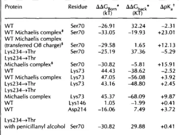
Table 2. Calculated differences in Born solvation and background
charge interaction energies with corresponding pKashifts.
Protein Residue AAGBorn* AAGback*
(kT) (KT) WT WT Michaelis complex* WT Michaelis complex (transferred 08 charge)* Lys234-Thr Lys234.-Thr Michaelis complex* WT WT Michaelis complex Lys234-Thr Lys234-sThr Michaelis complex WT WT Ser70 Ser70 Ser7 Ser70 -26.91 -33.05 -29.58 -25.19 Ser70 -30.82 Lys73 44.43 Lys73 47.05 Lys73 43.16 Lys73 Lys146 Asp214 Lys234-+Thr
with penicillanyl alcohol Ser70
45.37 1.05 -16.06 32.24 -19.93 1.65 37.36 -5.81 -38.62 -56.08 48.80 ApKat -2.31 +23.01 +12.13 -5.29 +15.91 -2.52 +3.92 +2.45 -68.09 +9.87 -1.99 +0.41 7.49 +3.72 -30.82 29.88 +0.41 *The differences are taken as the energy for the protonated state minus that for the unprotonated state. tApKais the shift of the side chain pKa
in the protein compared with the free amino acid. The Ser70 pKa in the Michaelis complexes is poorly estimated by the computations (see text). For an explanation of the terminology, see the Materials and
methods section.
Electrostatic potential maps
The enzyme is surrounded by a cloud of negative electro-static potential which is disrupted in places by several small positive lobes, generated by solvent-accessible lysine and arginine residues at the protein-solvent interface. This mainly negative envelope could be expected since the cal-culations were performed with a charge set corresponding to a pH value about two units above the isoelectric point of 5.4. At pH 7.8, the protein carries a net negative charge and presents an evenly distributed negative poten-tial on its surface. On a +0.5 kTe- 1contour level, solvent
Table 1. Electrostatic interaction energies (not including
desolvation effects) in the native WT enzyme and its Michaelis complex.
Interaction WT enzyme* Michaelis complex*
(kT) (kT) Ser70-Lys73t -45.75 -51.62 Ser70-Glu166 18.56 23.41 Ser70-H2 a-helix* -6.86 -8.31 Lys73-GIu166t -27.53 -31.75 Lys73-Lys234t 14.83 19.57 Asp214-Asp233 28.09 28.81 Ser70-COO- (Pen G) - 42.25 Lys73-COO- (Pen G)t - -23.06 Lys234-COO- (Pen G) - -66.04 Arg244-COO- (Pen G) - -9.20
*The interaction energies between complete side chains were computed with each side chain in its ionized state. Interior and exterior dielectrics were set to 3 and 80, respectively. tProtonated Lys73 is relevant in the Lys234-->Thr enzyme. *The H2 a-helix was assigned a charge distribution (overall neutral) only to its backbone atoms. The requirement of lysine or arginine at position 73 for measurable activity [43] indicates that the interaction between Ser70 and the H2 a-helix has a minor role in the activation of Ser70.
Electrostatic analysis of I3-lactamase Swaren et al. 605
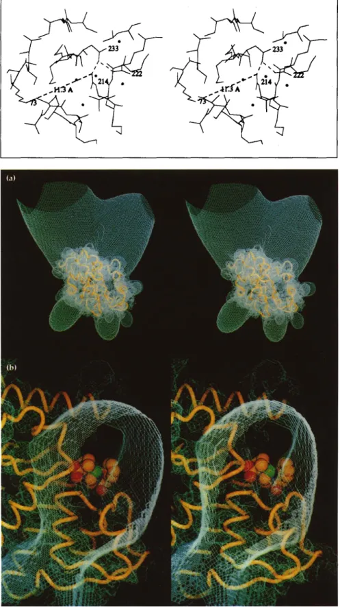
shielding prevents the potential from extending further into solution except for a large funnel-shaped positive lobe, which forms a continuous path from bulk solvent towards the substrate-binding site (Fig. 2). Since Lys215 is the only basic residue found in this region, this feature may indicate a pathway dedicated to attracting the substrate into its binding site with maximal efficiency.
Fig. 1. Stereoview of the environment around Asp214. The interaction between Asp214 and Asp233 and the salt bridge between Arg222 and Asp233 are both shown with dashed lines as well as the distance between the active site Lys73 and Asp214. Crystallographic waters are indicated by dots. (Figures 1 and 4 gener-ated using MOLSCRIPT [44].)
One important feature of the electrostatic potential is its bipolar distribution in the area of the substrate-binding site (Fig. 3). Most of the domain organized about the [-strands is immersed into a +20 kTe-1 continuous posi-tive potential: residues 234-238 from strand S3, residues 241-245 from strand S4, the first half of the C-terminal helix (residues 270-280), the H2 310-helix (residues
Fig. 2. (a) Map at +0.5 kTe-1 of the elec-trostatic potential of TEM1 1-lactamase, obtained after the second focusing step (see the Materials and methods section). (b) Enlarged perpendicular view of (a) showing the continuous path towards the substrate-binding site. Bound Pen G is shown as a space-filling model. (Figures 2, 3, 6, and 7 generated using 0 130].)
606 Structure 1995, Vol 3 No 6
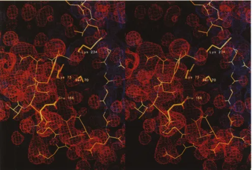
Fig. 3. Bipolar distribution of the electro-static potential in the area of the sub-strate-binding site. The +20 kTe-1contour is blue and the -20 kTe-1contour is red. The invariant residues of the active site
are labelled.
69-71), and the hinge region 213-218. This positive potential arises mainly from residues Lys215, Arg222, Lys234, Arg244, and Arg275. At the other side of the substrate cavity, a -20 kTe- 1continuous negative poten-tial surrounds the side-chains of residues 104, 105, 109, 110, 131-133, 136, 163, 166, 168, 170, 179, and 240 and the main-chain oxygen atoms of residues 103-106, 125, 129-132, 161, 167, 169, 170, and 179. As a conse-quence, the thiazolidine and 13-lactam rings of the sub-strate, whose re face is oriented towards the negative potential when bound to the enzyme, are located in a narrow and low potential area (-5 to +5 kTe-l) between the positive and negative potential domains. In agreement with the kinetic data [15,16], and as already discussed from the X-ray structure [9], before the [-lactam ring opening, the carboxylate group at C3 of the thiazolidine ring is oriented in the Michaelis complex towards the positive potential area and is 2.7 A and 3.9 A from Lys234 and Arg244, respectively (Fig. 4a). In the acyl-enzyme complex, after the -35° rotation about the N4-C5-C6-C7 torsion angle following the hydrolysis of the P-lactam ring, the carboxylate was observed at 3.5 A and 2.7 A from Lys234 and Arg244, respectively [8] (Fig. 4b).
Active-site residues
Kinetic studies demonstrated that lysines 73 and 234 are essential amino acids in the mechanism of the enzyme [17-20], and many hypotheses about their functions have been put forward. The pH dependence of kcat in the TEM1 enzyme suggests that a group at about pKa 10 must be protonated for maximal activity. Analysis of the X-ray structure shows that the only candidate in the substrate-binding area is Lys234.
Since all steps in catalysis involve proton transfers and charged groups, their respective locations and electrosta-tic environments were examined. We calculated the PKa of Lys73 in the free WT enzyme to be 8.0, which sug-gests that two enzyme populations (unprotonated and
protonated Lys73) are able to form Michaelis complexes at pH 7.8. The 5.6 A distance between Lys73 and Lys234 N5 atoms and the inaccessibility to bulk solvent contribute to this PKa shift from 10.5, for the free amino acid (Table 2). The shift does not, however, explain why acylation occurs, as it describes the physico-chemical state of the enzyme in equilibrium.
We therefore computed the PKa shift that occurs in the Michaelis complex (Fig. 4a) and found that, upon bind-ing, the benzylpenicillin (Pen G) substrate increases the PKa of Lys73 to 14.4 prior to any proton transfer. This shift essentially occurs because one negative charge, located 4.5 A from Lys73 N5, is introduced by the car-boxylate at C3 of the thiazolidine ring (Table 2). About 15 kT (9 kcalmol-1) are provided by protonation of a group at PKa 14.4, compared with protonation of the same group at PKa 8.0, and we propose that protonation of Lys73 at the Michaelis complex stage may contribute by lowering the energy barrier for Ser70 Oy deprotona-tion and thereby favour the concerted nucleophilic attack on the substrate C7 atom by the Ser70 Oy free electron pair (Fig. 5a). This proposal leads to a single-proton-transfer mechanism for acylation, which has been favoured by free energy considerations [21] and semi-empirical molecular orbital calculations [22] on serine proteases. The bell-shaped kcat/KM versus pH profile for the WT enzyme, with phenoxymethylpenicillin as a sub-strate, shows two ionizations, at pK1=5.0 and pK2=8.6,
between which maximum activity is attained [18]. pK2is
in good agreement with the calculated pKa of Lys73 (pKa=8.0), while pK1is close to the expected PKa of the
Glu166 side chain.
A second Michaelis complex population is formed with protonated Lys73. However, in this kinetic pathway all elementary rate constants are 10 to 100-fold lower [3,17] and negligible compared with those provided by the unprotonated Lys73 population in the WT enzyme [3].
Electrostatic analysis of -lactamase Swaren et al. 607
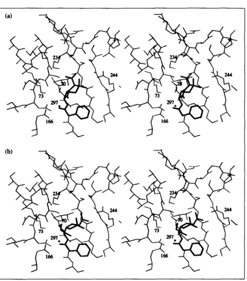
Fig. 4. Stereoviews of the Pen G sub-strate (thick lines) and the catalytic site of TEM1 (thin lines) (a) in the Michaelis complex and (b) in the acyl-enzyme complex. The deacylating water mole-cule, Wat297, is represented by a dot. In (b), the ester bond and the side-chain of Ser70 have also been drawn with thick lines.
This pathway becomes the only possible one with site-directed mutant enzymes where residue 73 is protonated, and will be discussed later.
Ser70 Oy is located only 3.5 A from the substrate car-boxylate, which, in the computation of pKa shifts within
the Michaelis complex, resulted in the Ser70 pKa being raised from 13.7 in the free enzyme to 39.0 after sub-strate binding and prior to proton transfer to Lys73. This suggests that the Ser70 Oy should be strongly deactivated in the Michaelis complex and a poor nucleophile in the acylation reaction, which is in contrast to the observed fast formation of the acyl-enzyme complex. The electro-static approach in evaluating the PKa of Ser70 in the Michaelis complex may be inappropriate for several rea-sons. First, Ser70 Oy is at bond formation distance from the substrate C7 atom. Second, local variations in dielec-tric properties, which may be important at the reaction centre, are ignored in the continuum approach. Third, a force field charge distribution that implies an identical
description of all residues is used, which might also be incorrect at the reaction centre. And, fourth, the charge distribution of the -lactam C7-08 carbonyl group could be different in free versus enzyme-bound Pen G. The last two points were addressed by changing the charge distribution of the C7-08 atoms and of the main-chain NH bonds of Ser70 and Ala237 in a way that
transfers the 08 partial charge onto the main-chain nitrogen atoms of residues 70 and 237, through the hydrogen bonds (2.7 A and 3.0 A) that comprise the oxyanion hole [8]. This appeared to be the simplest way to estimate the effect of the significant local dipoles gen-erated by the Met69-Ser70 and Gly236-Ala237 peptide bonds which are nearly parallel to the oxyanion hydrogen bonds. In addition, this observation may explain why the (4,lir) dihedral angles of residue 69 are found outside the low-energy areas of the Ramachandran plot in all class A 13-lactamase structures [9]. Transferring the 08 partial charge decreases the Ser70 PKa by 10.9 pH units (Table 2), in line with the proposal that each of these hydrogen bonds contributes about 5 kcalmol-1 [21], and does not significantly affect the pKa of Lys73, located 3.0 A away. The kinetic data of mutant proteins in which Lys234 is substituted by threonine, alanine, glutamate or histidine [18-20] indicates that the enzyme activity is reduced by 100-fold. One can expect that these mutations affect the pKas of Lys73 and Ser70O. The pKa of Lys73 was calculated to be 13.0 in the Lys234--Thr TEM1 enzyme and increases to 20.4 upon Pen G binding (Table 2). This indi-cates that Lys73 is already protonated in the free mutant protein and this is confirmed by the identical enzyme activity measured for the Lys73-Arg 3-lactamase I mutant enzyme [17,19].
608 Structure 1995, Vol 3 No 6 (a) Ser130-O\ H n 237 70 HzO," Wat297 H H H Ls73 \/\ H HO W2H H ' r "IH 0-Glut66 Michaelis complex a1li66 Tetrahedrl intermediate 37 A H/SeH 7'o 0_oWaa97
K
H'H o' Michaelis complex 0-hlr66 Acyl-enzyme intamediate Sr30-O Lys73\/\i H GlulA6 Tetrahedral intermediate Gl166 Acyl-enzyme intermediateFig. 5. Schematic representation of the two acylation reactions. (a) In the WT enzyme where Lys73 is unprotonated in the free enzyme [8]. (b) In the site-directed Lys73-->Arg and Lys234-*Thr mutant proteins where residue 73 is protonated in the free enzyme.
In the Lys234--Thr and Lys73--Arg mutant enzymes, Lys73 cannot be the proton acceptor of the Ser70 hydroxyl group and another proton acceptor has to be identified. The active-site structure suggests that Glu166, through the buried water molecule Wat297 located between Glu166 and Ser70 (2.8 A distant from each), can play this role. This water molecule could act as a pro-ton relay and assist the nucleophilic attack on the 3-lac-tam C7 carbonyl carbon atom by Ser70 Oy, through the concerted abstraction of the Ser70 hydroxyl group pro-ton and propro-tonation of Glu166 (Fig. 5b). In this case, the protonated Lys73 becomes involved in the formation of a tetrahedral intermediate, through a proton shuffle between Serl30, Lys73, Wat297, and Glu166, leading to the formation of an acyl-enzyme complex ready for de-acylation. The proton acceptor Glu166/Wat297 was sug-gested by Lamotte-Brasseur et al. [23] who had assumed that Lys73 was protonated in the WT enzyme. This acyl-ation mechanism appears less efficient than in the WT enzyme because, in addition to the loss of the 15 kT that
may lower the energy barrier for Ser70 deprotonation, it involves a larger number of proton transfers that may also contribute to the 100-fold reduction of the acylation rate constant, k2, measured for the Lys234--Thr and
Lys73-Arg mutant enzymes [17]. The respective loca-tions and distances between the carboxylate side chain of Glu166, the water, and the Ser70 Oy partners are also of importance in this process and k2 should, therefore, be
reduced upon a Glu166--Asp mutation. It was indeed found that the double mutant Lys73-Arg/Glu166-4Asp displays an additional 100-fold decrease in k2 compared
with that of the single mutant Lys73-Arg [17].
The relatively low pKa of 10.7 for Ser70 in the free Lys234-4Thr TEM1 mutant enzyme (Table 2) may indi-cate that Ser70 is more reactive than in the WT enzyme. Binding of Pen G increases the pKa of Ser70 by 21.2 pH units in this mutant and therefore deactivates Ser70 Oy, whereas a substrate devoid of the carboxylate group on the thiazolidine ring, such as penicillanyl alcohol, only
(b) Ser130-0o Lys73 \ +. IH I'H 37 'S70
Electrostatic analysis of P-lactamase Swaren et al. 609
increases the Ka of Ser70 by 5.7 pH units (Table 2). This substrate should therefore more readily form an acyl-enzyme with such mutant proteins and indeed, it displays a kcat value eightfold higher than that of phenoxymethylpenicillin [18]. These kinetic data show that the substrate carboxylate effect on the Ser70 pKa is poorly estimated, as already suspected: in the above case, it amounts to 15.5 pH units (21.2-5.7) and, if true, would lead to a larger difference in the kcat values between penicillanyl alcohol and phenoxymethyl-penicillin. The mechanism depicted in Figure 5b emphasizes the importance of the Glu166 and Lys73 pro-tonation states. This is also indicated by the bell-shaped kcat/KM versus pH profile for the Lys234-Ala protein
with penicillanyl alcohol as a substrate, which shows two ionizations, at pK1=5 and pK2=11 [18], in agreement
with the expected pKa of Glu166 and the calculated pKa of Lys73 (PKa=13.0).
The natural mutants of TEM1, identified from bacteria that are resistant to antibiotic treatment, have accord-ingly extended substrate specificities, but no mutation occurs at position 234, and none of the others which occur should affect the protonation state of Lys73. A charged residue at position 73 (i.e. Lys73---Arg or Lys234-X, where X is not arginine), is not known to occur spontaneously but was produced by site-directed mutagenesis. For these mutant enzymes we suggest an alternative mechanism for acylation (Fig. 5b). The idea of alternative mechanisms for a given enzyme may seem unlikely, but a_coenzyme analogue of liver alcohol de-hydrogenase, designed from the enzyme X-ray structure to interfere with the alcohol substrate-binding site, has been shown to induce a different mechanism [24]. The 'ordered bi-bi mechanism', expressing the sequential binding of NAD+ and the conformational change of the protein prior to substrate binding, became a 'rapid equi-librium random bi-bi mechanism' in the presence of this coenzyme analogue.
The deacylation step
Deacylation is promoted by a proton transfer from the crystallographic water molecule, Wat297, to the car-boxylate group of Glu166. This generates the nucle-ophilic hydroxyl group that attacks the ester carbonyl carbon of the acyl-enzyme intermediate. Electrostatic calculations on the acyl-enzyme complex show that the potential difference between Glu166 and the ester car-bonyl carbon of the acyl-enzyme amounts to about 150 kTe-' (-125 kTe-l at the carboxylate and +25 kTe- at the ester carbonyl carbon) (Fig. 6). Hence, the water molecule, located at 2.8 A from the Glu166 carboxylate in the direction of the ester carbonyl carbon, is experi-encing a steep potential gradient amounting to 30 kTe-1 A-' that will aid in polarizing the water molecule and direct the proton and hydroxyl groups towards Glu166 and the ester carbonyl carbon, respectively. To evaluate the contribution of Glu166 to this gradient, we also computed the electrostatic potential in the acyl-enzyme complex with no atomic charges assigned to Glu166. The carboxylate group was now at a -15 kTe-l potential surface, which arises from the main-chain oxy-gen atoms of residues 104, 105, 130, 131, 165, and 167, and the side chains of residues 104, 106, 131, 136, 168, 170, and 240. This generates an 8 kTe-1'A- gradient between Glu166 and the acyl-enzyme carbonyl carbon, similar to that computed for the Glu166--Asn TEM1 mutant protein (10 kTe-lA-'). This mutant protein, which was used to solve the acyl-enzyme structure and not reported to display any structural differences from the WT enzyme, lacks a defined proton acceptor. Nev-ertheless it deacylates, albeit slowly [11]. This might be due to the 10 kTe-1A-' gradient, present in this pro-tein. In -lactamase I, k3 is 1000 times lower upon a
Glu166--Asp substitution [17]. In TEM1, the same mutation leads to accumulation of the acyl-enzyme and poor deacylation [11], and the calculated gradient is 20 kTe- 1A-'. It thus seems that the active site structure provides a small intrinsic electrostatic potential gradient
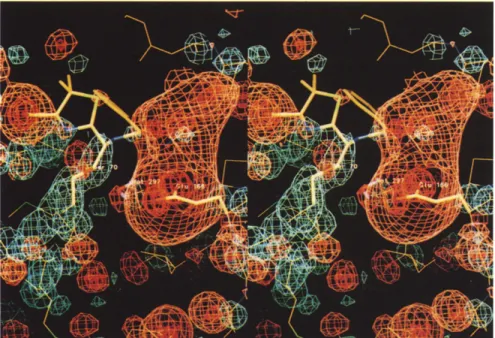
Fig. 6. Map of the electrostatic potential
generated by the acyl-enzyme drawn at different contour levels varying from -125 kTe-1(red) at Glu166 to +25 kTe- 1 (blue) at the ester carbonyl carbon. The deacylating Wat297, in the centre of the picture, is located between Glu166 and the ester bond (Ser70) at a -45 kTe- 1 potential surface (orange).
610 Structure 1995, Vol 3 No 6
Fig. 7. Comparison of the +20 kTe- 1
potential maps around residue 131 in the structure of the WT enzyme (dark
blue) and in the modelled Aspl31-->Asn mutant (light blue). The backbone of the enzyme is shown in yellow, helix H3 in
red, and helix H5 in green.
and that the proper positioning of an acidic group naturally increases its magnitude to an extent that might contribute to the high deacylation rate constant
(k3=1500 s-1). The function of Aspl 31
The buried and invariant Aspl31 is immersed in the -20 kTe- 1 negative electrostatic potential as described above, and its carboxylate group is hydrogen bonded to the main-chain nitrogen atoms of residues 108, 109, 133, and 134. These residues are at the N termini of helices H3 (109-111) and H5 (132-142) [9]; the stabi-lization of those local dipoles could explain the invari-ance and the structural role of Asp131. This hypothesis is reminiscent of the sulphate-binding protein of
Salmo-nella typhimurium where site-directed mutagenesis
demonstrated the role of local dipoles from three helices in binding of the sulphate ion [25]. The electrostatic calculation performed after a modelled substitution of Aspl31 by the most conservative residue, asparagine, showed the emergence of a very broad +20 kTe-1 posi-tive potential that immersed the N termini of helices H3 and H5 (Fig. 7). Although we are aware that the microscopic situation in this region could be improp-erly described by the macroscopic continuum calcu-lation [26], the inversion of potential to such a large extent in the Aspl31-*Asn protein compared with the WT enzyme indicates that strong favourable charge-helix interactions may be involved in the WT enzyme. Such interactions have been demonstrated in thermostable T4 lysozyme mutants [27] and in model peptides [28], and are consistent with the very poor expression of the Aspl31--Gly mutant protein [29]. In addition, since the calculations showed that the poten-tial gradient between Glu166 and the ester carbonyl carbon of the acyl-enzyme is not sensitive to the substi-tution, we may conclude that the major function of Aspl31 is to stabilize protein folding.
Biological implications
With a kcat/KM value of about 107 M-l s- t,
cataly-sis by class A -lactamases is nearly a diffusion-controlled reaction. There has been evolutionary pressure on -lactamases to broaden their sub-strate specificities but neither engineered mutants nor those isolated so far from natural sources have shown improved catalytic efficiency on the benzylpenicillin (Pen G) substrate. Hence, it seems that optimal conditions are met within the original Escherichia coli TEM1 enzyme, a penicilli-nase belonging to the class A -lactamases. The physico-chemical and electrostatic features shown in this work support this contention.
Several mechanisms for the class A 13-lactamases have been postulated. Kinetic characterization of
site-directed mutants and X-ray structure determi-nations of the TEM1 enzyme were important steps towards the rational understanding of the struc-ture-function relationships. Although amino acids essential for catalysis have been identified (Ser70, Lys73, Glu166, and Lys234), their exact mode of action (except for the acylating Ser70O) and inter-relations have been a matter of debate. The elec-trostatic potential distributions and pKa shifts, both in the free enzyme and in the Michaelis and acyl-enzyme complexes of the wild-type enzyme, suggest an essential role for the unprotonated and
solvent-shielded Lys73. We propose that substrate binding triggers the acylation reaction because it raises the pKa of the active site Lys73 (by 6.4 pH units for Pen G). Its protonation in the Michaelis complex then provides 15 kT (9 kcalmol 1) that may contribute by lowering the energy barrier for Ser70 deprotonation and thereby favour the nucleophilic attack on the substrate. Hydrolysis of
Electrostatic analysis of 13-lactamase Swartn et al. 611
the acyl-enzyme complex is promoted by a proton transfer from the crystallographic water molecule, Wat297, to Glu166. This generates the nucleophilic hydroxyl group that attacks the ester carbonyl car-bon of the acyl-enzyme. A steep electrostatic potential gradient, amounting to about 30 kTe-1 A-1, aids in polarizing the water molecule
and directs the proton and hydroxyl groups. In enzymes where the gradient is of smaller magni-tude, the deacylation step is considerably slower than in the wild-type enzyme.
In mutant proteins where protonation of residue 73 is imposed, the acylation pathway described for the wild-type enzyme cannot operate. Instead, an alternative and catalytically less effi-cient pathway, involving the Glu166/Wat297 cou-ple acting as proton acceptor, can be used in these enzymes. This situation, which has been revealed by site-directed mutagenesis, may not be specific to the Class A P-lactamases and it might be interesting to consider the possibility of alter-native mechanisms in other cases.
Materials and methods
Enzyme coordinatesThe atomic coordinates of TEM1 were taken from the 1.8 A
resolution refined X-ray structure [9] (Brookhaven Data Bank entry: 1BTL). The structure includes 199 water molecules and one sulphate ion provided by the crystallization conditions. This ion has no biological significance and was discarded from the calculations as well as 33 water molecules that do not exchange hydrogen bonds with protein atoms. All other water molecules were considered as part of the protein and have an average temperature factor of 14.6 A2. The different mutants were modelled on the structure of the WT enzyme with the program O [30]. Polar hydrogen positions were 'built and energy-minimized using the program X-PLOR [31]: new sets were generated for each mutant and each protonation state. None of the mutations introduced steric constraints into the protein structure. Solvent accessibilities [32] were computed with the program SURFACE from the CCP4 suite [33]. Modelling of complexes
The acyl-enzyme model was constructed according to the description given by Strynadka et al. [8]. The Michaelis complex was derived from the acyl-enzyme complex by closing the P-lactam ring by rotation of the N4-C5-C6-C7 torsion angle to its known value (5.40) [34]. Structural constraints and interac-tions between protein and substrate are such that this rotation could only alter the position of the thiazolidine ring so as to maintain the positions of the -lactam ring carbonyl and of the NHCO group at C6. In these models, five crystallographic waters (Wat323, Wat391, Wat404, Wat422, and Wat472) that occupy the substrate-binding site in the free enzyme were removed. Coordinates and charges of free Pen G were obtained by energy minimization in PCMODEL (Serena Software, Bloomington, IN). For the acyl-enzyme complex, the charges were obtained by energy minimization of penicilloic ethyl ester.
Numerical treatment
For evaluation of potential fields, we used the DelPhi package [35,36] which solves the linearized Poisson-Boltzmann
equation by a finite difference method. The TEM1 structure has a length of about 57
A
which, together with the original DelPhi grid (65 x 65 x 65 grid points), gives a resolution of about one grid point per A. This was considered insufficient and the grid was extended to 129 x 129 x 129 points. Two dummy atoms were introduced to control the position of the molecule within the grid. At a 'perfil' of 100%, these dummy atoms are located at opposite ends of the diagonal of the cubic grid in such a way that the longest dimension of the protein molecule, including its solvent-accessible surface (computed with a probe radius of 1.4 A), falls exactly within the grid. The atoms were assigned Connolly radii provided with the DelPhi package. A 2.0 A thick ion exclusion layer was added and the ionic strength of the bulk was set to 145 mM. The protein moiety was assigned a relative dielectric constant of 3.0 while that of the bulk was set to 80. Atomic charges were taken from the "tophl9.pro" file in X-PLOR [31,37] and distributed over the grid points according to an algorithm described by Gilsonet al. [38]. This algorithm gives more accurate potentials at
close distances than a traditional distribution over eight closest grid points. Charges of neutral lysines and anionic serines were obtained with PCMODEL. Analysis of the structure implied that all residues, except those stated in this work, could be kept in their normal protonation state at pH 7.8. Independence from chosen grid origin was verified by repeating the calcula-tions with the geometric centre of protein plus dummy atoms placed at seven different grid points (the grid centre itself and its six closest neighbours). A three-step focusing protocol (15%, 60%, and 100% 'perfils') was used with the Debye-Hiickel potential of the equivalent dipole to the molecular charge dis-tribution as boundary condition in the initial step [36]. The grid size was 0.52
A
(1.92 grid points perA)
in the last step.Calculation of pKas
Three types of electrostatic energy contributions must be considered to evaluate the difference in the titration behaviour of an ionizable group in a protein and the same group in a model compound such as a free amino acid in bulk water
[39,40]: first, the interaction of the group with the polarization that its charges induce in the protein and in the model (Born solvation energy, AAGBom); second, the interaction of the group with partial charges, such as the peptide dipoles (back-ground charge interactions, AAGback); third, the interaction of the group with other titrating groups in the protein. A rigorous treatment of titration should consider that the protonation state of a group depends on the protonation states of other titrating groups, which themselves are also pH dependent. Therefore, those interaction energies should be evaluated and a Boltzmann sum made over all possible energy states, which will be 2N,
where N equals the number of titratable sites in the protein. The process has been thoroughly outlined [40,41]. This statisti-cal mechanistatisti-cal approach still has to cope with the inaccuracy of the protein dielectric, treated as a constant although it is a function of coordinates. The simplification we have made was based on a careful analysis of the TEM1 structure whereupon the protonation states of the other titrating sites were simply included in the background charge interaction energies when evaluating the pKa of a certain residue. That is:
pKa=pKmodel-(kT In 10)-(AAGBom+AAGback)
where pKam°del is the side chain pKa of a free amino acid [42] (the side chain of a free serine was assumed to have pKa=16, like ethanol). This simplification, rigorously valid when the investigated group is the only one that titrates in the analyzed pH range, was confirmed by the consistency with experimental
612 Structure 1995, Vol 3 No 6
data. The model compounds were constructed by isolating the actual side chain, together with its backbone (the used charge set gives a neutral charge distribution of all backbones except termini), from the structure.
The calculation made to evaluate how the Ka of Ser70 may
be affected by the charge distribution of the 3-lactam C7-08 carbonyl group and of the NH bonds of residues 70 and 237 was cared out as follows. To get an estimate of the effect of the oxyanion hole in cancelling out the 08 partial charge, this was transferred in equal parts to the main-chain nitrogen atoms of residues 70 and 237. No additional polarization of the sub-strate carbonyl group compared with that of unbound Pen G was assumed. The charge distribution was, in free enzyme and Pen G: C7 +0.3; 08 -0.5; N70 -0.35; H70 +0.25; N237 -0.35; H237 +0.25; and after charge transfer: C7 +0.3; 08 0.0; N70 -0.60; H70 +0.25; N237 -0.60; H237 +0.25.
Hardware
The program was run on an Evans and Sutherland ESV/30-33 and PCMODEL on an IBM RS/6000 worksta-tion. All other software packages were run on a Digital DEC 3000/400 Alpha workstation (96 Mb RAM). A PKa calcula-tion required about one hour of CPU time.
Acknowledgements: We thank Dr Emmanuel Courcelle for his excellent assistance on informatics and Dr Martin Welch for correct-ing the English of the manuscript. We are also grateful to ProfJean Devillers for general discussions on energy minimization. This work was financed, in part, by the Institut National de la Sante et de la Recherche Medicale (contract no. 930612) and by ARC (contract no. 6564). The Service Scientifique de l'Ambassade de France Stockholm is gratefully acknowledged for financial support to PS.
References
1. Ambler, R.P., et al., & Waley, S.G. (1991). A standard numbering scheme for the class A j3-lactamases. Biochem. J. 276, 269-272. 2. Bush, K. (1989). Characterization of 3-lactamases. Antimicrob.
Agents Chemother. 33, 259-263.
3. Christensen, H., Martin, M.T. & Waley, S.G. (1990). P-lactamases as fully efficient enzymes. Biochem. J. 266, 853-861.
4. Dideberg, O., et al., & Ghuysen, J.-M. (1987). The crystal structure of the -lactamase of Streptomyces albus G at 0.3 nm resolution.
Biochem. J. 245, 911-913.
5. Herzberg, O. (1991). Refined crystal structure of 13-lactamase from
Staphylococcus aureus PC1 at 2.0 A resolution. . Mol. Biol. 217, 701-719.
6. Knox, J.R. & Moews, P.C. (1991). -lactamase of Bacillus
licheni-formis 749/C. Refinement at 2 A resolution and analysis of
hydra-tion. J. Mol. Biol. 220, 435-455.
7. Jelsch, C., Lenfant, F., Masson, J.-M. & Samama, .-P. (1992). P-lac-tamase TEM1 of E. coli. Crystal structure determination at 2.5 A res-olution. FEBS Lett. 299, 135-142.
8. Strynadka, N.C.J., et a., & James, M.N.G. (1992). Molecular struc-ture of the acyl-enzyme intermediate in p3-lactam hydrolysis at 1.7 A resolution. Nature 359, 700-705.
9. Jelsch, C., Mourey, L., Masson, J.-M. & Samama, J.-P. (1993). Crystal structure of Escherichia coli TEM1 1-lactamase at 1.8 A resolution.
Proteins 16, 364-383.
10. Herzberg, O. & Moult, J. (1991). Penicillin-binding and degrading enzymes. Curr. Opin. Struct. Biol. 1, 946-953.
11. Adachi, H., Ohta, T., Matsuzawa, H. (1991). Site-directed mutants, at position 166, of RTEM-1 -lactamase that form a stable acyl-enzyme intermediate with penicillin. J. Biol. Chem. 266, 3186-3191. 12. Oefner, C., et a., & Winkler, F.K. (1990). Refined crystal structure of
-lactamase from Citrobacter freundii indicates a mechanism for 13-lactam hydrolysis. Nature 343, 284-288.
13. Dao-pin, S., Anderson, D.E., Baase, W.A., Dahlquist, F.W. & Matthews, B.W. (1991). Structural and thermodynamic conse-quences of burying a charged residue within the hydrophobic core of T4 lysozyme. Biochemistry 30, 11521-11529.
14. Hedges, R.W., Datta, N., Kontomichalou, P. & Smith, J.Y. (1974).
Molecular specificities of R factor determined beta-lactamases: correlation with plasmid compatibility. J. Bacteriol. 117, 56-62.
15. Delaire, M., Labia, R., Samama, .-P. & Masson, J.-M. (1992). Site-directed mutagenesis at the active site of E coli TEM1 -lactamase.
J. Bio/. Chem. 267, 20600-20606.
16. Zafaralla, G., Manavathu, E.K., Lerner, S.A. & Mobashery, S. (1992). Elucidation of the role of arginine-244 in the turnover processes of class A -lactamases. Biochemistry 31, 3847-3852.
17. Gibson, R.M., Christensen, H. & Waley, S.G. (1990). Site-directed mutagenesis of 3-lactamase . Single and double mutants of Glu 166 and Lys 73. Biochem. J. 272, 613-619.
18. Ellerby, L.M., Escobar W.A., Fink, A.L., Mitchinson, C. & Wells, J.A. (1990). The role of lysine 234 in -lactamase catalysis probed by site-directed mutagenesis. Biochemistry 29, 5797-5806.
19. Lenfant, F., Labia, R. & Masson, J.-M. (1991). Replacement of lysine 234 affects transition state stabilization in the active site of 13-lacta-mase TEM1. J. Biol. Chem. 266, 17186-17194.
20. Brannigan, J., eta/., & Frere, J.-M. (1991). The mutation Lys234/His
yields a class A 3-lactamase with a novel pH-dependence.
Biochem. J. 278, 673-678.
21. Warshel, A., Naray-Szabo, G., Sussmann, F. & Hwang, J.-K. (1989). How do serine proteases really work? Biochemistry 28, 3629-3637. 22. Daggett, V., Schroder, S. & Kollman, P. (1991). Catalytic pathway of
serine proteases: classical and quantum mechanical calculations.
J. Am. Chem. Soc. 113, 8926-8935.
23. Lamotte-Brasseur, J., Dive, G., Dideberg, O., Charlier, P., Frere, J.-M. & Ghuysen, J.-M. (1991). Mechanism of acyl transfer by the class A serine -lactamase of Streptomyces albus G. Biochem. J.
279, 213-221.
24. Samama, J.-P., Hirsch, D., Goulas, P. & Biellmann, J.-F. (1986). Dependence of the substrate specificity and kinetic mechanism of horse-liver alcohol dehydrogenase on the size of the C-3 pyridinium substituent; 3-benzoylpyridine-adenine dinucleotide. Eur. J.
Biochem. 159, 375-380.
25. He, J.J. & Quiocho, F.A. (1993). Dominant role of local dipoles in stabilizing uncompensated charges on a sulfate sequestered in a periplasmic active transport protein. Protein Sci. 2, 1643-1647. 26. Aqvist, J., Luecke, H., Quiocho, F.A. & Warshel, A. (1991). Dipoles
localized at helix termini of proteins stabilize charges. Proc. Nat/.
Acad. Sci. USA 88, 2026-2030.
27. Nicholson, H., Anderson, D.E., Dao-pin, S. & Matthews, B.W. (1991). Analysis of the interaction between charged side-chains and the -helix dipole using designed thermostable mutants of phage T4 lysozyme. Biochemistry 30, 9816-9819.
28. Huyghues-Despointes, B.M.P., Scholtz, J.M. & Baldwin, L. (1993). Effect of a single aspartate on helix stability at different positions in a neutral alanine-based peptide. Protein Sci. 2, 1604-1611.
29. Jacob, F., Joris, B., Lepage, S., Dusart, . & Frere J.-M. (1990). Role of the conserved amino acids of the 'SDN' loop (Serl 30, Asp131 and Asn132) in a class A -lactamase studied by site-directed mutagen esis. Biochem. J. 271, 399-406.
30. Jones, T.A., Zou, J.-Y., Cowan, S.W. & Kjeldgaard, M. (1991). Improved methods for building protein models in electron density maps and the location of errors in these models. Acta Crystallogr. A 47,110-119.
31. Bronger, A.T. (1992). X-PLOR Version 3.1: A System for X-ray
Crys-tallography and NMR. Yale University, New Haven, CT.
32. Lee, B. & Richards, F.M. (1971). The interpretation of protein struc-tures: estimation of static accessibility. J. Mol. Bio/. 55, 379-400.
33. Collaborative Computational Project, Number 4 (1994). The CCP4 suite: programs for protein crystallography. Acta Crystallogr. D 50, 760-763.
34. Dexter, D.D. & van der Veen, J.M. (1978). Conformations of peni-cillin G: crystal structure of procaine penipeni-cillin G monohydrate and a refinement of the structure of potassium penicillin G. . Chem. Soc. (Perkin Trans. I), 185-190.
35. Nicholls, A. & Honig, B. (1991). A rapid finite difference algorithm, utilizing successive over-relaxation to solve the Poisson-Boltzmann equation. J. Comput. Chem. 12, 435-445.
36. Sharp, K.A. & Nicholls, A. (1989). DelPhi (v. 3.0) Manual. Dept. of Biochemistry and Molecular Biophysics, Columbia University, NY. 37. Brooks, B.R., Bruccoleri, R.E., Olafson, B.D., States, D.J.,
Swami-nathan, S. & Karplus, M. (1983). CHARMM: a program for macro-molecular energy, minimization and dynamics calculations.
J. Comput. Chem. 4, 187-217.
38. Gilson, M.K., Sharp, K.A. & Honig, B.H. (1987). Calculating the electrostatic potential of molecules in solution: method and error assessment. J. Comput. Chem. 9, 327-335.
39. Tanford, C. & Kirkwood, J.G. (1957). Theory of protein titration curves. . General equations for impenetrable spheres. . Am. Chem. Soc. 79, 5333-5339.
Electrostatic analysis of 13-lactamase Swaren et al. 613
40. Bashford, D. & Karplus, M. (1990). pKa's of ionizable groups in proteins: atomic detail from a continuum electrostatic model.
Biochemistry 29, 10219-10225.
41. Yang, A.-S., Gunner, M.R., Sampogna, R., Sharp, K. & Honig, B. (1993). On the calculation of pKas in proteins. Proteins 15, 252-265.
42. Dawson, R.M.C., Elliott, D.C., Elliott, W.H. & Jones, K.M. (1986).
Data for Biochemical Research. (3rd edn), pp. 1-31, Clarendon
Press, Oxford.
43. Lenfant, F. (1991). Etude du site actif de la -lactamase TEM-1
d'Escherichia co/i par mutagenese dirigee. [PhD thesis]. INSA, Toulouse, France.
44. Kraulis, P.J. (1991). MOLSCRIPT: a program to produce both detailed and schematic plots of protein structures. J. Appl.
Crystal-Iogr. 24, 946-950.
Received: 7 Nov 1994; revisions requested: 30 Nov 1994; revisions received: 19 Apr 1995. Accepted: 2 May 1995.
![Fig. 5. Schematic representation of the two acylation reactions. (a) In the WT enzyme where Lys73 is unprotonated in the free enzyme [8]](https://thumb-eu.123doks.com/thumbv2/123doknet/2343096.34263/7.918.93.833.88.717/fig-schematic-representation-acylation-reactions-enzyme-unprotonated-enzyme.webp)