POLYTECHNIQUE MONTRÉAL
affiliée à l’Université de MontréalSubtle changes in hyperelastic properties of myocardium with cardiotoxicity
remodeling from cardiac magnetic resonance
MARIANNA GAMBA Institut de génie biomédical
Mémoire présenté en vue de l’obtention du diplôme de Maîtrise ès sciences appliquées Génie biomédical
Mai 2019
POLYTECHNIQUE MONTRÉAL
affiliée à l’Université de MontréalCe mémoire intitulé :
Subtle changes in hyperelastic properties of myocardium with cardiotoxicity
remodeling from cardiac magnetic resonance
présenté par Marianna GAMBA
en vue de l’obtention du diplôme de Maîtrise ès sciences appliquées a été dûment accepté par le jury d’examen constitué de :
Farida CHERIET, présidente
Delphine PÉRIÉ-CURNIER, membre et directrice de recherche Caroline LAVERDIÈRE, membre externe
DEDICATION
ACKNOWLEDGEMENTS
First and foremost, I would like to sincerely thank my advisor, Prof. Delphine Périé-Curnier, for the patient guidance and support she has provided me throughout the course of my graduate studies. I am also very grateful for the opportunity I have had to work on this project and gain insights into the issue of doxorubicin-associated cardiotoxicity.
Furthermore, I would like to express my gratitude to the rest of the thesis examination committee, Prof. Farida Cheriet and Dr. Caroline Laverdière, for their interest, consideration, and time devoted to reviewing this manuscript.
I certainly owe a great deal to the team of researchers of the PETALE project for providing me the experimental data as well as the opportunity to conduct this complementary analysis on survivors of childhood acute lymphoblastic leukemia. Thanks also go to Prof. Alistair Young and his team of programmers at the Auckland MRI Research Group, who has kindly made the Cardiac Imaging Modeling software available to us. Without this tool, we could not have achieved the objectives of the thesis. I wish to acknowledge those students of Polytechnique Montreal who contributed to the development of this work by providing some input data.
On a more personal note, I would like to express my deepest gratitude towards my parents and my siblings, who have constantly supported and encouraged me to do my best. Moreover, I take this opportunity to thank Maria Lacruz, Yasamin Majedi, Evelyne Lechevalier Hubertise, and Ana Segovia; this journey would not have been nearly as great without all the laughs and tears we shared together. Finally, a huge and sincere thanks to my lifetime friends, Rossella, Gloria, Alessandra, Federico, and Eleonora, who helped me keep things in perspective.
RESUME
La doxorubicine (DOX) est un puissant agent antinéoplasique fréquemment administré dans le traitement de nombreux cancers pédiatriques, notamment la leucémie lymphoblastique aiguë (LLA). La doxorubicine possède une efficacité démontrée dans le traitement du cancer, mais elle engendre également un large spectre d'effets cardiaques indésirables. Les changements structurels du myocarde s'accompagnent de modifications progressives de la géométrie de la paroi du myocarde du ventricule gauche (VG). La détérioration de la fonction myocardique peut progresser silencieusement pendant des années et se manifester sans avertissement ou même ne devenir apparente que longtemps après la fin du traitement. Des doses cumulatives plus élevées de DOX augmentent le risque d'effets nocifs associés au traitement. La faisabilité de la résonnance magnétique cardiaque (RMC) a été établie et plusieurs logiciels de modélisation géométrique 3D du cœur ont été développés pour évaluer la fraction d'éjection, l'épaisseur de la parois, et le volume télésystolique et le volume télédiastolique du VG. La modélisation par éléments finis (EF) de la mécanique du ventricule gauche et les stratégies inverses d'identification des paramètres des matériaux ont ensuite été introduites pour tenir compte du comportement mécanique passif du tissu myocardique.
Compte tenu de cela, nous avons entrepris une étude visant à analyser en détail les subtils changements asymptomatiques dans la géométrie et dans la fonction du ventricule gauche chez 84 survivants de la LLA infantile traités par chimiothérapie utilisant la doxorubicine (dose faible à modérée). Étant donné le grand potentiel de la modélisation numérique du cœur, cette évaluation a été réalisée à l'aide d'une approche basée sur un modèle EF qui intègre la forme et le mouvement spécifiques de la chambre ventriculaire directement à partir des données RMC.
84 survivants de la LLA, âgés de 23 ± 7 ans, ont été recrutés prospectivement et répartis en deux groupes distincts, nommés respectivement risque standard (SR, n = 19) et risque élevé en fonction du risque de récidive. En outre, les sujets du groupe associé à un risque élevé de récidive ont été subdivisés en deux sous-groupes selon qu’ils avaient reçu (HRdex, n = 36) ou non (HR, n = 45) la thérapie cardioprotectrice (dexrazoxane) dans le but de réduire le risque de lésions cardiaques à long terme. Par ailleurs, à des fins de comparaison, 10 volontaires en santé, d’âge similaire aux survivants (22 ± 4 ans) et n’ayant jamais été atteints de leucémie aiguë ou de cardiomyopathie, ont formé un groupe témoin.
Dans le cadre de l'étude de la cardiotoxicité tardive, tous les sujets ont subi une imagerie RMC, une échocardiographie transthoracique et des tests d'effort. À partir des données RMC acquises, un opérateur formé a extrait la forme et le mouvement spécifiques du ventricule gauche à l'aide d'un cadre de points de repère (CIM v8.1, University of Auckland, Nouvelle-Zélande). Les bordures intérieures et extérieures des parois du ventricule gauche ont été dessinées semi-automatiquement à partir de six points de guidage placés par l'opérateur, puis corrigés manuellement en cas d'erreurs d'alignement. À partir de ce tracé, on a calculé la fraction d'éjection, le volume de course, la masse myocardique, l'épaisseur de la paroi, le volume diastolique final et le volume systolique final pour chacun des participants de l'étude. Par la suite, la reproductibilité inter- et intra-observateurs des résultats de la segmentation a été quantifiée par des coefficients de corrélation intra-classe (ICC) sur quatre reconstructions de 15 survivants du cancer, chacune étant réalisée par quatre opérateurs formés, et sur trois reconstructions du même sujet, chacune étant effectuée par un seul opérateur. Pour un sous-ensemble de la population des survivants de la leucémie (n = 48), un modèle 3D conçu par éléments finis a été utilisé pour calculer la propriété hyperélastique (C1) à partir d’une
stratégie d'identification des paramètres inverses des matériaux basés sur la géométrie du VG lors de la diastase. Au départ, nous avons supposé des valeurs physiologiquement raisonnables de 0.75, 1.0 et 1.25 kPa comme contraintes de charge de pression pour tous les participants. Une fois les pressions ventriculaires spécifiques au sujet (au repos et à l'exercice maximal) connues, nous avons incorporé ces données dans le modèle et répété l'analyse aux éléments finis. Les valeurs résultantes de C1 ont été notées et comparées entre les groupes pour chacun des cinq scénarios de charge. Les
comparaisons statistiques ont été effectuées par analyse de variance à sens unique (ANOVA) sur les paramètres géométriques globaux et régionaux et par analyse de variance à mesures répétées bidirectionnelles sur les paramètres géométriques dépendants du temps. Enfin, une analyse de sensibilité a été effectuée pour évaluer la dépendance de la propriété hyperélastique de la diastase et de la pression.
Dans le cas de notre étude, la reproductibilité intra-observateur était bonne pour les paramètres géométriques régionaux (ICC, 0.60-0.74) et excellente pour les paramètres géométriques globaux (ICC, 0.75-1.00), alors que la reproductibilité inter-observateur était excellente pour les deux types de paramètres (ICC, 0.75-1.00). Aucune différence significative entre les quatre groupes n’a été observée relativement à la fraction d’éjection, au débit systolique, à la masse, au volume systolique final et au volume diastolique final. Quelques différences de volume épicardique ont été observées,
mais seulement entre la phase finale de la systole et la diastase (p<0.01) à l’intérieur des groupes HRdex et HV ou SR, et parmi les groupes HV et HR. Il est intéressant de noter que la propriété hyperélastique pour les pressions standard était légèrement plus faible dans le groupe HR comparativement au groupe HRdex ou SR et aussi relativement au groupe contrôle (p<0.05). En revanche, aucune différence appréciable n’a pu être détectée dans la propriété hyperélastique à des pressions intraventriculaires au repos (p>0.5) et à l’exercice maximal (p>0.6).
Il ressort clairement de cette analyse que les paramètres géométriques globaux et régionaux ne sont pas suffisamment sensibles pour détecter les changements subtils induits par la cardiotoxicité tardive de la doxorubicine dans la structure et la fonction du ventricule gauche. Toutefois, les paramètres globaux dépendants du temps constituent des preuves préliminaires que l’exposition à la doxorubicin a un effet plus néfaste sur la diastole précoce que sur la systole ou la diastole tardive. La propriété hyperélastique, plus faible dans le groupe HR, suggère un tissu myocardique plus enclin à la dilatation si une pression intra-ventriculaire plus élevée est appliquée, comparativement aux autres groupes d'étude. Cette constatation concorde avec ce qui a été observé chez les survivants à long terme ayant eu la leucémie adulte et traités par chimiothérapie à forte dose à base de doxorubicine. Il convient toutefois de noter que ces estimations ne peuvent tenir compte que des effets géométriques du remodelage du myocarde sur la mécanique ventriculaire passive, puisque la pression intra-ventriculaire gauche spécifique au sujet n'était pas incluse dans ces simulations. Des résultats similaires ont été obtenus lors de l'application de pressions intra-ventriculaires mesurées au repos et pendant l'exercice maximal.
En conclusion, cette étude démontre que la cardiotoxicité subclinique de la doxorubicine peut être évaluée par l’analyse du comportement mécanique du ventricule gauche sur des images RMC. Nos résultats doivent toutefois être confirmés par des analyses additionnelles avant d’en tirer une conclusion solide sur la signification pronostique, à long terme, des altérations de la raideur myocardique et de leur relation avec le statut de risque dans la cohorte de survivants étudiée ou dans une cohorte similaire. Il serait très pertinent, dans le cadre d’études ultérieures, d’inclure la simulation de la mécanique ventriculaire, avec la méthode des éléments finis, pendant la diastole précoce et la systole afin de mieux analyser les changements de rigidité dans les groupes. Dans le même but, il pourrait être utile d'augmenter la résolution temporelle des données d'imagerie chez les survivants de la leucémie et les sujets témoins.
Mots-clés : survivants de la leucémie lymphoblastique aiguë, cardiotoxicité induite par la doxorubicine, imagerie par résonance magnétique cardiaque, rigidité passive du myocarde, modélisation par éléments finis personnalisée, mécanique ventriculaire gauche.
ABSTRACT
Doxorubicin (DOX) is a potent chemotherapeutic agent routinely administered in the treatment of several pediatric malignancies, including acute lymphoblastic leukemia (ALL). Despite its efficacy to improve the outlook of cancer patients, doxorubicin is known to cause a wide spectrum of cardiac adverse effects. The structural changes in the myocardium are accompanied by progressive changes in LV myocardial wall geometry. The deterioration of myocardial function can progress silently for many years before the manifestation of clinical symptoms and become apparent even long time after completion of treatment. Higher cumulative doses of this agent increase the risk for late cardiac complications. The feasibility of CMR imaging has been established and a variety of software for 3D geometric modeling of the heart have been developed to assess wall thicknesses, ejection fraction, end-systolic and end-diastolic volumes. Finite element (FE) modelling and inverse material parameter identification strategies were then introduced to take into account the passive mechanical behavior of the myocardial tissue.
In light of the above, we undertook a study to assess the asymptomatic changes in LV structure and function in a group of long-term survivors of childhood ALL treated with low to moderate doses of doxorubicin therapy. Given the high potential of numerical cardiac modeling, this evaluation was conducted using a FE model-based approach that integrates the subject-specific shape and motion of the ventricular chamber directly from imaging data.
Eighty-four ALL survivors (23±7 years old) were prospectively enrolled and stratified into two different groups, designated as standard-risk (SR, n=19) and high-risk groups, according to their risk of tumor recurrence. Subjects treated for high-risk ALL were further divided into two groups depending upon whether they did (HRdex, n=36) or did not (HR, n=45) receive the protective therapy (dexrazoxane) in an attempt to reduce the likelihood of late cardiotoxicity. Furthermore, for purposes of comparison, 10 healthy volunteers (HV, 22±4 years), with no prior history of cancer or cardiac pathologies and similar in age to the survivors, were used as controls.
As a part of the investigation of late-onset cardiotoxicity, all subjects underwent CMR imaging, transthoracic echocardiography, and exercise stress testing. From the acquired CMR data, a trained operator extracted the subject-specific shape and motion of the LV using a guide-point framework (CIM v8.1, University of Auckland, New Zealand). The inner and outer borders of the LV walls were semi-automatically drawn from six guidepoints placed by the operator at end-systole and then
manually corrected for mis-registration errors. From this tracing, ejection fraction, stroke volume, myocardial mass, wall thickness, end-diastolic and end-systolic volumes were computed for each of the study participants. After that, inter- and intraobserver repeatability of the segmentation results were quantified by intra-class coefficients (ICC) on four reconstructions of 15 leukemia survivors, each by four trained operators, and on three reconstructions of the same subject, each by a single operator.
For a subset of the leukemia survivor population (n=48), a 3D finite element model was used to quantify the hyperelastic property (C1) from inverse material parameters identification strategies
based on the LV geometry at diastasis. This biomechanical parameter was initially calculated by assuming physiologically realistic values of 0.75, 1.0, and 1.25 kPa as pressure loading constraints for all participants. Once the subject-specific LV pressures (at rest and peak exercise) became available, we incorporated such data in the model and repeated the FE analysis. The resulting values of C1 were reported and compared between groups for each of the five loading scenarios.
Statistical comparisons were performed by one-way analysis of variance (ANOVA) on global and regional geometrical parameters, and two-way repeated-measures ANOVA on time-dependent geometrical parameters. Ultimately, a sensitivity analysis was conducted to evaluate the dependence of the hyperelastic property on the diastasis frame and the pressure load.
In our experience, inter-observer repeatability was good for regional geometrical parameters (ICC, 0.60-0.74) and excellent for global geometrical parameters (ICC, 0.75-1.00), while intra-observer repeatability was excellent for both regional and global parameters (ICC, 0.75-1.00). Groups had similar LV function values. No significant differences were observed among the four study groups in ejection fraction, stroke volume, mass, end-diastolic or end-systolic volumes. Some differences were detected in epicardial volume only between end-systole and diastasis phases (p<0.01) among the HRdex and HV or SR groups, and among the HV and HR groups. Interestingly, the hyperelastic property for standard pressures was slightly lower in the HR group when compared against the HRdex or SR group, and also when compared against the control group (p<0.05). In contrast, no appreciable difference could be noted in the hyperelastic property for intra-ventricular pressures at rest (p>0.5) and peak exercise (p>0.6).
From this analysis, it is clear that the global and regional geometrical parameters are not sufficiently sensitive to capture the subtle changes induced by late doxorubicin cardiotoxicity in LV structure
and function. Nevertheless, the time-dependent global parameters provided preliminary evidence that early diastole was more affected by doxorubicin exposure than systole or late diastole. The smaller hyperelastic property in the high-risk group suggested a myocardial tissue more prone to dilatation if increased intra-ventricular pressure is applied than in the other study groups. This finding is consistent with what has been observed in long-term survivors of adult leukemia treated with high-dose doxorubicin-based chemotherapy. It should be noted, however, that these estimates could account only for the geometrical effects of myocardial remodeling on the passive ventricular mechanics since the subject-specific LV cavity pressure was not included in these simulations. Similar results were obtained when applying intra-ventricular pressures at rest and peak exercise. In conclusion, this study demonstrated that the subclinical cardiotoxicity of doxorubicin can be non-invasively assessed through the mechanical behavior analysis of the LV on CMR images. Additional investigations will be necessary to confirm our results and draw a firm conclusion about the long-term prognostic significance of alterations in myocardial stiffness and their relationship with ALL risk status in this or similar cohort of survivors. In future works, we hope toinclude the FE simulation of the left ventricular mechanics during systole and early diastole in order to better capture the changes in stiffness across groups. To the same purpose, it might be convenient to increase the temporal resolution of the image data in both leukemia survivors and control subjects.
Keywords: acute lymphoblastic leukemia survivors, doxorubicin-mediated cardiotoxicity, cardiac magnetic resonance imaging, passive myocardial stiffness, personalized FE modeling, in vivo left ventricular mechanics.
TABLE OF CONTENTS
DEDICATION ... III ACKNOWLEDGEMENTS ... IV RESUME ... V ABSTRACT ... IX TABLE OF CONTENTS ...XII LIST OF TABLES ... XV LIST OF FIGURES ... XVII LIST OF SYMBOLS AND ABBREVIATIONS... XX
CHAPTER 1 INTRODUCTION ... 1
1.1 Background ... 1
1.2 General and specific research objectives ... 4
1.3 Research hypotheses ... 5
1.4 Outline of the thesis... 5
CHAPTER 2 FUNDAMENTALS ... 7
2.1 The heart ... 7
2.1.1 Basic cardiac anatomy ... 7
2.1.2 The cardiac cycle ... 9
2.2 Cardiotoxicity of doxorubicin in the treatment of childhood ALL ... 11
2.2.1 Mechanisms of doxorubicin cytotoxic action ... 12
2.2.2 Histopathological features of doxorubicin cardiotoxicity ... 13
2.2.3 Macroscopic features of doxorubicin-related cardiotoxicity ... 15
2.2.4 Prevention and reduction of doxorubicin-related cardiotoxicity... 16
2.3 Analysis of the mechanical properties of the passive LV myocardium ... 19
2.3.1 Mechanical characterization of passive myocardium ... 20
2.3.2 Modeling ventricular myocardium ... 22
2.3.3 Passive stiffness of the left ventricular myocardium ... 26
2.4 Key points ... 28
CHAPTER 3 MATERIALS AND METHODS ... 30
3.1 Study population ... 30
3.2 Clinical measurements ... 33
3.2.1 Cardiac magnetic resonance imaging ... 33
3.2.2 Exercise stress testing... 33
3.3 Image segmentation and finite element model generation ... 34
3.3.1 Global analysis of LV geometry and function ... 37
3.3.2 Regional analysis of LV geometry and function ... 39
3.4 Finite element analysis ... 40
3.4.1 Input parameters of the ventricular mechanics model ... 41
3.4.2 Passive mechanics simulation for standard diastasis pressures ... 44
3.4.3 Estimation of in vivo myocardial stiffness ... 45
3.5 Repeatability analysis ... 46
3.6 Sensitivity analysis of the model parameters ... 48
3.7 Statistical analysis of data ... 49
3.7.1 Statistical methods... 49
3.7.2 Correlation between myocardial stiffness and CMR relaxation time ... 50
CHAPTER 4 RESULTS... 51
4.2 Results of the sensitivity analysis... 51
4.3 Global and regional 3D geometrical parameters ... 54
4.4 Global time-dependent 3D geometrical parameters ... 55
4.5 Estimates of myocardial stiffness for standard diastasis pressures ... 59
4.6 Estimates of in vivo myocardial stiffness ... 63
4.7 Results of the correlation analysis ... 65
CHAPTER 5 GENERAL DISCUSSION ... 67
5.1 Comment ... 67
5.2 Limitations of the study... 71
CHAPTER 6 CONCLUSION AND RECOMMENDATIONS ... 73
6.1 Highlights ... 73
6.2 Concluding remarks and future developments ... 74
LIST OF TABLES
Table 2.1. In vivo estimates of passive myocardial stiffness (mean standard deviation) for the three groups of study participants. Asterisks denote difference with respect to the healthy group at p<0.05 significance level. ... 28
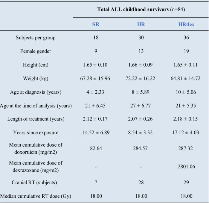
Table 3.1. Select demographic and clinical characteristics of the study population are reported as means with standard deviation. Data were abstracted from the medical records of the participants. ... 31 Table 3.2. A summary of the cardiac indices employed for LV structure and function analysis at a global scale (Young et al., 2000; Peng et al., 2016). ... 38 Table 3.3. Shown are the input parameters used for the mechanics simulations on a subset of ALL survivors (P, n=18) and healthy volunteer (HV, n=3) studies. The numbers featuring in the subjects’ identifiers are random and were assigned during the image loading process to avoid biases in the segmentation. ... 43 Table 3.4. List of the frames corresponding to the diastasis for each of the subjects analyzed here.
... 48
Table 4.1. Percentage change in C1 from the value at the true diastasis frame and those predicted
when the previous frame (left) or the next frame (right) was used as diastasis (first row). Percentage change from the C1 value at 1.0 kPa and those predicted when the pressure was
set at 0.75 kPa (left) or 1.25 kPa (right) (second row). ... 53 Table 4.2. Global geometrical parameters estimated from the subject-specific LV models in CIM. Data are expressed as mean standard deviation and presented separately for the SR, HR, HRdex, and HV groups. ... 54 Table 4.3. Summary of the average values and standard deviations of the FE-predicted results for each of the study group. This table shows that the estimated hyperelastic property increased as the LV cavity pressure increased. ... 60
Table 4.4. summarized the average values and standard deviation for the LV stiffness at rest and during exercise in the three groups of ALL survivors. ... 64
Table 5.1. Comparison of in vivo myocardial stiffness for the three groups analyzed by Wang et al. and the three groups of childhood ALL survivors assessed in the current investigation. Data are presented as means with standard deviation... 70
LIST OF FIGURES
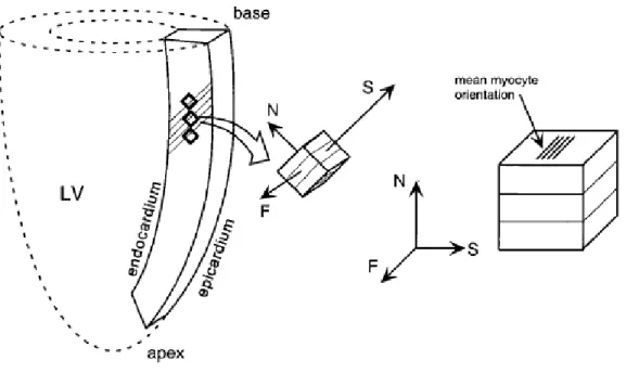
Figure 2.1. Anatomy of the inside of the heart, showing the four cavities and the roots of the great vessels, the other elements of the heart. ... 8 Figure 2.2. Shown is the temporal correlation between the left atrial and ventricular pressures (mm Hg), aortic pressure (mm Hg), left ventricular volume (mL), surface electrocardiogram (ECG), and phonocardiogram. ... 10 Figure 2.3. Chemical structure of the most commonly used anthracyclines. From top to bottom: doxorubicin (DOX), daunorubicin (DNR), epirubicin (EPI), and idarubicin (IDA). ... 11 Figure 2.4. Myocardial sections collected from the doxorubicin-exposed and placebo groups at day 7 after injection: normal (top left); altered cardiac histology (top right). ... 13 Figure 2.5. Risk factors for cardiac abnormalities after treatment with doxorubicin in long-term survivors of childhood and adult cancer. ... 15 Figure 2.6. Diagram showing the main directions along which the load is applied to the tissue sample during the mechanical tests: the fiber axis (F) which is the direction of the muscle fibers; the sheet axis (S) which is the direction transverse to the fiber axis; the normal axis (N) which is perpendicular to the specimen (Holger, 2006)... 21 Figure 2.7. Computational models of the left ventricle showing the boundaries of the FE mesh: model of Wenk et al. (2011) (a); model of Lee et al. (2013) (b). ... 26
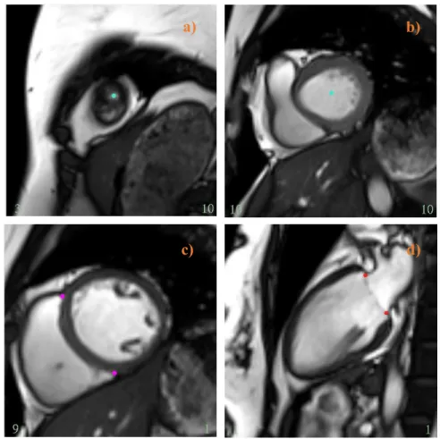
Figure 3.1. Shows the locations of apex point (a), basal point (b), RV insertion points (c), and base plane points (d) at end-systolic frames on a series of CMR images. ... 35 Figure 3.2. An example of short-axis (top) and long-axis (bottom) MRI images of a heart with the left ventricular endocardial and epicardial borders denoted, respectively, by green and blues lines. ... 36 Figure 3.3. Shows a representative CIM’s 16-element 3D model of the LV (4-chamber long-axis view) reconstructed at three different phases of the heart cycle: diastasis (a), end-diastole (b),
and end-systole (c). In green and blue wireframes are the 3D endocardial and the 3D epicardial surface. Observe how the model convincingly reproduces the apex-to-base shortening experienced by the LV during the systolic phase. ... 37 Figure 3.4. Standard representation of the 16-segment model of the American Heart Association. In the short-axis view, the left ventricle is depicted as a series of three concentric rings: the basal segments are in the outer ring, the midventricular segments are in the middle ring, and the apical segments are in the inner ring. RV stands for right ventricle. Diagram modified from Lang et al. (2015). ... 39 Figure 3.5. The three consecutive CMR frames (4-chamber long-axis view) display the movement of the mitral valve throughout the imaged portion of the cardiac cycle: the valve, initially open to permit the slow filling of the left ventricle (frame 17), switches in its closed position (frame 18), then moves again towards reopening to allow the refilling of the ventricle in atrial systole (frame 19). Frame 18 of the image series was therefore selected as diastasis. The red line identifies the position of the mitral valve, the red arrows point toward the leaflets of the valve (viewed in cross-section). ... 42 Figure 3.6. The evolution of the intra-ventricular pressure simulated at rest (top) and peak exercise (bottom) as a function of time for a typical case, before (left) and after the temporal synchronization (right). The red dot identifies the end-diastolic frame, which is the beginning of the cardiac cycle in the segmentation software CIM. ... 46
Figure 4.1. Subject-specific FE-predicted results plotted over the range of variation of the diastasis frame number (a) and the range of variation of the pressure load (b). ... 52 Figure 4.2. Comparisons of LV epicardial volume (a), endocardial volume (b), and wall thickness (c) among the SR, HR, HRdex and HV groups. For the sake of clarity, the corresponding standard deviations are not shown in the graph. The abscissa represents the phases of the heart cycle. Recall that the end-diastole corresponds to the first frame in this cycle, the end-systole is located at frame 9 or 10, and the diastasis lies around frames 16-17. Indeed, the curves for the HV group and those for the survivor group achieve their minimal (maximal) values at different frames in the cardiac cycle (shift of one phase). Also, note the different scales on each graph. ... 57
Figure 4.3. Global stiffness of LV myocardium for the HR, SR, HRdex, and HV groups estimated at diastasis under three different pressure boundary constraints: 0.75 (a), 1.0 (b), and 1.25 kPa (c). The boxplots show mean, median, standard deviation, and outliers for each of the four data sets. ... 60 Figure 4.4. Global stiffness of LV myocardium measured at rest (a) and peak exercise (b) for the HR, SR, and HRdex groups. Outliers, mean, median, and standard deviation values are illustrated for each data set. ... 63 Figure 4.5. Plot of C1 versus partition coefficient λ of gadolinium. The blue dashed line represents
the predictions of the multiple linear regression model, while the circles indicate the results of the FE simulations for each of the 39 subjects. ... 65
LIST OF SYMBOLS AND ABBREVIATIONS
3D Three-dimensional
AHA American Heart Association ALL Acute lymphoblastic leukemia ANOVA Analysis of variance
CIM Cardiac Image Modeling CMR Cardiac magnetic resonance DEX Dexrazoxane
DFCI Dana Faber Cancer Institute
DICOM Digital Imaging and Communication in Medicine DOX Doxorubicin
DS Diastasis
ED End-diastole
EDV End-diastolic volume EF Ejection fraction
ES End-systole
ESV End-systolic volume
FE Finite element
GUI Graphical user interface
HR High-risk
HRdex High-risk exposed to dexrazoxane HV Healthy volunteers
LV Left ventricle
LVM Left ventricular mass
MRI Magnetic resonance imaging
PETALE Prévenir les Effets Tardifs de la Leucémie Lymphoblastique Aigüe chez l’Enfant RMSE Root mean square error
RV Right ventricle SD Standard deviation SR Standard-risk group
CHAPTER 1
INTRODUCTION
1.1 Background
Acute lymphoblastic leukemia (ALL) is a malignant disease that arises from lymphocytes, a class of white blood cells, and is marked by the abnormal proliferation and accumulation of these cells in the bone marrow, blood and other tissues (Terwilliger et Abdul-Hay, 2017). In children and adolescents younger than 20 years, ALL is regarded as the most common tumor. In fact, 19% of the total cancer diagnoses and nearly 80% of the total leukemia cases in this age group are ALL (Rahman, Yusuf, et Ewer, 2007; Bhojwani, Yang, et Pui, 2015). The incidence of this neoplastic condition varies according to age at diagnosis, with the vast majority of cases occurring in the child’s first decade of life and peak prevalence in subjects between 2 and 5 years of age (Belson, Kingsley, et Holmes, 2006). Today, children with a diagnosis of ALL have a favorable prognosis. Recent estimates indicate that about 95% of patients are still alive and cancer-free at 5 years after cessation of therapy, while more than 80% attain long-term remission (DeAngelo et al., 2015). This success was brought about by advances made in the management and treatment of the disease over the past five decades. In particular, the survival rates were influenced favorably by the introduction of the anthracycline doxorubicin and the asparaginase as chemotherapeutic agents, as well as by the treatment of the central nervous system (Yeoh et al., 2002; Hunger et Mullighan, 2015; Vrooman et Silverman, 2016). Personalized treatment regimens carefully tailored to the risk of tumor recurrence have been recently applied to reduce the intensity of therapy while optimizing its leukemic response (Yeoh et al., 2002; Vrooman et Silverman, 2016)
Although improving the survival outcomes, exposure to doxorubicin is linked with a broad range of cardiotoxicity which can seriously affect the quality and duration of life of ALL patients (Lipshultz, Cochran, Franco, et Miller, 2013). In a study… More than 50% of the ALL survivors has been reported to experience cardiac abnormalities arising from the treatment (Lipshultz et al., 2005). Morphological and functional changes in the myocardial tissue are accompanied by progressive alterations in the geometry of the left ventricle. This cardiotoxicity seems to manifest itself by a persistent and, often, progressive deterioration of the cardiac function, which may continue for years after the initial exposure to doxorubicin (Lipshultz et al., 2005). These harmful effects may become apparent months to years after completion of therapy. The manifestations of the spectrum of cardiac toxicity range from subtle abnormalities in LV structure and function to
more serious complications, such as cardiomyopathies and congestive heart failure (Wouters, Kremer, Miller, et Herman, 2005; Chang et Wang, 2018). Interestingly, cardiotoxicity is more likely to occur in ALL survivors who received high doses of doxorubicin throughout the course of treatment (Lipshultz et al., 2013). In particular, doses greater than 400-450 mg/m are associated with a more severe myocardial damage. Other factors, such as exposure to radiotherapy, younger age at diagnosis or female gender, increase the risk of developing late cardiovascular complications (Lipshultz et al., 2013).
In response to concerns about the cardiac health of ALL patients, a variety of protective strategies have been proposed to attenuate the toxicity of doxorubicin during and after therapy. Among them, the cardioprotective agent dexrazoxane has proven to be very effective in reducing the incidence of myocardial injury. However, at present, none of the available measures completely eliminates the potential for health complications, making the issue of DOX-induced cardiotoxicity even more concerning (McGowan, 2017).
Although a vast body of data has accumulated on DOX-induced cardiotoxicity in survivors of childhood ALL, relatively little is understood of the long-term effects of modern chemotherapy programs on cardiovascular health (Hashimoto et al., 1999; Hudson et al., 2007; Vandecruys et al., 2012). Specifically, limited information is available on the cardiac status of these individuals at 10 years or more after completion of therapy (Adams et Lipshultz, 2005). Such data are important to accelerate progress towards improving the prevention of myocardial injury to enhance the quality of long-term survival of patients (Gianni et al., 2008). It has been suggested that a better knowledge of this cardiotoxicity may help researchers and clinicians refine management strategies and develop more effective cardioprotective measures (Gianni et al., 2008; Lipshultz et al., 2013; Hudson, Link, et Simone, 2014). This calls for more research on the long-term effects of chemotherapy associated with modern treatment regimens.
Imaging techniques are routinely used in the assessment of cardiac function for detection and screening of doxorubicin-induced cardiotoxicity (Dodos, Halbsguth, Erdmann, et Hoppe, 2007). Echocardiography has been the preferred method for such an evaluation in daily clinical practice (Jurcut et al., 2008; Da Silveira et al., 2016). Clinicians typically analyze the alterations occurred in experimental measurements of the diastolic and systolic LV function with respect to baseline values. Ejection fraction, stroke volume, and shortening fraction are examples of these indices (Jurcut et al., 2008; Tham et al., 2013; Khouri, Klein, Velazquez, et Jones, 2015). Nevertheless,
this imaging modality has poor sensitivity, which makes the current echocardiographically-derived indices less suitable for detection and surveillance of subclinical myocardial toxicity (Briant et al., 2007; Jain, Russell, Schwartz, Panjrath, et Aronow, 2017). CMR imaging offers a valid alternative to echocardiography for assessing the cardiac health of long-term ALL survivors. In fact, investigations in CMR imaging have demonstrated it to be a highly accurate modality for use in evaluating ventricular performance (Dong and Chen, 2018). Software for 3D geometric remodeling of the heart allow the determination of LV volumes, ejection fraction and mass from MRI scans (Young, Cowan, Thrupp, Hedley, et Dell’Italia, 2000; Suinesiaputra et al., 2014). Due to the ability of CMR to provide clear definitions of the LV wall contours, ejection fraction estimates measured on CMR images result to be more reliable and reproducible than those obtained with the aid of other imaging modalities, including echocardiography (Fallah-Rad, 2011; Gusso et al., 2012; Loganathan, Bilgen, Al-Hafez, et Smirnova, 2005).
Another parameter that provides a non-invasive indication of global left ventricular function is myocardial tissue stiffness (Hsu, Bouchard, Dumont, et Wolf, 2005). Many investigators have examined muscle stiffness to gain some insight into the mechanisms underlying cardiomyopathies, heart failure, hypertension, and other cardiac disorders (Lee et al., 2013; Mojsejenko et al., 2015). These studies showed that changes in muscle are accompanied by alterations in tissue stiffness during filling, with potential consequences for the mechanical function of the myocardium (Gupta, Ratcliffe, Fallert, Edmunds, et Bogen, 1994; Wang et al. 2013). Their outcomes also suggest that this biomechanical parameter can yield useful information on the structural and functional LV changes following late-onset cardiotoxicity.
A non-invasive estimate of myocardial stiffness can be obtained through the use of magnetic resonance imaging in connection with 3D finite element models of the LV chamber (Mojsejenko et al., 2015). Numerical modeling of the heart provides a powerful tool to investigate how changes in the myocardial tissue at small scale affect the structure and function of the ventricle. This tool has been extensively used in research for estimating the in vivo mechanical properties of the LV myocardium in a non-invasive fashion. By incorporating the subject-specific imaging data into the FE modelling framework, this technique offers the potential to construct anatomically realistic model of the LV for a wide range of medical applications (Wang et al., 2015). In research setting, biomechanical models of the LV have been employed to elucidate the impact of various cardiac
pathologies and/or their treatments on cardiac functionality and myocardial mechanics (Sermesant, Delingette, et Ayache, 2006).
1.2 General and specific research objectives
Several studies have been carried out over the past years in an attempt to draw a comprehensive picture for doxorubicin-related cardiotoxicity in survivors of childhood ALL. Among them, a work of particular relevance for the current research is the PETALE project (Prévenir les Effets Tardifs de la Leucémie Lymphoblastique Aigüe chez l’Enfant). Initiated in 2010, this multidisciplinary study assessed 250 subjects who had previously undergone therapy with doxorubicin at the Sainte-Justine University Hospital Center in Montreal. This study was designed to characterize the delayed adverse effects of antileukemia treatment on various organs and systems, including the heart, in a young population of acute leukemia survivors. The current investigation develops from this study and was conducted on a subgroup of the PETALE cohort.
As part of the PETALE project, our study sought to assess the subclinical changes in LV structure and function in a group of long-term survivors of childhood ALL who received low-to-moderate doses of doxorubicin-based chemotherapy. Given the high potential of numerical cardiac modeling, this evaluation was conducted using a FE model-based approach that integrates subject-specific LV shape and motion directly from CMR scans.
In particular, the following specific objectives were addressed:
1. to extract from each patient’s CMR images the geometric and function information required to build personalized biomechanical FE models of the LV and to calculate a set of relevant 3D geometrical parameters.
2. to examine the changes occurred in LV geometry and function after doxorubicin exposure (at both global and regional scales) by comparing the calculated geometrical parameters between cancer survivors and their matched healthy controls.
3. to simulate the mechanical events occurring during mid-diastole on the MRI-reconstructed FE models and to compute the hyperelastic property of the transversely-isotropic constitutive law used to describe the passive mechanical behavior of the myocardium.
4. to gain some insight into the long-term effects of DOX therapy on the passive LV mechanics by comparing the estimated hyperelastic property between cancer survivors and controls.
1.3 Research hypotheses
The current analysis was designed around a proper set of methodological assumptions and research hypotheses. In detail, we assumed that the administration of low-to-moderate cumulative doses of doxorubicin induced dose-dependent, subtle, and asymptomatic alterations in LV geometry and function in the study participants. As far as concern the hypotheses addressed by the study, we first speculated that the cardiac abnormalities can be detected and evaluated by means of LV myocardial mass, ejection fraction, and other selected 3D geometrical parameters. Furthermore, we tested the hypothesis that the hyperelastic property (as a measure of the in vivo myocardial stiffness) was able to unmask the subclinical myocardial injury caused by exposure to doxorubicin
1.4 Outline of the thesis
The present thesis is structured into six chapters and a bibliography as follows:
Chapter 1 provides the reader with a brief overview of the clinical context of the project, paying particular attention to the side effects of doxorubicin on the cardiovascular system and the methods for assessing this cardiotoxicity. The general and specific research objectives are also outlined in this section, along with the hypotheses underpinning the study.
Chapter 2 introduces the necessary background for a deep understanding of the issue documented. It details the anatomy and physiology of the heart and describes the main in vitro experiments to characterize the mechanical behavior of the heart muscle in relaxed and contracted conditions. Subsequently, it compares most pertinent examples of existing models of cardiac mechanics. Chapter 3 describes the cardiac image analysis and modeling tools employed in the assessment of LV geometry and function on a subject-specific basis. This chapter also includes information about the study population, data acquisition protocols, and statistical methods applied in the data analysis. Chapter 4 presents the results obtained by applying the methods outlined in survivors of childhood ALL. In particular, it reports estimates of 3D geometrical and mechanical properties of the LV are illustrated with the support of graphs and tables.
Chapter 5 analyzes and interprets the results in light of current knowledge about doxorubicin-induced long-term cardiotoxicity in ALL survivors. Furthermore, it discusses the limitations of the proposed methodology.
Chapter 6 concludes with a reiteration of the main findings of the study and their implications for the detection of late doxorubicin cardiotoxicity. Project implementations, as well as possible future directions for research on this topic, are also provided.
CHAPTER 2
FUNDAMENTALS
This chapter provides an overview of the basic cardiac anatomy and physiology. We also describe the mechanisms by which doxorubicin exerts its cytotoxic action and the features of this toxicity on the heart muscle. After that, we review the imaging techniques and the intervention strategies currently available to minimize the risk for early and late myocardial injury. In addition, we present the experimental techniques for the mechanical characterization of the myocardium. Numerical modeling of the heart is then introduced as a tool for estimating the passive mechanical properties of cardiac tissue and for evaluating cardiac dysfunction. We compare the most pertinent examples of existing models of cardiac mechanics and we conclude by making a point of the situation.
2.1 The heart
The heart, central organ of the cardiovascular system, is a complex structure that generates the adequate force for perfusing tissues throughout the body. In such a way, the heart provides cells all over the body and tissues with enough nutrients and oxygen (Volpe et Makaryus, 2019). In the following, the cardiac function, as well as its internal morphology, are shortly described.
2.1.1 Basic cardiac anatomy
The heart is a hollow muscular organ located in the center of the thoracic cavity, between the two lungs, in a luminal space known as mediastinum (Volpe et Makaryus, 2019). Grossly, the heart consists of four distinct chambers: the two posterosuperior ones are namely (left and right) atria and the two anteroinferior ones are namely (left and right) ventricles. A central wall (septum) separates the left atrium and ventricle from their right-sided counterparts. Figure 2.1 illustrates the internal anatomy of the cardiac pump. In the human cardiocirculatory system, the heart and blood vessels are configured in two separate circuits in series, the pulmonary and circulatory circulations, that function at the same time to ensure the transports of nutrients and oxygen to all body tissue. Two valves connect each atrium to the corresponding ventricle. As well as allowing the communication between the chambers, the heart valves guarantee the unidirectionally of the blood flow and prevent any chance of backward flow. In particular, the atria communicate to the corresponding ventricles through the atrioventricular valves (mitral and tricuspid), whereas the ventricles communicate to the great arteries through the semilunar valves (aortic and pulmonary). Although atria and ventricles share a primary structural organization, their internal morphology
reveals distinctive anatomical features that are linked to the different function the two chambers deploy. Essentially, the atrium collects the blood entering the heart, while the ventricle pumps the blood into the circulations (Volpe et Makaryus, 2019).
Figure 2.1. Anatomy of the inside of the heart, showing the four cavities and the roots of the great vessels, the other elements of the heart.
(Copyright 2010 by ZooFari. Reproduced with permission from Wikipedia Commons)
The cardiac wall is composed of three layers, each of which exerts a specific function. Moving from the external to the inner surface of the heart, it is possible to find epicardium, myocardium and endocardium. The myocardium is the cardiac muscle composed by myocytes which are behind the cardiac contraction. Endocardium and epicardium are, respectively, the inner and outer walls of the myocardium. It is interesting to note that the structure of the cardiac wall does not change along the length of the chambers but remains the same for the four chambers. Some differences are observed in the thickness of the cardiac wall among the chambers. The ventricles, which have to propel the blood through the systemic and pulmonary circulations facing higher pressures, exhibit
a more developed myocardium as compared with the atria. Additional differences were observed between the left and right ventricles. Specifically, the myocardium was shown to be thicker in the left ventricle, with wall thickness ranging from 6 to 16 mm (Petitjean et Dacher, 2011). The right ventricle (RV) presents a myocardial layer that is almost three to six times thinner than that of the left ventricle (Petitjean et Dacher, 2011). In fact, the right ventricle is responsible for pumping deoxygenated blood to the pulmonary circulation at lower physiological pressure.
2.1.2 The cardiac cycle
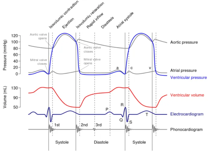
From a mechanical standpoint, the heart consists of two distinct pumps, operating in series, and whose function is to ensure blood transportation throughout the body by contracting alternatively with an average frequency of 75 beats/min (Baldissera, 2000). Indeed, atria and ventricles alternate between phases of contraction (systole), in which they propel the blood into the pulmonary and systemic circuits, and phases of relaxation (diastole), in which they are refilled with blood. The time interval between one diastole (or systole) and the beginning of the subsequent one is referred to as the cardiac cycle. In healthy individuals at rest, this sequence lasts 0.8 s. Figure 2.2 (Wiggers diagram) illustrates the complete series of events occurring during a cycle in the left heart. For purposes of discussion, the cycle of cardiac contraction can be decomposed into the following consecutive phases:
1. Ventricular isovolumetric contraction (preload): the contraction of the left ventricle causes an increase in intra-ventricular pressure, which results into the closure of the tricuspid and mitral valves and subsequently into an increase of atrial pressure. During isovolumetric contraction, the volume of the LV chamber remains constant. This phase terminates when the LV pressure attains 80 mmHg.
2. Ventricular ejection: when the LV pressure exceeds 80 mmHg (which is the aortic pressure), the aortic valve opens and the ventricle pumps blood into the aortic arch. The outflow is rapid up to a peak of 125 mmHg, followed by a slow outflow that stops at a pressure of 100 mmHg. The end of this phase coincides with the end of systole and the closure of the aortic valve. 3. Isovolumetric relaxation: the LV pressure drops to 0 mmHg, with 40% of the diastolic blood
4. Rapid diastolic ventricular filling: when the LV pressure decrease below the atrial pressure, the mitral valve opens allowing a quantity of blood to flow inside the ventricle.
5. Slow diastolic ventricular filling: this is the phase in which the LV pressure equals the atrial pressure, then following the systole, the mitral valve closes and the cardiac cycle resumes. At the end of the ventricular systole, a new cycle begins: the ventricles begin the diastole and the semilunar valves close due to the high pressure in the aorta and pulmonary arteries. The atria are still in diastole, so overall the inner pressure is low throughout the entire heart.
Figure 2.2. Shown is the temporal correlation between the left atrial and ventricular pressures (mm Hg), aortic pressure (mm Hg), left ventricular volume (mL), surface electrocardiogram (ECG), and phonocardiogram.
2.2 Cardiotoxicity of doxorubicin in the treatment of childhood ALL
Anthracyclines, such as doxorubicin, daunorubicin or epirubicin, are a class of anticancer agents that are routinely administered in the treatment of acute lymphoblastic leukemia (McGowan, 2017). Within this group, doxorubicin is the most commonly employed agent in virtue of its ability to significantly improve the prognosis of ALL patients (Vejpongsa et Yeh, 2014; Rahman et al., 2007). Survivors of ALL have been reported to experience a wide range of cardiovascular complications after doxorubicin exposure. Arrhythmias, pericarditis, myocarditis, left ventricular dysfunction, and heart failure are some examples of the toxic side-effects associated with chemotherapy (Lipshultz et al., 2013). According to recent estimates, more than 50% of these patients experience cardiovascular events within 6 years since the completion of treatment (Dong et Chen, 2018).
Figure 2.3. Chemical structure of the most commonly used anthracyclines. From top to bottom: doxorubicin (DOX), daunorubicin (DNR), epirubicin (EPI), and idarubicin (IDA).
(Reproduced with permission from: J. V., McGowan, R., Chung, A., Maulik, et al. Anthracycline
chemotherapy and cardiotoxicity. Cardiovascular drugs and therapy. 2017; 31(1), 63-75.
The harmful cardiac effects of doxorubicin can severely affect the extent and the quality of life of leukemia survivors (Dong et Cheng, 2018). This is true especially for survivors of childhood ALL who are more sensitive to these modifications (Lipshultz et al., 2013).
In the next four sub-sections, we describe the features of doxorubicin-mediated cardiotoxicity in greater detail as required for the current study.
2.2.1 Mechanisms of doxorubicin cytotoxic action
The mechanisms by which doxorubicin exerts its cytotoxic function have been extensively studied. Several molecular pathways have been proposed as modes of action but the exact global mechanism by which doxorubicin acts in cancer cells has not been fully elucidated to date (Yang, Teves, Kemp, et Henikoff, 2014).
The cytotoxic antileukemic action of this anthracycline has been primarily attributed to its ability of interfering with nucleic acid synthesis and, more specifically, with the catalytic function of the enzyme DNA topoisomerase IIα (Lynch, Guinee, et Holden, 1997; Lyu et al., 2007). This enzyme, which is a marker of proliferation expressed in proliferating cells and in neoplastic tissues, plays a critical role in several processes of the cell cycle, including DNA replication and RNA transcription It catalyze the unwinding of DNA: binds DNA supercoils, breaks both strands of one DNA duplex, passes the other duplex through the resulting gap, and reseals the break (Yang et al., 2014; Burden et Osheroff, 1998; Lyu et al., 2007). Under physiological conditions (i.e., in the absence of doxorubicin) the topoisomerase-DNA complex has reversible nature: the DNA breaks double-stranded cut generated in the process of DNA replication are resealed (Swift, Rephaeli, Nudelman, Phillips, & Cutts, 2006). In chemotherapy, the relegation of the cleaved DNA is prevented by the intercalation of doxorubicin. Doxorubicin traps the target and stabilizes the enzyme-DNA complex by cross-links the topoisomerase to tumor DNA molecules (Burden et Osheroff, 1998; Lyu et al., 2007; Chang et Wang, 2018). The formation of topoisomerase-doxorubicin-DNA complexes generates double-stranded cut in the genome of leukemia cells, which, if not repaired, ultimately trigger an apoptotic response (Swift et al., 2006; Lyu et al., 2007). Other molecular mechanisms that have been proposed to explain doxorubicin-induced cell death include inhibition of DNA and RNA synthesis, formation of formaldehyde-mediated doxorubicin-DNA adducts, ceramide overproduction, and generation of free radicals (Swift et al., 2006; Yang et al., 2014). It is important to stress that the enlisted mechanisms are not mutually exclusive (Swift et al., 2006). Furthermore,
it has been suggested that the generation of free radicals has an important role in the toxicity of doxorubicin within cardiomyocytes (Meiners, Shenoy, et Zordoky, 2018). Neoplastic cells require a higher uptake of iron compared to the myocytes. Doxorubicin is an iron chelator, binds the cellular iron and boosts the production of myocardial reactive oxygen species (ROS). These species damage the DNA which in turn can contributing to the generation of damage.
2.2.2 Histopathological features of doxorubicin cardiotoxicity
The nature of the histological cardiac injury caused by doxorubicin has been investigated in animal models and in humans primarily by biopsy of the ventricular myocardium (Aissiou, 2016). In such analyses, tissue specimens collected from the ventricular walls were processed and then attentively examined by the electron microscope in order to identify the lesions occurred at the cellular level.
Figure 2.4. Myocardial sections collected from the doxorubicin-exposed and placebo groups at day 7 after injection: normal (top left); altered cardiac histology (top right).
(Reproduced with permission from: D., Rea et al. Strain analysis in the assessment of a mouse
model of cardiotoxicity due to chemotherapy: sample for preclinical research. in vivo. 2016; 30(3),
With this experimental technique, Rea et al. (2016) showed that myocardial injury manifests itself as vacuolization of the cytoplasm and an amplified apoptotic death of cardiomyocytes in the early days after doxorubicin exposure in mice (dose of 2.17 mg/kg for 7 consecutive days). Biopsy data acquired one-week post-injection revealed that treatment was also accompanied by an increase in the content of myocardial collagen and an enlargement of the remaining cardiomyocytes. Greater fibrosis was observed in myocardial samples from the treated animals when compared against the placebo group (Figure 2.4).
Similar results were found by Zhou et al. (2001) in a study to evaluate the effects of doxorubicin on the cardiovascular system of rats with 4 to 8 weeks of daily injections (2 mg/kg). Evidence of mitochondrial and myofibrillar damage and loss, and cellular edema were present in the treated animals. Furthermore, this study suggested that the degree of cytoplasmic vacuolization and, thus, the extent of cell injury relate to the duration of doxorubicin exposure. Mice with injections for 6 weeks exhibited myocytes with small cytoplasmic vacuoles, damage more diffuse after 8 weeks of injection. Histological examination also revealed that myocardial cell injury did not heal over time, but rather progressed for weeks after discontinuation of treatment. At 5 weeks after exposure to doxorubicin, damaged cardiomyocytes accounted for about 20% of the total number in rats with 6 weeks of injections. This percentage increased to 30% when the duration of exposure was 8 weeks. Similar histological alterations were documented by Lipshultz et al. (2012) in a group of pediatric ALL survivors (median cumulative dose of doxorubicin, 300 mg/m2) when measuring biochemical biomarkers for myocardial injury. The blood levels of these biomarkers indicated that doxorubicin induced death of cardiac myocytes during treatment and for few months after the last exposure. Moreover, it was shown that the reduction in the number of functional cardiomyocytes caused an increase in the diameter of the remaining cells (as was seen in the doxorubicin-exposed mice) at 4 years after chemotherapy (Lipshultz et al., 2012). These myocytes were subjected to a greater workload which force them to compensate so as to maintain the normal heart structure (Sabbah et Goldstein, 1993; Lipshultz, Alvarez, et Scully, 2008).
2.2.3 Macroscopic features of doxorubicin-related cardiotoxicity
If sufficiently large, the loss of functional cardiac units results into a higher workload on the viable tissue, which in turn triggers a series of compensatory structural events (within the myocardium) aimed to preserve the normal ventricular structure (Sabbah et al., 1993; Adams et Lipshultz; 2005; Schunke, Coyle, Merrill, et Denhardt, 2013; Coviello, 2018). Throughout this process, the left ventricle undergoes progressive changes in geometry. Such alterations are referred to as ventricular chamber remodeling. The wall of the ventricle becomes progressively thinner, while the diameter increases, possibly leading to the dilation of the chamber. As a result of these changes in cavity size and shape, ventricular afterload and myocardial wall stress increase (Lipshultz et al., 1991; Wouters et al., 2005; Lipshultz et al., 2008). The loss of contractile tissue following doxorubicin therapy may also cause a reduction in myocardial contractility (Lipshultz et al., 1991; Giantris et al., 1998). This decreased contractility, together with an elevated afterload, in turn, can profoundly affect the global function of the LV, giving rise to give rise to compromised diastolic filling.
Figure 2.5. Risk factors for cardiac abnormalities after treatment with doxorubicin in long-term survivors of childhood and adult cancer.
(Reproduced with permission from: Lipshultz, S. E., Cochran, T. R., Franco, V. I., & Miller, T. L.
Treatment-related cardiotoxicity in survivors of childhood cancer. Nature reviews Clinical
LV chamber remodeling is progresses gradually over time and may turn into a pathologic process promoting the development of cardiomyopathy or congestive heart failure (Wouters et al., 2005). The deterioration of the ventricular function can continue for years and become apparent even 4 to 20 years after completion of treatment (Huang et al., 2010; Lipshultz et al., 2012; Vachhani et al., 2017). The manifestations of the spectrum of cardiotoxicity range from subtle alterations in the structure and function of LV to more severe complications, which are congestive heart failure and cardiomyopathy.
The degree and progression of doxorubicin-induced cardiotoxicity are associated with several well-established risk factors (Lipshultz et al., 2008). Clinical studies in childhood and adult leukemia survivors have demonstrated that the likelihood of developing late cardiovascular complications increases with the cumulative dose of doxorubicin administered during therapy (Meiners et al., 2018). In particular, doses of this agent greater than 450-550 mg/m2 are linked with more serious
complications. Higher rate of administration, female gender, very young and very old age at the time of therapy, and exposure to radiation have also been reported as risk factors for delayed cardiotoxicity (Bansal, Amdani, Lipshultz, et Lipshultz, 2017).
2.2.4 Prevention and reduction of doxorubicin-related cardiotoxicity
In response to concern about the cardiac health of cancer survivors, researchers and clinicians have implemented a variety of potential protective measures directed to prevent or minimize the harms of doxorubicin on the myocardial tissue (McGowan et al., 2017).
Given the close relationship between cumulative doxorubicin dose and risk for late cardiotoxicity, current preventive strategies primarily focus on limiting the total dose of doxorubicin delivered during treatment to 450-550 mg/m2 so as to reduce the risk for short- and long-term cardiotoxicity
without compromising the tumor-killing activity of doxorubicin (Dong et Chen, 2018). Lipshultz et al. (2012) have shown that the administration of doxorubicin doses above this threshold reduced significantly the incidence of heart failure. However, other authors observed that cardiotoxicity occurs even at lower doses, leading to conclude that no safe dose exists (Harake, Franco, Henkel, Miller, et Lipshultz, 2012; Silverman, 2014; Bassareo et al., 2016). This strategy does not guarantee that the cardiac injury will not occur and, therefore, needs to be complemented by other measures. Clinicians have investigated the efficacy of different dosing schedules designed to lower the myocardial concentration of doxorubicin or its metabolites and, thereby, reduce the risk for cardiac
toxicity. In particular, continuous administration of doxorubicin was tested in both children and adults. The results of these studies were then compared with those obtained for the bolus injection. Continuous infusion had significant benefits in preventing cardiac functional deterioration over the bolus-infusion on a short-term basis in adult. No cardioprotective benefit was proven in children (Adams et Lipshultz, 2005). A study of the efficacy of continuous infusion in preventing cardiac functional deterioration reinforced this claim. No difference in LV structure and function was documented between the two schedules in children with high-risk ALL at 8 years after cessation of treatment (Lipshultz et al., 2012; Jain et al., 2017).
Another management strategy for toxicity involves the administration of structural analogues or alternative formulations of doxorubicin that are deemed to be less harmful to the cardiac tissue. Epirubicin, idarubicin, mitoxantrone, and liposomal doxorubicin are examples of such anticancer drugs (Giantris et al., 1998). Epirubicin and idarubicin induced fewer clinical and subclinical cardiac alterations compared with the same doses of doxorubicin, but they were also less potent as anticancer agents (Hande, 1998; Smith et al., 2010). Liposomal doxorubicin has yielded promising results in preliminary studies in survivors of adult ALL, reducing the incidence of cardiac complications significantly (Rahman et al., 2007). Studies of the efficacy and the late cardiac effects of this alternative formulation are underway in childhood ALL survivors (Silverman, 2014).
A deeper understanding of the mechanisms by which doxorubicin interacts with healthy and tumor cells has led to the development of a broad range of cardioprotective agents. Various beta-blockers, free-radical scavengers, antioxidants, and inhibitors of the renin-angiotensin-aldosterone system, have been considered as a means to reduce the incidence of doxorubicin-related myocardial injury during and after therapy (Wouters et al., 2005; Harake et al., 2012; McGowan et al., 2017). The cardioprotective efficacy of these agents and their side effects on heart have been evaluated in several clinical trials on survivors of adult and childhood cancers: some agents were able to provide some degree of protection, others failed in reducing cardiotoxicity (Vejpongsa et Yeh, 2014). At the current time, the only cardioprotectant approved for use in oncological practice is dexrazoxane (Sharalaya and Collier, 2018). Developed initially as an antitumor agent, this free-radical scavenger is recommended as part of the standard of care for adult patients assigned to receive large doses of doxorubicin, due to its ability to reduce the incidence of myocardial injury without compromising the chemotherapeutic value of doxorubicin (Hasinoff et Herman, 2007; Sepe, Ginsberg, et Balis, 2010; Harake, 2012). The protective activity of dexrazoxane has been primarily ascribed to its
affinity for intracellular iron, which is believed to be stronger than that showed by doxorubicin or the other anthracyclines. Once inside the myocyte, dexrazoxane binds free iron or sequestrates the metal complexed to the chemotherapeutic, thereby reducing the generation of iron-catalyzed ROS and lowering cellular damages associated to cardiac injury (Hasinoff et Herman, 2007; Bryant et al., 2007; Harake, 2012; Lipshultz, Franco, Miller, Colan, et Sallan, 2013).
2.2.5 Monitoring and Detection
Despite the proven efficacy in reducing the incidence of doxorubicin-related myocardial injury, none of the strategies currently available confer complete protection against the adverse effects of chemotherapy (Silverman, 2014). Taken together with the observation that cardiotoxicity may not manifest until weeks or years after the end of therapy, the lack of complete protection emphasizes the importance of screening and effective detection of toxicity (Jurcut et al., 2008; Dong et Chen, 2018). Monitoring the cardiac status of children who underwent treatment with doxorubicin is of great importance especially during the silent phase of left ventricular dysfunction. Intervention before the manifestation of clinical symptoms allows to prevent or inhibit the progression of tissue damage to a more advanced stage (Ali et al., 1994; Khouri, 2015; Coviello, 2018).
There are currently multiple invasive and non-invasive clinical techniques for follow-up and detection of doxorubicin-related cardiotoxicity. Traditionally, endomyocardial biopsy has been the preferred method for such an evaluation in virtue of its high sensitivity to structural changes in the myocardial tissue (Jain et al., 2000; Jain et al., 2017). In a study by Friedman et al. (1978) endomyocardial biopsy was able to detect the subclinical myocardial injury induced by a DOX dose of 180 mg/m2. However, this procedure is invasive, with the risk of severe side effects (e.g.
hematoma, damage to the tricuspid valve…), and thus not feasible for use in daily clinical practice (Rahman et al., 2007; Jurcut et al., 2008; Tham et al., 2013).
Echocardiography is the current imaging modality of choice for evaluating the structural and functional cardiac abnormalities associated with chemotherapy in a non-invasive fashion (Jurcut et al., 2008; Da Silveira et al., 2016). Various parameters of systolic (LV ejection fraction, shortening fraction, and systolic wall thickening) and diastolic function (mitral inflow pattern E/A ratio, isovolumic relaxation time, and pulmonary venous flow pattern) are measured in ALL survivors (Jurcut et al., 2008). The alterations in the experimental measurements of these clinical indices with respect to baseline values are then analyzed by the clinicians (Khouri et al., 2015). Advantages
of the echocardiography include the non-invasiveness of the measurements, the absence of ionizing radiation, the high portability and the easy availability of the acquisition apparatus (Rahman et al., 2007). Nevertheless, this imaging modality has poor sensitivity in detecting structural and functional cardiac changes during the silent phase, which makes the echocardiographically-derived indices less suitable to evaluate subclinical myocardial toxicity (Briant et al., 2007; Levis, Binkley et Shapiro, 2017; Jain et al., 2017).
Another imaging modality to assess the cardiotoxic impact of doxorubicin in cancer survivors is cardiac magnetic resonance (Lunning et al., 2015). CMR is a more sensitive test for monitoring cardiotoxicity than echocardiography (Fallah-Rad, 2011). The feasibility of CMR for cardiac toxicity evaluation has been recently established. Software for 3D geometric modeling of the cardiac chambers allow determination of LV volumes, ejection fraction, wall thicknesses and mass from CMR scans (Levis et al., 2017; Jain et al., 2017). These estimates were proven to be more reliable, precise and accurate compared to echocardiographically-derived indices due to the ability of CMR to provide clear definitions of the LV wall contours (Loganathan et al., 2005; Fallah-Rad, 2011; Gusso et al., 2012). Nonetheless, the routine use of CMR imaging is restricted by its limited availability, the patient tolerance to the examination, as well as the high costs associated with this procedure (Jurcut et al., 2008).