HAL Id: pastel-00005635
https://pastel.archives-ouvertes.fr/pastel-00005635
Submitted on 17 Dec 2009
HAL is a multi-disciplinary open access
archive for the deposit and dissemination of sci-entific research documents, whether they are pub-lished or not. The documents may come from teaching and research institutions in France or abroad, or from public or private research centers.
L’archive ouverte pluridisciplinaire HAL, est destinée au dépôt et à la diffusion de documents scientifiques de niveau recherche, publiés ou non, émanant des établissements d’enseignement et de recherche français ou étrangers, des laboratoires publics ou privés.
Multiscale explanation of elasticity and strenght of bone
and bone replacement materials made of hydroxyapatite,
glass-ceramics, or titanium : a continuum
micromechanics approach
Andreas Fritsch
To cite this version:
Andreas Fritsch. Multiscale explanation of elasticity and strenght of bone and bone replacement materials made of hydroxyapatite, glass-ceramics, or titanium : a continuum micromechanics approach. Engineering Sciences [physics]. Ecole des Ponts ParisTech, 2009. English. �pastel-00005635�
D O C T O R A L
T H E S I S
MULTISCALE EXPLANATION OF ELASTICITY AND
STRENGTH OF BONE AND BONE REPLACEMENT
MATERIALS MADE OF HYDROXYAPATITE,
GLASS-CERAMICS, OR TITANIUM: A CONTINUUM
MICROMECHANICS APPROACH
——————————————————————
D I S S E R T A T I O N
MEHRSKALIGE ERKL ¨
ARUNG DER ELASTIZIT ¨
AT UND
FESTIGKEIT VON KNOCHEN UND
KNOCHENERSATZMATERIALIEN AUS
HYDROXYAPATIT, GLAS-KERAMIK ODER TITANIUM:
EIN KONTINUUMSMIKROMECHANISCHER ANSATZ
——————————————————————
ausgef¨uhrt zum Zwecke der Erlangung des akademischen Grades eines Doktors der technischen Wissenschaften
eingereicht an der Technischen Universit¨at Wien Fakult¨at f¨ur Bauingenieurwesen
von
Dipl.-Ing. Andreas Fritsch Matrikelnummer 9925071
Hießgasse 4/22, A - 1030 Wien, ¨Osterreich
Pr¨ufer und Betreuer: Ao.Univ.Prof. Dr. Christian Hellmich Prof. Dr. Luc Dormieux
Pr¨ufer: O.Univ.Prof. Dr. Hans Irschik Gutachter: Prof. Dr.-Ing. Dietmar Gross
Prof. Dr. Alain Molinari
Pr¨ufer und Betreuer: Ao.Univ.Prof. Dr. Christian Hellmich
Institut f¨ur Mechanik der Werkstoffe und Strukturen, Technische Universit¨at Wien, ¨Osterreich
Prof. Dr. Luc Dormieux
UR Navier, ´Ecole des Ponts ParisTech, Marne-la-Vall´ee, Frankreich
Pr¨ufer: O.Univ.Prof. Dr. Hans Irschik Institut f¨ur Technische Mechanik,
Johannes Kepler Universit¨at Linz, ¨Osterreich Gutachter: Prof. Dr.-Ing. Dietmar Gross
Instiut f¨ur Mechanik,
Technische Universit¨at Darmstadt, Deutschland Prof. Dr. Alain Molinari
Laboratoire de Physique et M´ecanique des Mat´eriaux, Universit´e de Metz, Frankreich
Diese Dissertation entstand durch eine Kooperation zwischen der ´Ecole Nationale des Ponts et Chauss´ees (ENPC), Marne-la-Vall´ee, Frankreich, und der TU Wien. Das fran-z¨osische Cotutelle-Programm erm¨oglichte mir, als Doktorand von den Vorz¨ugen von zwei angesehenen Hochschul- und Forschungseinrichtungen zu profitieren und unterschiedliche Wissenschaftskulturen kennenzulernen.
Mein Dank geb¨uhrt meinen Betreuern Prof. Dr. Luc Dormieux (Unit´e de recherche Navier, ENPC) und Ao.Prof. Dr. Christian Hellmich (Institut f¨ur Mechanik der Werkstoffe und Strukturen, IMWS) f¨ur ihre hervorragende und engagierte Betreuung sowie ihre intensive Zusammenarbeit, von der ich außerordentlich profitieren konnte.
Die vorliegende Arbeit w¨are nicht m¨oglich gewesen ohne die finanzielle F¨orderung von beiden Seiten: Die ENPC gew¨ahrte mir ein Stipendium (Allocation de recherche), welches mir einen Aufenthalt von 18 Monaten an dieser Hochschule erm¨oglichte. Das Institut f¨ur Mechanik der Werkstoffe und Strukturen der TU Wien f¨orderte den zweiten Teil durch meine Anstellung als Projektassistent.
Mein Dank gilt weiters der Institutsleitung des IMWS, Univ.-Prof. Dr. Josef Eberhardstei-ner und Univ.-Prof. Dr. Herbert Mang, sowie der Administration, insbesondere Mag.(FH) Martina P¨oll und Dipl.-Ing. Dominik Dejmek. Auf franz¨osischer Seite bin ich Prof. Dr. Patrick de Buhan (Institutsvorstand) und Dr. Denis Garnier (Administration) f¨ur ihre Unterst¨utzung sehr verbunden. F¨ur das angenehme und produktive Arbeitsklima m¨ochte ich mich bei allen meinen Kolleginnen und Kollegen an beiden Instituten bedanken. Weitere Unterst¨utzungen gew¨ahrten der ¨Osterreichische Akademische Austauschdienst, welcher mit dem Programm Wissenschaftlich-Technische Zusammenarbeit zwei Reisen nach Frankreich erm¨oglichte, sowie die Fakult¨at f¨ur Bauingenieurwesen der TU Wien, welche durch ein F¨orderungsstipendium den Ankauf von Laborger¨at erm¨oglichte und ebenfalls Reisekosten ¨ubernahm. Desweiteren wurde diese Arbeit im Rahmen des EU-Exzellenznetzwerkes Knowledge-based Multicomponent Materials for Durable and Safe Performance (KMM-NoE), Nr. NMP3-CT-2004-502243, gef¨ordert.
Mein Dank gilt weiters meinen Koautoren Julien Sanahuja von der ENPC, Herbert W. M¨ullner, Christoph Kohlhauser und Roland Reihsner vom IMWS, Dirk Godlinski und Astrid Rota vom Fraunhofer-Institut f¨ur Fertigungstechnik und Angewandte Mate-rialforschung in Bremen, Robert Slesinski, Tomasz Brynk und Zbigniew Pakiela von der Technischen Universit¨at Warschau sowie Andrea Malasoma und Chiara Vitale-Brovarone vom Politecnico di Torino.
Beim Erstellen des Kooperationsabkommens f¨ur mein Doktoratsstudium waren mir fre-undlicherweise Dr. Sabine Kiesel-Szontagh von der Rechtsabteilung der TU Wien sowie Marine Daniel von der Abteilung f¨ur Doktoratsangelegenheiten der ENPC sehr behilflich.
Mein besonders herzlicher Dank gilt meinen Eltern und meiner Partnerin Sylvia Goluch, die mir stets R¨uckhalt waren und mich durch manch anstrengende Phase begleitet haben.
Knochen ist ein hierarchisch aufgebautes Material, gekennzeichnet durch eine erstaunliche Variabilit¨at und Diversit¨at. Knochenersatz- oder Biomaterialien sind wichtige Kompo-nenten f¨ur k¨unstliche Organe und werden auch als Ger¨uste f¨ur Tissue Engineering einge-setzt. Das Ziel dieser Dissertation ist die Vorhersage der Festigkeit von Knochen und Knochenersatzmaterialien auf Grund ihrer Zusammensetzung und Mikrostruktur mit-tels Mehrskalenmodellen. Die theoretischen Entwicklungen werden durch umfangreiche Experimente an kortikalen Knochen sowie an Biomaterialien aus Hydroxyapatit, Glas-Keramik und Titanium untermauert.
Kapitel A untersucht verschiedene morphologische Konzepte (Kugel vs. Nadeln) f¨ur die Homogenisierung der linear elastischen Eigenschaften von por¨osen Polykristallen, wie sie in der Mineralphase des Knochens vorkommen.
In Kapitel B wird ein erster Versuch zur Modellierung der Festigkeit von Hydroxyapatit-Biomaterialien vorgeschlagen, aufbauend auf einer mikromechanischen Beschreibung der Steifigkeit und des spr¨oden Versagens der Kontaktfl¨ache (Interface) zwischen isotropen, kugelf¨ormigen Kristallen. Um Optimierungsverfahren zur R¨uckbestimmung der Eigen-schaften der Kontaktfl¨ache zu vermeiden (wie sie in Kapitel B verwendet werden), wurde ein alternativer Ansatz (Kapitel C) entwickelt, wo die nichtkugelf¨ormige Form von Hy-droxyapatitkristallen ber¨ucksichtigt wurde. Die Verwendung von Nadeln impliziert einen 1D-Spannungszustand im soliden Kristall in Nadelrichtung, und diese Spannung kann als relevant f¨ur die Spannungen an der Kontaktfl¨ache zwischen den Kristallen erachtet werden.
Kapitel D pr¨asentiert ein experimentell gest¨utztes mikromechanisches Modell zur Er-kl¨arung der Festigkeit des kortikalen Knochens, basierend auf einer neuen Sichtweise f¨ur dessen Versagen: Gegenseitiges duktiles Gleiten von Hydroxyapatit-Mineralkristallen ent-lang von geschichteten Wasserfilmen geht dem Reißen des Kollagens voran. Es wird gezeigt, dass das mehrskalige mikromechanische Modell die Festigkeiten f¨ur verschiedene Knochen von verschiedenen Arten vorhersagen kann, auf der Grundlage ihres Mineral- und Kollagengehalts, ihrer Porosit¨aten und der Steifigkeit und Festigkeit von Hydroxyapatit und (molekularen) Kollagen.
Experimentelle Untersuchungen und Modellierungen von zwei weiteren Arten von Bio-materialien begleiten die theoretischen Entwicklungen: In Kapitel E werden por¨ose Titaniumproben akustisch und mechanisch getestet und die entsprechenden mechan-ischen Eigenschaften, Steifigkeit und Festigkeit, von einem poromikromechanmechan-ischen Mod-ell vorhergesagt. In Kapitel F wird eine mikromechanische Beschreibung von bioresor-bierbaren por¨osen Glas-Keramik-Materialien pr¨asentiert. Ein validiertes Materialmodell ist im Stande, Beziehungen zwischen der Porosit¨at und der Steifigkeit oder Festigkeit vorherzusagen.
Abstract
Bone is a hierarchically organized material, characterized by an astonishing variability and diversity. Bone replacement or biomaterials are critical components in artificial organs, and they are also used as scaffolds in tissue engineering. The aim of this thesis is the prediction of the strength of bone and bone replacement materials, from their composition and microstructure, by means of multiscale models. The theoretical developments are supported by comprehensive experiments on cortical bone and on biomaterials made of hydroxyapatite, glass-ceramic, and titanium.
Chapter A investigates different morphological concepts (spheres vs. needles) for ho-mogenization of linear elastic properties of porous polycrystals, as can be found in the mineral phase of bone.
Chapter B proposes a first attempt to model the strength properties of hydroxyapatite biomaterials, based on a micromechanical description of the elasticity and brittle failure of interfaces between isotropic, spherical crystals. In order to avoid optimization proce-dures for back-analysis of interface properties (as used in Chapter B), we developed an alternative approach (Chapter C) where we considered the non-spherical shape of the hydroxyapatite crystals. Using needles implies a 1D stress state in the bulk phase related to the needle direction, and this stress can be regarded as relevant for the stresses at the interface between crystals.
Chapter D presents an experimentally supported micromechanical explanation of corti-cal bone strength, based on a new vision on bone material failure: mutual ductile sliding of hydroxyapatite mineral crystals along layered water films is followed by rupture of colla-gen crosslinks. The multiscale micromechanics model is shown to be able to satisfactorily predict the strength characteristics of different bones from different species, on the basis of their mineral/collagen content, their porosities, and the elastic and strength properties of hydroxyapatite and (molecular) collagen.
Experimental investigations and modeling of two other classes of biomaterials accom-pany the theoretical developments: In Chapter E, porous titanium samples are tested acoustically and mechanically, and the corresponding mechanical properties, stiffness and strength, are predicted by a poro-micromechanical model. Chapter F presents a mi-cromechanical description of bioresorbable porous glass ceramic scaffolds. Again, a ma-terial model predicting relationships between porosity and elastic/strength properties is developed and validated.
Introductory remarks 1
Presentation of investigated materials . . . 1
Hypotheses and limits . . . 4
Original contributions to the field of micromechanical modeling . . . 8
Reading guide . . . 9
A Porous polycrystals built up by uniformly and axisymmetrically ori-ented needles: Homogenization of elastic properties (Fritsch et al. 2006) 13 A.1 Introduction . . . 14
A.2 Uniform orientation distribution of needles . . . 14
A.3 Axisymmetric orientation distribution of needles . . . 16
A.4 Discussion . . . 17
A.5 Appendix: Hill tensor for arbitrarily oriented cylindrical inclusions embed-ded in a transversely isotropic material . . . 19
B Micromechanics of crystal interfaces in polycrystalline solid phases of porous media: fundamentals and application to strength of hydroxya-patite biomaterials (Fritsch et al. 2007a) 22 B.1 Introduction . . . 23
B.2 Fundamentals of continuum micromechanics – representative volume ele-ment . . . 28
B.3 Micromechanics of polycrystal with weak interfaces . . . 28
B.3.1 Micromechanical representation . . . 28
B.3.2 Constitutive behavior of interfaces and single crystals . . . 30
B.3.4 Upscaled failure properties of polycrystal with weak interfaces . . 34
B.4 Micromechanics of porous material with polycrystalline skeleton . . . 38
B.5 Application to hydroxyapatite biomaterials . . . 42
B.5.1 Materials processing and uniaxial mechanical testing . . . 42
B.5.2 Micromechanical representation of hydroxyapatite biomaterials . . 43
B.5.3 Elastic properties of single crystals of hydroxyapatite . . . 43
B.5.4 Biomaterial-independent properties of interfaces between hydroxya-patite crystals, α, h, κ – back-analysis . . . 43
B.5.5 Brittle versus ductile failure of solid matrix in porous medium . . 46
B.6 Appendix: solution of matrix-inclusion problem with compliant interface (‘generalized Eshelby problem’, Fig. B.3) . . . 46
C Mechanical behavior of hydroxyapatite biomaterials: An experimen-tally validated micromechanical model for elasticity and strength (Fritsch et al. 2009a) 49 C.1 Introduction . . . 51
C.2 Fundamentals of continuum micromechanics . . . 54
C.2.1 Representative volume element and phase properties . . . 54
C.2.2 Averaging – Homogenization . . . 55
C.3 Micromechanical representation of porous biomaterials made of hydroxy-apatite – stiffness and strength estimates . . . 57
C.3.1 Stiffness estimate . . . 57
C.3.2 Strength estimate . . . 59
C.4 Model validation . . . 61
C.4.1 Strategy for model validation through independent test data . . . 61
C.4.2 Universal mechanical properties of (biomaterial-independent) hy-droxyapatite – Experimental set I . . . 61
C.4.3 Biomaterial-specific porosities – Experimental set IIa . . . 62
C.4.4 Biomaterial-specific elasticity experiments on hydroxyapatite bio-materials – Experimental set IIb-1 . . . 62
C.4.5 Comparison between biomaterial-specific stiffness predictions and corresponding experiments . . . 63
C.4.6 Biomaterial-specific strength experiments on hydroxyapatite bioma-terials – Experimental set IIb-2 . . . 66
C.4.7 Comparison between biomaterial-specific strength predictions and
corresponding experiments . . . 66
C.5 Discussion . . . 68
C.6 Appendix: Nomenclature . . . 70
D Ductile sliding between mineral crystals followed by rupture of collagen crosslinks: experimentally supported micromechanical explanation of bone strength (Fritsch et al. 2009b) 73 D.1 Introduction . . . 74
D.2 A new proposition for bone failure: layered water-induced ductile sliding of minerals, followed by rupture of collagen crosslinks . . . 76
D.3 Fundamentals of continuum micromechanics – random homogenization of elastoplastic properties . . . 77
D.3.1 Representative volume element . . . 77
D.3.2 Upscaling of elastoplastic properties . . . 78
D.3.3 Matrix-inclusion based estimation of concentration and influence tensors . . . 79
D.4 Application of microelastoplastic theory to bone . . . 82
D.4.1 Elastic properties of hydroxyapatite, collagen, and water . . . 82
D.4.2 Failure properties of hydroxyapatite crystals and collagen . . . 84
D.4.3 Homogenization over wet collagen . . . 86
D.4.4 Homogenization over mineralized collagen fibril . . . 87
D.4.5 Homogenization over extrafibrillar space (hydroxyapatite foam) . 87 D.4.6 Homogenization over extracellular bone matrix . . . 88
D.4.7 Homogenization over extravascular bone material . . . 89
D.4.8 Homogenization over cortical bone material . . . 89
D.5 Algorithmic aspects . . . 90
D.6 Experimental validation of multiscale model for bone strength . . . 92
D.6.1 Experimental set providing tissue-specific volume fractions as model input . . . 93
D.6.2 Experimental set providing tissue-specific strength values for model testing . . . 97
D.6.3 Comparison between tissue-specific strength predictions and corre-sponding experiments . . . 97
D.7 Discussion of model characteristics . . . 98
D.8 Conclusion and Perspectives . . . 105
D.9 Appendix: Hill tensors P . . . 107
D.9.1 Hill tensor for homogenization over wet collagen . . . 107
D.9.2 Hill tensors for homogenization over mineralized collagen fibril . . 107
D.9.3 Hill tensors for homogenization over extrafibrillar space . . . 109
D.9.4 Hill tensor for homogenization over extracellular bone matrix . . . 110
D.9.5 Hill tensor for homogenization over extravascular bone material . 110 D.9.6 Hill tensor for homogenization over cortical bone material . . . . 110
E Acoustical and poromechanical characterization of titanium scaffolds for biomedical applications (M¨ullner et al. 2008) 116 E.1 Introduction . . . 119
E.2 Materials . . . 120
E.3 Mechanical testing . . . 121
E.3.1 Identification of triaxial tests as poromechanical tests . . . 122
E.3.2 Determination of strength properties . . . 124
E.4 Acoustical Testing . . . 126
E.4.1 Equipment for transmission through technique . . . 126
E.4.2 Theoretical basis of ultrasonic measurements . . . 127
E.4.3 Determination of elastic properties . . . 128
E.5 Prediction of mechanical properties by means of poro-micromechanics – microstructure-property relationships . . . 129
E.5.1 Stiffness . . . 131
E.5.2 Strength . . . 132
E.6 Conclusions . . . 133
F Micromechanics of bioresorbable porous CEL2 glass ceramic scaffolds for bone tissue engineering (Malasoma et al. 2008) 135 F.1 Introduction . . . 139
F.2 Processing and microstructural characterisation of CEL2 biomaterials be-fore and after bioactivity treatment . . . 141
F.3.1 Fundamentals of continuum micromechanics – representative
vol-ume element . . . 143
F.3.2 Micromechanical representation of CEL2-based biomaterial . . . . 144
F.3.3 Constitutive behaviour of CEL2 and pores . . . 145
F.3.4 Homogenisation of elastic properties . . . 145
F.3.5 Upscaling of failure properties . . . 147
F.4 Model validation . . . 150
F.4.1 Strategy for model validation through independent test data . . . 150
F.4.2 ‘Universal’ mechanical properties of dense CEL2 glass ceramics – experimental set I . . . 150
F.4.3 Sample specific porosities of CEL2-based biomaterials – experimen-tal set IIa . . . 151
F.4.4 Sample specific elasticity experiments on CEL2-based biomateri-als – experimental set IIb-1 . . . 151
F.4.5 Comparison between sample specific stiffness predictions and cor-responding experiments . . . 153
F.4.6 Sample specific strength experiments on CEL2-based biomaterials – experimental set IIb-2 . . . 154
F.4.7 Comparison between sample specific strength predictions and cor-responding experiments . . . 155
F.5 Conclusions . . . 155
Concluding remarks 157
Introductory remarks
Presentation of investigated materials
Bone
Bone materials are characterized by an astonishing variability and diversity. Their hi-erarchical organizations are often well suited and seemingly optimized to fulfill specific mechanical functions. This has motivated research in the fields of bionics and biomimetics. The aforementioned optimization is primarily driven by selection during the biological evo-lution process. However, apart from the fact that selection is quite unlikely to push bone skeletal and material design to a well-defined optimum (Nowlan and Prendergast 2005), it is of great importance to notice that selection is realized at the level of the individual plant or animal (and not at the material level). Therefore, material optimization in the strictest sense of the word does not take place. Rather, ‘architectural constraints’ (Seilacher 1970; Gould and Lewontin 1979) merely due to once chosen material constituents and their physical interactions imply the fundamental hierarchical organization patterns or basic building plans, which remain largely unchanged during biological evolution. These build-ing plans are expressed by typical morphological features which can be discerned across all bone materials. Katz et al. (1984) distinguish five levels of hierarchical organization, which have been quite generally accepted in the scientific community:
• The macrostructure at an observation scale of several mm to cm, where cortical (or compact) bone and trabecular (or spongy) bone can be distinguished [Fig. 1(a) and (b)];
• The microstructure at an observation scale of several 100 µm to several mm, where cylindrical units called osteons build up cortical bone, and where the single trabec-ular struts or plates can be distinguished [Fig.1(c) and (d)];
• The ultrastructure (or extracellular solid bone matrix) at an observation scale of several µm, comprising the material building up both trabecular struts and osteons [Fig.1(e)].
(f)
(a)
(c)
(e)
(b)
10 mm
0.15 mm
(g)
5 cm
400 nm
100 nm
1 mm
(d)
Figure 1: Hierarchical organization of bone: (a) whole long bone (macrostruc-ture)(+); (b) section through long bone (macrostruc(macrostruc-ture)(+); (c) os-teonal cortical bone (microstructure)(o); (d) trabecular spaceframe (microstructure)(2); (e) ultrastructure(×); (f) hydroxyapatite crystals (elementary components)(+); (g) collagen molecules (elementary com-ponents)(+); (+). . . From (Weiner and Wagner 1998), reprinted, with permission, from the Annual Review of Materials Science 28, c 1998 by Annual Reviews, www.annualreviews.org; (o). . . Reprinted with permis-sion from Lees et al. (1979a). 1979, American Institute of Physics;c (2). . . reprinted from (Ding and Hvid 2000), with permission from Else-vier; (×) . . . With kind permission from Springer Science+Business Me-dia: (Prostak and Lees 1996, p.478, Fig. 5a).
• Within the ultrastructure, rich domains [light areas in Fig.1(e)] and collagen-free domains [dark areas in Fig.1(e)] can be distinguished at an observation scale of several hundred nanometers. Commonly, these domains are referred to as fibrils and extrafibrillar space.
• Finally, at an observation scale of several ten nanometers, the so-called elementary components of mineralized tissues can be distinguished. These are
– Plate or needle-shaped mineral crystals consisting of impure hydroxyapatite (HA;
Ca10[PO4]6[OH]2) with typical 1 to 5 nm thickness, and 25 to 50 nm length
(Weiner and Wagner 1998) [Fig.1(f)];
– Long cylindrically shaped collagen molecules with a diameter of about 1.2 nm and a length of about 300 nm (Lees 1987a), which are self-assembled in stag-gered organizational schemes (fibrils) with characteristic diameters of 50 to 500 nm (Cusack and Miller 1979; Miller 1984; Lees et al. 1990, 1994; Weiner et al. 1997; Weiner and Wagner 1998; Rho et al. 1998; Prostak and Lees 1996), [Fig.1(g)]; several covalently bonded fibrils are sometimes referred to as fibers; – Different non-collagenous organic molecules, predominantly lipids and proteins
(Urist et al. 1983; Hunter et al. 1996); and – Water.
The present thesis extends a previously published multi-scale model for bone elasticity (Fritsch and Hellmich 2007) to bone strength, with emphasis on the material ‘cortical bone’ (see Chapter D).
Biomaterials and tissue engineering scaffolds
Biomaterials are critical components in artificial organs, and they are also used as scaf-folds in tissue engineering (see next paragraph for more details). Biomaterial production includes metals, ceramics, polymers, and biocomposites. Metals such as stainless steel, cobalt alloys, titanium and titanium alloys are preferred for orthopedic applications due to their high strength and toughness. Ceramics are solid materials composed of inorganic, non-metallic substances. They are produced at high temperatures above 500◦C and are
characterized by their brittleness and high hardness. Bioceramics are used for implants and in the repair and reconstruction of diseased or damaged body parts. Examples of bioceramics are alumina, zirconia, titania, tricalcium phosphate, hydroxyapatite, calcium aluminates, bioactive glasses and glass-ceramics.
Tissue engineering is the laboratory-based design and construction of living, functional components that can be used for the regeneration of malfunctioning tissues (Buttery and
Bishop 2005). Ideally, stem cells are extracted from a patient, seeded on a scaffold in vitro, and with the help of biological signals a tissue will grow. In more detail, the term scaffold refers to a structure, realized with natural or synthesized materials, which is able to pro-mote cellular regeneration and to guide bone regeneration. Therefore, synthetic scaffolds may be seeded with carefully chosen biological cells and/or growth factors. Within this concept, the main role of a scaffold is to assure a mechanical support to the growing tissue, to guide this growth and to induce correct development of the bony organ. Due to their stimulating effects on bone cells, ceramics (such as hydroxyapatite, β-tricalcium phos-phate, bioactive glasses, or glass ceramics) are identified as expressly promising materials for fabrication of tissue engineering scaffolds.
However, the design of such scaffolds is still a great challenge since (at least) two competing requirements must be fulfilled:
1. on the one hand, the scaffold must exhibit a sufficient mechanical competence, i.e. stiffness and strength comparable to natural bones;
2. on the other hand, once the scaffold would be implanted into the living organism, it should be continuously resorbed and replaced by natural bones. This typically requires a sufficient pore space (pore size in the range of hundred micrometers and porosity of more than 50-60% (Cancedda et al. 2007)), which discriminates the aforementioned mechanical properties, and therefore competes with the first requirement.
As concerns biomaterials, the present work focuses on modeling the macroscopic mechan-ical properties (elasticity and strength) of hydroxyapatite biomaterials as their properties are very similar to those of one major component of natural bone, namely bone mineral (see Chapters B and C). In particular the third paper (Chapter C) lays the foundation for a micromechanical description of the extracellular mineral, relevant for bone (dealt with in Chapter D).
In addition, mechanical characterization through acoustic, uniaxial, and triaxial testing as well as application of micromechanical models is shown for porous titanium biomaterials (see Chapter E) and porous glass-ceramic scaffolds (see Chapter F).
Hypotheses and limits
Morphology
The real morphology of bone mineral crystals is still an open question. Observations with atomic force microscopy (Eppell et al. 2001; Tong et al. 2003; Hassenkam et al. 2004), scanning electron microscopy (SEM) and transmission electron microscopy (TEM) (Traub
et al. 1989; Su et al. 2003) reveal a rather plate-shaped morphology, being in contrast to a needle-like crystal shape observed with TEM (Lees et al. 1994) or X-ray small angle scattering (Fratzl et al. 1996).
The same ambiguity can be found for artificially produced hydroxyapatite biomaterials. There is evidence for spherical crystals from SEM (De With et al. 1981; Liu 1997), but also for rather elongated morphologies (Martin and Brown 1995). These hydroxyapatite ceramics are typically produced by sintering at temperatures above 500◦C with resulting
crystal size in the micrometer range. There are only few attempts to synthesize hydrox-yapatite at physiological temperatures (Martin and Brown 1995; Tadic and Epple 2003), and only the latter study produced nanosized crystals.
Given the absence of a confirmed morphological description of hydroxyapatite crystals in artificial biomaterials as well as in natural bone, different hypotheses were tested. The aim was to identify a morphological description being sufficient for prediction of the mechanical properties of both materials.
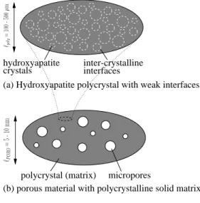
In Chapter B, hydroxyapatite biomaterials are envisioned as porous polycrystals with a non-porous matrix. This matrix consists of spherical crystals with weak interfaces. A second approach is presented in Chapter C: Based on the morphological description of a polycrystal developed in Chapter A, hydroxyapatite biomaterials are represented as a polycrystal consisting of uniformly distributed crystal needles and spherical pores. The experimental validation for elasticity and strength indicates the superiority of the latter model.
Brittle versus ductile behavior of crystals
In Chapter C, a brittle behavior of the hydroxyapatite crystal needles within biomaterials is considered, whereas in Chapter D, we propose a (layered water-induced) ductile behav-ior for interfaces between the hydroxyapatite crystals as part of natural collagenous bone tissue. The reason for the different behaviors may well lie in the characteristic size of the crystals, and hence of the nature of their contact surfaces, the crystals in collagenous bone tissue being much smaller than the biomaterial crystals. In the same sense, in low or non-collagenous tissues, such as specific whale bones (Zioupos et al. 1997), the minerals grow larger, and also these tissues exhibit a brittle failure behavior. The idea of increased ductility due to increased activity of layered water films is also supported by the fact (Nyman et al. 2008) that bound water content is correlated to bone toughness; and this idea fits well with the suggestions of Boskey (2003), that larger crystals (implying less layered water films per crystal content) would lead to a more brittle behavior of bone materials.
Mechanical properties of elementary constituents
Validation of the micromechanical predictions for macroscopic mechanical properties (elas-ticity and strength) of bone and biomaterials is based on ‘universal’ micro/nanoscopic mechanical properties of the elementary constituents of the considered material. These properties are tissue and biomaterial-independent, and they are derived from experimental investigations. These ‘universal’ properties are the stiffness and strength characteristics of hydroxyapatite crystals and their interfaces (see Chapters B and C for the case of artificial biomaterials as well as Chapter D for the case of natural bone), of (molecular) collagen and of water (see Chapter D), of pure titanium (see Chapter E for the case of metallic biomaterials), and of a dense glass ceramic matrix (see Chapter F for ceramic biomaterials).
Concerning the tissue-independent elastic phase properties of bone (Chapter D), we con-sider the following experiments: Tests with an ultrasonic interferometer coupled with a solid media pressure apparatus (Katz and Ukraincik 1971; Gilmore and Katz 1982) reveal the isotropic elastic properties of hydroxyapatite powder, which, in view of the largely disordered arrangement of minerals (Lees et al. 1994; Fratzl et al. 1996; Peters et al. 2000; Hellmich and Ulm 2002a), are sufficient for the characterization of the mineral phase (Hellmich and Ulm 2002b; Hellmich et al. 2004b; Fritsch et al. 2006). Given the absence of direct measurements of (molecular) collagen, its elastic properties are approximated by those of dry rat tail tendon, a tissue consisting almost exclusively of collagen. By means of Brillouin light scattering, Cusack and Miller (1979) have determined the respective five independent elastic constants of a transversely isotropic material (Table D.1). We assign the standard bulk modulus of water (Table D.1) to phases comprising water with mechanically insignificant non-collageneous organic matter.
Concerning the biomaterial-independent elastic properties of artificial hydroxyapatite crystal (Chapters B and C) we adapt those chosen for bone mineral.
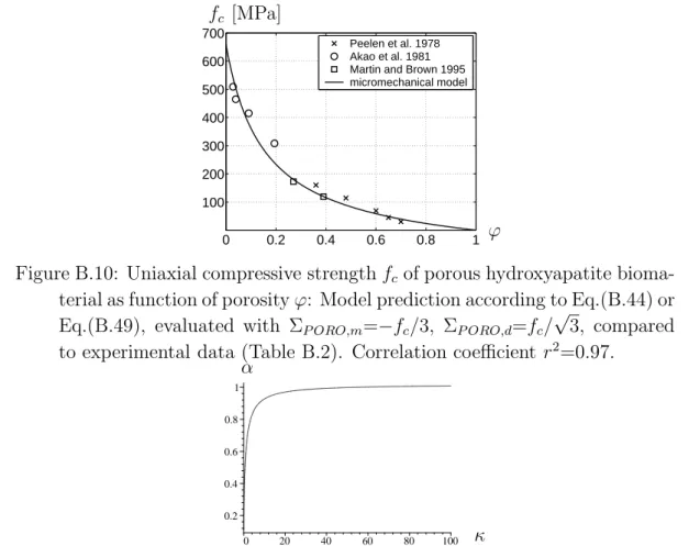
The approach proposed in Chapter B relies on three ‘universal’ material properties of in-terfaces between single hydroxyapatite crystals represented, for mathematical tractability, as spheres. The interface properties are difficult to be directly accessed, namely the fric-tion angle α, the cohesion h, and a dimensionless quantity κ of the interfaces. Therefore, these phase properties are determined by means of an optimization procedure providing the closest match of model predictions to experimentally determined uniaxial compressive strength data of hydroxyapatite biomaterials. Applying an evolution algorithm yields a set of solution vectors which are equal in terms of the highly satisfactory correlation coef-ficient between the respective model predictions and the corresponding experimental data for uniaxial compressive strength (see Section B.5.4).
In order to avoid such an optimization procedure for back-analysis of interface properties, we developed an alternative approach where we considered the non-spherical shape of the hydroxyapatite crystals. Using needles suggests a predominant stress state related
to the needle direction, and given this virtual 1D situation, this stress can be regarded as relevant for the stresses at the interface between crystals. In this sense, the approach proposed in Chapter C relies on the strength properties of interfaces between needle-shaped hydroxyapatite crystals, expressed by the bulk phase ‘hydroxyapatite’, namely its tensile and shear strength, σult,tHA and σHAult,s. We are not aware of direct strength tests on pure hydroxyapatite (with φ = 0). Therefore, we consider one uniaxial tensile test and one uniaxial compressive test on the densest samples available. From these two tests, we back-calculate the universal tensile and shear strength of pure hydroxyapatite relevant for crystal interfaces (Table C.2). It is interesting to note that consideration of the normal stress alone proved to be not sufficient for predicting macroscopic failure, in particular for low porosities in uniaxial compression. Only the ‘mixed’ formulation of the failure criterion taking into account normal and shear stresses (see Section C.3.2) inside the needles delivers satisfying macroscopic strength predictions.
Experimental data for model validation
The micromechanical models presented in Chapters B-F are based on experimentally de-termined elasticity and strength properties of the elementary material components. The models predict, for each set of tissue or biomaterial-specific volume fractions (e.g. porosi-ties), the corresponding tissue or biomaterial-specific elasticity and strength properties at all observation scales. Thus, a strict experimental validation of the mathematical model is realized as follows: (i) different sets of volume fractions are determined from compo-sition experiments on different bone or biomaterial samples; (ii) these volume fractions are used as model input, and (iii) corresponding model-predicted stiffness and strength values (model output) are compared to results from stiffness and strength experiments on the same or very similar bone or biomaterial samples.
Elastic macroscopic properties of biomaterials can be determined through uniaxial quasi-static mechanical tests, ultrasonic techniques or resonance frequency tests (Chapters C, E and F). Typical sample geometries include cylinders (diameter 5 mm, length 10 mm) for titanium samples (Chapter E) and glass ceramic scaffolds (Chapter F), and millimeter or centimeter-sized cylinders, bars or discs for hydroxyapatite biomaterials (see Chapter C and Table C.1). In case of ultrasonic testing, the length of the propagating wave has to be taken into account: If the wavelength is considerably smaller than the diameter of the specimen, a (compressional) ‘bulk wave’, i.e. a laterally constrained wave, propagates in a quasi-infinite medium. On the other hand, if the wavelength is considerably larger than the diameter of the specimen, a ‘bar wave’ propagates, i.e. the specimen acts as one-dimensional bar without lateral constraints.
Macroscopic uniaxial strength properties of bone and biomaterial samples can be deter-mined through quasi-static tensile, compressive and bending tests (Chapters C-F). Typi-cal sample geometries include cylinders (diameter 5 mm, length 10 mm) for titanium
sam-ples (Chapter E) and glass ceramic scaffolds (Chapter F), millimeter or centimeter-sized cylinders, bars or discs for hydroxyapatite biomaterials (see Chapter C and Table C.1) and millimeter or centimeter-sized cylinders or parallelepipeds, often with reduced cross section, for bone (see Chapter D and Table D.4).
Original contributions to the field of micromechanical
modeling
Effect of morphology in self-consistent schemes
The classical self-consistent scheme (Hershey 1954; Kr¨oner 1958; Hill 1963) is often used for modeling the overall elastic properties of porous polycrystals. It consists in embedding spherical inclusions into a matrix with stiffness of the homogenized material. This ap-proach predicts a vanishing overall stiffness (‘percolation threshold’) for porosities greater than 50%.
In Chapter A, it is proposed to replace spherical solid inclusions by a set of infinitely many uniformly oriented cylindrical inclusions (needles). All these needles are identical with respect to shape and material behavior, while being oriented in all directions in space. This has two implications: (i) the stiffness tensor related to a single crystal is a function of the Euler angles, while the components are orientation-independent in a local base frame, and (ii) the (overall) effective stiffness tensor of the porous polycrystal is isotropic.
Interfaces
Interfaces are often believed to play a role in the mechanical behavior of mineralized biological and biomimetic materials (Bhowmik et al. 2007). In Chapter B, porous hy-droxyapatite biomaterials are represented as a (dense) polycrystal with weak interfaces, which serves as the skeleton of a porous material defined one observation scale above. In detail, isotropic single crystals of typically quasi-spherical shape are separated from each other by very thin (essentially 2D) interfaces. The interface stiffness tensor exhibits an infinite normal component and a positive tangential component, and its load bearing capacity is characterized by a Coulomb-type law, considering the tangential and normal components of the traction force acting on the interface (see Section B.3 for details). In order to determine the effective failure properties resulting from local (brittle) failure characteristics and from the interactions between interfaces and bulk single crystals, the local interface forces have to be related to the ‘macroscopic’ stresses. The tangential and normal traction forces occurring in the interface failure criterion are non-homogeneously distributed across the interfaces. Failure will occur where relatively high tangential trac-tion forces encounter a relative low resistance due to relatively low normal tractrac-tion forces.
Instead of trying to model the actual force fields across the interfaces, we estimate the effect of the actual force distribution through so-called effective traction forces, as it is commonly done for stress, strain, or force fields in the context of continuum micromechan-ics (Suquet 1997a; Dormieux et al. 2007). In this line, we represent the failure-inducing interplay between moderate normal traction forces and tangential traction force peaks by means of two different effective measures for the normal and the tangential traction forces, respectively: (i) first-order moments of normal forces, and (ii) second-order mo-ments (also called quadratic average) of tangential forces, in the line of (Kreher 1990; Kreher and Molinari 1993; Dormieux et al. 2002). The relation between the quadratic average and the macroscopic stress is established through energy considerations. Remark-ably, the second-order moment of tangential tractions over all interfaces within the RVE is proportional to the ‘macroscopic’ equivalent deviatoric stress, and local, Coulomb-type brittle failure in the interfaces implies Drucker-Prager-type (brittle, elastic limit-type) failure properties at the scale of the polycrystal.
It is also interesting to note that the elastic, brittle failure criterion is quasi-identical to the yield surface of a porous medium obtained through non-linear homogenization (Dormieux 2005; Dormieux et al. 2006b) which is related to failure of a ductile solid matrix obeying a Drucker-Prager criterion. The ductile criterion is even identical to the elastic domain for incompressible solid matrices, see Section B.5 for a detailed discussion.
Organization of the thesis
The overall aim of this thesis is the prediction of bone strength from its composition and microstructure. Classically, the strength of bone materials is thought to be related to the strength properties of hydroxyapatite and collagen, and/or interfaces between these constituents. Chapters B and C concentrate on the failure properties of artificial hydrox-yapatite biomaterials which are very similar to natural bone mineral, based on a morpho-logical concept presented in Chapter A. A micromechanical model for bone strength is presented in Chapter D, while some experimental investigations and modeling of bioma-terials accompany the theoretical developments (Chapters E and F).
Chapter A is dedicated to the homogenization of linear elastic properties of porous polycrystals built up of needle-like platelets or sheets. Such microstructures can be found in a number of biological and man-made materials such as the mineral phase of bone, the cement paste of concrete or gypsum. Within a self-consistent scheme the solid phase is represented by cylindrical inclusions (needles). Uniform and axisymmetrical orientation distribution of linear elastic, isotropic as well as anisotropic needles is considered and the results are compared to the classical ones related to spherical inclusions. As a key result, a porosity lower than 0.4 is shown to result in the (overall) elastic properties of the polycrystal with uniformly oriented needles, which are quasi-identical to those of
a polycrystal with solid spheres. However, as opposed to the sphere-based model, the needle-based model does not predict a percolation threshold for inclusions with infinite aspect ratio.
Chapter B proposes a first attempt to model the strength properties of hydroxyapatite biomaterials, based on a micromechanical description of the elasticity and brittle failure of interfaces between isotropic crystals in a (dense) polycrystal, which serves as the skeleton of a porous material defined one observation scale above. Equilibrium and compatibility conditions, together with a suitable matrix-inclusion problem with a compliant interface, yield the homogenized elastic properties of the polycrystal, and of the porous material with polycrystalline solid phase. Incompressibility of single crystals guarantees finite shear stiffness of the polycrystal, even for vanishing interface stiffness, while increasing the latter generally leads to an increase of polycrystal shear stiffness. Corresponding elastic energy expressions give access to effective stresses representing the stress heterogeneities in the microstructures, which induce brittle failure. Thereby, Coulomb-type brittle failure of the crystalline interfaces implies Drucker-Prager-type (brittle, elastic limit-type) failure properties at the scale of the polycrystal. At the even higher scale of the porous material, high interfacial rigidities or low interfacial friction angles may result in closed elastic do-mains, indicating material failure even under hydrostatic pressure. This micromechanics model can satisfactorily reproduce the compressive experimental strength data of differ-ent (brittle) hydroxyapatite biomaterials, across largely variable porosities. Thereby, the brittle failure criteria can be well approximated by micromechanically derived criteria referring to ductile solid matrices, both criteria being even identical if the solid matrix is incompressible.
A second approach for modeling the strength properties of hydroxyapatite biomaterials is addressed in Chapter C, with the aim to predict uniaxial compressive and tensile failure. Thereby, these biomaterials are envisioned as porous polycrystals consisting of (isotropic) hydroxyapatite needles and spherical pores, in the line of Chapter A. Failure possibly occurs at the interfaces of the crystal needles, but modeling interfaces between non-spherical objects is extremely complex. Therefore, the effect of ‘micro’-interface be-havior of elongated 1D particles on the overall ‘macroscopic’ material is mimicked by equivalent ‘bulk’ failure properties of the crystal needles. Validation of respective mi-cromechanical models relies on two independent experimental sets: Biomaterial-specific macroscopic (homogenized) stiffness and uniaxial (tensile and compressive) strength pre-dicted from biomaterial-specific porosities, on the basis of biomaterial-independent (‘uni-versal’) elastic and strength properties of hydroxyapatite, are compared to corresponding biomaterial-specific experimentally determined (acoustic and mechanical) stiffness and strength values. The good agreement between model predictions and the corresponding experiments underlines the relevance of this approach.
Chapter D proposes an experimentally supported micromechanical explanation of corti-cal bone strength, based on a new vision on bone material failure: mutual ductile sliding of
hydroxyapatite mineral crystals along layered water films is followed by rupture of collagen crosslinks. In order to cast this vision into a mathematical form, a multiscale continuum micromechanics theory for upscaling of elastoplastic properties is developed, based on the concept of concentration and influence tensors for eigenstressed microheterogeneous materials. The model reflects bone’s hierarchical organization, in terms of representative volume elements for cortical bone, for extravascular and extracellular bone material, for mineralized fibrils and the extrafibrillar space, and for wet collagen. In order to get access to the stress states at the interfaces between crystals, the extrafibrillar mineral is resolved into an infinite amount of cylindrical material phases oriented in all directions in space in the line of Chapter C. The multiscale micromechanics model is shown to be able to satisfactorily predict the strength characteristics of different bones from different species, on the basis of their mineral/collagen content, their intercrystalline, intermolecular, la-cunar, and vascular porosities, and the elastic and strength properties of hydroxyapatite and (molecular) collagen.
In Chapter E, titanium with different porosities, produced on the basis of metal pow-der and space holpow-der components, is investigated as bone replacement material. For the determination of mechanical properties, i.e. strength of dense and porous titanium sam-ples, two kinds of experiments were performed – uniaxial and triaxial tests. The triaxial tests were of poromechanical nature, i.e. oil was employed to induce the same pressure both at the lateral surfaces of the cylindrical samples and inside the pores. The stiffness properties were revealed by acoustic (ultrasonic) tests. Different frequencies give access to different stiffness components (stiffness tensor components related to high-frequency-induced bulk waves versus Young’s moduli related to low-frequency-high-frequency-induced bar waves), at different observation scales; namely, the observation scale the dense titanium with around 100 µm characteristic length (characterized through the high frequencies) versus that of the porous material with a few millimeters of characteristic length (characterized through the low frequencies). Finally, the experimental results were used to develop and validate a poro-micromechanical model for porous titanium, which quantifies material stiffness and strength from its porosity and (in the case of the aforementioned triaxial tests) its pore pressurization state.
Chapter F presents a micromechanical description of bioresorbable porous glass ceramic scaffolds used for bone tissue engineering. Based on continuum micromechanics, a ma-terial model predicting relationships between porosity and elastic/strength properties is employed. The model, which mathematically expresses the mechanical behavior of a ceramic matrix (based on a glass system of the type SiO2-P2O5-CaO-MgO-Na2O-K2O;
called CEL2) in which interconnected pores are embedded, is carefully validated through a wealth of independent experimental data. The remarkably good agreement between porosity-based model predictions for the elastic and strength properties of CEL2-based porous scaffolds and corresponding experimentally determined mechanical properties un-derlines the great potential of micromechanical modeling for speeding up the biomaterial
and tissue engineering scaffold development process – by delivering reasonable estimates for the material behavior, also beyond experimentally observed situations.
Publication
A
Porous polycrystals built up by
uniformly and axisymmetrically
oriented needles: Homogenization of
elastic properties (Fritsch et al.
2006)
Authored by Andreas Fritsch, Luc Dormieux, and Christian Hellmich Published in Comptes Rendus Mecanique, Volume 334, pages 151–157
Porous polycrystal-type microstructures built up of needle-like platelets or sheets are characteristic for a number of biological and man-made materials. Herein, we consider (i) uniform, (ii) axisymmetrical orientation distribution of linear elastic, isotropic as well as anisotropic needles. Axisymmetrical needle orientation requires derivation of the Hill tensor for arbitrarily oriented ellipsoidal inclusions with one axis tending towards infinity, embedded in a transversely isotropic matrix; therefore, Laws’ integral expression of the Hill tensor is evaluated employing the theory of rational functions. For a porosity lower 0.4, the elastic properties of the polycrystal with uniformly oriented needles are quasi-identical to those of a polycrystal with solid spheres. However, as opposed to the sphere-based model, the needle-sphere-based model does not predict a percolation threshold. As regards axisymmetrical orientation distribution of needles, two effects are remarkable: Firstly, the sharper the cone of orientations the higher the anisotropy of the polycrystal. Secondly,
for a given cone, the anisotropy increases with the porosity. Estimates for the polycrystal stiffness are hardly influenced by the anisotropy of the bone mineral needles. Our results also confirm the very high degree of orientation randomness of crystals building up mineral foams in bone tissues.
A.1
Introduction
Porous polycrystal-type microstructures built up of needle-like platelets or sheets can be found in a number of biological and man-made materials; such as bone (Hellmich et al. 2004a; Hellmich and Ulm 2002a) or eggs (Silyn-Roberts and Sharp 1986), or at the cement paste level of concrete (Baroughel-Bouny 1994). We here deal with homogenization of their overall (linear) elastic properties, by means of self-consistent schemes. Thereby, the solid phase (needles) is represented by cylindrical inclusions (a cylinder being the limit case of a prolate spheroid with its long axis being very much larger than its spherical axis), and the (empty) pore inclusions (drained conditions) are spherical; extension to pressurized pores according to Chateau and Dormieux (2002) is straightforward. Subsequently, we consider (i) uniform, (ii) axisymmetrical orientation distribution of isotropic as well as anisotropic needles with elasticity tensor Cs.
A.2
Uniform orientation distribution of needles
Uniformly oriented needles result in isotropic elastic properties of the polycrystal. The corresponding stiffness estimate C
SCS reads as C SCS = (1− φ) Cs:< [I+P SCS cyl : (Cs−C SCS)]−1 >: {(1 − φ) < [I+P SCS cyl : (Cs−C SCS)]−1 > +φ ( I−P SCS sph :C SCS)−1 }−1(A.1) with < [I+P SCS cyl : (Cs−C SCS)]−1 >= 2π Z ϕ=0 π Z ϑ=0 [I+P SCS cyl (ϑ, ϕ) (Cs−C SCS)]−1sin ϑ dϑ dϕ 4π (A.2)
where I, Iijkl = 1/2(δikδjl+ δilδkj), is the fourth-order unity tensor, δij is the Kronecker
delta, φ denotes the porosity,P
SCS sph andP
SCS
cyl are the fourth-order Hill tensors for spherical
and cylindrical inclusions, respectively. The Hill tensor for spherical inclusions, P
SCS sph , is
widely available in the open literature (Eshelby 1957; Suvorov and Dvorak 2002). The components of the Hill tensor for cylindrical inclusions embedded in an isotropic medium are given for a base frame coinciding with the long axis of the cylinder (Eshelby 1957). Transformation of Hill tensors related to differently oriented cylindrical inclusions, to one
reference frame can be expressed by Euler angles ϑ and ϕ, rendering P = P
SCS
cyl (ϑ, ϕ) in
Eqn.(A.2).
The numerical solution of (A.1) shows that the effective Young’s modulus ESCS is
prac-tically independent of the needles’ Poisson’s ratio νs.
The question arises whether uniform orientation of needles can be appropriately consid-ered by representing the solid phase simply by spherical inclusions. The corresponding self-consistent estimateC
SCS for identical shape and orientation of inclusions reads as (see
e.g. (Zaoui 1997a))
C SCS = (1 − φ)Cs :{I+P SCS sph : (Cs−C SCS) }−1 (A.3) In case of an incompressible solid phase (with bulk modulus ks → ∞), (A.3) can be solved
analytically: µSCS = µs 3(1− 2φ) 3− φ , k SCS = 4(1− φ) 3φ µ SCS (A.4)
where kSCS and µSCS are the effective bulk and shear moduli, and µ
sis the shear modulus
of the isotropic solid. This scheme shows a percolation threshold exactly equal to φ = 1 2,
for any value of the Poisson’s ratio νs of the solid phase. As for a compressible solid
phase, the homogenized Young’s modulus ESCS can still be approximated by the affine
expression Es(1− 2φ) with an error of at most 4 % relative to the exact solution, i.e.
ESCS is quasi-independent of Poisson’s ratio.
On the entire porosity range, 0 < φ < 1, the self-consistent stiffness estimates based on uniformly oriented solid needles are quasi-identical for both isotropic and anisotropic needle behavior [Fig. A.1 (a) and (b), see Fig. A.1(c) for elastic constants (Katz and Ukraincik 1971) of hydroxyapatite crystals building up porous foams in bone (Hellmich and Ulm 2002a)]. In addition, on the interval 0 < φ < 0.4, these estimates are quasi-identical to those based on isotropic solid spheres [Fig. A.1 (a) and (b)]. ¿From a physical viewpoint, one may argue that, at a sufficiently high concentration, both spherical as well as isotropic or anisotropic needle-type particles build up similar contiguous matrices. Particularly, in the vicinity of φ = 0, the first-order expansions of the homogenized elastic constants with respect to the porosity are identical for the two models with an isotropic solid phase, reading as:
ESCS Es = 1−3 2 (1− νs)(5νs+ 9) 7− 5νs φ ; νSCS = νs+ φ 3(1− 5νs)(1− νs2) 2(7− 5νs) (A.5) kSCS ks = 1−3 2 1− νs 1− 2νs φ ; µ SCS µs = 1− 15 1− νs 7− 5νs φ (A.6)
However, as opposed to the sphere-based model, the needle-based model does not predict any percolation threshold, i.e. ESCS, kSCS and µSCS → 0 only if the volume fraction of the
0 0.2 0.4 0.6 0.8 1 20 40 60 80 100 120 0 0.2 0.4 0.6 0.8 1 0.1 0.2 0.3
0.4 Anisotropic (transversely isotropic)
elasticity of hydroxyapatite [GPa]: C1111= C2222= 137, C1122= 42.5, C1133= 54.9, C3333= 172, C1313= C2323= 39.6. Isotropic elasticity of hydroxyapatite: E = 114 GPa, ν = 0.27 (a) (b) (c) ESCS[GPa] νSCS φ φ
Figure A.1: (a) Young’s modulus and (b) Poisson’s ratio of isotropic porous polycrystals, predicted by the sphebased and needle-based models, re-spectively (isotropic spheres . . . dashed lines, uniformly oriented isotropic needles . . . solid lines, uniformly oriented anisotropic needles . . . dash–dot lines); (c) Anisotropic and isotropic elasticity of hydroxyapatite (Katz and Ukraincik 1971).
solid phase becomes very small (φ → 1). From an intuitive viewpoint, this is consistent with the ‘rice grain effect’: As compared to spheres, needles are more likely to contact each other, especially at low volume fraction (φ → 1). A first-order expansion in the vicinity of φ = 1 of µSCS (resp. kSCS) can be sought in the form µSCS ∼ m(1 − φ) [resp.
kSCS ∼ k(1 − φ)]. As regards isotropic needles, analytical expressions for m and k can be
derived and proven to be independent of νs :
m = 71− 2 √ 79 1575 , k = −8 + 2√79 189 (A.7)
Accordingly, the limit of νSCS when φ tends towards 1 is independent of ν
s as well : lim φ→1ν SCS = 17− √ 79 35 (A.8)
A.3
Axisymmetric orientation distribution of needles
Axisymmetrically oriented needles result in transversely isotropic elastic properties of the polycrystal. With ϑ being measured with respect to the symmetry axis of the orientation distribution, we consider (i) uniform needle distribution in the cone [0, ϑmax], and (ii)
Gaussian needle distribution around ϑmax/2 with standard deviation sϑ; both expressed
in terms of a distribution function F (ϑ). The corresponding stiffness estimate still obeys (A.1), while (A.2) now reads as
< (I+P SCS cyl : δC) −1 >= 2π Z ϕ=0 ϑZmax ϑ=0 F (ϑ) [I+P SCS cyl (ϑ, ϕ) (Cs−C SCS)]−1 sin ϑ dϑ dϕ 2π(1− cos ϑmax) (A.9) and while the Hill tensorsP
SCS cyl andP
SCS
sph now refer to inclusions in a transversely isotropic
material.
Expressions for P
SCS
sph can be found in Hellmich et al. (2004a), and for determination of
P
SCS
cyl we evaluate Laws’ double integral expression of the Hill tensor (Laws 1977) for
arbitrarily oriented cylindrical inclusions embedded in a transversely isotropic material, employing the theory of rational functions. Thereby, we arrive at a single-integrated expression allowing for efficient computational evaluation (see Appendix).
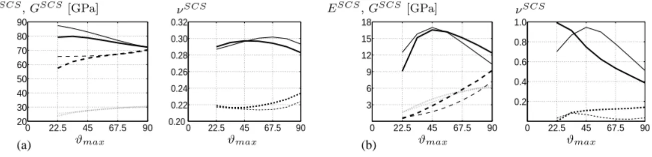
We evaluated Eqn.(A.9) for a uniform distribution of needles between 0 and a maxi-mum angle ϑmax as well as for a Gaussian distribution with different standard deviations
around ϑmax/2, see Fig. A.2 and A.3. Two effects are remarkable (Fig. A.2): Firstly, as
expected, the sharper the cone of orientations the higher is the anisotropy of the polycrys-tal. Secondly, the higher the porosity the more pronounced is the effect of the non-uniform needle orientation distribution, on both the Young’s modulus and the Poisson’s ratio. As compared to uniform needle distribution between ϑ = 0 and ϑ = ϑmax, the Gaussian
distribution around ϑmax/2 with standard deviation sϑ significantly affects the effective
Poisson’s ratio (compare Fig. A.2 and A.3), while differences in Young’s and shear moduli are, on the average, less than 7 % for the investigated distributions (Fig. A.2 and A.3).
A.4
Discussion
The present results are also noteworthy from a biomechanical viewpoint: In the ultra-structure of bones and mineralized tissues hydroxyapatite crystals build up a contiguous network or mineral foam (Hellmich and Ulm 2002a; Hellmich et al. 2004a). Single crystals have typical dimensions of 50 nm average length, 25 nm average width, and 1 to 7 nm thickness (Weiner and Wagner 1998; Fratzl et al. 1996). In a first approximation, they are often characterized as needles (Fratzl et al. 1996; Sasaki 1991; Fratzl et al. 1991). This renders the homogenization schemes developed here as appropriate for mineral foams occuring in bones. In particular, agreement between homogenized elastic properties of uniformly oriented needles with those of spheres for a porosity lower 0.4 (Fig. A.1) confirms the use of self-consistent schemes with spherical inclusions for hydroxyapatite polycrystals (Hellmich et al. 2004a), which have been validated by the experimental data of (Lees et al. 1983; Lees 1987a). At higher porosities, however, the needle-based scheme seems to be superior to the sphere-based scheme, since the former accounts for contigu-ity of the crystals, leading to non-zero homogenized stiffness, while the latter exhibits a
0 22.5 45 67.5 90 20 30 40 50 60 70 80 90 0 22.5 45 67.5 90 0.20 0.22 0.24 0.26 0.28 0.30 0 22.5 45 67.5 90 3 6 9 12 15 18 0 22.5 45 67.5 90 0.2 0.4 0.6 0.8 (a) (b) ESCS, GSCS[GPa] νSCS ESCS, GSCS[GPa] νSCS ϑmax ϑmax ϑmax ϑmax
Figure A.2: Effect of axisymmetric distribution of anisotropic needles (uni-formly distributed between ϑ = 0 and ϑ = ϑmax) on the longitudinal and
transverse Young’s moduli, Poisson’s ratios, and shear modulus for dif-ferent porosities [(a) φ = 0.2, (b) φ = 0.6]. Longitudinal components are shown as solid lines, transversal components as dashed lines, and the shear modulus as dotted line.
0 22.5 45 67.5 90 20 30 40 50 60 70 80 90 0 22.5 45 67.5 90 0.20 0.22 0.24 0.26 0.28 0.30 0.32 0 22.5 45 67.5 90 3 6 9 12 15 18 0 22.5 45 67.5 90 0.2 0.4 0.6 0.8 1.0 (a) (b) ESCS, GSCS[GPa] νSCS ESCS, GSCS[GPa] νSCS ϑmax ϑmax ϑmax ϑmax
Figure A.3: Effect of axisymmetric distribution of anisotropic needles (Gaussian–type distributed around ϑmax/2 with standard deviation sϑ)
on the longitudinal and transverse Young’s moduli and Poisson’s ratios for different porosities [(a) φ = 0.2, (b) φ = 0.6] and different standard deviations (sϑ= 2.5o . . . thick lines, sϑ= 12o . . . thin lines). Longitudinal
components are shown as solid lines, transversal components as dashed lines, and the shear modulus as dotted line.
percolation threshold beyond which the homogenized stiffness vanishes. Indeed, elasticity experiments (Lees and Page 1992) reveal that mineral crystals do contribute to the overall stiffness of low-mineralized turkey leg tendon, with a mineral foam porosity larger than 50%.
The present results also confirm the pronounced randomness of crystal orientation in bone tissues, revealed already by chemical (Peters et al. 2000) or mechanical (Hellmich and Ulm 2002a) means: Any pronounced orientation of needles leads to high anisotropy ratios Etran/Elong far beyond two, and up to ten (Fig. A.2). In real bone ultrastructure,
Ulm 2002a).
A.5
Appendix: Hill tensor for arbitrarily oriented
cylindrical inclusions embedded in a transversely
isotropic material
The starting point is Laws’ classical expression for the Hill tensor (see for instance (Laws 1977, 1985)) : P = ω2ω3 4π Z |ξ|=1 Γ (ξ· AT · A · ξ)3/2 dS(ξ) (A.10)
ξ is the unit length vector pointing from the origin of the sphere to the surface element dS(ξ). The second-order tensor A describes the shape of the ellipsoid, with base vectors w1, w2 and w3 pointing in the principal directions of the ellipsoid,
A = w1⊗ w1+ ω2w2⊗ w2+ ω3w3⊗ w3, ω3 ≫ 1 (A.11)
The fourth-order tensor Γ is defined as
Γ = ξ⊗ Ks −1 ⊗ ξ, K = ξ ·s
C· ξ (A.12)
The second-order tensor K is the acoustic tensor, C is the stiffness tensor of the
trans-versely isotropic matrix. ⊗ denotes the symmetrized tensor product.s
The technique presented hereafter adapts the ideas presented in (Gruescu et al. 2005) and (Suvorov and Dvorak 2002) to cylindrical inclusions. First, we consider the denominator of expression (A.10). The unit vector ξ can be expressed in spherical coordinates Φ ∈ [0, 2π] and Θ ∈ [0, π] as ξ1 = sin Θ cos Φ, ξ2 = sin Θ sin Φ and ξ3 = cos Θ, so that
dS = sin Θ dΦ dΘ. Since
ξ· AT · A · ξ = ω32cos2Θ + sin2Θ (cos2Φ + ω22sin2Φ) we find with x = cos Θ and γ2 = 1
ω2 3(cos 2Φ + ω2 2sin2Φ) P= ω2 4π 2π Z 0 1 Z −1 γ2 [x2+ (1− x2)γ2]3/2 Γ(x, Φ) cos2Φ + ω2 2sin2Φ (−dx) dΦ (A.13)
lim γ→0 γ2 [x2+ (1− x2) γ2]3/2 = 2 δ(x), Z δ(x) f (x) dx = f (0) (A.14) yields, with ω2 = 1, P= 1 2π 2π Z 0 Γ(Θ =π 2, Φ) dΦ (A.15)
Next, we consider the numerator of Eqn. (A.10), Γ = ξ ⊗ Ks −1⊗ ξ. Expressing ξ and Ks
in terms of the base vectors w1 and w2, while adopting z = cot Φ, yields
ξ = cos Φ w1 + sin Φ w2 = sin Φ(z w1+ w2) (A.16)
K = ξ·C· ξ = = sin2Φ ((z w1+ w2)·C· (z w1+ w2)) sin 2Φ (z2Q + z(R + RT) + T) | {z } K′(z) (A.17)
when having introduced the second-order tensors Q, R and T as
Q = w1·C· w1, R = w1·C· w2, T = w2·C· w2 (A.18) K(Φ) = sin2Φ (z2Q + z(R + RT) + T) | {z } K′(z) (A.19) K′
(z) is a second-order polynomial. In order to obtain the inverse of K′
(z), we use the matrix of cofactors (algebraic complements) co K′,
(K(z))−1 = 1 sin2Φ(K ′ )−1 = 1 sin2Φ 1 det K′ (co K ′ ) (A.20) The determinant of K′ , det K′
, is a sixth-order polynomial. Thus
Γ = ξ ⊗ Ks −1 ⊗ ξ =s 1 sin2Φ 1 det K′ (ξ s ⊗ (co K′)⊗ ξ) =s = 1 sin2Φ 1 det K′ (sin 2Φ (z w 1+ w2) s ⊗ (co K′)⊗ (z ws 1+ w2)) (A.21)
P = 1 2π 2π Z Φ=0 Γ dΦ = 1 2π2 ∞ Z z=−∞ Γ dz 1 + z2 (A.22) = 1 π ∞ Z −∞ (z w1+ w2) s ⊗ (co K′)⊗ (z ws 1+ w2) (det K′ ) (1 + z2) dz (A.23)
The integrand in (A.23) is a rational fraction with a sixth-order polynomial in the nu-merator and an eighth-order polynomial in the denominator. Hence, the integration can be based on the Residue Theorem:
∞ Z −∞ f (z) dz = 2iπX j Res(f, zj), (A.24)
Publication
B
Micromechanics of crystal interfaces
in polycrystalline solid phases of
porous media: fundamentals and
application to strength of
hydroxyapatite biomaterials (Fritsch
et al. 2007a)
Authored by Andreas Fritsch, Luc Dormieux, Christian Hellmich, and Julien Sanahuja Published in Journal of Materials Science, Volume 42, pages 8824–8837
Interfaces are often believed to play a role in the mechanical behavior of mineralized bio-logical and biomimetic materials. This motivates the micromechanical description of the elasticity and brittle failure of interfaces between crystals in a (dense) polycrystal, which serves as the skeleton of a porous material defined one observation scale above. Equi-librium and compatibility conditions, together with a suitable matrix-inclusion problem with a compliant interface, yield the homogenized elastic properties of the polycrystal, and of the porous material with polycrystalline solid phase. Incompressibility of single crystals guarantees finite shear stiffness of the polycrystal, even for vanishing interface stiffness, while increasing the latter generally leads to an increase of polycrystal shear stiffness. Corresponding elastic energy expressions give access to effective stresses
rep-resenting the stress heterogeneities in the microstructures, which induce brittle failure. Thereby, Coulomb-type brittle failure of the crystalline interfaces implies Drucker-Prager-type (brittle, elastic limit-Drucker-Prager-type) failure properties at the scale of the polycrystal. At the even higher scale of the porous material, high interfacial rigidities or low interfacial fric-tion angles may result in closed elastic domains, indicating material failure even under hydrostatic pressure. This micromechanics model can satisfactorily reproduce the ex-perimental strength data of different (brittle) hydroxyapatite biomaterials, across largely variable porosities. Thereby, the brittle failure criteria can be well approximated by mi-cromechanically derived criteria referring to ductile solid matrices, both criteria being even identical if the solid matrix is incompressible.
B.1
Introduction
Interfaces are believed to often play a fundamental role in the mechanical behavior of hierarchically organized biological materials. Accordingly, much attention has been paid to the polymer-filled interfaces between ceramic tablets in nacre (Gennes and Okumura 2000; Okumura and Gennes 2001; Katti and Katti 2001; Katti et al. 2001; Okumura 2002, 2003; Barthelat et al. 2007), but the importance of interfacial behavior was also discussed for other classes of biological materials, such as bone (Tai et al. 2006).
To gain insight into these material systems, material/microstructure models have been developed within different theoretical frameworks, such as fracture mechanics and scaling laws (Gennes and Okumura 2000; Okumura and Gennes 2001; Okumura 2002, 2003), large-scale elastoplastic Finite Element analyses (Katti and Katti 2001; Katti et al. 2001; Tai et al. 2006), or periodic homogenization on the basis of a unit cell discretized by Finite Elements (Barthelat et al. 2007).
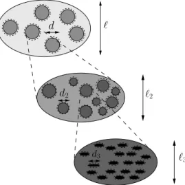
In addition to such periodic, FE-based (‘computational’) homogenization approaches, analytical and/or semianalytical approaches of random homogenization (continuum mi-cromechanics (Zaoui 1997b, 2002)) have been recently used as to effectively predict the elastic properties of complicated hierarchically structured material systems (such as bone (Hellmich and Ulm 2002b; Hellmich et al. 2004b,a; Fritsch and Hellmich 2007), wood (Hofstetter et al. 2005, 2006), concrete (Bernard et al. 2003; Ulm et al. 2004; Hellmich and Mang 2005), or shale (Ulm et al. 2005)), from the elasticity and the mechanical interactions – over different observation scales – of nanoscaled elementary components. Thereby, not every single detail of the highly random microstructures, but only the essen-tial morphological features are considered, in terms of homogeneous subdomains (material phases) inside representative volume elements (RVEs, Fig. B.1), their volume fractions, their elasticity, and their mechanical interaction. Theoretically, it has been recently well understood how to extend these homogenization techniques to the ductile failure of (bulk) phases (Dormieux and Maghous 2000; Bernaud et al. 2002; Barth´el´emy and Dormieux
ℓ ℓ3 d3 d ℓ2 d2
Figure B.1: Multistep homogenization: Properties of phases (with character-istic lengths of d and d2, respectively) inside RVEs with characteristic
lengths of ℓ or ℓ2, respectively, are determined from homogenization over
smaller RVEs with characteristic lengths of ℓ2 ≤ d and ℓ3 ≤ d2,
respec-tively.
2003, 2004; Dormieux et al. 2006c,a) (while applications to real materials (Lemarchand et al. 2002) are more rare than for the elastic case). In comparison, the treatment of brittle failure and of interfaces in the framework of random homogenization is still a very open field: It is the focus of this paper – both fundamentally, and in view of the failure of biomimetic hydroxyapatite biomaterials.
Extending very recent results (Sanahuja and Dormieux 2005; Dormieux et al. 2007), where inclusion coatings and interfaces in porous polycrystals were modeled, we here tackle the description of the elasticity and failure of interfaces between crystals in a (dense) polycrys-tal, which serves as the skeleton of a porous material defined one observation scale above (Fig. B.2). Thereby, we show characteristic features of a corresponding new micromechan-ics model, which is based on matrix-inclusion problems with compliant interfaces (Hashin 1991; Herv´e and Zaoui 1993; Zhong and Meguid 1997), and which turns out to reasonably explain the behavior of porous hydroxyapatite biomaterials, especially for their brittle failure in the compressive regime.

![Figure C.6: Comparison between model predictions [Eqs. (C.14)-(C.25)] and experiments for tensile strength of different porous biomaterials made of hydroxyapatite, as a function of porosity φ.](https://thumb-eu.123doks.com/thumbv2/123doknet/2961041.81444/79.892.252.612.325.591/comparison-predictions-experiments-strength-different-biomaterials-hydroxyapatite-porosity.webp)
