O
pen
A
rchive
T
oulouse
A
rchive
O
uverte
(OATAO)
OATAO is an open access repository that collects the work of some Toulouse
researchers and makes it freely available over the web where possible.
This is
an author's
version published in:
https://oatao.univ-toulouse.fr/23078
Official URL :
https://doi.org/10.1016/j.ejrad.2017.10.010
To cite this version :
Any correspondence concerning this service should be sent to the repository administrator:
[email protected]
Borel, Christophe and Larbi, Ahmed and Delclaux, Stephanie and Lapegue, Franck and
Chiavassa-Gandois, Helene and Sans, Nicolas and Faruch-Bilfeld, Marie Diagnostic value
of cone beam computed tomography (CBCT) in occult scaphoid and wrist fractures. (2017)
European Journal of Radiology, 97. 59-64. ISSN 0720-048X
OATAO
Diagnostic
value of cone beam computed tomography (CBCT) in occult
scaphoid
and wrist fractures
Christophe
Borel
a,⁎,
Ahmed Larbi
b,
Stephanie Delclaux
c,
Franck Lapegue
a,
Helene
Chiavassa-Gandois
a,
Nicolas Sans
a,
Marie Faruch-Bilfeld
aa Service d’imagerie, CHU de Toulouse-Purpan, Bâtiment Pierre Paul Riquet, TSA 40031-31059 Toulouse, France b Service d'imagerie médicale - CHU de Nîmes - 4 Rue du Professeur Robert Debré, 30029 Nîmes, France
c Service de chirurgie d’orthopédie et de traumatologie, CHU de Toulouse-Purpan, Bâtiment Pierre Paul Riquet, TSA 40031-31059 Toulouse, France
A R T I C L E I N F O
Keywords: Scaphoid fracture Wrist fracture Occult fracture
Cone beam computed tomography Magnetic resonance imaging
A B S T R A C T
Objective: Evaluate the diagnostic value of cone beam computed tomography (CBCT) for scaphoid and wrist fractures that are missed on standard radiographs.
Materials and methods: Between September 2014 and October 2015, we prospectively enrolled 49 patients with a clinically suspected scaphoid fracture following an acute injury but had normal radiographs. Each patients underwent radiographs, CBCT and (magnetic resonance imaging) MRI within 7 days of the initial injury event. Both exam were evaluated independently by two radiologists.
Results: For scaphoid cortical fractures CBCT sensitivity is 100% (95% CI: 75%–100%), specificity 97% (95% CI: 83%–100%). CBCT diagnosed all 24 corticals wrist fractures, corresponding to a sensitivity of 100% (95% CI: 83%–100%), specificity of 95% (95% CI: 75%–100%). Kappa agreement rate between the two radiologists was K = 0.95 (95% CI: 0.85–1) for scaphoid fractures and K = 0.87 (95% CI: 0.73–1) for wrist fractures. Conclusions: CBCT is superior to radiographs for diagnosing occult cortical fractures. Because of its low radiation dose, we believe that CBCT can be used in current practice as a replacement or supplement to radiographs to detect these fractures and optimize the cost-effectiveness ratio by limiting the number of needless im-mobilizations.
1. Introduction
The scaphoid is the most frequently fractured carpal bone [1].
Scaphoid fractures typically occur in young, predominantly male pa tients. Early diagnosis is essential to limit disabling consequences such as nonunion, avascular necrosis, carpal instability and osteoarthritis
[2].
The combination of clinical examination and radiographic assess ment is often inadequate, as 14% to 23% of fractures are not detected according to studies using MRI (magnetic resonance imaging) as a gold standard[3,4]. The diagnostic value of standard radiographs is poor, as evidenced by low sensitivity values of 66% for scaphoid fractures,
57.8% for wrist fracture and 38.7% for carpal fractures [5]. With a
sensitivity of 97% and specificity of 99%[6], MRI is said to be the gold standard[7,8]for diagnosing occult scaphoid fractures.
Currently, in cases of clinically suspected scaphoid fracture with normal initial radiographs, there are three avenues of investigation[9]: perform another set of radiographs on Day 14, perform an MRI, or
perform a standard CT scan(computerized tomography). Thefirst so
lution, consisting of 14 days of immobilization, often leads to lost work time, even when there is no fracture. Since MRI is costly and not always available in an emergency context, a CT scan is typically performed; the latter modality has a sensitivity of 91% and specificity of 98% for the diagnosis of scaphoid fractures[6]. Bone scanning is not an effective
alternative in this context given its higher cost, long scan time and higher level of radiation.
Cone beam computed tomography (CBCT) was developed for dental
imaging purposes in the 1990s[10]. It has now replaced CT scanning
for dental patients because of the high resolution images that it pro
vides. The conical X ray source with a two dimensionalflat detector
covers the desired volume in a single rotation. The acquired data are processed with volume reconstruction algorithms to produce multi planar reconstructions. Due to this technicals differences, CBCT require less computing performance.
Application of dedicated CBCT for musculoskeletal diagnostics is quite recent, but CBCT has been used in otherfields.
⁎Corresponding author.
E-mail addresses:[email protected](C. Borel),[email protected](A. Larbi),[email protected](S. Delclaux),[email protected](F. Lapegue),
3. Results
In all, 16 scaphoid fractures (33% of patients, 16/49) were diag nosed: 15 cortical fractures (Fig. 1) and 1 trabecular fractures (Fig. 2) (Table 1). Furthermore, 11 other occult wrist fractures were discovered: 5 distal radius fractures (including 1 trabecular), 2 cortical metacarpal fractures, 1 cortical trapezium fracture, 1 trabecular trapezoid fracture, 1 cortical triquetrum fracture and 1 cortical pisiform fracture (Fig. 3) (Table 2). One trabecular fracture was classified as a cortical fracture on MRI. One patient had two fractures: 1 cortical radius fracture and 1 cortical scaphoid fracture. None of the fractures were displaced (Table 3).
3.1. CBCT
CBCT found all 15 cortical fractures of the scaphoid with one false positive. For this type of fracture, CBCT has a sensitivity of 100% (95% CI: 75% 100%), specificity of 97% (95% CI: 83% 100%), PPV of 94% (95% CI: 68% 100%), NPV of 100% (95% CI: 87% 100%). One tra becular fracture of the scaphoid was not detected on CBCT, resulting in a sensitivity of 94% (95% CI: 68% 100%) specificity of 97% (95% CI: 83% 100%), PPV of 94% (95% CI: 68% 100%), NPV of 97% (95% CI: 82% 100%) for all types of scaphoid fractures.
CBCT diagnosed all 24 cortical wrist fractures, corresponding to a CBCT have found application in ear, nose, and throat imaging,
especially in the imaging of small structures like middle ear. CBCT is used in C arm flat panel computed tomography for opera tive imaging and in image guided radiotherapy. CBCT is also used in ear, nose, and throat imaging, especially for imaging of small structures like middle ear [11].
In 2011, a study of the prototype of an musculoskeletal dedicated CBCT showed good results in terms of resolution and practicability. A study of this musculoskeletal dedicated CBCT showed higher spatial resolution for the CBCT compared to CT scan. The first clinical study of CBCT images for wrist fractures showed similar results for CBCT and CT scan in terms of spatial resolution, allowing fracture detection [11].
This technique results in precise evaluation of bone; it applies less radiation than a CT scan [12 16], has a fast volumetric acquisition,
good spatial resolution and high bone contrast. This was observed in several studies of the extremities [17 21], and the use of CBCT has
recently been extended to soft tissues [22].
A wrist CT scan has an effective dose of 0.03 mSv [23], which re
presents a low exposure to ionizing radiation relative to the average annual radioactivity exposure from natural sources of 2.4 mSv. In a study [19] CBCT for exploration of wrist has an even lower radiation exposure, estimated about 0.007 mSv. However, it should be remarked that effective radiation dose for CBCT and CT scan varies with tube current and voltage.
The objective of our study was to evaluate the diagnostic value of CBCT for scaphoid and wrist fractures that are missed on standard radiographs.
2. Materials and methods 2.1. Patients
Between September 2014 and October 2015, we prospectively en rolled 49 patients (31 men, 18 women) who were between 18 and 78 years of age (mean 36 ± 14 years) and had a clinically suspected scaphoid fracture following an acute injury but had normal radio graphs. All of these patients underwent radiographs, CBCT and MRI within 7 days of the initial injury event (mean 4.1 days).
The initial radiographs consisted of four views evaluated by two radiologists (with 4 and 9 years of experience in musculoskeletal ima ging), who also interpreted the MRI and CBCT exams. Only patients with normal initial radiographs were included in the study.
The local ethics committee approved this study and each patient received information about the study verbally.
2.2. Imaging techniques
Radiographs − The initial radiographs consisted of four images: A/P and lateral views of the wrist in neutral position and two views in ulnar deviation (Schneck 1 view and Larsen view). The digital images were obtained with a flat detector (ProGrade Eleva, Philips Medical Systems, Eindhoven, Netherlands); the constants were 50 kV and 4 mA. The images were read on a PACS workstation (McKesson Horizon Rad Station, McKesson Radiology, San Francisco, California).
MRI − The MRI images were obtained with a 3 T unit (Magnetom Skyra, Siemens Healthcare, Erlangen, Germany), and a dedicated 16 channel wrist coil (Hand/wrist 16, Siemens Healthcare, Erlangen, Germany). The protocol consisted of coronal slices with the following sequences: Dixon (repetition time/echo time: 3000/40 ms; 2.5 mm slice thickness) and T1 spin echo (repetition time/echo time: 1080/14 msec; 2.5 mm slice thickness). A 100 mm field of view with a 512 × 512 matrix was used for each sequence. The examination was carried out with the arm hanging at the side of the body. No gadolinium was in jected.
CBCT − All the CBCT images were obtained with same unit (Planmeca ProMax 3D mid, Helsinki, Finland). The acquisition was
carried out in the “high resolution” mode; the tube voltage was 90 kV and the charge was 120 mA for a 15 s acquisition time. The field of view was 90 × 90 mm and encompassed the distal end of the forearm, carpus and proximal third of the metacarpals. For the interpretation, bone reconstructions in the three planes in space and a fourth plane in the major axis of the scaphoid were performed using a 0.5 mm slice thickness [24]. No iodine contrast product was injected.
2.3. Image analysis
In a double blind set up, a junior radiologist and a senior radiologist interpreted the MRI and the CBCT exams. Each radiologist determined whether a fracture was present and whether the fracture was cortical or trabecular on the MRI images. The sequence in which the images were presented was randomized and varied between the two radiologists. The observers read the images on a PACS workstation for MRI (McKesson Horizon Rad Station, McKesson Radiology, San Francisco, California) and used the OsiriX software (OsiriX foundation, Geneva, Switzerland) for the CBCT exams. A fracture was defined as the pre sence of a cortical disruption on CBCT.
2.4. Reference examination
A cortical fracture was present on MRI if a cortical fracture line was identified as a hypo T1 signal [7,25,26]. The diagnosis of a trabecular
fracture was made when an area of intraosseous edema was visible as a hyper Dixon signal (water only sequence) and as a hypo T1 signal. 2.5. Statistical analysis
The sensitivity, specificity, positive predictive value (PPV), negative predictive value (VPN), 95% confidence intervals (CI) and kappa coefficient for inter rater agreement were calculated using SPSS soft ware (SPSS, Chicago, Ill, USA) with MRI as the gold standard. 2.6. Exclusion criteria
Patients were excluded if they met any of these criteria: less than 18 years of age, pregnant woman, severe polytrauma requiring intensive care, complex wrist fracture, history of scaphoid fracture and contra indication for MRI.
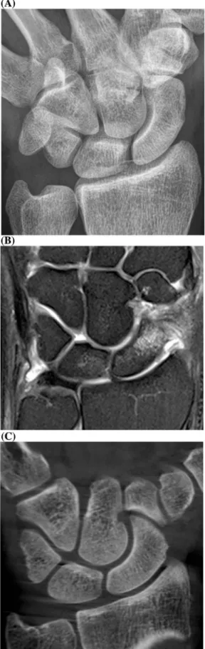
Fig. 1. Cortical scaphoid fracture in a 23-year-old man. a The initial coronal conventional ulnar deviation radiograph is normal. b The coronal reformation from CBCT shows a non-displaced fracture of the scaphoid (arrow). c The coronal T1-weighted (1080/14) MRI shows a cortical scaphoid fracture line that appears hypointense (arrow). d The same fracture line appears hyperintense on coronal Dixon water-only-weighted MRI (3000/40) (arrow).
Fig. 2. Trabecular scaphoid fracture in a 39-year-old-man. a The initial coronal con-ventional ulnar deviation radiograph is normal. b The coronal Dixon water-only-weighted (3000/40) MRI shows a large area of bone marrow edema that appears hyperintense. c Coronal reformation from CBCT revealed no cortical scaphoid fracture..
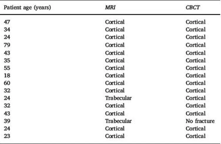
Table 1
Pattern of scaphoid fractures.
Patient age (years) MR1 CBCT
47 Cortical Cortical 34 Cortical Cortical 24 Cortical Cortical 79 Cortical Cortical 43 Cortical Cortical 35 Cortical Cortical 55 Cortical Cortical 18 Cortical Cortical 60 Cortical Cortical 32 Cortical Cortical 24 Trabecular Cortical 32 Cortical Cortical 43 Cortical Cortical 39 Trabecular No fracture 24 Cortical Cortical 23 Cortical Cortical
sensitivity of 1()0°/4 (95% CI: 83% 100%), specificity of 95% (95% CI: 75% 100%), PPV of 96% (95% CI: 78% 100%), and NPV of 100% (95% CI: 83% 100%). CBCT failed to detect two other trabecular fractures in the wrist (1 radial, 1 trapezoid) in addition to the scaphoid fracture, resulting in a sensitivity of 89% (95% CI: 70% 97%), speci ficity of 95% (95% CI: 75% 100%), PPV of 96% (95% CI: 78% 100%), and NPV of 88% (95% CI: 67% 97%) for ail wrist fractures.
3.2. Inter rater
agreement
for CBCTTwo of the cortical fractures were not diagnosed by the junior radiologist: one scaphoid fracture and one trapezium fracture. One examination was interpreted by the senior radiologist as a false posi tive: this was a cortical fracture of the scaphoid diagnosed on CBCT that was not confirmed on MRI. The agreement rate between the two radi ologists was K
=
O. 95 (95% CI: 0.85 1) for ail scaphoid fractures andK
=
0.87 (95% CI: 0.73 1) for all wrist fractures.4. Discussion
CBCT is a reliable, reproducible, low radiation technique for diag nosing occult cortical fractures of the scaphoid, as well as occult cortical fractures in the entire wrist, with 100% sensitivity and 95% specificity. The diagnostic performance of CBCT for investigating ail types of sca phoid fractures was similar to that of a conventional CT scan, which has a sensitivity of 94% and specificity of 97% (6).
Yin et al. (6) recently showed that performing a CT scan im mediately after the injury instead of standard radiographs was a better strategy for diagnosing scaphoid fractures and improved the cost ef fectiveness ratio. When specifically looking at the cost effectiveness ratio, the difference is not due to the cost of the imaging modalities themselves, but to the loss of productivity caused by the needless im mobilization secondary to false positive fractures, and also due to false negatives. In fact, these non diagnosed fractures cause complications that can later lead to costly surgery, hospitalization and time off work. Karl et al. (27) recently showed CT scanning to have a better cost effectiveness ratio when it was carried out immediately after the injury, instead of control radiographs carried out after 14 days of im mobilization. CBCT is now recognized to be at least equal to CT scan for investigating bone, as it provides very good spatial resolution, making multiplanar reconstructions possible.
Although our healthcare facility does not have a specific rating for CBCT when imaging the extremities, the costs of this deviœ are lower than those of a CT scan, which means the exams themselves should be less costly.
The diagnostic performance of CBCT is clearly better than that of
(A)
(B)
Fig. 3. Pisiform cortical fracture ln a 23-year-old-woman. a The Initial coronal oonven-tional ulnar devlation radiograph is negative. b The coronal reformation from CBCT shows a small non-displaced fracture Une ùt the pisiform bone (arrow). c This fracture Une appears hyperintense on coronal Dixon water-only-weighted MRI (3000/40) (arrow).
standard radiographs, which have sensitivity and specificity values of 66% and 97% for scaphoid fractures, 57.8% and 99.5% for wrist frac tures, and 38.7% and 99.5% for carpal fractures, respectively (5). The differences between the wrist and carpus can be explained by better analysis of the long bones on radiographs (radius, ulna, metacarpals)
than of carpal bones, which have a more complex geometry.
MRI is still the golden standard for most wrist fractures as stated by Edlund et al. (30), the sensitivity forfinding scaphoid fractures using MRI is higher compared to CBCT.
Ourfindings are consistent with a recent study by Edlund et al.[28], who found that CBCT was a reliable examination for the diagnosis of scaphoid fracture. However, in their study, not every patient underwent both types of imaging exams (CBCT and MRI), and in the MRI was carried out 15 days after the CBCT.
The main limitation of our study is directly related to the CBCT technique and its acquisition time of approximately 15 s; this makes it more sensitive to movement artefacts than a standard CT scan. This can explain the three instances where the two radiologists had a different diagnosis; the images in question had significant artefacts that hindered their interpretation. Since our CBCT unit is not a dedicated extremity imaging machine, we had to stabilize the arm with straps to limit movement artefacts. Other technical limitations included the fact that CBCT does not image soft tissues and has a smallerfield of view than a CT scan. Nevertheless, our study was designed to look at bone only and
thefield of view used was large enough to encompass the wrist in all
cases.
In our study, one trabecular fracture was classified as a cortical fracture on MRI. This type of false negative has previously been docu
mented [26]and could lead to needless surgery. Trabecular fractures
are much less common but cannot be seen on CBCT or on standard CT scans during the acute phase
Management of trabecular fractures is controversial, some studies have allowed for no treatment while others have recommended im mobilization for to 2 weeks depending on patients level of pain.
A study assessed 41 patients with acute scaphoid trabecular frac ture. These patients were immobilised for 6 weeks and the eight pa tients who remained symptomatic at 3 months all showed improvement of bruising on MRI. They concluded that bone bruising is a benign
injury with predictable recovery and questioned the need for prolonged immobilization[29].
Trabecular fracture are not complicated by nonunion and require no treatment other than immobilization. To our knowledge neither study assessed the natural history of bone bruising in the absence of im mobilization[30].
To conclude, our study found that CBCT was superior to radiographs for diagnosing occult cortical fractures in the carpus. Because of its low radiation dose, we believe that CBCT can be used in current practice as a replacement or supplement to radiographs to better detect these fractures and optimize the cost effectiveness ratio by limiting the number of needless immobilizations.
Conflict of interest
We wish to confirm that there are no known conflicts of interest
associated with this publication and there has been no significant fi nancial support for this work that could have influenced its outcome.
We confirm that the manuscript has been read and approved by all
named authors and that there are no other persons who satisfied the criteria for authorship but are not listed.
We further confirm that the order of authors listed in the manuscript has been approved by all of us.
We confirm that we have given due consideration to the protection of intellectual property associated with this work and that there are no impediments to publication, including the timing of publication, with respect to intellectual property.
In so doing we confirm that we have followed the regulations of our institutions concerning intellectual property.
We further confirm that any aspect of the work covered in this manuscript that has involved human patients has been conducted with the ethical approval of all relevant bodies and that such approvals are acknowledged within the manuscript.
We understand that the Corresponding Author is the sole contact for the Editorial process (including Editorial Manager and direct commu nications with the office). He is responsible for communicating with the
other authors about progress, submissions of revisions andfinal ap
proval of proofs.
We confirm that we have provided a current, correct email address which is accessible by the Corresponding Author and which has been configured to accept email from ([email protected]). References
[1] K. Bohndorf, R.F. Kilcoyne, Traumatic injuries: imaging of peripheral musculoske-letal injuries, Eur. Radiol. 12 (7) (2002) 1605–1616.
[2] A. Fotiadou, et al., Wrist injuries in young adults: the diagnostic impact of CT and MRI, Eur. J. Radiol. 77 (2) (2011) 235–239.
[3] M.J. Breitenseher, et al., Radiographically occult scaphoid fractures: value of MR imaging in detection, Radiology 203 (1) (1997) 245–250.
[4] C. Gabler, et al., Diagnosis of occult scaphoid fractures and other wrist injuries Are repeated clinical examinations and plain radiographs still state of the art? Langenbecks Arch. Surg. 386 (2) (2001) 150–154.
[5] A. Balci, et al., Wrist fractures: sensitivity of radiography, prevalence, and patterns in MDCT, Emerg. Radiol. 22 (3) (2015) 251–256.
[6] Z.G. Yin, J.B. Zhang, K.T. Gong, Cost-Effectiveness of diagnostic strategies for suspected scaphoid fractures, J. Orthop. Trauma 29 (8) (2015) e245–52. [7] S. Tibrewal, et al., Role of MRI in the diagnosis and management of patients with
clinical scaphoid fracture, Int. Orthop. 36 (1) (2012) 107–110.
[8] R.L. Baldassarre, T.H. Hughes, Investigating suspected scaphoid fracture, BMJ 346 (2013) f1370.
[9] A. Newberg, et al., Acute hand and wrist trauma. American College of Radiology. ACR Appropriateness Criteria, Radiology 215 (Suppl) (2000) 375–378. [10] P. Mozzo, et al., A new volumetric CT machine for dental imaging based on the
cone-beam technique: preliminary results, Eur. Radiol. 8 (9) (1998) 1558–1564. [11] H. Lang, et al., A retrospective: semi-quantitative image quality analysis of cone beam computed tomography (CBCT) and MSCT in the diagnosis of distal radius fractures, Eur. Radiol. 26 (12) (2016) 4551–4561.
[12] S. Kim, et al., Comparison of radiation doses between cone beam CT and multi detector CT: TLD measurements, Radiat. Prot. Dosimetry 132 (3) (2008) 339–345. [13] J. Koivisto, et al., Assessment of effective radiation dose of an extremity CBCT:
MSCT and conventional X ray for knee area using MOSFET dosemeters, Radiat.
Patient age (years) Type of bone fracture MRI CBCT
37 Triquetrum Cortical Cortical
40 Distal radius Cortical Cortical
23 Pisiform Cortical Cortical
52 Distal radius Cortical Cortical
30 Trapezium Cortical Cortical
42 Distal radius Cortical Cortical
45 Distal radius Trabecular No fracture
27 Metacarpal Cortical Cortical
32 Distal radius Cortical Cortical
22 Metacarpal Cortical Cortical
31 Trapezoid Trabecular No fracture
Table 3
CBCT diagnostic accuracy for occult scaphoid and wrist fractures. Sensitivity (95% CI) Specificity (95% CI) PPV (95% CI) NPV (95% CI) Scaphoid fractures Cortical fractures 100 (75–100) 97 (83–100) 94 (68–100) 100 (87–100) All fractures 94 (68–100) 97 (83–100) 94 (68–100) 97 (82–100) Wrist fractures Cortical fractures 100 (83–100) 95 (75–100) 96 (78–100) 100 (83–100) All fractures 89 (70–97) 95 (75–100) 96 (78–100) 88 67–97) CI: confidence interval.
PPV: positive predictive value. NPV: negative predictive value. Tableau 2
Prot. Dosimetry 157 (4) (2013) 515–524.
[14] R. Ramdhian-Wihlm, et al., Cone-beam computed tomography arthrography: an innovative modality for the evaluation of wrist ligament and cartilage injuries, Skeletal Radiol. 41 (8) (2012) 963–969.
[15] K. Tsiklakis, et al., Dose reduction in maxillofacial imaging using low dose Cone Beam CT, Eur. J. Radiol. 56 (3) (2005) 413–417.
[16] M. Loubele, et al., Comparison between effective radiation dose of CBCT and MSCT scanners for dentomaxillofacial applications, Eur. J. Radiol. 71 (3) (2009) 461–468. [17] A.J. Huang, et al., Using cone-beam CT as a low-dose 3D imaging technique for the
extremities: initial experience in 50 subjects, Skeletal Radiol. 44 (6) (2015) 797–809.
[18] N. Faccioli, et al., Finger fractures imaging: accuracy of cone-beam computed to-mography and multislice computed toto-mography, Skeletal Radiol. 39 (11) (2010) 1087–1095.
[19] S.K. Koskinen, et al., CT arthrography of the wrist using a novel: mobile, dedicated extremity cone-beam CT (CBCT), Skeletal Radiol. 42 (5) (2013) 649–657. [20] B.S. Pugmire, et al., Initial clinical experience with extremity cone-Beam CT of the
foot and ankle in pediatric patients, AJR Am. J. Roentgenol. 206 (2) (2016) 431–435.
[21] G.K. Thawait, et al., Extremity cone-beam CT for evaluation of medial tibiofemoral osteoarthritis: initial experience in imaging of the weight-bearing and non-weight-bearing knee, Eur. J. Radiol. 84 (12) (2015) 2564–2570.
[22] S. Demehri, et al., Assessment of image quality in soft tissue and bone visualization tasks for a dedicated extremity cone-beam CT system, Eur. Radiol. 25 (6) (2015) 1742–1751.
[23] D. Biswas, et al., Radiation exposure from musculoskeletal computerized tomo-graphic scans, J. Bone Joint Surg. 91 (8) (2009) 1882–1889.
[24] W.H. Mallee, et al., Computed tomography for suspected scaphoid fractures: com-parison of reformations in the plane of the wrist versus the long axis of the sca-phoid, Hand (N Y) 9 (1) (2014) 117–121.
[25] J.C. Hunter, et al., MR imaging of clinically suspected scaphoid fractures, AJR Am. J. Roentgenol. 168 (5) (1997) 1287–1293.
[26] M. Memarsadeghi, et al., Occult scaphoid fractures: comparison of multidetector CT and MR imaging?initial experience, Radiology 240 (1) (2006) 169–176. [27] J.W. Karl, E. Swart, R.J. Strauch, Diagnosis of occult scaphoid fractures: a
cost-Effectiveness analysis, J. Bone Joint Surg. Am. 97 (22) (2015) 1860–1868. [28] R. Edlund, et al., Cone-Beam CT in diagnosis of scaphoid fractures, Skeletal Radiol.
(2015).
[29] N. La Hei, et al., Scaphoid bone bruising–probably not the precursor of asympto-matic non-union of the scaphoid, J. Hand Surg. Eur. Vol. 32 (3) (2007) 337–340. [30] R. Ganeshalingam, K. Eng, R.S. Page, Magnetic resonance imaging in the acute
management of suspected scaphoid fractures: a review of the literature and as-sessment of treatment algorithm, OA Musculoskeletal Med. 18 (2013) p. 1(2):19.