R E S E A R C H
Open Access
Rat pulmonary responses to inhaled nano-TiO
2
:
effect of primary particle size and agglomeration
state
Alexandra Noël
1, Michel Charbonneau
2, Yves Cloutier
3, Robert Tardif
1and Ginette Truchon
3*Abstract
Background: The exact role of primary nanoparticle (NP) size and their degree of agglomeration in aerosols on the determination of pulmonary effects is still poorly understood. Smaller NP are thought to have greater biological reactivity, but their level of agglomeration in an aerosol may also have an impact on pulmonary response. The aim of this study was to investigate the role of primary NP size and the agglomeration state in aerosols, using well-characterized TiO2NP, on their relative pulmonary toxicity, through inflammatory, cytotoxic and oxidative stress effects in Fisher 344 male rats.
Methods: Three different sizes of TiO2NP, i.e., 5, 10–30 or 50 nm, were inhaled as small (SA) (< 100 nm) or large agglomerates (LA) (> 100 nm) at 20 mg/m3for 6 hours.
Results: Compared to the controls, bronchoalveolar lavage fluids (BALF) showed that LA aerosols induced an acute inflammatory response, characterized by a significant increase in the number of neutrophils, while SA aerosols produced significant oxidative stress damages and cytotoxicity. Data also demonstrate that for an agglomeration state smaller than 100 nm, the 5 nm particles caused a significant increase in cytotoxic effects compared to
controls (assessed by an increase in LDH activity), while oxidative damage measured by 8-isoprostane concentration was less when compared to 10–30 and 50 nm particles. In both SA and LA aerosols, the 10–30 nm TiO2NP size induced the most pronounced pro-inflammatory effects compared to controls.
Conclusions: Overall, this study showed that initial NP size and agglomeration state are key determinants of nano-TiO2 lung inflammatory reaction, cytotoxic and oxidative stress induced effects.
Keywords: TiO2nanoparticles, Inhalation, Agglomeration state, Primary particle size, Inflammation, Cytotoxicity, Oxidative stress
Background
In recent decades, nanoparticles (NP) (< 100 nm) have attracted the increased attention of the industrial sector due to their unique physico-chemical properties and nu-merous applications. Indeed, compared to their larger-sized counterparts, NP have improved and distinctive surface characteristics which are considered as the building blocks of nanotechnology. This fast developing field is expected to generate by 2014 more than 10 million jobs related to this technology [1]. Thus, an increasing number of workers are
going to be handling NP during the production and dis-posal of several consumer products [2]. Reliable testing strategies to investigate possible health effects caused by NP exposure are therefore urgently needed. Risk assess-ment is based on exposure expressed in terms of dosimetry and toxicological data [3]. Hence, research studies should examine the characteristics of NP or aerosols that deter-mine their ability to cause deleterious effects. Currently, the best metric, such as mass, surface area, number or size dis-tribution, to measure NP in order to evaluate risk and pre-vent the development of occupational diseases is still a matter of debate [3].
Several toxicological studies have addressed the micro versus the nano size effect. This has indicated at equivalent * Correspondence:[email protected]
3
Institut de recherche Robert-Sauvé en santé et en sécurité du travail (IRSST), 505 Boul. De Maisonneuve Ouest, Montréal, Québec H3A 3C2, Canada Full list of author information is available at the end of the article
© 2013 Noël et al.; licensee BioMed Central Ltd. This is an open access article distributed under the terms of the Creative Commons Attribution License (http://creativecommons.org/licenses/by/2.0), which permits unrestricted use, distribution, and reproduction in any medium, provided the original work is properly cited.
Noël et al. Particle and Fibre Toxicology 2013, 10:48 http://www.particleandfibretoxicology.com/content/10/1/48
mass concentration that agglomerated NP produce greater pulmonary inflammation responses than micron size parti-cles [4-8]. However, few studies have investigated in vivo the size-dependent effects of NP [9-11]. As NP (< 100 nm) size decreases, it is expected that the percentage of atoms and active sites at the surface, as well as the structural im-perfections, increase significantly [12,13]. In addition, it has previously been shown that metal oxide particles less than 30 nm in size, had enhanced interfacial reactivity [14,15]. Correspondingly, the biological reactivity of smaller NP is expected to be higher than that of larger NP [16]. More-over, it is at approximately 20 nm that the highest relative deposition efficiency of NP in the alveolar region occurs [17,18]. This suggests that NP of different initial sizes could produce different biological responses [16,19].
TiO2is a substance that is manufactured at a large scale, either as crude, fine or ultrafine powder. Given its import-ant industrial production, several countries use TiO2as a reference nanomaterial in research and in the assessment of workplace exposures [20-23]. However, to this day there is still limited consistent toxicological data and similarly limited epidemiological studies related to occupational ex-posures to this chemical [24-29]. This puts the emphasis on the need for more research on TiO2NP.
Our research group and other studies have previously reported that agglomeration is a process occurring in nano-TiO2 aerosol production [9,19,30-34]. Using different methods to generate NP aerosols in laboratory studies, we have shown that a given TiO2NP can agglomerate in differ-ent sizes and structures [33]. This observation is relevant and valuable for toxicological assessment since there is in-creasing evidence that aerosol characteristics can modulate NP interaction with biological systems [9,18,35-38]. Indeed, agglomeration of NP has multiple influences on dose char-acteristics and on their pulmonary toxicity and kinetics. It can affect: 1- the number concentration in the aerosol; 2-the deposition site in 2-the respiratory tract; 3- 2-the possibility of detection and subsequent phagocytosis by alveolar mac-rophages; 4- the fate in lung tissue and translocation to other extra-pulmonary compartments; and 5- the size, dens-ity, shape and structure of the resulting particle, being either loose or compact. Furthermore, once deposited in the re-spiratory tract, NP can interact with biological material (e.g., pulmonary surfactant, macromolecules and proteins), which can also have an impact by increasing, slowing or preventing the degree of agglomeration in the physiological environ-ment [36,39,40]. Taken together, it is highly relevant to characterize and consider the agglomeration state of NP, particularly metal oxides, in nanotoxicological studies.
We have previously reported that rats exposed by in-halation to small agglomerates (< 100 nm) of 5 nm TiO2 showed greater cytotoxic and oxidative stress responses than rats exposed to larger agglomerates (> 100 nm) of the same NP, which induced a slight inflammatory
reaction [41]. This suggested that biological responses to TiO2might depend on the dimension and concentration of the NP agglomerates. These results are in line with the current hypothetical mechanisms of NP pulmonary tox-icity, based on the inflammation and oxidative stress paradigm [17,42-44].
In inhalation studies, establishing the effect of primary NP size involves considering the size of the agglomerates, and only few studies have addressed this issue. In the early 90’s, several influential studies [4,5,45,46] were conducted and showed differences in toxicologically relevant responses and translocation of NP versus submicrometer particles ad-ministered as submicrometer-sized aerosols. A recent study conducted by Balasubramanian et al. [47] showed in rats that primary NP size of inhaled gold NP influenced the whole-body biodistribution. Gold NP of 7 or 20 nm pri-mary sizes were generated into aerosols of similar number concentration and size distribution. This showed that the agglomerates of the 7 nm NP were distributed in more or-gans than the 20 nm NP and that macrophage clearance was more effective for agglomerates of larger primary size. In a study by Pauluhn [11], rats exposed to aluminum oxide NP aerosols showed a clearance half-time which increase with decreased primary NP size (10 nm - 840 nm in aero-sols and 40 nm– 660 nm in aerosols), while the inflamma-tory response appeared to be determined by the size of the agglomerates. In a study conducted by Grassian et al. [9], mice exposed to 7 mg/m3of 5 nm TiO2NP (120 nm in aerosol) or 21 nm NP (139 nm in aerosol) showed that the latter was slightly, but significantly more toxic than the smaller NP. These authors suggested that the difference in the structure of the agglomerates, which was compact for the 5 nm TiO2versus loose for the 21 nm particles, could be partly responsible for the results. Collectively, these in-halation nanotoxicology studies suggest that smaller ini-tial NP size could distribute in more extra-pulmonary organs and might not always induce enhanced lung in-flammatory reactions when compared to larger nano-metric counterparts.
The exact role of primary NP size and their degree of agglomeration in aerosols in the determination of pul-monary effects is still poorly understood. Indeed, smaller NP are thought to have greater biological reactivity, but the level to which these NP agglomerate in aerosols may also have an impact on the pulmonary response. More-over, NP of different primary sizes could possibly bundle in agglomerates of various dimensions, shapes and struc-tures in aerosols, which can also influence their pulmon-ary toxicity and kinetics. Thus, the primpulmon-ary particle size and agglomeration state of NP can be of significance in toxicity and partly responsible for the distinct effects in-duced in lungs. To our knowledge, no previous study has addressed both factors simultaneously, i.e., the size-dependent effect of NP while also comparing the influence
of the agglomeration state. Elucidating the impact of these factors on the mechanism of toxicity will provide import-ant knowledge for NP risk assessment.
The aim of this study was to investigate the role of pri-mary NP size and agglomeration state in aerosols using well-characterized TiO2 NP and to compare their relative pulmonary toxicity, through inflammatory, cytotoxic and oxidative stress effects in rats. For this purpose, we used three different sizes of TiO2NP, i.e., 5, 10–30 or 50 nm in-haled as small (< 100 nm) or large agglomerates (> 100 nm) at a concentration of 20 mg/m3.
Results
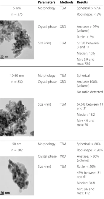
Characterization of initial NP powders
In this paper, we define nanoparticle (NP) as particle whose nominal diameter (geometric, mobility, aerodynamic, projected surface or other) is less than 100 nm [48]. The characterization of the three nano-TiO2 powders (as re-ceived by the manufacturers) which were used to produce the aerosols along with transmission electron microscopy (TEM) images, are presented in Table 1. X-ray spectromet-ric analysis (EDS) analysis showed that all powders were made of TiO2. The size distribution of the NP present in the powders was established by measuring the diameter of over 300 particles by TEM. These results showed that the median diameter of the NP present in the powder labeled by the manufacturer as 5 nm was 10.6 nm, while it was 18.2 nm and 34.8 nm for the 10–30 nm and 50 nm pow-ders, respectively. The analysis also showed that the size distributions of the three nano-powders slightly overlapped (data not shown). In addition, TEM analysis showed that, in the case of the 5 and 50 nm powders, particles had a spherical or rod-shaped morphology, while the 10–30 nm powder, showed only spherical particles. Analysis by X-ray diffraction (XRD) revealed that the 5 and 10–30 nm pow-ders were predominantly in the anatase form (> 97%), while the 50 nm powder showed a slightly higher presence of the rutile form (< 20%).
Measurement and characterization of NP aerosols
Throughout this article, we refer to small agglomerate (SA) aerosols as mainly composed of agglomerates with a size smaller than 100 nm and large agglomerate (LA) aero-sols as essentially composed of agglomerates larger than 100 nm. Aerosol’s characterization data carried out with the ELPI, DustTrak and gravimetric measurements are presented in Table 2. Cumulative size distributions based on number concentration measured with the ELPI are shown in Figure 1. Three values were used to estimate the number size distributions, namely the first quartile (D25), the midpoint (D50) or the median aerodynamic diameter based on the number concentration (NMAD), and the third quartile (D75). These results show that for each TiO2 NP, the size distributions obtained for SA and LA aerosols
are different (Table 2 and Figure 1). The targeted exposure concentration was 20 mg/m3for all exposed groups and the concentrations gravimetrically measured were between 18.8 and 22.0 mg/m3. As measured with the ELPI, the total particle number was higher in the aerosols composed of SA compared to the aerosols composed of LA.
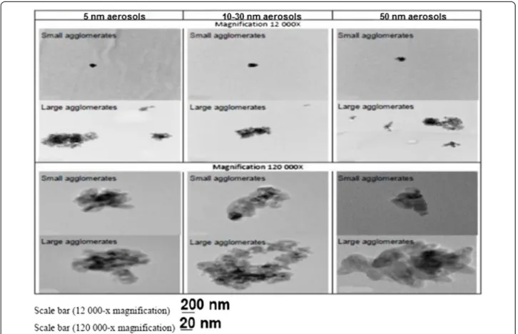
Figure 2 shows representative TEM images of particles from the aerosols composed of SA or LA. For the same initial particle size, TEM images (12 000-×) allowed the observation of qualitative differences in the size, shape
Table 1 TiO2NP powder characterization assessed by
transmission electron microscopy (TEM) and X-ray diffraction (XRD)
Parameters Methods Results 5 nm Morphology TEM Spherical: > 97%
n = 375 Rod-shape: < 3%
Crystal phase XRD Anatase: > 97% (volume) Rutile: < 3% Size (nm) TEM 53.3% between
3 and 11 Median: 10.6 Min: 3.9 and max: 75.6
10-30 nm Morphology TEM Spherical n = 330 Crystal phase XRD Anatase: 100%
(volume) No rutile detected
Size (nm) TEM 67.6% between 11 and 31
Median: 18.2 Min: 4.9 and max: 70
50 nm Morphology TEM Spherical: > 80%
n = 302 Rod-shape: < 20%
Crystal phase XRD Anatase: > 80% (volume) Size (nm) TEM Rutile: < 20%
47% between 31 and 61 Median: 34.8 Min: 8.6 and max: 112
Noël et al. Particle and Fibre Toxicology 2013, 10:48 Page 3 of 18 http://www.particleandfibretoxicology.com/content/10/1/48
and structure of the agglomerates present in the aerosols (Figure 2). Indeed, SA aerosols were mostly composed of small compact agglomerates, while LA aerosols had lar-ger agglomerates with void space. Higher magnification (120 000-×) allowed the observation of irregularly shaped small agglomerates (Figure 2).
Pulmonary deposition
Using the polydisperse diameter interquartile ranges presented in Table 2, the estimated respiratory tract de-position fraction of NP agglomerates was computed with the MPPD model for LA and SA aerosols. These results
are presented in Figure 3 and indicate that pulmonary deposition is different for the two types of aerosols.
Rat pulmonary response
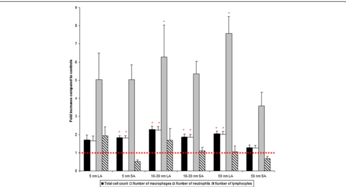
A transient increase in leukocytes from BALF is a natural lung defense mechanism following deposition of inhaled particles. In this manner, BALF cytology analysis from rats exposed to SA aerosols showed increases in total cell count, number of macrophages and neutrophils compared to the control group. Increases were slightly but statistically sig-nificant (p < 0.05) for total cell count and number of mac-rophages in the 5 and 10–30 nm groups compared to the
Table 2 Measurements and characterization of the NP aerosols
Experimental groups
Parameters Control 5 nm SAa 5 nm LAb 10-30 nm SA 10-30 nm LA 50 nm SA 50 nm LA
Average mass concentrationc(mg/m3) 0.05 18.77 19.30 22.04 21.99 21.38 21.94
Min and maxd(mg/m3) 0.03 and
0.27 17.12 and 22.47 17.43 and 22.18 18.83 and 27.99 20.48 and 25.42 19.66 and 25.34 20.45 and 30.09 Total particle numbere(/cm3) - 3 159 758 308 098 1 808 939 374 225 1 320 239 280 379 D25 e,f (nm) - 29 156 28 128 35 135 NMAD*or D50 e,f (nm) - 48 369 65 255 85 321 GSDg - 3.1 2.2 3.3 2.6 3.2 2.6 D75 e,f (nm) - 124 575 183 686 305 783 Fraction of NP agglomerates <100 nme(%) - 71 16 63 18 54 19 a
Aerosol composed of small agglomerates.
b
Aerosol composed of large agglomerates.
c
Average mass concentration determined by weight measurements.
d
Min and max concentration determined by a DustTrak.
e
Measurements made with the ELPI.
f
Aerodynamic diameters for which 25% (D25), 50% (D50or NMAD) or 75% (D75) of the particles in the aerosol are smaller than this value, based on number
concentration. D75– D25is the interquartile range and represents the size distribution where we find 50% of the particles. g
GSD: geometric standard deviation.
*
NMAD: number median aerodynamic diameter.
Figure 1 Cumulative distributions based on number concentration of the NP aerosols. NP aerosols cumulative size distributions based on number concentration measured with the ELPI. For each aerosol 5 thirty-minute samples were collected every hour of the experiment.
controls (Table 3, Figure 4). Rats exposed to LA aerosols also showed increases in total cell count, number of macro-phages and neutrophils compared to the control group. Re-sults were significant (p < 0.05) for total cell count, number of macrophages and neutrophils in the 10–30 and 50 nm groups compared to the controls (Table 3, Figure 4).
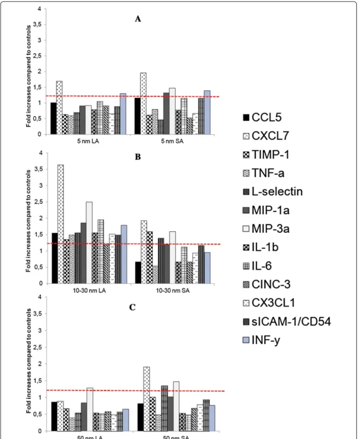
The profiles of inflammatory cytokines are shown in Figure 5. Thirteen of the 29 cytokines analyzed in the assay showed≥ 1.2-fold increases compared to the control group (CCL5, CXCL7, TIMP-1, TNF-α, L-selectin, 1α, MIP-3α, IL-1β, IL-6, CINC-3, CXCL1, sICAM, INF-γ). Notice-ably, the 10–30 nm LA aerosol showed a profile where all of the 13 cytokines were increased compared to the controls.
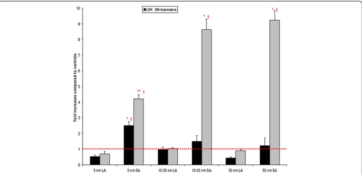
8-isoprostane, a marker of oxidative stress, showed a statis-tically significant increase for all SA aerosols compared to the controls or respective LA aerosols (Figure 6). There were also significant differences between the three SA aerosols. For the 5 nm SA aerosol the 8-isoprostane concentration was significantly (p < 0.05) lower compared to the 10–30 and 50 nm SA aerosols (Figure 6). For LDH activity, only the results for the 5 nm SA aerosol were statistically (p < 0.05) different from the controls (Figure 6). There was also a sig-nificant (p < 0.05) difference between the 5 nm SA and all LA aerosols (Figure 6).
Representative cell morphology of the BALF cytoprepa-rations from the control and exposed rats are shown in Figure 7. The control group showed typical BAL cells. For all nano-TiO2 exposed groups, the majority of the cells (macrophages, neutrophils and lymphocytes) were also typical, although giant cells (macrophages) and fragmented nucleus macrophages were observed (Figure 7).
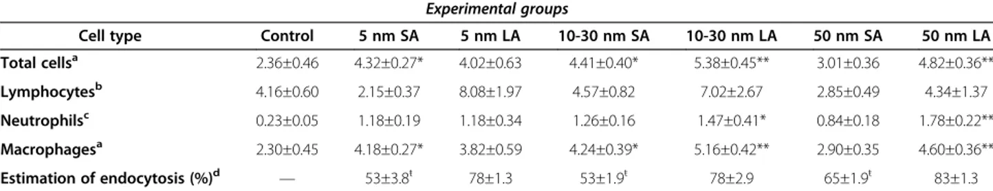
Estimation of NP endocytosis by macrophages
Figure 7 shows representative images of BAL macro-phages with (arrows) or without phagocytized NP ag-glomerates. All groups exposed to the LA aerosols had 79 ± 2% (standard error on the mean, SEM) of macro-phages that contained nano-TiO2agglomerates, whereas for the SA aerosols, it was 57 ± 3% (SEM) (Table 3). For each primary NP size, a significant difference was ob-served for the percentage of particle-laden macrophages between the LA and SA aerosols (Table 3).
Lung histopathology
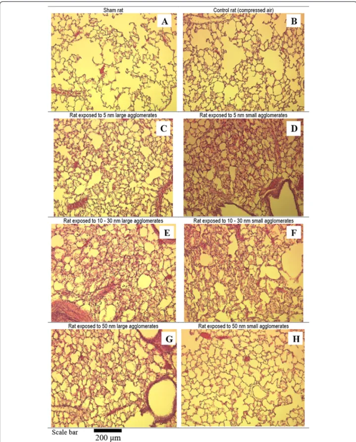
Figures 8 and 9 show images of haematoxylin and eosin stained lungs. The control group did not show any signs of inflammation. Noteworthy, the cellular influx ob-served were partial since these lungs were also used for
Figure 2 NP aerosol agglomerate structure observed by transmission electron microscopy. Air samples were collected on pre-carbon coated Formvar copper grids glued onto 25-mm polycarbonate filters. Characterization (shape, agglomeration degree and structure) of the nano-aerosols was performed by TEM.
Noël et al. Particle and Fibre Toxicology 2013, 10:48 Page 5 of 18 http://www.particleandfibretoxicology.com/content/10/1/48
BAL. Notwithstanding, morphological assessments of lung tissue responses to nano-TiO2were different in in-tensity compared to the controls, except for the 50 nm SA group (Figure 8). The lungs of rats exposed to 5 and 10–30 nm SA aerosols as well as rats exposed to 10–30 and 50 nm LA aerosols showed more leukocyte infiltra-tion compared to the control group (Figure 8). This is consistent with the cytological analysis (Table 3, Figure 4) and is considered to represent a normal macrophage clearance response. Figure 9, which is representative of all groups, shows TiO2 NP-laden macrophages in the 5 nm SA aerosol group.
Discussion
Since TiO2NP tends to agglomerate in aerosols [9,32-34], establishing the effect of these NP involves considering the primary particle size and that of the agglomerates [9]. The agglomeration state of NP influences the site of par-ticle deposition in the respiratory tract and affects lung clearance mechanisms, including endocytosis [9,19,45-53]. In general, the size distribution of a NP aerosol is com-posed of a less (< 100 nm) and highly (> 100 nm) agglom-erated fraction, with the percentage varying from one aerosol to another. The less agglomerated fraction, for which the size of the agglomerates is closer to the size of the primary NP, could possibly induce effects related to their interaction with lung tissue and epithelial cells at the site of pulmonary deposition. These small particles are also more readily available for translocation to the lymph nodes or bloodstream [17]. Larger agglomerates (> 100 nm) are thought to be more easily detected and removed by the lung macrophages [41,54-57].
In our study, the cellular pulmonary response observed for all exposed groups denoted by the increases in leuko-cytes from BALF compared to the controls (Table 3, Figure 4) could be considered as a normal immediate re-sponse to particle aggression [58]. The increase in the number of macrophages and neutrophils is thought to con-tribute to particle removal. Indeed, previous studies have shown that following inhalation of nano-TiO2this type of response is temporary and resolves rapidly [9,19,30,32]. Hence, after an acute exposure, this could be a defense mechanism [59].
NP aerosol characterization
For the LA aerosols, we observed that the 5 nm TiO2 pro-duced larger agglomerates than the 10–30 and 50 nm parti-cles (Table 2). This result could be partly explained by the fact that as particle size decreases, the attractive force per unit mass increases, which favors agglomeration [60]. In-deed, small particles that coagulate into agglomerates larger than superior-sized counterparts is a common finding pre-viously described in numerous inhalation studies, including ones using nano-TiO2[11,30,38,45,61].
Figure 3 Computed fractional deposition of NP agglomerates in the rat respiratory tract. Estimated deposition fraction for the aerosols at 20 mg/m3composed of SA or LA modeled for multiple diameters. TB = tracheobronchial; P = pulmonary. Functional residual capacity (FRC) volume 4 ml; head volume: 0.42 ml; nasal breathing route; tidal volume 2.1 ml; breathing frequency 110/min and inspiratory fraction 0.1.
Table 3 BALF cytology and estimation of NP endocytosis by macrophages following inhalation exposure
Experimental groups
Cell type Control 5 nm SA 5 nm LA 10-30 nm SA 10-30 nm LA 50 nm SA 50 nm LA
Total cellsa 2.36±0.46 4.32±0.27* 4.02±0.63 4.41±0.40* 5.38±0.45** 3.01±0.36 4.82±0.36** Lymphocytesb 4.16±0.60 2.15±0.37 8.08±1.97 4.57±0.82 7.02±2.67 2.85±0.49 4.34±1.37 Neutrophilsc 0.23±0.05 1.18±0.19 1.18±0.34 1.26±0.16 1.47±0.41* 0.84±0.18 1.78±0.22** Macrophagesa 2.30±0.45 4.18±0.27* 3.82±0.59 4.24±0.39* 5.16±0.42** 2.90±0.35 4.60±0.36** Estimation of endocytosis (%)d — 53±3.8ŧ 78±1.3 53±1.9ŧ 78±2.9 65±1.9ŧ 83±1.3 a number of cells x 106
± standard error on the mean (SEM);b
number of cells x 104
± SEM;c
number of cells x 105
± SEM;d
estimation of the extend of NP phagocytosis by macrophages. Mean values (n = 6 rats per group) significantly different from controls (ANOVA followed by a Tukey’s test) *p<0.05, **p<0.01,
LA aerosols
As shown in Figure 3, the estimated pulmonary deposition was different for the LA and SA aerosols. Also, given their different agglomeration states (Table 2), it can be assumed, as described by Oberdörster et al. [17] as well as Geiser and Kreyling [62], that the penetration of these aerosols into the various regions of the respiratory tract is different. Considering their size distribution characteristics (D25 = 128 nm to D75 = 783 nm; Table 2), particles of the LA aerosols could be more easily detected by immune system cells, including alveolar macrophages (Table 3, Figures 7 and 8). Indeed, the estimation of NP endocytosis showed for all LA aerosols that 79 ± 2% of macrophages contains nano-TiO2agglomerates (Table 3). In our study, activation of macrophages following phagocytosis of large NP ag-glomerates (Figure 7) is supported by a slight but signifi-cant increase in the total cell count, and number of macrophages and neutrophils compared to controls for two out of the three LA aerosols (10–30 and 50 nm) (Table 3 and Figure 4). However, the 50 nm LA aerosol did not increase the level of inflammatory cytokines, whereas interferon γ (INF-γ), chemokine C-X-C motif ligand 7 (CXCL7), interleukine-6 (IL-6), macrophage inflammatory protein 1α (MIP-1α) and MIP-3α were increased for the 10–30 nm LA aerosol when compared to the controls. These cytokines are produced by activated macrophages and act in host defense by promoting phagocytosis, resulting in chemotaxis, inflammatory cell recruitment and
activation at the site of injury [63-65]. In particular, MIP-1α activates granulocytes (neutrophils, eosinophils, basophils) which can lead to acute neutrophilic inflammation [63-65]. The detection and phagocytosis of these agglomerates by macrophages can prevent their interaction with lung cells and tissue [41,45,66,67]. This is also supported by our results, where no cytotoxicity or oxidative stress effects, evaluated through LDH activity and 8-isoprostane concen-tration, were observed for these LA aerosols (Figure 6). LDH is a cytoplasmic enzyme that is released by dead cells and is therefore a suitable marker of cell cytotoxicity, while 8-isoprostane is a biomarker of lipid peroxidation and thus an indicator of oxidative stress effects [68,69].
An increase in the number of neutrophils supports the presence of an inflammatory reaction [12,32,39,65]. Thus, a mild significant inflammatory (p < 0.05) response was ob-served following the 10–30 and 50 nm LA aerosols expo-sures. These results are consistent with the common finding of various nanotoxicological studies on the increases in the number of neutrophils following agglomerated nano-TiO2 exposures [9,19,32,41,45,70-72]. Previous studies have also observed that inhalation of agglomerated nano-TiO2, in mice and rats, caused slight inflammatory responses and long-term pulmonary inflammation [9,19,30,32,72-75].
SA aerosols
For the 5 and 10–30 nm SA aerosols, the cytological ana-lysis showed a statistically significant increase in total cell
Figure 4 BALF cytology analyzed by the cytospin method. Data were expressed as fold increases of exposed groups compared to controls. Bars represent the mean value and the standard error of the mean obtained for 6 rats in each exposure group. Statistical procedures: ANOVA followed by a Tukey’s test. *Mean value is statistically different from control level p < 0.05.
Noël et al. Particle and Fibre Toxicology 2013, 10:48 Page 7 of 18 http://www.particleandfibretoxicology.com/content/10/1/48
Figure 5 Relative levels of BALF pro-inflammatory cytokines. BALF pro-inflammatory cytokines were expressed as fold increases of exposed groups compared to controls. Samples from all rats of the same exposure group were pooled together. The ratio for each cytokine was calculated as described in materials and methods. Results with ratios≥1.2 were considered to represent a slight inflammation. (A) Aerosols with a primary NP size of 5 nm. (B) Aerosols with a primary NP size of 10–30 nm. (C) Aerosols with a primary particle size of 50 nm.
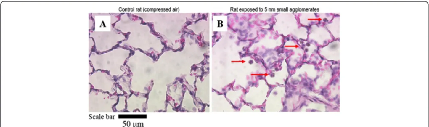
Figure 7 Representative cell morphology from BALF cytopreparations of rats. Optical microscopy (magnification 400-x) of cells cytopreparation collected in BALF of sham, controls (exposed to compressed air) and TiO2NP exposed rats. For all TiO2exposed groups, the
images show giant foamy macrophages and the distribution of macrophages containing phagocytized TiO2NP (arrows). Scale bar = 50μm.
Figure 6 BALF cytotoxicity (LDH) and oxidative stress (8-isoprostane) markers. Data were expressed as fold increases of exposed groups compared to controls. Bars represent the mean value and the standard error on the mean obtained for 6 rats in each exposure group. LDH assay for each rat was done in triplicate and 8-isoprostane was in duplicate. Statistical procedures: ANOVA followed by a Tukey’s test. * Mean value is statistically different from control level p < 0.05; ** Mean value is statistically different from control level and 10–30; 50 nm SA aerosols p < 0.05; ŧ Mean value is statistically different from all LA aerosol levels p < 0.05.
Noël et al. Particle and Fibre Toxicology 2013, 10:48 Page 9 of 18 http://www.particleandfibretoxicology.com/content/10/1/48
Figure 8 Optical microscopy images (100-x) of lung tissue sections. Morphological assessments of lung tissue stained with haematoxylin and eosin of sham (A), control (exposed to compressed air) and TiO2NP exposed rats by means of inhalation for 6 hours. Responses to nano-TiO2(C to G)
were different in intensity compared to the controls (B), except for the 50 nm SA group (H). The lungs of rats exposed to 5 and 10–30 nm SA aerosols (D and F) as well as rats exposed to 10–30 and 50 nm LA aerosols (E and G) showed more leukocyte infiltration compared to the control group (B).
count and number of macrophages (Table 3, Figure 4). Qualitatively, these results are consistent with the histo-pathological findings (Figure 8). The estimation of NP endocytosis showed that all SA aerosols had 57 ± 3% of macrophages containing nano-TiO2agglomerates (Table 3). Despite the NMAD values that were below 100 nm for these aerosols, the D75values ranged from 124 to 305 nm. Thus, the agglomerated (> 100 nm) fraction, which was en-countered for 29 to 46% of these aerosols could explain the NP endocytosis observed. Nonetheless, for each primary NP size, a significant difference was observed for the per-centage of particle-laden macrophages between LA and SA aerosols. Also, increases in the relative levels of CXCL7 and MIP-3α were observed in all SA aerosols, while it was also the case for the tissue inhibitor matrix proteinase 1 (TIMP-1), a glycoprotein involved in the degradation of the extra-cellular matrix, for the 10–30 nm SA aerosol. Considering the size distributions of the SA aerosols (D25= 28 nm to D75= 305 nm; Table 2) and as shown with the estimation of NP endocytosis, it can be assumed that these aerosols were not as well detected and phagocytized by alveolar macrophages as the LA aerosols. Thus, increased NP inter-action with biological materials (lung cells and tissue) may have occurred compared to the LA aerosols and could be expressed as cytotoxicity and oxidative stress effects. In our study, statistically significant increases were observed in LDH activity and 8-isoprostane concentration for the 5 nm SA aerosol compared to the controls and its respective LA aerosol, while only 8-isoprostane was significantly increased for the 10–30 and 50 nm SA aerosols (Figure 6). There-fore, overall, the results for the SA aerosols indicate clear trends of NP interaction with lung cells and tissue through oxidative stress damage and suggestive slight cytotoxic effects (Figure 6).
Effect of the agglomeration state
Overall, these results confirm, at a higher mass concentra-tion, what we had previously shown at 7 mg/m3 [41],
namely that an acute inhalation of nano-TiO2 with two distinct agglomeration states, smaller or larger than 100 nm, induced different mild pulmonary effects. An acute inflammatory response measured by an increase in the number of neutrophils was induced by exposure to two out of three LA (> 100 nm) aerosols, while significant oxi-dative stress effects were observed after exposures to all of the SA (< 100 nm) aerosols. With respect to hazard identi-fication, our results indicate that even though LA aerosols induced an acute inflammatory response, which is revers-ible according to the literature [9,19,32,45,70,72], it cannot be concluded that these aerosols induce toxicity through the same mechanisms as SA aerosols, which showed clear oxidative stress damage in BALF.
Effect of primary nanoparticle size
For the three initial TiO2NP sizes, we observed only one significant difference within the smaller than 100 nm ag-glomeration state aerosols. The significant difference was observed between the 5 nm and the two other SA aerosols for the 8-isoprostane concentration (Figure 6). This sug-gests that the larger 10–30 and 50 nm particles induced more lipid peroxidation and oxidative stress damage than the smaller 5 nm particles. Numerous inhalation studies have previously demonstrated that translocation of various type of NP, including TiO2, to extrapulmonary compart-ments occurred for small agglomerated NP (average diam-eter < 80 nm) in aerosols [37,76-83]. Collectively, these studies indicate that the penetration efficiency of NP through cellular membranes increases as the NP size de-creases and that the translocation time inde-creases with par-ticle size [84-86]. Thus, in our study, the smaller size of the 5 nm particles (D50 = 48 nm in aerosol) would facilitate their possible and rapid translocation from the lung epithe-lial cells, thereby reducing their availability and time to cause cellular membrane lipid peroxidation at the NP - cell interface. The larger size of the 10–30 and 50 nm particles (D50= 65 and 85 nm in aerosols, respectively) may on the
Figure 9 Optical microscopy images (400-x) of lung tissue sections. (A) Lung of control rat exposed to compressed air. (B) Lung of a rat exposed to small agglomerates of 5 nm NP. This figure demonstrates TiO2NP engulfed by alveolar macrophages (arrows).
Noël et al. Particle and Fibre Toxicology 2013, 10:48 Page 11 of 18 http://www.particleandfibretoxicology.com/content/10/1/48
other hand promote translocation to a lesser extent and over a longer period of time, resulting in increased inter-action of NP with the cellular membranes, which generates oxidative stress through membranolytic effects (Figure 6).
We observed that the LDH activity for the SA aerosols compared to controls was only significant for the 5 nm par-ticles (Figure 6). This same aerosol also showed a lower in-crease in 8-isoprostane concentration than the other two SA aerosols (Figure 6). This could possibly be explained by the higher cytotoxicity response observed in these animals. Indeed, LDH is an enzyme that leaks from damaged cells as a sign of membrane integrity lost [87] and as previously mentioned, is a suitable marker of cell death, particularly by necrosis. It could also be considered as evidence of NP penetration into cells [88]. NP penetration into cells leading to interactions with intracellular components is size-dependent [85,86]. Thus, the lower cytotoxicity observed for the larger NP (10–30 and 50 nm) could possibly be due to their less efficient penetration into cells. Interestingly, our data suggest that membrane damage by lipid peroxida-tion at the NP– cell membrane interface might not be the primary cause of cytotoxicity. Thus, the size-dependent ef-fect of nano-TiO2observed in our study in the smaller than 100 nm agglomeration state is supported by the literature. Also, these results are in line with Paulhun’s study [11] that reported that the clearance kinetics of NP was more dependent on their initial particle size.
50 nm TiO2NP
In addition, for the SA aerosols, there may be a few rea-sons that explain the lack of cellular and histopatho-logical changes with the 50 nm group compared to the 5 and 10–30 nm groups (Figures 4 and 8). First, consider-ing the NP powder characterization, approximately 20% of the crystal phase of the 50 nm powder was in the rutile form, while it was 3% or less for the two other powders (Table 1). It has already been reported that the rutile form of TiO2 NP is less toxic than the anatase crystal phase [38,40,89-92]. Thus, in the less than 100 nm agglomeration state, the presence of the rutile phase in the 50 nm powder may be partly responsible for the lower cellular toxicity ob-served. At equal mass concentration, as the NP size de-creases, the surface area per mass unit increases and leads to high surface to volume ratios, giving smaller NP en-hanced surface reactivity [12]. Studies have also shown that the surface adsorption and reactivity of smaller than 10 nm TiO2 NP were enhanced relatively to larger NP [19,93]. Hence, the size effect of the initial 5 nm particle size (D50= 48 nm in aerosol), which would be more toxic than the 50 nm particles (D50= 85 nm in aerosol), may also contribute to the cytological effects observed for the SA aerosols. Also, for these three aerosols, the total particle number concen-tration was elevated (Table 2). However, the 50 nm SA aerosol had the lowest total particle number concentration
by a factor of 2.4 and 1.4 compared to the 5 and 10–30 nm aerosols, respectively. Due to their small size, NP mainly contribute to number concentrations in aerosols and, to a much lesser degree, to mass concentration [94,95]. For identical masses, a larger number of NP can occupy the same space, and thus, in theory, increase the interactions with biological material [12,96]. Thus, all of these factors may also contribute to the toxicological results observed for the 50 nm SA aerosol.
Primary NP size-dependant effect
Overall, these results show that within a less than 100 nm agglomeration state, there may be a primary particle size-dependent effect of nano-TiO2. Even though the 10–30 and 50 nm particles induced significantly higher oxidative stress and pro-inflammatory damage than the 5 nm particles, it cannot be directly concluded that these larger TiO2 NP are more toxic. Our data, in line with the current literature, show that the smaller 5 nm parti-cles may potentially pose greater health risks by causing more cytotoxicity through necrosis.
It is noteworthy that a limitation to our study can be at-tributed to the fact that only data 16 hours after a 6-hour exposure were collected. Therefore, conclusions on pro-longed inflammation and cytotoxicity cannot be drawn from these data. However, our results indicate that for an acute exposure the 10–30 nm particles induced significant increases in the total cell count and number of macro-phages in both the SA and LA aerosols, while the number of neutrophils was significantly increased in the LA aerosol (Table 3, Figure 4), which also showed the highest fold in-creases in pro-inflammatory cytokine (Figure 5). Moreover, qualitatively comparing the agglomerate structure of the LA aerosols (Figure 2) we noticed that the 10–30 nm parti-cles agglomerated into loose structures with more void open spaces. The possibility of agglomeration and de-agglomeration of NP in physiological environments still remains an open question [3,9,38,45,47]. However, if de-agglomeration was to occur once deposited in the lungs, the loose agglomerate structure is thought to be more eas-ily de-agglomerated [9]. Moreover, when considering the geometry of similar agglomerates size, the loose type structure has a higher surface available to interact with biological materials and could hence increase their toxicity compared to the compact agglomerates. Therefore, the NP agglomerates structure may also play a role in toxicity. Overall, of the three NP sizes, the 10–30 nm TiO2 NP seemed to induce the most pronounced pro-inflammatory effects. These results are consistent with Grassian et al. [9] inhalation study in mice at 7 mg/m3where it was con-cluded, solely based on the inflammatory cell response, that the 21 nm nano-TiO2 particles (139 nm in aerosol) were slightly, but significantly more toxic than the 5 nm ones (120 nm in aerosol). Interestingly, the highest relative
deposition efficiency of NP in the alveolar region occurs at approximately 20 nm [17,18].
Conclusions
In summary, the results of this study suggest that the ini-tial NP size and the agglomeration state are key determi-nants of nano-TiO2lung inflammatory reaction, cytotoxic and oxidative stress induced effects. Acute exposure to 20 mg/m3 of nano-TiO2 in rats inhaled as LA aerosols induced an acute inflammatory response, noted by an increase in the number of neutrophils, while SA aerosols also produced oxidative stress damage and cytotoxicity in BALF. These results indicate that the toxicity modes of nano-TiO2are different when inhaled as two distinct ag-glomeration states, smaller or larger than 100 nm, and that within a smaller than 100 nm agglomeration state, there may be a primary particle size-dependent effect of nano-TiO2. The 5 nm particles caused increased cytotoxic effects while the oxidative damage was milder when com-pared to 10–30 and 50 nm particles. This suggests that smaller NP can cause cytotoxicity by penetrating more easily into cells and thereby reducing their interaction with the cellular membrane at the NP – cell interface, resulting in decreased oxidative stress effects. In addition, the most pronounced pro-inflammatory effects were induced by the 10–30 nm TiO2NP, which is also the size having the highest relative deposition efficiency in the alveolar region.
The overall observed responses for the LA and SA aerosols as well as the primary particle size effect in the less than 100 nm agglomeration state must be
investigated in future studies incorporating multiple doses, time points and nano-TiO2 of different crystal phases, as well as systemic and translocation effects to better elucidate the impact of these factors on the NP kinetics and mechanism of toxicity.
Methods
General experimental study design
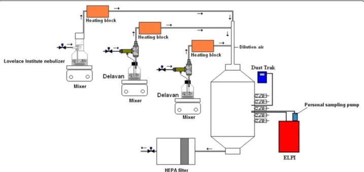
Animal inhalation exposures were performed in a cubic stainless steel 500-L inhalation chamber (Unifab, Kalamazoo, MI) adapted for nose-only. Ports were placed on a single wall of the inhalation chamber so that only the nose of the animal was exposed to the aerosol (see Figure 10). Six groups of rats (n = 6 per group) were exposed to TiO2aerosols for 6 hours; one control group (n = 6) was exposed to clean air for the same duration. One sham group (n = 6) was not exposed and remained housed in the animal facility. No anesthetic treatment was given to the rats prior to the exposures. The aerosols were composed of either 5, 10–30 or 50 nm primary particle size TiO2. Each primary particle size of TiO2was gener-ated in aerosols with two distinct size distributions consisting of large (> 100 nm) or small agglomerates (< 100 nm) at mass concentrations of 20 mg/m3. Small agglomerate (SA) aerosols were produced by a wet gener-ation method and large agglomerate (LA) aerosols by a dry powder dispersion technique. The animals were sacrificed 16 hours after the end of the exposure period, and bronchoalveolar lavages (BAL) were performed to de-termine cellular markers of pulmonary toxicity. Lungs were also fixed for histopathology observations.
Figure 10 Experimental set-up for the generation of small agglomerate aerosols. Experimental set-up using three nebulizers in parallel to produce aerosols essentially composed of small NP agglomerates. ELPI: electrical low pressure impactor; HEPA filter: high-efficiency particulate air filter. Noël et al. Particle and Fibre Toxicology 2013, 10:48 Page 13 of 18 http://www.particleandfibretoxicology.com/content/10/1/48
TiO2nanoparticles
Anatase TiO2with average particle sizes of 5 nm (Stock# 5420MR) and 10–30 nm (Stock# 5420HT), specific area of 200–220 m2
/g and near spherical morphologies were purchased from Nanostructured and Amorphous Mate-rials Inc. (Texas, USA). Anatase 50 nm TiO2 (MK-TiO2-A050) was purchased from MKnano (Ontario, Canada). All the NP were stored in a desiccator placed in a fume hood prior to use. These NP were characterized by TEM (field emission gun (FEG) JEOL JEM-2100F), EDS and XRD (Philips, model: X’Pert, Lelyweg, The Netherlands). These characterization methods were previously des-cribed in Noël et al. [33].
Animals
Forty-eight 8-week-old male CDF (F344)/CrlBR (Charles River Breeding Laboratories, St. Constant, Québec, Canada) rats with an average weight on the day of sacrifice of 164 ± 12 g were housed at the animal care facilities of the Université de Montréal. The rats were placed two per cage and had access to water and food ad libitum. The animals were exposed to a 12-h/12-h day/night cycle from 6:00 am to 6:00 pm. The animals were acclimatized to the inhalation chamber for a period of six days prior to NP exposure. Weight gain for each animal was recorded daily. The re-search project was approved by the Université de Montréal’s Ethics Committee on Animal Experiments.
Generation of TiO2NP aerosols
All aerosols were generated using compressed air that first passed though a Donaldson high-efficiency indus-trial filter equipped with a coalescing filter (Donaldson DFSP, Series Model-DF 0070 ZU, Ultra-Filter Superplus, Donaldson Company, Inc., Norcross, GA, USA). The average temperature and relative humidity in the inhal-ation chamber were 23.07 ± 0.72°C and 39.3 ± 16.6% RH. A fan mixed the air in the inhalation chamber. For all exposures, the total air flow entering the chamber was between 3.6 and 4.5 m3per hour.
Generation of small agglomerate aerosols– Nebulization of TiO2NP
Aerosols composed of small agglomerates were generated using a Lovelace-type nebulizer (In-Tox products, Albu-querque, New Mexico) placed in parallel with two Delavan siphon spray nebulizers (Part number 30609–2 used with an adapter, part number DLN 17147, Delavan Spray Tech-nologies, Goodrich Corporation, Montréal, Canada) (Fig-ure 10). We used three nebulizers in parallel to reach the targeted concentration of 20 mg/m3. Indeed, the type and number of nebulizers used constitute important factors contributing to the ultrafine size distribution of the aero-sols [33]. The high air flow that passes through the Delavan nebulizer, helps to reduce the size of the droplets
and by this means the use of two Delavan allowed the NMAD to be lowered by increasing the number of small NP agglomerates in the aerosol [33]. A 7 g/L (for the 5 nm experiment) or a 5 g/L (for the 10–30 nm and 50 nm experiments) NP suspension in distilled water (Milli-Q reference A+ system, water purification system with total oxidizable carbon indicator, Millipore Corporation) was filtered on Whatman 41 filter paper (Piscataway, NJ, USA) to remove large agglomerates, and sonicated for 10 mi-nutes (Bransonic tabletop ultrasonic cleaner, model 5510, Branson, Danbury, CT, USA). This suspension was poured into the Delavan devices. Since the filtered suspension did not allow the targeted mass concentration to be reached, 2.5 g (for the 5 nm experiment) or 1.5 g (for the 10– 30 and 50 nm experiments) of the TiO2powder was placed in the Lovelace-type nebulizer prior to the addition of the original suspension (350 ml) to completely fill the device. This new suspension was not sonicated. The suspension was agitated for the entire generation period using mag-netic stirring plates placed under each nebulizer. The flow rate used for each nebulizer was 5, 25 and 10–13 L/min for the Lovelace-type nebulizer, the first and second Delavan, respectively. The pressure applied to each nebulizer was between 30 and 35 psi. Dual-element heating tapes (624 watts, 120 VAC, Cole-Parmer, Canada) wrapped around a copper tube were used to dry the aero-sol, prior to its dispersion in the inhalation chamber. The flow rate of the dilution air was 25–30 L/min and served to reduce the relative humidity in the chamber created by the water vapor content of the aerosol. No charge neutralization was performed on these aerosols.
Generation of large agglomerate aerosols–Powder dispersion of TiO2NP
The aerosols composed of large agglomerates were pro-duced using a Fluidized Bed 3400A device (TSI Inc., Shoreview, MN, USA). The exposure concentration for each primary particle size was achieved by adjusting the various feed rates of the Fluidized Bed. The pressure ap-plied to this instrument was between 33 and 38 psi. No charge neutralization was performed.
TiO2aerosol sampling and characterization
TiO2 NP aerosols sampling and characterization methods have been described in detail elsewhere [33]. Briefly, air samples were collected throughout the experiment on cas-settes (Sure Seal, SKC Inc.) using 37-mm polyvinyl chloride (PVC) filters at a flow rate of 4 L/min to subsequently de-termine the average mass concentration by gravimetric ana-lysis. The mass concentrations were followed and adjusted in real time using a Model 8520 Dust Trak Aerosol Moni-tor (TSI Inc., Shoreview, MN, USA) previously calibrated with TiO2by comparison with the gravimetric method. Air samples were also collected at a flow rate of 1 L/minute on
pre-carbon coated Formvar copper grids glued onto 25-mm polycarbonate filters. The glue used was a current cyanoacrylate (Loctite superglue gel, Henkel, Boucherville, Canada). The sampling durations were 2.5 and 5 min for all of the aerosols. Characterization (shape, agglomeration degree and structure) of the aerosols sampled on these grids was performed by TEM (Philips CM200 equipped with a digital camera: Corel Corp. AMTV600 2K×2K, 80 kV). Numbers and particle size distributions were monitored in real time with an electrical low pressure impactor (ELPI) (Dekati Ltd., Tampere, Finland) which was operated at a flow rate of 10 L/minute in the filter stage configuration. The sintered impaction substrates were oiled to prevent or reduce particle bounce. Cumu-lative size distributions based on number concentration were monitored through 5 thirty-minute samples col-lected every hour of the experiment. The ELPI was also used to determine the number median aerodynamic diameter NMAD and the geometric standard deviation (GSD). Air samples were all collected in the area of the inhalation chamber corresponding to the breathing zone of the animals.
Pulmonary deposition modeling
The rats’ airway particle dosimetry was estimated using the Multiple-Path Particle Dosimetry Model (MPPD) (software version 2.11, Applied Research Associates Inc., Albuquer-que, NM, USA). The respiratory tract deposition of NP ag-glomerates was estimated for LA and SA aerosols.
Bronchoalveolar lavages
The animals were anaesthetized with isoflurane and sacrificed by exsanguination. BAL fluids (BALF) were col-lected with 0.9% saline. The BAL techniques were previ-ously described elsewhere [97]. Briefly, five 5-ml washes were pooled and placed on ice. The collected BALF were centrifuged at 929 × g at a temperature of 4°C for 10 mi-nutes. After centrifugation, the supernatant was removed and frozen at−80°C. These supernatants were aliquoted and used for cytotoxicity and oxidative stress analysis. Multiple freeze–thaw cycle was avoided to prevent loss of enzyme activity. These supernatants were used for cyto-toxicity and oxidative stress analysis. The cells were resuspended in 500μl of saline. 100-μl aliquots were fixed with formalin for cell count (1:1). The lungs were fixed in situwith buffered formalin.
Analysis of the pulmonary response
Cell suspensions were mixed 1:1 with methylene blue to de-termine the total cell counts using a hemacytometer. The cytospin cell staining method using Hema 3 solutions (Fisher Diagnostics cat. nos. 911A, 911B and 122-911C) were used to obtain differential cell counts for lymphocytes, neutrophils and macrophages. Slides were
observed using a photonic microscope with a magnification of 400-× (Leica DM 1000). Cytotoxicity was evaluated by determining levels of lactate dehydrogenase (LDH) activity (Cytotoxicity Detection Kit for LDH, Roche Applied Sci-ence, Laval, QC, Canada). The oxidative stress response was evaluated by measuring 8-isoprostane concentration (8-isoprostane EIA kit, Cayman Chemical, Ann Arbor, MI, USA). Cytokines in BAL were analyzed using Rat Cytokine Array Panel A (R & D Systems, Minneapolis, MN, USA) to determine the relative levels of 29 cytokines (CINC-1, CINC-2α/β, CINC-3, CNTF, Fractalkine (CX3CL1), GM-CSF, sICAM-1 (CD54), IFN-γ, IL-1α, IL-1β, IL-1ra. IL-2, IL-3, IL-4, IL-6, IL-10, IL-13, IL-17, IP-10 (CXCL10), LIX, L-Selectin (CD62L/LECAM-1), MIG (CXCL9), MIP-1α (CCL3), MIP-3α (CCL20), RANTES (CCL5), Thymus Che-mokine (CXCL7), TIMP-1, TNF-α, VEGF). To perform this assay, samples from the same exposure group were pooled together. Data from this assay were analyzed by chemilu-minescent signals of cytokines/chemokines present in the BALF and were detected on Kodak X OMAT-RA film, as described in Ratthe et al. [98] and Gonçalves et al. [98]. Results were reported as described in Gonçalves et al. [99]. Briefly, cytokines with≥ 1.2-fold increased compared to the control group were considered to represent a slight inflam-mation. All assays were performed as specified by the re-spective manufacturers.
Estimation of NP endocytosis by macrophages
The cytospin slides were used to visually estimate the extent of NP phagocytosis by counting 200 macro-phages for the presence or absence of NP agglomerates inside the cytoplasm. In this way, the percentage of particle-laden macrophages was established. Slides were observed using a photonic microscope (Leica DM 1000).
Lung histopathology
The lungs were fixed in situ with buffered formalin. Sub-sequently, caudal right lobe lung sections were cut into thin slices (3 to 5 mm thick) using a clean scalpel. Samples were placed inside histology cassettes before processing. Wax infiltration (Sakura Tissue Tek VIP E150) was achieved following dehydration through 3 alcohol baths (70, 85 and 90%) and cleared through 3 toluene baths. For embedding, tissue was oriented inside a mold filled with hot paraffin (Embedding station ESBE EC350). Tissue sec-tioning (4μm thick) was done with a microtome (Micro-tome LEICA RM2255). Sections were then colored using haematoxylin and eosin staining standard protocol. Images were acquired using an Olympus BX51 optical microscope.
Statistical procedures
BALF cytology, pulmonary cytotoxicity, oxidative stress markers as well as NP endocytosis by macrophages were
Noël et al. Particle and Fibre Toxicology 2013, 10:48 Page 15 of 18 http://www.particleandfibretoxicology.com/content/10/1/48
analyzed using ANOVA and Tukey’s test. Statistical sig-nificance was achieved when p < 0.05. Statistical analyses were performed using the Statistical Package for the So-cial Sciences (SPSS, version 17.0, SPSS Inc.).
Competing interest
The authors declare that they have no competing interests. Authors’ contributions
AN, MC, YC, RT and GT were involved in the conception and design of the study. AN and YC developed the NP aerosols generation and
characterization methods. AN and RT were responsible of the inhalation exposures. AN and MC were responsible of the BALF analysis. AN, MC, YC, RT and GT were involved in the analysis and interpretation of data. AN drafted the manuscript. All authors read and approved the final manuscript. Acknowledgement
This work was supported by a grant from the Réseau de recherche en santé environnementale du Québec (RRSE) and the Institut de recherche en santé publique de l’Université de Montréal (IRSPUM). Alexandra Noël is a recipient of Ph.D. scholarships from the Institut de recherche Robert-Sauvé en Santé et en Sécurité du Travail (IRSST) and from the Fonds de la Recherche en Santé du Québec (FRSQ). Special thanks to Ginette Charest-Tardif of the Université de Montréal and Guylaine Lassonde of the INRS-Institut Armand-Frappier, Université du Québec for technical support.
Author details
1Département de santé environnementale et de santé au travail, Institut de
recherche en santé publique, Université de Montréal, C.P. 6128 Succursale Centre-Ville, Montréal, Québec H3C 3J7, Canada.2INRS-Institut
Armand-Frappier, Université du Québec, 531 Boul. des Prairies, Laval, Québec H7V 1B7, Canada.3Institut de recherche Robert-Sauvé en santé et en sécurité du
travail (IRSST), 505 Boul. De Maisonneuve Ouest, Montréal, Québec H3A 3C2, Canada.
Received: 11 March 2013 Accepted: 1 October 2013 Published: 4 October 2013
References
1. Lux Research: The Nanotech Report; 2004. http://www.luxresearchinc.com/ 2004. 2. McIntyre RA: Common nano-materials and their use in real world
applications. Sci Progress 2012, 95:1–22.
3. Donaldson K, Seaton A: A short history of the toxicology of inhaled particles. Part Fibre Toxicol 2012, 9:13.
4. Oberdorster G, Ferin J, Gelein R, Soderholm SC, Finkelstein J: Role of the alveolar macrophage in lung injury: studies with ultrafine particles. Environ Health Perspect 1992, 97:193–199.
5. Oberdorster G, Ferin J, Lehnert BE: Correlation between particle size, in vivo particle persistence, and lung injury. Environ Health Perspect 1994, 102(Suppl 5):173–179.
6. Brown DM, Wilson MR, MacNee W, Stone V, Donaldson K: Size-dependent proinflammatory effects of ultrafine polystyrene particles: a role for surface area and oxidative stress in the enhanced activity of ultrafines. Toxicol Appl Pharmacol 2001, 175:191–199.
7. Renwick LC, Brown D, Clouter A, Donaldson K: Increased inflammation and altered macrophage chemotactic responses caused by two ultrafine particle types. Occup Environ Med 2004, 61:442–447.
8. Duffin R, Mills NL, Donaldson K: Nanoparticles-a thoracic toxicology perspective. Yonsei Med J 2007, 48:561–572.
9. Grassian VH, Adamcakova-Dodd A, Pettibone JM, O’Shaughnessy PT, Thorne PS: Inflammatory response of mice to manufactured titanium dioxide nanoparticles: comparison of size effects through different exposure routes. Nanotoxicol 2007, 1:211–226.
10. Kobayashi N, Naya M, Endoh S, Maru J, Yamamoto K, Nakanishi J: Comparative pulmonary toxicity study of nano-TiO(2) particles of different sizes and agglomerations in rats: different short- and long-term post-instillation results. Toxicology 2009, 264(1–2):110–118.
11. Pauluhn J: Pulmonary toxicity and fate of agglomerated 10 and 40 nm aluminum oxyhydroxides following 4-week inhalation exposure of rats:
toxic effects are determined by agglomerated, not primary particle size. Toxicol Sci 2009, 109:152–167.
12. Oberdorster G, Maynard A, Donaldson K, Castranova V, Fitzpatrick J, Ausman K, Carter J, Karn B, Kreyling W, Lai D, Olin S, Monteiro-Riviere N, Warheit D, Yang H: Principles for characterizing the potential human health effects from exposure to nanomaterials: elements of a screening strategy. Part Fibre Toxicol 2005, 2:8.
13. Nel A, Xia T, Madler L, Li N: Toxic potential of materials at the nanolevel. Science 2006, 311:622–627.
14. Auffan M, Rose J, Bottero JY, Lowry GV, Jolivet JP, Wiesner MR: Towards a definition of inorganic nanoparticles from an environmental, health and safety perspective. Nat Nanotechnol 2009, 4:634–641.
15. Liang G, Pu Y, Yin L, Liu R, Ye B, Su Y, Li Y: Influence of different sizes of titanium dioxide nanoparticles on hepatic and renal functions in rats with correlation to oxidative stress. J Toxicol Environ Health A 2009, 72:740–745.
16. Liu W, Wu Y, Wang C, Li HC, Wang T, Liao CY, Cui L, Zhou QF, Yan B, Jiang GB: Impact of silver nanoparticles on human cells: effect of particle size. Nanotoxicology 2010, 4:319–330.
17. Oberdorster G, Oberdorster E, Oberdorster J: Nanotoxicology: an emerging discipline evolving from studies of ultrafine particles. Environ Health Perspect 2005, 113:823–839.
18. Landsiedel R, Ma-Hock L, Haussmann HJ, van Ravenzwaay B, Kayser M, Wiench K: Inhalation studies for the safety assessment of nanomaterials: status quo and the way forward. Wiley Interdiscip Rev Nanomed. Nanobiotechnol 2012, 4:399–413.
19. Grassian VH, O’shaughnessy PT, Mcakova-Dodd A, Pettibone JM, Thorne PS: Inhalation exposure study of titanium dioxide nanoparticles with a primary particle size of 2 to 5 nm. Environ Health Perspect 2007, 115:397–402. 20. United-States Environmental Protection Agency (EPA): Nanomaterial
Research Strategy. Washington, D.C: Office of Research and Development U. S. Environmental Protection Agency; 2009. Report no EPA620/K-09/011. 21. Organization for Economic Co-operation and Development (OECD): List of
manufactured nanomaterials and list of endpoints for phase one of the sponsorship programme for the testing of manufactured nanomaterials: revision. Environment, Health and Safety Publications Series on the Safety of Manufactured Nanomaterials. Series on the Safety of Manufactured Nanomaterials No. 27 ENV/JM/MONO (2010)46 http://search.oecd.org/officialdocuments/displaydocumentpdf/? cote=env/jm/mono(2010)46&doclanguage=en.
22. Boutou-Kempf O, Marchand JL, Radauceanu A, Witschger O, Imbernon E: Development of a French epidemiological surveillance system of workers producing or handling engineered nanomaterials in the workplace. J Occup Environ Med 2011, 53(6 Suppl):S103–S107.
23. National Institute for Occupational Safety and Health (NIOSH): Occupational Exposure to Titanium Dioxide. Department of Health and Human Services, Centers for Disease Control and Prevention: DHHS; 2011. Publication No. 2011–160. Current intelligence bulletin 63. http://www.cdc.gov/niosh/docs/ 2011-160/pdfs/2011-160.pdf.
24. Fryzek JP, Chadda B, Marano D, White K, Schweitzer S, McLaughlin JK, Blot WJ: A cohort mortality study among titanium dioxide manufacturing workers in the United States. J Occup Environ Med 2003, 45:400–409. 25. Hext PM, Tomenson JA, Thompson P: Titanium dioxide: inhalation
toxicology and epidemiology. Ann Occup Hyg 2005, 49:461–472. 26. Hsu LY, Chein HM: Evaluation of nanoparticle emission for TiO2
nanopowder coating materials. J Nanopart Res 2007, 9:157–163. 27. Demou E, Stark WJ, Hellweg S: Particle emission and exposure during
nanoparticle synthesis in research laboratories. Ann Occup Hyg 2009, 53:829–838.
28. Curwin B, Bertke S: Exposure characterization of metal oxide nanoparticles in the workplace. J Occup Environ Hyg 2011, 8:580–587. 29. Lee JH, Kwon M, Ji JH, Kang CS, Ahn KH, Han JH, Yu IJ: Exposure
assessment of workplaces manufacturing nanosized TiO2and silver. Inhal
Toxicol 2011, 23:226–236.
30. Bermudez E, Mangum JB, Wong BA, Asgharian B, Hext PM, Warheit DB, Everitt JI: Pulmonary responses of mice, rats, and hamsters to subchronic inhalation of ultrafine titanium dioxide particles. Toxicol Sci 2004, 77:347–357.
31. Ma-Hock L, Gamer AO, Landsiedel R, Leibold E, Frechen T, Sens B, Linsenbuehler M, van Ravenzwaay B: Generation and characterization of test atmospheres with nanomaterials. Inhal Toxicol 2007, 19:833–848. 32. Ma-Hock L, Burkhardt S, Strauss V, Gamer AO, Wiench K, Van Ravenzwaay B,
using nano-titanium dioxide as a model substance. Inhal Toxicol 2009, 21:102–118.
33. Noël A, Cloutier Y, Wilkinson KJ, Dion C, Halle S, Maghni K, Tardif R, Truchon G: Generating Nano-Aerosols from TiO(2) (5 nm) Nanoparticles Showing Different Agglomeration States. Application to Toxicological Studies. J Occup Environ Hyg 2013, 10:86–96.
34. Noël A, L’Esperance G, Cloutier Y, Plamondon P, Boucher J, Philippe S, Dion C, Truchon G, Zayed J: Assessment of the contribution of electron microscopy to nanoparticle characterization sampled with two cascade impactors. J Occup Environ Hyg 2013, 10:155–172.
35. Warheit DB, Hoke RA, Finlay C, Donner EM, Reed KL, Sayes CM:
Development of a base set of toxicity tests using ultrafine TiO2particles
as a component of nanoparticle risk management. Toxicol Lett 2007, 171:99–110.
36. Zook JM, Maccuspie RI, Locascio LE, Halter MD, Elliott JT: Stable nanoparticle aggregates/agglomerates of different sizes and the effect of their size on hemolytic cytotoxicity. Nanotoxicology 2011, 5:517–530. 37. Kreyling WG, Semmler-Behnke M, Takenaka S, Moller W: Differences in the
Biokinetics of Inhaled Nano- versus Micrometer-Sized Particles. Acc Chem Res 2013, 46:714–722.
38. Warheit DB, Webb TR, Reed KL, Frerichs S, Sayes CM: Pulmonary toxicity study in rats with three forms of ultrafine-TiO2particles: differential
responses related to surface properties. Toxicology 2007, 230:90–104. 39. Warheit DB, Webb TR, Sayes CM, Colvin VL, Reed KL: Pulmonary instillation
studies with nanoscale TiO2rods and dots in rats: toxicity is not dependent
upon particle size and surface area. Toxicol Sci 2006, 91:227–236. 40. Jiang J, Oberdorster G, Elder A, Gelein R, Mercer P, Biswas P: Does
Nanoparticle Activity Depend upon Size and Crystal Phase? Nanotoxicology 2008, 2:33–42.
41. Noël A, Maghni K, Cloutier Y, Dion C, Wilkinson KJ, Halle S, Tardif R, Truchon G: Effects of inhaled nano-TiO2aerosols showing two distinct
agglomeration states on rat lungs. Toxicol Lett 2012, 214:109–119. 42. Donaldson K, Stone V, Clouter A, Renwick L, MacNee W: Ultrafine particles.
Occup Environ Med 2001, 58:211–216. 199.
43. Scherbart AM, Langer J, Bushmelev A, van Berlo D, Haberzettl P, van Schooten FJ, Schmidt AM, Rose CR, Schins RP, Albrecht C: Contrasting macrophage activation by fine and ultrafine titanium dioxide particles is associated with different uptake mechanisms. Part Fibre Toxicol 2011, 8:31.
44. Xia T, Kovochich M, Brant J, Hotze M, Sempf J, Oberley T, Sioutas C, Yeh JI, Wiesner MR, Nel AE: Comparison of the abilities of ambient and manufactured nanoparticles to induce cellular toxicity according to an oxidative stress paradigm. Nano Lett 2006, 6:1794–1807.
45. Ferin J, Oberdorster G, Penney DP: Pulmonary retention of ultrafine and fine particles in rats. Am J Respir Cell Mol Biol 1992, 6:535–542. 46. Oberdörster G, Cox C, Gelein R: Intratracheal instillation versus
intratracheal-inhalation of tracer particles for measuring lung clearance function. Exp Lung Res 1997, 23:17–34.
47. Balasubramanian SK, Poh KW, Ong CN, Kreyling WG, Ong WY, Yu LE: The effect of primary particle size on biodistribution of inhaled gold nano-agglomerates. Biomaterials 2013, 34:5439–5452.
48. Hervé-Bazin B: Les nanoparticules un enjeu majeur pour la santé au travail?. France: EDP Sciences; 2007:701.
49. Sager TM, Kommineni C, Castranova V: Pulmonary response to intratracheal instillation of ultrafine versus fine titanium dioxide: role of particle surface area. Part Fibre Toxicol 2008, 5:17.
50. Muhlfeld C, Mayhew TM, Gehr P, Rothen-Rutishauser B: A novel quantitative method for analyzing the distributions of nanoparticles between different tissue and intracellular compartments. J Aerosol Med 2007, 20:395–407.
51. Muhlfeld C, Rothen-Rutishauser B, Vanhecke D, Blank F, Gehr P, Ochs M: Visualization and quantitative analysis of nanoparticles in the respiratory tract by transmission electron microscopy. Part Fibre Toxicol 2007, 4:11. 52. Muhlfeld C, Rothen-Rutishauser B, Blank F, Vanhecke D, Ochs M, Gehr P:
Interactions of nanoparticles with pulmonary structures and cellular responses. Am J Physiol Lung Cell Mol Physiol 2008, 294:L817–L829. 53. Seipenbusch M, Binder A, Kasper G: Temporal evolution of nanoparticle
aerosols in workplace exposure. Ann Occup Hyg 2008, 52:707–716. 54. Kanapilly GM, Wolff RK, DeNee PB, McClellan RO: Generation,
characterization and inhalation deposition of ultrafine aggregate aerosols. Ann Occup Hyg 1982, 26:77–91.
55. Pratten MK, Lloyd JB: Pinocytosis and phagocytosis: the effect of size of a particulate substrate on its mode of capture by rat peritoneal macrophages cultured in vitro. Biochim Biophys Acta 1986, 881:307–313. 56. Oberdörster G: Lung clearance of inhaled insoluble and soluble particles.
J Aerosol Med 1988, 1:289–329.
57. Yang W, Peters JI, Williams RO III: Inhaled nanoparticles–a current review. Int J Pharm 2008, 356:239–247.
58. Cho WS, Duffin R, Poland CA, Howie SE, MacNee W, Bradley M, Megson IL, Donaldson K: Metal oxide nanoparticles induce unique inflammatory footprints in the lung: important implications for nanoparticle testing. Environ Health Perspect 2010, 118:1699–1706.
59. Warheit DB, Reed KL, Delorme MP: Embracing a weight-of-evidence approach for establishing NOAELs for nanoparticle inhalation toxicity studies. Toxicol Pathol 2012:.
60. Preining E: The physical nature of very, very small particles and its impact on their behaviour. J Aerosol Sci 1998, 29:481–495.
61. Bermudez E, Mangum JB, Asgharian B, Wong BA, Reverdy EE, Janszen DB, Hext PM, Warheit DB, Everitt JI: Long-term pulmonary responses of three laboratory rodent species to subchronic inhalation of pigmentary titanium dioxide particles. Toxicol Sci 2002, 70:86–97.
62. Geiser M, Kreyling WG: Deposition and biokinetics of inhaled nanoparticles. Part Fibre Toxicol 2010, 7:2.
63. Cavaillon JM: Les cytokines. Paris: Masson; 1993:412. ISBN 2-225-84174-8. 64. Nicola NA: Guidebook to cytokines and their receptors. A Sambrook and Tooze
publication. Oxford University Press; 1994:261. ISBN 0-19-859946-3. 65. Goldsby RA, Kindt TJ, Osborne BA: Immunologie le cours de Janis Kuby.
New-York and Basingstoke: W.H. Freeman and Company; 2000:660. 66. Oberdorster G, Finkelstein JN, Johnston C, Gelein R, Cox C, Baggs R, Elder
AC: Acute pulmonary effects of ultrafine particles in rats and mice. Res Rep Health Eff Inst 2000, 96:5–74.
67. Tetley TD: Health effects of nanomaterials. Biochem Soc Trans 2007, 35:527–531.
68. Beck-Speier I, Dayal N, Karg E, Maier KL, Schumann G, Schulz H, Semmler M, Takenaka S, Stettmaier K, Bors W, Ghio A, Samet JM, Heyder J: Oxidative stress and lipid mediators induced in alveolar macrophages by ultrafine particles. Free Radic Biol Med 2005, 38:1080–1092.
69. Fahmy B, Cormier SA: Copper oxide nanoparticles induce oxidative stress and cytotoxicity in airway epithelial cells. Toxicol In Vitro 2009, 23:1365–1371.
70. Johnston HJ, Hutchison GR, Christensen FM, Peters S, Hankin S, Stone V: Identification of the mechanisms that drive the toxicity of TiO(2) particulates: the contribution of physicochemical characteristics. Part Fibre Toxicol 2009, 6:33.
71. Rossi EM, Pylkkanen L, Koivisto AJ, Vippola M, Jensen KA, Miettinen M, Sirola K, Nykasenoja H, Karisola P, Stjernvall T, Vanhala E, Kiilunen M, Pasanen P, Makinen M, Hameri K, Joutsensaari J, Tuomi T, Jokiniemi J, Wolff H, Savolainen K, Matikainen S, Alenius H: Airway exposure to silica-coated TiO2nanoparticles induces pulmonary neutrophilia in mice. Toxicol Sci
2010, 113:422–433.
72. Halappanavar S, Jackson P, Williams A, Jensen KA, Hougaard KS, Vogel U, Yauk CL, Wallin H: Pulmonary response to surface-coated nanotitanium dioxide particles includes induction of acute phase response genes, inflammatory cascades, and changes in microRNAs: a toxicogenomic study. Environ Mol Mutagen 2011, 52:425–439.
73. Van Ravenzwaay B, Landsiedel R, Fabian E, Burkhardt S, Strauss V, Ma-Hock L: Comparing fate and effects of three particles of different surface properties: nano-TiO(2), pigmentary TiO(2) and quartz. Toxicol Lett 2009, 186:152–159.
74. Lindberg HK, Falck GC, Catalan J, Koivisto AJ, Suhonen S, Jarventaus H, Rossi EM, Nykasenoja H, Peltonen Y, Moreno C, Alenius H, Tuomi T, Savolainen KM, Norppa H: Genotoxicity of inhaled nanosized TiO(2) in mice. Mutat Res 2012, 745:58–64.
75. McKinney W, Jackson M, Sager TM, Reynolds JS, Chen BT, Afshari A, Krajnak K, Waugh S, Johnson C, Mercer RR, Frazer DG, Thomas TA, Castranova V: Pulmonary and cardiovascular responses of rats to inhalation of a commercial antimicrobial spray containing titanium dioxide nanoparticles. Inhal Toxicol 2012, 24:447–457.
76. Takenaka S, Karg E, Roth C, Schulz H, Ziesenis A, Heinzmann U, Schramel P, Heyder J: Pulmonary and systemic distribution of inhaled ultrafine silver particles in rats. Environ Health Perspect 2001, 109(Suppl 4):547–551.
Noël et al. Particle and Fibre Toxicology 2013, 10:48 Page 17 of 18 http://www.particleandfibretoxicology.com/content/10/1/48