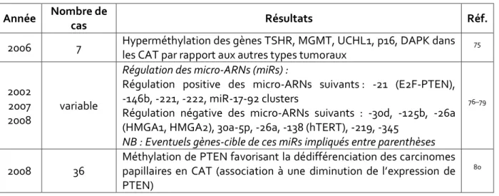
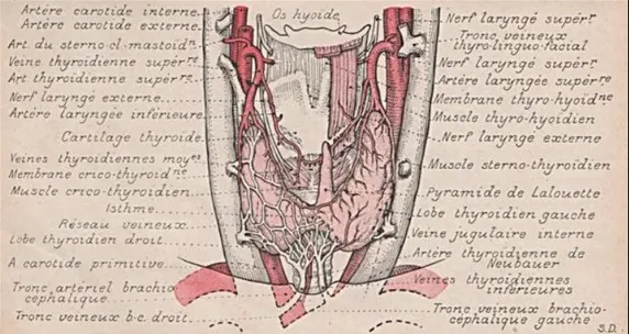
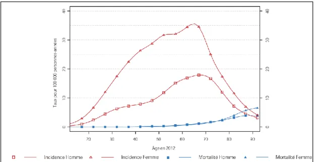
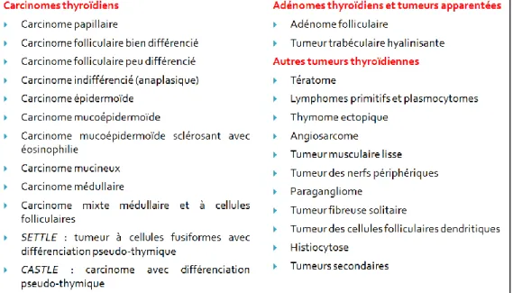
Pathologie moléculaire des carcinomes anaplasiques thyroïdiens : étude rétrospective à propos de 144 cas
Texte intégral
Figure

Documents relatifs
While the absolute values of the radiation intensity and carrier velocity will affect the final temperature of the device, of interest is their effect on the
The main contributions in Chapters 3 and 4 of this thesis are the formulation of a general power control problem for time varying wireless networks, the characterization of the
that applies in typical network operating regimes. This will give us an intuitive sense of the relationship between these quantities and the network parameters such as the traffic
In low-risk DMR patients, surgical repair will remain the standard of care for many years, with TMVRep and TMVI playing a role in high- risk or inoperable patients, who are not
Furthermore, a planet located in the white area would have induced a TTV signal amplitude above 3 min, the approximative expected TTV for an Earth-mass planet in mean motion
Also, they will improve the source inversions of vlp events when used for tilt correction of horizontal seismograms, as this cor- rection includes strain-tilt coupling in contrast
In an effort to determine the pattern of chromatin condensation and recombination at meiosis in an Imprinted region, fine scale genetic mapping in the approximately 4
In Figur 2 sind die verarbeite- ten Spektren der gemessenen Phosphorkerne aus einer Transversalschicht durch das menschliche Gehirn dargestellt.. Die Messungen wurden auf ei- nem 1.5
