Journal of the Hellenic Veterinary Medical Society
Vol. 69, 2018
Effects of GnRH or hCG on day 11 after artificial insemination in cows luteal activity
Besbaci Mohamed Laboratory of Biotechnology Related to Animals
Reproduction, Institute of Veterinary Sciences
Abdelli A. Laboratory of Biotechnology
Related to Animals Reproduction, Institute of Veterinary Sciences
Belabdi I. Laboratory of Biotechnology
Related to Animals Reproduction, Institute of Veterinary Sciences
Benabdelaziz A Nuclear Research Center of
Draria-Algiers
Khelili R. Nuclear Research Center of
Draria-Algiers
Mebarki M. High National School
Veterinary El Harrach Kaidi R.
http://dx.doi.org/10.12681/jhvms.16128
Copyright © 2019 mohamed mohamed besbaci
To cite this article:
Besbaci, M., Abdelli, A., Belabdi, I., Benabdelaziz, A., Khelili, R., Mebarki, M., & Kaidi, R. (2019). Effects of GnRH or hCG on day 11 after artificial insemination in cows luteal activity. Journal of the Hellenic Veterinary Medical Society, 69(4), 1227-1234. doi:http://dx.doi.org/10.12681/jhvms.16128
Effects of GnRH or hCG on day 11 after artificial
insemination in cows luteal activity
M. Besbaci1,2*, A. Abdelli1, I. Belabdi1 , A. Benabdelaziz3,
R. Khelili3, M. Mebarki2, R. Kaidi1
1Laboratory of Biotechnology Related to Animals Reproduction, Institute of Veterinary Sciences, Blida, Algeria. 2High National School Veterinary El Harrach, Algiers, Algeria.
3Nuclear Research Center of Draria-Algiers, Algeria
Corresponding Author:
M. Besbaci
Laboratory of Biotechnology Related to Animals Reproduction, Institute of Veterinary Sciences, Blida Soumaa, Algeria.
Date of initial submission: 23-4-2018 Date of revised submission: 14-6-2018 Date of acceptance: 27-6-2018
Ερευνητικό άρθρο
ΠΕΚΕ 2018, 69(4): 1227-1234
ABSTRACT. In order to optimize luteal function, human chorionic gonadotrophin (hCG) or gonadotrophin releasing hormone (GnRH) were used on day 11 after artificial insemination (AI). 33 cows synchronized by the Ovsynch and di-vided into 3 groups according to the type of treatment: 1) hCG (1500 IU, n=11); 2) GnRH (100 µg, n=11) and 3) control (2 mL of saline, n=11). Blood samples were collected from all animals every 3 days from day 5 to day 23 to determine progesterone concentration. Ultrasonography was used to monitor the luteal surface structures at the time of blood sam-ple collection. An accessory corpus luteum (CL) formed in 63.63% of cows treated with GnRH or hCG, resulting in an increase in the total luteal tissue area compared with the controls. Compared with the controls, the principal CL area was increased by hCG but not by GnRH. Additionally, compared with the control group, hCG-treated cows had increased progesterone concentrations (p<0.0001), while GnRH-treated cows had P4 similar to that of controls cows.
1228 M. BESBACI, A. ABDELLI, I. BELABDI , A. BENABDELAZIZ, R. KHELILI, M. MEBARKI, R. KAIDI
J HELLENIC VET MED SOC 2018, 69(4) ΠΕΚΕ 2018, 69(4)
INTRODUCTION
E
arly embryonic loss is the principal cause of low pregnancy rate (Inskeep and Dailey 2005). Indeed, embryonic and foetal mortalities are estimated to be 50% in dairy cows and between 70 and 80% of these losses occur in the first 16 days after AI (Diskin et al., 2011). One of the causes of embryonic loss is related to delayed embryo development, which reduces signalling for the maternal recognition of pregnancy (Mann and Lamming 2001). This condition may be due to a lower progesterone concentration in dairy cows (Mann and Lamming 2001; Wiltbank et al., 2011). Progesterone plays an important role in regulating changes in the uterine environment conducive to the development of the embryo and maintenance of pregnancy (Geisert et al., 1992). Increased milk production in dairy cows is connected with increased general metabolism as well as with increased progesterone metabolism in the liver, which decreases progesterone concentration in peripheral blood (Rhinehart et al., 2009). Progesterone insufficiency can be an important factor responsible for the decrease of the conception rate and is associated with abnormal early embryo development (Mann and Lamming 2001), while high progesterone concentration favours embryo development and interferon-τ secretion (Lonergan 2011). Several approaches have been used to increase the concentration of progesterone in the blood in order to reduce the occurrence of embryo death (Campanile et al., 2007). In different studies, proges-terone has been administered via implants or intra-vag-inal devices with inconsistent results (Wiltbank et al., 2014). Other studies have shown that administration of natural sequence GnRH, GnRH agonists or hCG after AI can stimulate CL function which induces accessory CL formation, increases progesterone concentration, and reduces oestradiol production with a consequent positive effect on embryonic survival (Thatcher et al., 2003). Administration of GnRH, a GnRH agonist or hCG after AI at specific times, coincident with the presence of the dominant follicle of the first and second follicular waves, may stimulate CL function, induce accessory CL formation, increase progesterone concentration (Schmitt et al., 1996a, Stevenson et al., 2007) and reduce oestrogen production with a con-sequent positive effect on pregnancy rate or embryo survival (Thatcher et al., 2003; Šuluburić et al., 2017). Several studies have investigated the effects of GnRHadministered during mid-estrous cycle (day 12, 13 or 14 after AI) (Szenci et al., 2006; Lopez-Gatius et al., 2006). Beneficial effects of treatment with GnRH at day 11-12 after AI on fertility have been observed in lactating dairy cows during heat stress (Lopez-Gatius et al., 2006), but very few studies that have attempted to compare GnRH and hCG injected in the mid-cycle to improve luteal function after AI. We hypothesized that cows receiving GnRH in day 11 of estrous would have concentrations of progesterone closer to cows receiv-ing hCG. The objective of this study was to compare the effects of GnRH and hCG administration at day 11 after AI on CL development and function, in terms of progesterone secretion in cows.
MATERIALS AND METHODS Ethical approval
Experimental procedures were approved by the Institu-tional Animal Care Committee of the NaInstitu-tional Adminis-tration of the Algerian Higher Education and Scientific Research (Ethical approval number: 98-11, Law of August 22, 1998).
Animals and treatments
The experiment was undertaken at a commercial dairy farm in Mitija (longitude 36° and latitude 3°), Algeria from January to April. Animals were housed in free-stall barns and fed a diet consisting of grass and clover supplemented with a commercial concentrate (18% digestible raw protein), as well as roughly crushed maize grains, soybean meal, barley and vitamin–min-eral mixture. Cows were selected by clinical examina-tion before AI and only those in a healthy reproductive status were included in the study. A total of 33 prim-iparous Holstein lactating cows (ranging in age from 2-3 years old) were selected. Cows were synchronized by the Ovsynch protocol initiated with Gonadorelin (Cystoreline® CEVA, France) followed by an injection
of prostaglandin PGF2𝛼 analogue at day 7 (Enza-prost® CEVA, France), and two days later, the cows
received a second Gonadorelin injection. All animals were inseminated 12–16 h after the second injection of Gonadorelin (day 0). Eleven days after AI (day 11), cows were randomly assigned to one of the three treat-ment groups, administered a single injection of 1500 IU hCG (hCG group, n=11, Chorulon® Intevet Laboratories
Ltd Holland given i.v.), 100 µg GnRH (GnRH group, n=11, Cystoréline® CEVA France given i.m.) or 2 mL of 0.9% saline (n=11, control group).
Blood collection procedures and progesterone quan-tification
Blood samples were collected via venipuncture of the median caudal vein or artery into 5 mL evacuated tubes (VACUETTE® Blood Collection Tubes) every 3 days from day 5 to day 23 after timed AI (Fig 1) for later analysis of serum progesterone; the first sample collected immediately before administration of treat-ments. Blood samples were allowed to clot for 24 h at 4°C, then were centrifuged (1,935×g for 15 min), and serum was harvested and stored at −20°C until assayed for progesterone using a solid-phase, no-extraction radioimmunoassay.
Ultrasonography
Ultrasound examination of the ovaries was performed, using a Chison 600VET Ultrasound (Japan) equipped with a linear array 5 MHz transrectal transducer, to determine ovulatory response to Ovsynch and post-in-semination treatments. Ultrasound examinations were performed at day -10 and at day 0 and day 1 after AI to confirm ovulation. Cows were considered to have syn-chronized ovulation after the second GnRH injection of Ovsynch when 1 or more follicles ≥10 mm were present at AI and were absent at an ultrasound examination
conducted 2 days later. Ultrasound examinations were continued every 3 days from day 5 to day 23 after AI (Fig 1). Pregnancy status was determined at day 45 after AI by transrectal ultrasonography.
Statistical analysis
Because progesterone and CL area were measured over time, a repeated measures approach using ANOVA with mixed linear models (fixed effects of treatment, day and their interaction, random effect of cow) was used in SAS (Version 9.1.3; SAS Institute Inc., Cary, NC). For clarity, corresponding means ± SEM of the non-transformed data are presented in the results. Additionally, a mixed general linear model was fitted using the MIXED procedure of SAS (random effect of cow) to evaluate the effect of treatment on pregnancy rate. A Pearson correlation coefficient between pro-gesterone concentrations and total area of luteal tissue and between dominant follicle diameter on day 11 and accessory CL area at day 17 after AI ware calculated by the PROC CORR procedure in SAS.
RESULTS Pregnancy rate
The pregnancy rate was 45.45% (5/11) in either control or GnRH-treated cows. However, this rate was 54.54% (6/11) in hCG-treated cows. There was no significant difference (p ˃ 0.05) between groups.
Fig 1: Schematic diagram of activities and treatment schedules of control group (Saline), day 11 GnRH group
(go-nadoréline 100 μg) day 11 hCG group (hCG, 1500 IU) in cows (AI: artificial insemination; BS: blood sample d: day; PG: prostaglandin; US: ultrasonography).
1230 M. BESBACI, A. ABDELLI, I. BELABDI , A. BENABDELAZIZ, R. KHELILI, M. MEBARKI, R. KAIDI
J HELLENIC VET MED SOC 2018, 69(4) ΠΕΚΕ 2018, 69(4)
Ovarian status of cows at day 11 after AI
The ultrasound examination at day 11 after AI (the day of GnRH or hCG injection to induce ovulation or luteinisation of the dominant follicle) revealed the presence of one CL (no double ovulations were detected after AI) and one dominant follicle. The mean diameter of the dominant follicle on day 11 after AI was 11.74 ± 1.34, 12.06 ± 1.31 and 11.86 ± 1.31 mm in the hCG group, GnRH group and control group, respectively. In cows treated with GnRH or hCG, the mean (± S.E.M.)
diameter of the dominant follicle for cows that subse-quently formed an accessory CL (14/22) was 14.52± 0.61 mm (maximum diameter: 18.6 mm, minimum diameter: 10 mm). The mean follicular diameter for cows that have not formed an accessory CL (8/22) was 7.28 ± 0.4 mm (maximum diameter: 9 mm, minimum diameter: 5 mm). There was a significant difference in the diameter of dominant follicle between cows that have formed an accessory CL and cows that have not formed an accessory CL (p=0.004).
Fig 2:Sonogram image of an ovary at day 24 post AI showing two CL after GnRH treatment: (A) principal CL, (B)
GnRH-induced accessory CL
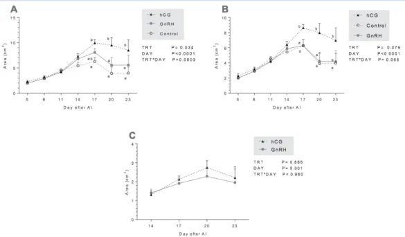
Fig 3: Mean (± SEM) of area of luteal tissue (cm2) in control cows that did not receive treatment, in cows that received
1500 IU of human chorionic gonadotrophin (hCG) and in cows treated with GnRH. (A) Mean (± SEM) of total area of luteal tissue (accessory CL+ principal CL. (B) Mean (± SEM) of principal CL area. (C) Mean (± SEM) of accessory CL area. (TRT: type of treatment, DAY: day of measurement, TRT*DAY: interaction treatment by day).
Corpora lutea
Treatment at day 11 induced ovulation of the dominant follicle and formation of an accessory CL in 63.63% (7/11) of cows treated with GnRH and in 63.63% (7/11) of cows treated with hCG. However, none of the con-trol cows formed an accessory CL (Fig 2). Thus, there was an acute disappearance of the dominant follicle and subsequent appearance of an accessory CL at the same location on the ovary in GnRH and hCG treated cows. Total CL areas was significantly higher at day 20 and 23 in hCG-treated cows compared to GnRH and control groups (Fig 3.A). Furthermore, principal CL areas was higher at day 17, 20 and 23 in hCG group compared to GnRH and control group (Fig 3B). There were significant effects of measurement time on total and principal CL areas (p˂0.0001) (Fig 3. A and B), and there was an effect of group-by time interaction (P=0.0003) for total CL area (Fig. 3A). However, the
accessory CL area induced by GnRH and hCG showed no significant change through the time and no group-by time interaction was observed (Fig 3. C). A significant correlation (r=0.83, p< 0.001) was found between dom-inant follicle diameter on day 11 and accessory CL area at day 17 after AI.
Plasma concentration of progesterone
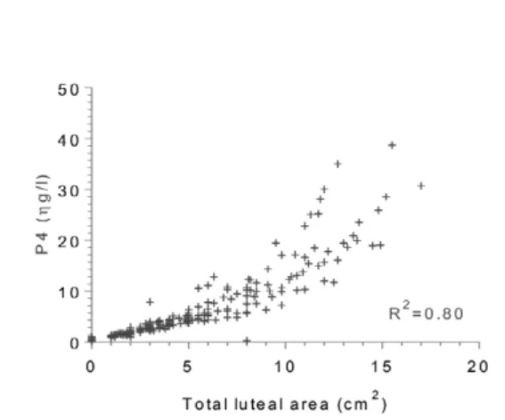
Cows treated with hCG presented significantly increased circulating progesterone concentrations from day 17 to day 23 after AI relative to the control and GnRH groups (Fig 4). There was an effect of group-by time interaction (p<0.0001). A significant correlation (r=0.89, p< 0.001) was found between progesterone level and total CL area (principal CL+ accessory CL) for each day (Fig 5). DISCUSSION
Previous studies showed that administration of GnRH, a GnRH agonist or hCG after AI at specific times coincident with the presence of the dominant follicle of the first and second follicular waves may stimulate CL function, induce accessory CL formation, increase progesterone concentration and reduce oestrogen pro-duction with a consequent positive effect on embryo survival (Stevenson et al., 2007).
In the present study, the diameter of the dominant follicle was higher in cows that formed an accessory CL compared to those that did not (p=0.004). Similarly to our results, Musilová et al (2014) observed that the incidence of two CL was higher in cows bearing the largest follicles from 10 to 20 mm in diameter compared to cows bearing only follicles ≤ 9 mm. Whether cows respond to GnRH may depend upon the characteristics of follicular growth; a follicle must reach 10 mm in diameter to ovulate in response to LH (Sartori et al., 2001). In the current study, 63.63% (n=14/22) cows treated with hCG or GnRH at day 11 after AI had formed an accessory CL; these results were higher than that (33.5%) reported by Musilová et al (2014) follow-ing a al t on the fact that others have used different dose of hCG? GnRH injection on day 11-13. Stevenson et al (2007) found an almost similar (60% for GnRH-treated cow) or grater (77.5% for hCG-treated cows) accessory CL formation after treatment of the cows between day 4 and day 9 after AI. This finding may be explained by the timing of treatment.
Fig 4:Mean (± SEM) of progesterone concentration in
cows (ng/mL) .
Fig 5. Correlation between progesterone concentrations
and total area of luteal tissue. Both principal and acces-sory CLs were taken into account
1232 M. BESBACI, A. ABDELLI, I. BELABDI , A. BENABDELAZIZ, R. KHELILI, M. MEBARKI, R. KAIDI
J HELLENIC VET MED SOC 2018, 69(4) ΠΕΚΕ 2018, 69(4)
progesterone until it reaches its final size.
The longer half-life of hCG in the blood and the slower turnover of the hCG-LH receptor complex on the surface of granulosa cells are probably responsible for an increased gonadotropic stimulation on the day 5 ovulatory follicle (De Rensis et al., 2010; Lonergan 2011) and its subsequent differentiation into a principal CL with a greater progesterone secreting capacity. The half-life and therefore the LH-like effect of hCG on the ovarian cells may last for 30 h after treatment (Schmitt et al., 1996a); in contrast, administration of 10mg buserelin increases LH concentrations in serum for approximately 5 h (Chenault et al., 1990). Moreover, the CL formed after ovulation induced by GnRH may not be fully functional (Santos et al., 2001; Schmitt et al., 1996b). Thus, hCG treatment usually elevates proges-terone concentrations more than GnRH treatment does (Schmitt et al., 1996a; Stevenson et al., 2007).
It has been suggested that the increase in progester-one after hCG administration is due to the progesterprogester-one produced by the induced CL. In the study of Schmitt et al (1996b), plasma progesterone did not differ between the control and hCG groups after removal of the acces-sory CL on day 13. Thus, other evidence indicates that the increased progesterone cannot be entirely attributed to the induced CL. For example, administration of hCG leads to an increase in the area of luteal tissue in the principal CL in addition to the increase in total luteal tissue associated with the presence of the accessory CL (Rizos et al., 2012).
In conclusion, the injection of 1500 IU hCG or 100 μg GnRH in dairy cows at day 11 after AI results in the formation of an accessory CL, in cows with a dominant follicle with a diameter ≥ 10 mm. Compared with the control group, only hCG-treated cows had increased progesterone production.
ACKNOWLEDGEMENTS
This research did not receive any specific grant from funding agencies in the public, commercial, or not-for-profit sectors.
CONFLICT OF INTEREST
The authors declare that they have no conflict of inter-est.
The type of hormone (hCG or GnRH) injected at day 11 after AI did not really influence the size of the accessory CL (p=0.88). However, the principal CL area was significantly larger in cows treated with hCG com-pared to that of cows treated with GnRH and the control cows. There was no significant difference between the area of principal CL in cows treated with GnRH and control cows. Despite difference in doses, our results match with those found by Stevenson et al (2007), Stevenson and Pulley (2012) and Rizos et al (2012). Previous studies in dairy cows have reported similar frequencies of newly formed luteal structures when hCG was administered post AI at doses ranging from 1,000 to 3,300 IU (Santos et al., 2001; Stevenson et al., 2007). The increase of luteal tissue area arises from the LH-like action of hCG on luteal cells (Schmitt et al., 1996a). In the present study, administration of hCG not only resulted in ovulation of the dominant follicle present on day 11 and the formation of an accessory CL but also stimulated the principal CL in treated cows, leading to an increase in the area of luteal tissue in that CL, in addition to an increase in luteal tissue associated with the presence of the accessory CL.
The main objective of administration of GnRH or hCG is to improve the fertility of post-partum dairy cows by increasing progesterone concentration (Santos et al., 2001; Stevenson et al., 2007; Rizos et al., 2012; Maillo et al., 2014). In the present study, injection of hCG on day 11 increased plasma concentrations of progesterone compared with that of controls or GnRH treated cows. Several authors (Franco et al., 2006; Cruz et al., 2009, Ataman et al., 2011; Musilová et al., 2014) detected higher progesterone concentrations in dairy cows after induction of an accessory CL with GnRH or hCG administered between day 11 and 15 after AI. It is worth mentioning that the stimulation of progesterone secretion by the principal CL after treatment with hCG was higher compared with that of GnRH. This finding is supported by the observation of an increase of prin-cipal CL area in hCG- treated cows compared with GnRH-treated cows with a strong positive and close correlation between the total luteal area and the proges-terone level in the current study (r=0.89, p<0.0001) and in previous studies (Kastelic et al., 1990; Herzogn et al., 2010) between the total luteal area and the progesterone level. Indeed, Mann (2009) showed that progesterone level is related to the diameter of the CL that secretes
centration in cows. Anim Reprod Sci 115: 296-299.
Musilová D, Bartoněk J, Čech S, Páleník T, Doležel R (2014) Induction of accessory corpus luteum in cows by gonadotro-phin-releasing hormone administrated after insemination. Acta Vet Brno 83: 107-111.
Rhinehart JD, Starbuck-Clemmer MJ, Flores JA, Milvae RA, Yao J, Poole DH, Inskeep EK (2009) Low peripheral progesterone and late embryonic/early fetal loss in suckled beef and lactating dairy cows. Theriogenology 71: 480-490.
Rizos D, Scully S, Kelly AK, Ealy AD, Moros R, Duffy P, Al Naib A, Forde N, Lonergan P (2012) Effects of human chorionic gonadotrophin administration on Day 5 after oestrus on corpus luteum characteristics, circulating progesterone and conceptus elongation in cattle. Reprod Fertil Dev 24: 472-481.
Santos JE, Thatcher WW, Pool L, Overton MW (2001) Effect of human chorionic gonadotrophin on luteal function and repro-ductive performance of high-producing lactating Holstein dairy cows. J Anim Sci 79: 2881-2894.
Sartori R, Fricke PM, Ferreira JC, Ginther OJ, Wiltbank MC (2001) Follicular deviation and acquisition of ovulatory capacity in bovine follicles. Biol Reprod 65: 1403-1409.
Schmitt EJ, Barros CM, Fields PA, Fields MJ, Diaz T, Kluge JM, Thatcher WW (1996a). A cellular and endocrine characterization of the original and induced corpus luteum after administration of a gonadotrophin-releasing hormone agonist or human chorionic gonadotrophin on Day 5 of the oestrous cycle. J Anim Sci 74: 1915-1929.
Schmitt EJ, Diaz T, Barros CM, De La Sota RL, Drost M, Fredriksson EW, Staples CR, Thorner R, Thatcher WW (1996b) Differential response of the luteal phase and fertility in cattle following ovulation of the first-wave follicle with human chorionic gonad-otrophin or an agonist of gonadgonad-otrophin-releasing hormone. J Anim Sci 74: 1074-1083.
Stevenson JS, Portaluppi MA, Tenhouse DE, Lloyd A, Eborn DR, Kacuba S, De Jarnette JM (2007) Interventions after artificial insemination: conception rates, pregnancy survival, and ovarian responses to gonadotrophin-releasing hormone, human chorionic gonadotrophin, and progesterone. J Dairy Sci 90: 331-340. Stevenson JS, Pulley SL (2012) Characteristics and retention of
luteal structures, extended postinsemination cycle, progester-one, and pregnancy-specific protein B in serum after human chorionic gonadotrophin treatment of dairy cows. J Dairy Sci 95: 4396-4409.
Šuluburić A, Milanović S, Vranješ-Đurić S, Jovanović I B, Barna T, Stojić M, Fratrić N, Szenci O, Gvozdić D (2017) Progesterone concentration, pregnancy and calving rate in Simmental dairy cows after oestrus synchronisation and hCG treatment during the early luteal phase. Acta Vet Hung 65: 446-458.
Szenci O, Takács E, Sulon J, de Sousa NM, Beckers JF (2006) Eval-uation of GnRH treatment 12 days after AI in the reproductive performance of dairy cows. Theriogenology 66: 1811-1815. Thatcher WW, Guzeloglu A, Meikle A, Kamimura S, Bilby T,
Kowalski AA,Badinga L, Pershing R, Bartolome J, Santos JEP (2003) Regulation of embryo survival in cattle. Reprod (Suppl): Ataman MB, Erdem H, Bülbül B, Ümütlü S, Çolak M (2011) The
effect of buserelin injection 12 days after insemination on selected reproductive characteristics in cows. Acta Veterinaria Brno 80: 171-177.
Campanile G, Di Palo R, Neglia G, Vecchio D, Gasparrini B, Prandi A, Galiero G, D’Occhio MJ (2007) Corpus luteum function and embryonic mortality in buffaloes treated with a GnRH agonist, hCG and progesterone. Theriogenology 67: 1393-1398. Chenault JR, Kratzer DD, Rzepkkowski RA, Goodwin MC (1990)
LH and FSH response of Holstein heifers to fertirilin acetate, gonadorelin and buserelin. Theriogenology 34:81–98. Cruz V, Elizondo V, Ulloa A, FernándezG (2009) The effect of GnRH
after insemination on progesterone concentrations and conception rates in repeat-breeding Holstein cows under heat stress condi-tions. Téc Pecu Méx. 47: 107-115.
De Rensis F, Lopez-Gatius F, García-Ispierto I, Techakumpu M (2010) Clinical use of human chorionic gonadotrophin in dairy cows: an update. Theriogenology 73: 1001-1008.
Diskin MG, Parr MH, Morris DG (2011) Embryo death in cattle: an update. Reprod Fertil Dev 24: 244-251.
Franco M, Thompson PM, Brad AM, Hansen PJ (2006) Effectiveness of administration of gonadotrophin-releasing hormone at Days 11, 14 or 15 after anticipated ovulation for increasing fertility of lactating dairy cows and non-lactating heifers. Theriogenology 66: 945-954.
Geisert RD, Morgan GL, Short EC, Zavy MT (1992) Endocrine events associated with endometrial function and conceptus development in cattle. Reprod Fertil Dev 4: 301-305. Herzogn K, Brockhan-Ludemann M, Kaske M, Beindorff N, Paul
V, Niemann H, Bollwein H (2010) Luteal blood flow is a more appropriate indicator for luteal function during the bovine estrous cycle than luteal size. Theriogenology 73: 691-697.
Inskeep EK, Dailey RA (2005) Embryonic death in cattle. Vet Clin North Am Food Anim Pract 21: 437-461.
Kastelic JP, Bergfelt DR, Ginther OJ (1990) Relationship between ultrasonic assessment of the corpus luteum and plasma proges-terone concentration in heifers. Theriogenology 33: 1269-1278.. Lonergan P (2011) Influence of progesterone on oocyte quality and
embryo development in cows. Theriogenology 76: 1594-1601. López-Gatius F, Santolaria P, Martino A, Delétang F, De Rensis, F
(2006). The effects of GnRH treatment at the time of AI and 12 days later on reproductive performance of high producing dairy cows during the warm season in northeastern Spain. Theriog-enology, 65: 820-830.
Maillo V, Duffy P, O’Hara L, De Frutos C, Kelly AK, Lonergan P, Rizos D (2014) Effect of hCG administration during corpus lute-um establishment on subsequent corpus lutelute-um development and circulating progesterone concentrations in beef heifers. Reprod Fertil Dev 26: 367-374.
Mann GE, Lamming GE (2001) Relationship between maternal endocrine enviroment, early embryo development and inhibition of the luteolytic mechanism in cows. Reproduction 121: 175-180. Mann GE (2009) Corpus luteum size and plasma progesterone
1234 M. BESBACI, A. ABDELLI, I. BELABDI , A. BENABDELAZIZ, R. KHELILI, M. MEBARKI, R. KAIDI
J HELLENIC VET MED SOC 2018, 69(4) ΠΕΚΕ 2018, 69(4)
lactating dairy cattle. Reprod Fertil Dev 24: 238-243. Wiltbank MC, Souza AH, Carvalho PD, Cunha AP, Giordano JO,
Fricke PM, Diskin MG (2014) Physiological and practical effects of progesterone on reproduction in dairy cattle. Animal. 8 : 70-81. 253-266.
Wiltbank MC, Souza AH, Carvalho PD, Bender RW, Nascimento AB (2011) Improving fertility to timed artificial insemination by manipulation of circulating progesterone concentrations in