University de Sherbrooke
CONCOM ITANT TREATMENT OF COLORECTAL CANCER W ITH PLATINUM- BASED CHEM OTHERAPY AND RADIATION: STUDIES ON CYTOTOXICITY, PHARM ACOKINETICS AND CONCOMITANT IN VITRO AND IN VIVO EFFECTS
B y
Thititip TIPPAYAMONTRI
Departement de Medecine Nucleaire et Radiobiologie
Thesis submitted to the Faculte de medecine et des sciences de la santy for the Degree of
Doctor of Philosophy (Ph.D.) in Radiation Sciences and Biomedical Imaging
Thesis Examiner Thesis Examiner Thesis Examiner Thesis Supervisor Thesis Supervisor Thesis Supervisor
Sherbrooke, Quebec, Canada August 2013
Doctoral Committee:
Prof. Thierry M. Muanza, McGill University Prof. Johannes van Lier, University de Sherbrooke Prof. David Mathieu, University de Sherbrooke Prof. Rami Kotb, University de Sherbrooke Prof. Leon Sanche, University de Sherbrooke Prof. Benoit Paquette, University de Sherbrooke
Université de Sherbrooke
CONCOMITANT TREATMENT OF COLORECTAL CANCER WITH
PLATINUM-BASED CHEMOTHERAPY AND RADIATION: STUDI~S ON CYTOTOXICITY,
PHARMACOKINETICS AND CONCOMITANT IN VITRO AND IN VIVO EFFECTS
By
Thititip TIPPAY AMONTRI
Département de Médecine Nucléaire et Radiobiologie
Thesis submitted to the Faculté de médecine et des sciences de la santé for the Degree of
Do~tor of Philosophy (Ph.D.) in Radiation Sciences and Biomedical Imaging
Thesis Examiner Thesis Examiner .Thesis Examiner Thesis Supervisor Thesis Superviser Thesis Supervisor
Sherbrooke, Québec, Canada August 2013
Doctoral Committee:
Prof. Thierry M. Muanza, McGill University Prof. Johannes van Lier, Université de Sherbrooke Prof. David Mathieu, Université de Sherbrooke Prof. Rami Kotb, Université de Sherbrooke Prof. Leon Sanche, Université de Sherbrooke Prof. Benoit Paquette, Université de Sherbrooke
1+1
Library and Archives Canada Published Heritage Branch Bibliotheque et Archives Canada Direction du Patrimoine de I'edition 395 Wellington Street Ottawa ON K 1A0N 4 Canada 395, rue Wellington Ottawa ON K1A 0N4 CanadaYour file Votre reference ISBN: 978-0-499-00430-7 Our file Notre reference ISBN: 978-0-499-00430-7
NOTICE:
The author has granted a non
exclusive license allowing Library and Archives Canada to reproduce, publish, archive, preserve, conserve, communicate to the public by
telecomm unication or on the Internet, loan, distrbute and sell theses
worldwide, for commercial or non commercial purposes, in microform, paper, electronic and/or any other formats.
AVIS:
L'auteur a accorde une licence non exclusive permettant a la Bibliotheque et Archives Canada de reproduire, publier, archiver, sauvegarder, conserver, transmettre au public par telecomm unication ou par I'lnternet, preter, distribuer et vendre des theses partout dans le monde, a des fins com merciales ou autres, sur support microforme, papier, electronique et/ou autres formats.
The author retains copyright ownership and moral rights in this thesis. Neither the thesis nor substantial extracts from it may be printed or otherwise reproduced without the author's permission.
L'auteur conserve la propriete du droit d'auteur et des droits moraux qui protege cette these. Ni la these ni des extraits substantiels de celle-ci ne doivent etre imprimes ou autrement
reproduits sans son autorisation.
In compliance with the Canadian Privacy A ct some supporting forms may have been removed from this thesis.
W hile these forms may be included in the document page count, their removal does not represent any loss of content from the thesis.
Conform em ent a la loi canadienne sur la protection de la vie privee, quelques
form ulaires secondaires ont ete enleves de cette these.
Bien que ces form ulaires aient inclus dans la pagination, il n'y aura aucun contenu manquant.
Canada
NOTICE:
Library and Archives Canada Published Heritage Branch 395 Wellington Street Ottawa ON K1A 0N4 Canada
The author has granted a
non-exclusive license allowing Library and Archives Canada to reproduce, publish, archive, preserve, conserve, communicate to the public by
telecommunication or on the Internet, loan, distrbute and sell theses
worldwide, for commercial or non-commercial purposes, in microform, paper, electronic and/or any other formats.
The author retains copyright ownership and moral rights in this thesis. Neither the thesis nor substantial extracts from it may be printed or otherwise reproduced without the author's permission.
ln compliance with the Canadian Privacy Act some supporting forms may have been removed from this thesis.
While these forms may be included in the document page count, their removal does not represent any loss of content from the thesis.
C d ...
ana.a
Bibliothèque et Archives Canada Direction du Patrimoine de l'édition 395, rue Wellington Ottawa ON K1A 0N4 Canada AVIS:Your file Votre référence ISBN: 978-0-499-00430-7 Our file Notre référence ISBN: 978-0-499-00430-7
L'auteur a accordé une licence non exclusive permettant à la Bibliothèque et Archives Canada de reproduire, publier, archiver, sauvegarder, conserver, transmettre au public par télécommunication ou par l'Internet, prêter, distribuer et vendre des thèses partout dans le monde, à des fins commerciales ou autres, sur support microforme, papier, électronique et/ou autres formats.
L'auteur conserve la propriété du droit d'auteur et des droits moraux qui protege cette thèse. Ni la thèse ni des extraits substantiels de celle-ci ne doivent être imprimés ou autrement
reproduits sans son autorisation.
Conformément à la loi canadienne sur la protection de la vie privée, quelques
formulaires secondaires ont été enlevés de cette thèse.
Bien que ces formulaires aient inclus dans la pagination, il n'y aura aucun contenu manquant.
ABSTRACT
CONCOMITANT TREATMENT OF COLORECTAL CANCER WITH PLATINUM- BASED CHEMOTHERAPY AND RADIATION: STUDIES ON CYTOTOXICITY, PHARMACOKINETICS AND CONCOMITANT IN VITRO AND IN VIVO EFFECTS
By
Thititip Tippayamontri
Department of Nuclear Medicine and Radiobiology
Thesis submitted to the Facultd de medecine et des sciences de la sant6 for the graduation of philisophiae doctor (Ph.D.) in sciences des radiations et imagerie biom&iicale, Faculty de mddecine et des sciences de la sante, University de Sherbrooke, Sherbrooke, Quebec, Canada
J1H 5N4
Advances in curing rectal cancer came from successful chemoradiotherapy. Platinum- based drugs such as oxaliplatin have also been studied and integrated in treatment strategies against rectal cancer. Although platinum-based drugs can act as radiosensitizers, their radiosensitizing activity is limited by their narrow therapeutic index which avoids the dose escalation. In addition, it is important also to optimize the schedule of drug administration with radiation treatment to gain advantage of drug-radiation interactions and maximize tumor response. We evaluated the new liposomal formulation of cisplatin and oxaliplatin (Lipoplatin™ and Lipoxal™, respectively) that should increase the anticancer effectiveness while minimizing the side effects.
We investigated different chemoradiation schedules to assess the best antitumor efficacy with regard to our hypothesis of the “true” concomitant chemoradiotherapy which consist in the addition of radiation at the time o f maximum accumulation o f platinum in the DNA of cancer cells. We performed in vitro studies using human colorectal carcinoma HCT116, and in vivo using nude mice HCT116 xenograft. Pharmacokinetic studies on platinum accumulation were measured by inductively coupled plasma mass spectrometry. Regarding the results o f DNA-platinum concentrations, die synergy with radiation was assessed for in vitro and in vivo studies. Cytotoxicity was determined by a colony formation assay, while the resulting tumor growth delay in animal model was correlated to induction of apoptosis and histophatology analyses. The synergism of combined treatments was evaluated using the combination index method.
In this study, a radiosensitizing enhancement was observed with combining radiation treatment with cisplatin, oxaliplatin and their liposomal formulations in both in vitro and in vivo studies. Variations of platinum accumulation with incubation time in normal and tumor tissues and in different cell compartments, as well as platinum-DNA were measured. A higher level of synergism was observed when radiotherapy was performed in vitro at 8 h o f exposure and in vivo at 4 and 48 h after drug administration, which corresponded to the times of maximal platinum binding to tumor DNA. These results were correlated to a highest induction o f apoptosis and a low mitotic activity. In conclusion, the optimal treatment schedule of chemoradiotherapy is dependent on the time interval between drug administration and radiation, which was closely associated to the kinetics o f platinum accumulation to DNA and the intracellular concentration of the platinum drugs. Regarding our hypothesis, administered radiotherapy to the time intervals of maximum synergism could improve efficacy of chemoradiation treatment. This should be confirmed in clinical trials.
Keywords: Cisplatin, oxaliplatin, Lipoplatin™, Lipoxal™, radiotherapy, pharmacokinetic, colorectal cancer, concomittance.
ABSTRACT
CONCOMITANT TREATMENT OF COLORECTAL CANCER WITH
PLATINUM-BASED CHEMOTHERAPY AND RADIATION: STUDIES ON CYTOTOXICITY,
PHARMACOKINETICS AND CONCOMITANT IN VITRO AND IN VIVO EFFECTS
By
Thititip Tippayamontri
Department ofNuclear Medicine and Radiobiology
Thesis submitted to the Faculté de médecine et des sciences de la santé for the graduation of philisophiae doctor (Ph.D.) in sciences des radiations et imagerie biomédicale, Faculté de médecine et des sciences de la santé, Université de Sherbrooke, Sherbrooke, Québec, Canada
JlH 5N4
Advances in curing rectal cancer came from successful chemoradiotherapy. Platinum-based drugs such as oxaliplatin have also been studied and integrated in treatment strategies against rectal cancer. Although platinum-based drugs can act as radiosensitizers, their radiosensitizing activity is limited by their narrow therapeutic index which avoids the dose escalation. In additioQ., it is important also to optimize the schedule of drug administration with radiation treatment to gain advantage of drug-radiation interactions and maximize tumor response. We evaluated the new liposomal formulation of cisplatin and oxaliplatin (Lipoplatin™ and Lipoxal™, respectively) that should increase the anticancer effectiveness while minimizing the side effects.
We investigated different chemoradiation schedules to assess the best antitumor efficacy with regard to our hypothesis of the ''true" concomitant chemoradiotherapy which consist iµ the addition of radiation at the time of maximwn accumulation of platinum in the
DNA of cancer cells. We performed in vitro studies using human colorectal carcinoma HCT116, and in vivo using nude mice HCT116 xenograft. Pharmacokinetic studies on platinum a~cumulation were measured by inductively coupled plasma mass spectrometry. Regarding the results of DNA-platinum concentrations, the synergy with radiation was assessed for in vitro and in vivo studies. Cytotoxicity was determined by a colony formation assay, while the resulting tumor growth delay in animal model was correlated to induction of · apoptosis and histophatology analyses. The synergism of combined treatments was evaluated using the combination index method.
In
this study, a radiosensitizing enhancement was observed with combining radiation treatment with cisplatin, oxaliplatin and their liposomal formulations in both in vitro and in vivo studies. Variations of platinum accumulation with incubati~n time in normal and tumor tissues and in different cell compartments, as well as platinum-DNA were measured. A higher level of synergism was observed when radiotherapy was performed in vitro at 8 h of exposure and in vivo at 4 aI)d48
h after drug administration, which corresponded to the times of maximal platinum binding to tumor DNA. These results were correlated to a highest induction of apoptosis and a low mitotic activity. In conclusion, the optimal treatment schedule of chemoradiotherapy is dependent on the time interval between drug administration and radiation, which was closely associated to the kinetics of platinum accumulation to DNA and the intracellular concentration of the platinum drugs. Regarding our hypothesis, administered radiotherapy , to the time intervals of maximum synergism could improve efficàcy of chemoradiation treatment. This should be confirmed in clinical trials.Keywords: Cisplatin, oxaliplatin, Lipoplatin™, Lipoxal™, radiotherapy, pharmacokinetic, colorectal cancer, concomittance.
RESUME
Traitem ent concomitant du cancer rectal avec la chimiotherapie basee su r des derives de platin et la radiotherapie: Etudes sur la cytotoxicity, la pharm acocinetique et l'effet
concomitant in vitro et in vivo Par
Thititip Tippayamontri
Departement de medecine nucleaire et radiobiologie
Thyse pr6sentye a la Faculty de mddecine et des sciences de la sante pour l’obtention du grade de philisophiae doctor (Ph.D.) en sciences des radiations et imagerie biom&licale, Faculty de mydecine et des sciences de la santy, University de Sherbrooke, Sherbrooke, Quybec, Canada
J1H 5N4
Les progrys des traitements du cancer colorectal proviennent principalement des succys en chimio-radiothyrapie. Toutefois, 1’augmentation de l'activity radiosensibilisante du cisplatine et de l'oxaliplatine est principalement limitye par leur toxicity ce qui limite la dose administrye. Par consequent, l’optimisation de la syquence et de la cydule d’administration entre la chimiothyrapie et la radiothyrapie devient essentielle pour maximiser l’interaction du rayonnement ionisant et de la chimiothyrapie et ainsi optimiser la ryponse tumorale. Notre objectif est de dyvelopper une cydule optimale de la chimio-radiothyrapie pour obtenir la meilleure efficacity anti-tumorale tout en minimisant les effets secondaires aux tissus sains. Pour atteindre ce dernier objectif, nous avons ygalement yvalud la nouvelle formulation liposomale de cisplatine et d’oxaliplatine (Lipoplatin™ et Lipoxal™, respectivement) qui ont pour but d'accroitre l'efficacity anticancyreuse tout en ryduisant les effets secondaires.
Nous avons ytudiy la syquence optimale de radio-chimiothyrapie en lien avec a notre hypothyse de "vraie" concomitance qui favorise l'ajout de rayonnement au moment de 1'accumulation maximale de platine dans l'ADN des cellules cancyreuses. Notts avons effectuy des ytudes in vitro utilisant le carcinome colorectal humain HCT116, et in vivo sur des souris nue HCT116 xenogreffe. Les ytudes pharmacocinytiques sur 1'accumulation de platine ont yty mesuryes par spectromytrie de masse couplee au plasma induit. La cytotoxicity a yty dyterminye par un essai de formation de colonie, tandis que le retard de croissance tumorale obtenue en modyie animal est corryie a l'apoptose et analyses histopathologiques. La synergie des traitements combinys a yty evaluye en utilisant l'indice de combinaison.
Dans cette ytude, nous avons observy une amyiioration de la radiothyrapie combinye avec le cisplatine, l'oxaliplatine et leurs formulations de liposomes a la fois in vitro et in vivo. Des variations entre 1'accumulation du platine dans les cellules cancyreuses, de tissus normaux et tumoraux, ainsi que des adduits du platine k ADN en fonction de la cydule d'administration du mydicament ont yty observyes. Un fort effet concomitant in vitro a yty observy lorsque la radiothyrapie a yty dyiivrye k 8 h et in vivo a 4 et 48 h apr£s l'administration du mydicament, ce qui correspondait au temps de liaison maximale du platine k l'ADN tumoral. L'augmentation de la radiosensibilisation a yty corryiye a une yiyvation de l’apoptose et une ryduction de l’activity mitotique. La cydule de traitement optimal de la chimio- radiothyrapie dypend de l'intervalle de temps entre l'administration de la radiation et de la drogue, ce qui a yty ytroitement associy k la cinytique d’accumulation de platine k l’ADN et de Jeurs concentrations intracellulaires. En conclusion, les meilleurs rysultats in vitro et in vivo pourraient etre ultyrieurement confirmys en essai clinique pour valider ces concepts.
Mots-ciys: Cisplatine, oxaliplatine, Lipoplatin™, Lipoxal™, radiothyrapie, pharmacocinytique, cancer colorectal, concomittance
RÉSUMÉ
Traitement concomitant du cancer rectal avec la chimiothérapie basée sur des dérivés de platin et la radiothérapie: Études sur la cytotoxicité, la pharmacocinétique et l'effet
concomitant
in vitro
etin
vivo
Par
Thititip Tippayamontri
Département de médecine nucléaire et radiobiologie
Thèse présentée à la Faculté.de médecine et des sciences de la santé pour l'obtention du grade de philisophiae doctor (Ph.D.) en sciences des radiations et imagerie biomédicale, Faculté de médecine et des sciences de la santé, Université de Sherbrooke, Sherbrooke, Québec, Canada
JlH 5N4
Les progrès des traitements du cancer colorectal proviennent principalement des succès en chimio-radiothérapie. Toutefois, l'augmentation de l'activité radiosensibilisante du cisplatine et de l'oxaliplatine est principalement limitée par leur toxicité ce qui limite la dose administrée. Par conséquent, l'optimisation de la séquence et de la cédule d'administration entre la chimiothérapie et la radiothérapie devient essentielle pour maximiser l'interaction du rayonnement ionisant et de la chimiothérapie et ainsi optimiser la réponse tumorale. Notre objectif est de développer une cédule optimale de la chimio-radiothérapie pour obtenir la meilleure efficacité anti-tumorale tout en minimisant les effets secondaires aux tissus sains. Pour atteindre ce dernier objectif, nous avons également évalué la nouvelle formulation liposomale de cisplatine et d'oxaliplatine (Lipoplatin™ et Lipoxal™, respectivement) qui ont pour but d'accroître l'efficacité anticancéreuse tout en réduisant les effets secondaires.
Nous avons étudié la séquence optimale de radio-chimiothérapie en lien avec à notre hypothèse de "vraie" concomitance qui favorise l'ajout de rayonnement au moment de l'accumulation maximale de platine dans l'ADN des cellules cancéreuses. Nous avons effectué des études in vitro utilisant le carcinome colorectal humain HCTI 16, et in vivo sur des souris nue HCTI 16 xénogreffe. Les études pharmacocinétiques sur l'accumulation de platine ont été mesurées par spectrométrie de masse couplée au plasma induit. La cytotoxicité a été déterminée par un essai de formation de colonie, tandis que le retard de croissance tumorale obtenue en modèle animal est corrélé à l'apoptose et analyses histopathologiques. La synergie des traitements combinés a été évaluée en utilisant l'indice de combinaison.
Dans cette étude, nous avons observé une amélioration de la radiothérapie combinée avec le cisplatine, l'oxaliplatine et leurs formulations de liposomes à la fois in vitro et in vivo. Des variations entre l'accumulation du platine dans les cellules cancéreuses, de tissus normaux et tumoraux, ainsi que des adduits du platine à ADN en fonction de la cédule d'administration du médicament ont été observées. Un fort effet concomitant in vitro a été observé lorsque la radiothérapie a été délivrée à 8 h et in vivo à 4 et 48 h après l'administration du médicament, ce qui correspondait au temps de liaison maximale du platine à l'ADN tumoral. L'augmentation de la radiosensibilisation a été corrélée à une élévation de l'apoptose et une réduction de l'activité mitotique. La cédule de traitement optµnal de la chimio-radiothérapie dépend de l'intervalle de temps entre l'administration de la radiation et de la drogue, ce qui a été étroitement associé à la ciri.étique d'accumulation de platine à l' ADN et de Jeurs concentrations intracellulaires. En conclusion, les meilleurs résultats in vitro et in vivo pourraient être ultérieurement confirmés en essai clinique pour valider ces concepts.
Mots-clés: Cisplatine, oxaliplatine, Li pop latin™,
ACKNOWLEDGEMENTS
I would like to thank to the Department of Nuclear Medicine and Radiobiology, Faculty of Medicine and Health Sciences, University de Sherbrooke, to give me the opportunity to perform my Ph.D.
I would like to express my thankful to Prof. Leon Sanche, Prof. Benoit Paquette and Dr. Rami Kotb, for their supervision, encouragement, and kindness throughout the course of this work. I am thankful to Prof. Sherif Abou Elela, co-supervisor o f my comiti d'encadrement.
I would like to thank Dr. Theirry Muanza, Dr. David Mathieu, Prof. Johan E. Van Lier, for having accepted to be on my thesis committee.
I wish to thank the professors of the Deparment of Nuclear Medicine and Radiobiology for their great courses and suggestions. I am thankful to Dr. Ana-Maria Crous- Tsanaclis for her kindly advices and interesting discussions.
I am grateful to Gabriel Charest, Helene Therriault and Rosalie Lemay for their advices and help to begin my experimental work in the laboratory. I also appreciated the help, friendship and cheerful support of all my colleagues in our laboratory and in the department.
Most importantly, my warmest thanks go to my always-supporting family for their generosity and understanding, their encouragement, and their great love.
This research was supported by the Canadian Institute of Health Research (grant # 81356). Prof. L6on Sanche, Prof. Benoit Paquette and Dr. Rami Kotb are members of the Centre de recherche Clinique-Etienne Lebel supported by the Fonds de la Recherche en Santd du Quebec. Sanofi-Aventis Canada has offered a partial unrestricted grant to support this project. Regulon has provided liposomal formulation drugs (Lipoplatin™ and Lipoxal™) for this project.
ACKNOWLEDGEMENTS
I would like to thank to the Department of Nuclear Medicine and Radiobiology, Faculty of Medicine and Health Sciences, Université de Sherbrooke, to give me the opportunity to perform my Ph.D.
I would like to express my thankful to Prof. Léon Sanche, Prof. Benoit Paquette and Dr. Rami Kotb, for their supervision, encouragement, and kindness throughout the course of this work. I am thankful to Prof. Sherif Abou Elela, co-supervisor of my comité d'encadrement.
I would like to thank Dr. Theirry Muanza, Dr. David Mathieu, Prof. Johan E. V an Lier, for having accepted to be on my thesis committee.
I wish to thank the professors of the Deparment of Nuclear Medicine and Radiobiology for their great courses and suggestions. I am thankful to Dr. Ana-Maria Crous-Tsanaclis for her kindly advices and interesting discussions.
I am grateful to Gabriel Charest, Hélène Therriault and Rosalie Lemay for their advices and help to begin my experimental work in the laboratory. I also appreciated the help, friendship and cheerful support of all my colleagues in our laboratory and in the department.
Most importantly, my warmest thanks go to my always-supporting family for their generosity and understanding, their encouragement, and their great love.
This research was supported by the Canadian Institute of Health Research (grant # 81356). Prof. Léon Sanche, Prof. Benoit Paquette and Dr.
Rami
Kotb are members of the Centre de recherche Clinique-Étienne Lebel supported by the Fonds de la Recherche en Santé du Québec. Sanofi-Aventis Canada has offered a partial unrestricted grant to support this project. Regulon has provided liposomal formulation drugs (Lipoplatin™ and Lipoxal™) for this project.TABLE OF CONTENTS
ABSTRACT..:... i
RESU M E... ii
ACKNOW LEDGEM ENTS... iii
LIST OF ILLUSTRATIONS (TABLES AND FIGURES)... vi
LIST OF ABBREVIATIONS/SYMBOLS... x
CHAPTER I - IN TRO D U CTIO N ... 1
Colorectal cancer... 1
Platinum (Pt) compounds -Cisplatin and Oxaliplatin...6
History ...6
Chemical structure... ;... 7
Chemistry... 8
Mechanism of action... :... 9
Interaction with the cell...9
Interaction with the D N A ... 11
Liposomal formulation ofplatinum compounds... 15
Lipoplatin™ and Lipoxal™...16
Mechanism of action ...17
Preclinical and clinical studies o f Lipoplatin™ and Lipoxal™... 18
Chemoradiation treatment... 19
Objectives o f this study... 28
CHAPTER II - ARTICLE NO. 1... 29
CHAPTER III - ARTICLE NO. 2 ... 51
CH APTER IV - ARTICLE NO. 3 ... 80
CHAPTER V - ARTICLE N O.4 ...115
CHAPTER VI - DISCUSSION... 147
The scope o f this thesis... :.. 147
TABLE OF CONTENTS
ABSTRACT .. : ...
iRÉ
SUME ...
, .. 11ACKNOWLEDGEMENTS ... : ...
iii
LIST OF ILLUSTRATIONS (TABLES AND FIGURES) ...
viLIST OF ABBREVIATIONS/SYMBOLS ...
x
CHAPTER
I --INTRODUCTION ...
1Colorectal cancer ... ; ... 1
Platinum (Pt) compounds -Cisplatin and Oxaliplatin ... 6
History ... : ... · ... 6
Chemical structure ... ; ... 7
Chemistry ... '. ...
8t Mechanism of action ... : ... 9
Interaction with the ce/1 ... 9
Interaciion with the DNA ... 11
Liposomal formulation of platinum compounds ... 15
Lipoplatin™ and Lipoxal™ ... 16
Mechanism of action ... '. ... 17
Preclinical and clinical studies of Lipoplatin ™ and Lipoxal™ ... .' ... 18
Chemoradiation treatment ... 19
Objectives ofthis study ... 28
CHAPTER
II--ARTICLE NO.
l ... 29CHAPTER
III --ARTICLE NO. 2 ...
51CHAPTER IV -- ARTICLE NO. 3 ...
80CHAPTER V -- ARTICLE NO.4 ... · ...
115CHAPTER VI -- DISCUSSION ... 147
The scope of this thesis ... : .. 141 Cytotoxicity potential of cisplatin and oxaliplatin ...
14 7
Pharmacokinetics o f platinum drugs... 148
Platinum-based radiosensitizer and cell cycle progression to chemoradiotherapy in human colorectal cancer HCT116 cell...151
TXyf Tk/f Radiosensitizing activity o f Lipoplatin and Lipoxal ...156
Histopathological analysis... ... ...156
Emerging strategies fo r improvement in chemoradiotherapy: Clinical aspects... 166
CHAPTER VII - CONCLUSIONS... 171
BLIBIOGRAPHY ...173
Pharmacokinetics of platinum
drugs ... 148Platinum-based radiosensitizer and ce/1 cycle progression to chemoradiotherapy
in human colorectal cancer HCTJ 16
cell ... 151R d.
a zosenszhzzng activzty o zpop atm · an zpoxa ... .
· · ·
· ·
1r·
z ·
rMdL.
zrM · 156Histopathological ana/ysis ... : ... 156
Emerging strategies for improvement in chemoradiotherapy;' Cliniéal aspects ... 166
CHAPTER VII -- CONCLUSIONS ... : ... 171
LIST OF ILLUSTRATIONS (TABLES AND FIGURES)
CHAPTER I - INTRODUCTION
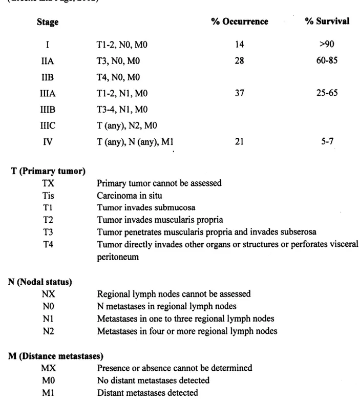
Table 1. Staging for colorectal cancer, percentage of occurrence and percentage of survival ..2
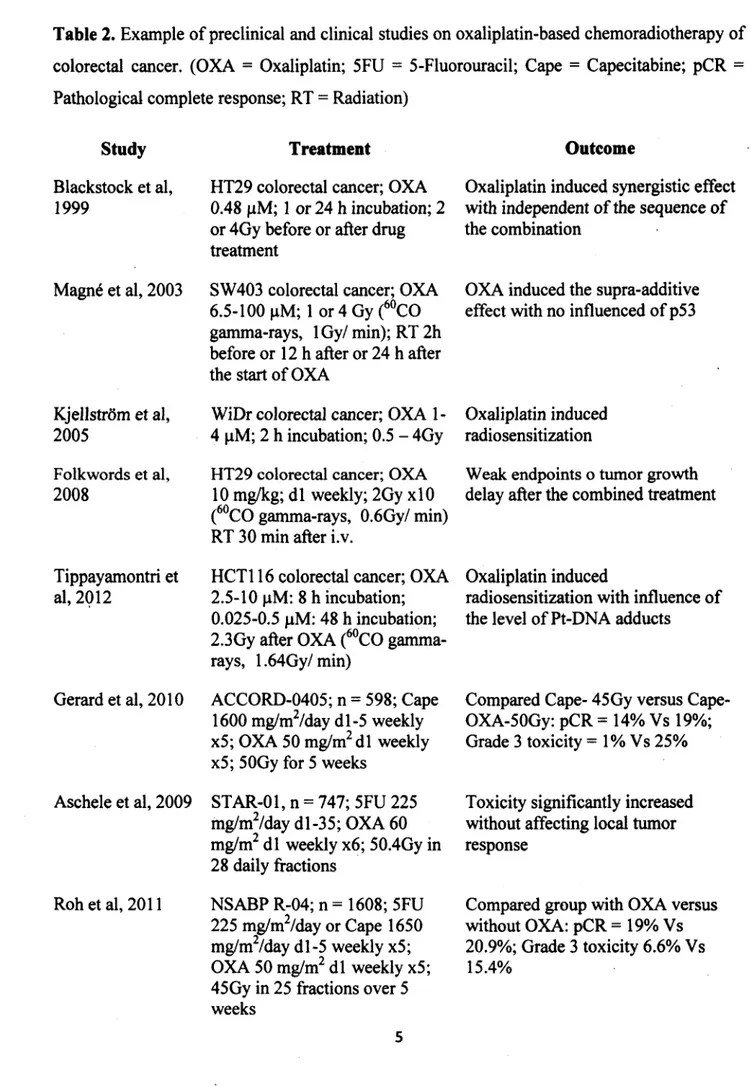
Table 2. Example of preclinical and clinical studies on oxaliplatin-based chemoradiotherapy of colorectal cancer - ... 5-6 Figure 1. The chemical structures of cisplatin and oxaliplatin... 7
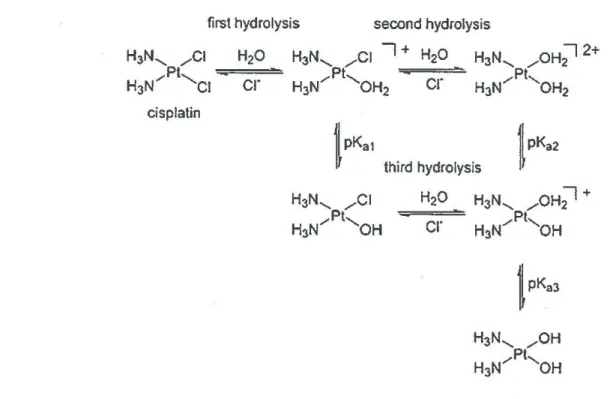
Figure 2. The hydrolysis reaction scheme of cisplatin... 8
Figure 3. The hydrolysis reaction scheme of oxaliplatin...9
Figure 4. Mechanism o f platinum uptake and efflux... 10
Figure 5. Formation of cisplatin- and oxaliplatin-DNA adducts... 12
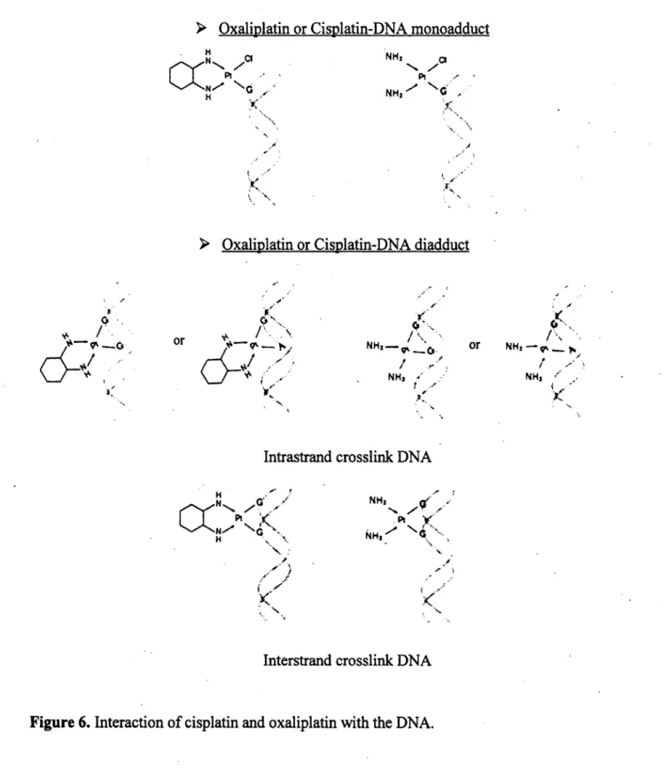
Figure 6. Interaction of cisplatin and oxaliplatin with the DNA... 13
Figure 7. Mechanism of cell death after platinum chemotherapy... 15
Figure 8. Depiction of a liposomal formulation... 17
Figure 9. Liposome-cell interactions... 18
Figure 10. The theoretical framework for the prediction of the outcome o f combined treatments with cytotoxic chemotherapy and radiaotherapy...21
Table 3. The mechanisms of chemotherapy and radiation interaction... . 22
Figure 11. Comparison o f the yield of SSB and DSB as a function of different cisplatin to plasmid DNA ratios... 24
Table 4. Example of previous studies of combined treatment of cisplatin, oxaliplatin, Lipoplatin™ and Lipoxal™ plus radiation... 26-27
LIST OF ILLUSTRA TI ONS (TABLES AND FIGURES)
CHAPTER I -- INTRODUCTION
Table 1. Staging for colorectal cancer, percentage of occurrence and percentage of survival .. 2
Table 2. Example of preclinical and clinicat studies on oxaliplatin-based
chemoradiotherapy of colorectal cancer ... ·-··· .. ··· ... 5-6
Figure
1.The chemical structures of cisplatin and oxaliplatin ... ; ... 7
Figure 2. The hydrolysis reaction scheme of cisplatin ...
8
Figure 3. The hydrolysis reaction scheme of oxaliplatin ... 9
Figure 4. Mechanism of platinum uptake and efflux ... 10
Figure 5. Formation of cisplatin- and oxaliplatin-DNA adducts ... 12
Figure 6. Interaction of cisplatin and oxaliplatin with the DNA ... 13
Figure 7. Mechanism of cell death after platinum chemotherapy ... 15
Figure 8. Depiction of a liposomal formulation ... , ... 17
Figure 9. Liposome-cell interactions ... 18
Figure 10. The theoretical framework for the prediction of the outcome of combined
treatments with cytotoxic chemotherapy and radiaotherapy ... 21
Table 3. The mechanisms of chemotherapy and radiation interaction ... : .... 22
Figure 11. Comparison of the yield of SSB and DSB as a function of different cisplatin
to plasmid DNA ratios ... 24
Table 4. Example of previous studies of combined treatment of cisplatin, oxaliplatin,
CHAPTER II - ARTICLE No.l
Figure 1. Chemical structures o f platinum-based drugs studied and schematic of
a liposome... 34
Table 1. IC50 values o f the platinum compounds for the HCT116 cells...37
Figure 2, Time course of platinum derivatives accumulation in HCT116 cells... 38
Figure 3. Cell proliferation in presence of the platinum derivatives !...39
Figure 4. Amount o f platinum bound to DNA after exposing the HCT116 cells to platinum drugs for 4 and 24 h ... 40
Figure 5. Time course o f platinum binding to DNA after exposing the HCT116 cells ... 41
Figure 6. Distribution of platinum compounds in cytoplasm or bound to DNA... 42
CHAPTER III - ARTICLE No.2 Figure 1. Dose response curve for radiotherapy alone on HCT116 cells... 61
Figure 2. Response curves for chemoradiotherapy on HCT116 cells at 8 h incubation time.. 62
Figure 3. Response curves for chemoradiotherapy on HCT116 cells at 48 h incubation time...63
Figure 4. The IC50 vialues of cisplatin, oxaliplatin and their liposomal formulation (Lipoplatin™ and Lipoxal™) in combination with radiation... 64
Figure 5. The combination index (Cl) of chemoradiotherapy on HCT116 ce lls...6 6 Figure 6. Flow cytometric measurement of cell-cycle distribution of the HCT116 c e ll... 69
TV /I * r w Figure 7. Induction of apoptosis by cisplatin, oxaliplatin, Lipoplatin and Lipoxal combination with radiation... 70
CHAPTER II -- ARTICLE No.1
Figure 1. Chemical structures of platinum-based drugs studied and schematic of a liposome ... : ... 34Table 1. IC50 values of the platinum compounds for the HCTI 16 cells ... 37
Figure 2 .. Time course of platinum derivatives accumulation in HCTI 16 cells ... 38
Figure 3. Cell proliferation in presence of the platinum derivatives ... : ... 39
Figure 4. Amount ofplatinum bound to DNA after exposing the HCTI 16 cells to platinum drugs for 4 and 24 h ... : ... 40
Figure 5. Tune course of platinum binding to DNA after exposing the ~CTl 16 cells ... 41
Figure 6. Distribution of platinum compounds in cytoplasm or bound to DNA. ... 42
CHAPTER
III --ARTICLE No.2
Figure 1. Dose response curve for radiotherapy alone on HCTI 16 cells ... : ... ; 61Figure 2. Response curves for chemoradiotherapy on HCTl 16 cells at 8 h incubation ti:qie .. 62
Figure 3. Response curves for chemoradiotherapy on HCTl 16 cells at 48 h incubation time ... 63
Figure 4. The ICso values of cisplatin, oxaliplatin and their liposomal fonnulation (Lipoplatin TM and LipoxalTM) in combination with radiation ... 64
Figure 5. The combination index (CI) of chemoradiotherapy on HCTI 16 cells ... 66
Figure 6. Flow cytometric measurement of cell-cycle distribution of the HCTl 16 cell ... 69
Figure 7. Induction of apoptosis by cisplatin, oxaliplatin, Li pop latin TM and Lipoxal
™
combination with radiation ... 70CHAPTER IV - ARTICLE No.3
Table 1. Change in body weight of male nu/nu nude mice treated with i.v. oxaliplatin
or Lipoxal™ in combination with GK irradiation... 91 Figure 1. The experiment setup for chemoradiotherapy in nu/nu nude mice model... 92 Figure 2. Pharmacokinetic profiles of platinum concentration in blood, tumor tissues
TK/C
and normal tissues after a single i.v. injection of oxaliplatin or Lipoxal ... 95-96 Figure 3. Time dependence of platinum concentration in cytoplasm, nucleus and
platinum-DNA adducts obtained after a single i.v. injection of oxaliplatin or Lipoxal™ 99 Figure 4. The tumor response after nu/nu nude mice were treated initially with
chemotherapy and followed by radiotherapy...1 0 0 Table 2. In vivo efficacy of platinum-based chemotherapy alone and in combination
with radiation treatment... 101
Figure 5. The combination index of chemoradiotherapy in nude mice model ...103 Figure 6. TUNEL assay after chemoradiotherapy of oxaliplatin and Lipoxal™... 104
CHAPTER V - ARTICLE No.4
Table 1. Change in body weight of male nu/nu nude mice treated with i.v. cisplatin or TK/f
Lipoplatin in combination with Gamma Knife irradiation... 124 Figure 1. Pharmacokinetic profiles o f platinum concentration in whole blood,
serum, kidney, liver, muscle, and tumor after a single i.v. injection of cisplatin
or Lipoplatin™... 126-127 Figure 2. Time dependence of platinum concentration in cytoplasm, nucleus and
platinum-DNA adducts obtained after a single i.v. injection of cisplatin or Lipoplatin™...129 Figure 3. The tumor growth delay after initial treatment of nude mice bearing HCT116 colorectal tumor with cisplatin and Lipoplatin™ followed by radiotherapy... 131
CHAPTER IV -- ARTICLE No.3
Table 1. Change in body weight of male nu/nu nude mice treated with i.v. oxaliplatin
or Lipoxal™ in combination with GK irradiation ... 91 Figure 1. The experiment setup for chemoradiotherapy in nu/nu nude mice model.. ... 92 Figure 2. Pharmacokinetic profiles of platinum concentration in blood, tumor tissues
and nonnal tissues after a single i.v. injection of oxaliplatin or Lipoxal™ ... 95-96 Figure 3. Time dependence of platinum concentration in cytoplasm, nucleus and
platinum-DNA adducts obtained after a single i.v. injection of oxaliplatinor Lipoxal™ ... 99 Figure 4. The tumor response after nu/nu nude mice were treated initially with
chemotherapy and followed by radiotherapy ... 100 Table 2.
In vivo
efficacy of platinum-based chemotherapy alone and in combination.with radiation treatment ... : ... 101 Figure S. The combination index of chemoradiotherapy in nude mice model. ... 103
. .
Figure 6. TUNEL assay after chemoradiotherapy of oxaliplatin and Lipoxal™ ... 104
CHAPTER V -- ARTICLE No.4
Table 1. Change in body weight of male nu/nu nude mice treated with i.v. cisplatin or
Lipoplatin™ in combination with Gamma Knife irradiation ... , ... 124 Figure 1. Pharmacokinetic profiles of platinum concentration in whole blood,
serum, kidney, liver, muscle, and tumor after a single i.v. injection of cisplatin
or Lipoplatin ™ ... .-.. ... 126-127 Figure 2. Time dependence of platinum concentration in cytoplasm, nucleus and
pla~um-DNA adducts obtained after a single i.v. injection of cisplatinor Lipoplatin™ ... 129 Figure 3. The tumor growth delay after initial treatment of nude mice bearing HCTl 16
Table 2. Efficacy of platinum-based chemotherapy alone and in combination with
radiation treatment...133 Figure 4. Assessment of apoptosis after chemoradiotherapy of cisplatin and
Lipoplatin™ ... 134
CHAPTER VI - DISCUSSION
Table 1. The platinum-DNA binding after expousred HCT116 to cisplatin, oxaliplatin,
and their liposomal formulation for 4 h and 24 h of drug incubation... 151 Figure 1. Diagram illustrated phases of cell cycle and their relative radiosensitivity or
radioresistance... 156 Figure 2. Induction of the mitochondrial apoptotic pathway by cisplatin...161 Figure 3. Histophatologic changes o f HCT116 human colorectal cancer tumors
treated with platinum drugs and radiation... 163-166 Figure 4. Possible improvement for clinical chemoradiation treatment apply from our
preclinical studies... 168
Table 2. Lists of other chemotherapeutic agents that are commonly combined with
platinum drugs... 170
Table 2. Efficacy of platinum-based chemotherapy alone and in combination with
radiation treatment ... 133
Figure
4.Assessment of apoptosis after chemoradiotherapy of cisplatin and
Li pop latin™ ... 134
CHAPTER VI -- DISCUSSION
Table 1. The platinum-DNA binding after expousred HCTl 16 to cisplatin, oxaliplatin,
and their liposomal formulation 'for 4 h and 24 h of drug incubation ... 151
Figure
1.Diagram illustrated phases of cell cycle and their relative radiosensitivity or
radioresistance ... 156
Figure 2. Induction of the mitochondrial apoptotic pathway by cisplatin ... 161
Figure 3. Histophatologic changes ofHCTl 16 human colorectal cancer tumors
treated with platinum drugs and radiation ... 163-166
Figw:e
4.Possible improvement for clinical chemoradiation treatment apply from our
preclinica/ studies ...
168Table
2.Lists of other chemotherapeutic agents that are commonly combined with
LIST OF ABBREVIATIONS/SYMBOLS Pt Platinum DACH Diaminocyclohexane CDDP Cisplatin 5-FU 5-Fluorouracil FA Folinic acid
DPPG Dipalmitoyl phosphatidyl glycol PEG Polyethylene glycol
NER Nucleotide excision repair
MMR Mitmatch repair
HMG High-mobility group
OCT Organic cation transporter CTR Copper treansporter
GSH Glutathione
MT Metallothionein
DNA Dioxyribonucleic acid
RNA Ribonucleic acid
G Gaunine
A Adenine
SSB Single strand break DSB Double strand break
ICP-MS Inductively coupled plasma mass spectrometer
IR Ionizing radiation 60c o Cobalt-60 GK GammaKnife
Pt
DACH
CDDP
5-FU
FA
DPPG
PEG
NER MMRHMG
OCT
CTR
GSH
MT
DNA
RNA G ASSB
DSB
ICP-MSIR
6oco
GK
Gy
LIST OF ABBREVIATIONS/SYMBOLS
Platinum
Diaminocyclohexane
Cisplatin
5-Fluorouracil
Folinic acid
Dipalmitoyl phosphatidyl glycol
Polyethylene glycol
Nucleotide excision repair
Mitmatch repair
High-mobility group
Organic cation transporter
Copper treansporter
Glutathione
Metallothionein
Dioxyribonucleic acid
Ribonucleic acid
Gaunine
Adenine
Single strand break
Double strand break
Inductively coupled plasma mass spectrometer
Ionizing radiation
Cobalt-60
GammaKnife
eV Electronvolt tl/2 Half-life h Hour Micromolar Hg Microgram ng Nanogram mg/kg Milligram/kilogram
Td Tumor growth delay
eV
Electronvolt
t112Half-life
hHour
µM
Micromolar
µg
Microgram
ng
Nanogram
mg/kg
· Milligram/kilogram
CHAPTER I
INTRODUCTION
Colorectal cancer
Colorectal cancer occurs when abnormal cells grow in the lining of both the colon and rectum. Colorectal cancer is one of the most common types of cancer worldwide, accounting for a significant percentage of cancer mortality (International Agency for Research on Cancer- World Health Organization, www.iarc.fr). In Canada, mortality rates continue to decline in both sexes by 2.6% per year since 2003, however, colorectal cancer is still the second leading cause of death from cancer, behind lung cancer in men and breast cancer in women. The Canadian Cancer Society has estimated that there will be about 23,300 new cases o f colorectal cancer in 2012. On average, 64 Canadians will be diagnosed with colorectal cancer and 25 Canadians will die o f this disease each day (Canadian Cancer Society, www.cancer.ca).
Colorectal cancer
CHAPTERI
INTRODUCTION
Colorectal cancer occurs when abnonnal cells grow in the lining of both the colon and rectum. Colorectal cancer is one of the most common types of cancer worldwide, accounting for a significant percentage of cancer mortality (International Agency for Research on Cancer-World Health Organization, www.iarc.fr).
In
Canada, mortality rates continue to decline in both sexes by 2.6% per year since 2003, however, colorectal cancer is still the second leading cause of death from cancer, behind lung cancer in men and breast cancer in women. The Canadian Cancer Society has estimated that there will be about 23,300 new cases of colorectal cancer in 2012. On average, 64 Canadians will be diagnosed with colorectal cancer and 25 Canadians will die ofthis disease each day (Canadian Cancer Society, www.cancer.ca).Table 1. Staging for colorectal cancer, percentage of occurrence and percentage o f survival
(Greene and Page, 2002)
Stage % Occurrence % Survival
I Tl-2, NO, MO 14 >90 IIA T3, NO, MO 28 60-85 IIB T4, NO, MO IIIA Tl-2, N I, MO 37 25-65 IIIB T3-4, N I, MO IIIC T (any), N2, MO IV T (any), N (any), M l 21 5-7 T (Primary tumor)
TX Primary tumor cannot be assessed Tis Carcinoma in situ
T1 Tumor invades submucosa
T2 Tumor invades muscularis propria
T3 Tumor penetrates muscularis propria and invades subserosa
T4 Tumor directly invades other organs or structures or perforates visceral peritoneum
N (Nodal status)
NX Regional lymph nodes cannot be assessed NO N metastases in regional lymph nodes
N 1 Metastases in one to three regional lymph nodes N2 Metastases in four or more regional lymph nodes
M (Distance metastases)
MX Presence or absence cannot be determined MO No distant metastases detected
M l Distant metastases detected
Table 1. Staging for colorectal cancer, percentage of occurrence and percentage of survival
(Greene and Page, 2002)
Stage
I
HA % Occurrence Tl-2, NO, MO T3, NO, MO HB T4, NO, MO HIA Tl-2, NI, MO HIB T3-4, NI, MO HIC IV T (Primary tumor) TX Tis Tl T2 T (any), N2, MO T (any), N (any), MlPrimary tumor cannot be assessed Carcinoma in situ
Tumorinvadessubmucosa
Tumor invades muscularis propria 14 28 37 21 % Survival >90 60-85 25-65 5-7
T3 Tumor penetrates muscularis propria and invades subserosa T4 N (Nodal status)
NX
NO NI N2Tumor directly invades other organs or structures or perforates visceral peritoneum
Regional lymph nodes cannot be assessed N metastases in regional lymph nodes
Metastases in one to three regional lymph nodes Metastases in four or more regional lymph nodes
M (Distance metastases)
MX Presence or absence cannot be determined MO No distant metastases detected
Table 1 shows the colorectal cancer staging, the percentage of occurrence and percentage of survival (Greene and Page, 2002). Surgical resection at an early stage is still the only chance for a cure in patients with colorectal cancer (Canadian Cancer Society, www.cancer.ca). The role o f adjuvant chemotherapy (5-fluorouracil) in stage II (T3/4, NO) is debatable, there is a minimal benefit. For patients with stage III (TX, N I) rectal cancer, adjuvant chemoradiotherapy was considered the standard treatment because it improved local control and overall survival compared with surgery alone or surgery plus radiation (Krook et al., 1991). Neoadjuvant chemoradiotheraapy protocols were mainly used for locally advanced rectal cancers, aiming to achieve subsequent curative resection by decreasing tumor size and decrease the risk of local recurrence (Kapiteijn et al., 2001). The radiotherapy is the use of carefully measured dose of radiation (e.g. adjuvant radiotherapy is typically given 1.8 Gy per day for 5 days per week for a number of weeks, total dose of 50.4 Gy). When the dose per day is increased, the total number of days is correspondingly decreased to limit the risks for injury to normal tissue from the increasing dose per fraction (Robertson, 2008). Fractionation radiotherapy allows normal cells to recover and also allows tumor cells that were in a relatively radio-resistant phase of the cell cycle during one treatment to cycle into a sensitive phase o f the cycle before the next fraction is given (Hall and Giaccia, 2005). Palliative therapy is recommended for patients with stage IV (Andre and Schmiegel, 2005).
Three recent completed clinical phase III trials (ACCORD-0405, STAR-01 and NSABPR-04) that added oxaliplatin to 5-FU or capecitabine treatment have failed to improve results of chemoradiotherapy (Aschele et al., 2011; Gerard et al., 2010 and Roh et al., 2009), with only the results from the German CAO/ARO/AIO-04 study (Rodel et al., 2012) that showed an increase in the treatment efficacy when added oxaliplatin to 5-FU plus radiation
Table 1 shows the colorectal cancer staging, the percentage of_ occurrence and percentage of survival (Greene and Page, 2002). Surgical resection at an early stage is still the only chance for a cure in patients with colorectal cancer (Canadian Cancer Society, www.cancer.cà). The role of adjuvant chemotherapy (5-fluorouracil) in stage II (T3/4, NO) is debatable, there is a minimal benefit. For patients with stage III (TX, Nl) rectal cancer, adjuvant chemoradiotherapy was conside~ed the standard treatment because it improved local control and overall survival compared with surgery alone or surgery plus radiation (Krook et al., 1991). Neoadjuvant chemoradiotheraapy protocols were mainly used for locally advanced rectal cancers, aiming to achieve subsequent curative resection by decreasing tumor size and decrease the risk of local recurrence (Kapiteijn et al., 2001). The i:adiotherapy is the use· of carefully measured dose of radiation ( e.g. adjuvant radiotherapy is typically given 1.8 Gy per dày for 5 days per week for a number ofweeks, total dose of 50.4 Gy). When the dose per day is increased, the total number of days is correspondingly decreased to limit the risks for injury to normal tissue from the increasing dose per fraction (Robertson, 2008). Fractionation radiotherapy allows normal cells to recover and also allows tumor cells that were in a relatively radio-resistant phase of the cell cycle during one treatment to cycle into a sensitive phase of the cycle before the next fraction is given (Hall and Giaccia, 2005). Palliative therapy is recommended for patients with stage IV (Andre and Schmiegel, 2005).
Three recent completed clinical phase III trials (ACCORD-0405, STAR-01. and NSABPR-04) that added oxaliplatin to 5-FU or capecitabine treatment have failed to improve results of chemoradiotherapy (Aschele et al., 2011; Gerard et al., 2010 and Roh et al., 2009), with only the results from the German CAO/ARO/AIO-04 study (Rodel et al., 2012) that showed an increase in the treatment efficacy when added oxaliplatin to 5-FU plus radiation
(Table 2). From the various treatment studies, non-optimal schedule for rectal cancer treatment may be one of the important factors in clinical treatment failure. Despite the fact that approximately 70-80% of patients are eligible for curative surgical resection at the time of diagnosis (Faivre-Finn et al., 2002), 5 years overall survival rate is only 50-60% et al., 2003). Some 6 6% of patients who undergo curative resection will experience local recurrence or distant metastases. In 85% of people, relapse was diagnosed within the first 2.5 years after surgery (Gill et al., 2004). Upon diagnosis o f metastatic disease, patients have a median survival rate of only 6 months and the 5-year overall survival rate was less than 10% (Greene and Page, 2002).
Importantly, severe side effects of either chemotherapy or radiotherapy can limit the potential for dose escalations. Chemotherapy is known to cause severe adverse reactions including renal toxicity, gastrointestinal toxicity, peripheral neuropathy, asthenia and ototoxicity (Howell, 1991; Kweekel et al., 2005 and O'Dwyer et al., 2000). Radiotherapy potentially can cause skin irritation, nausea, diarrhea, trouble controlling o f bowels, rectal or bladder irritation, and tiredness (Hall and Giaccia, 2005). This will limit the anticancer efficiency of both modalities. Moreover, the optimal schedule for adding platinum-based drugs to the standard chemoradiotheapy for treating rectal cancer is not yet determined. Investigations to improve the chemoradiation schedule for rectal adenocarcinoma to attain the maximum concomitant effect, while minimizing the systemic toxicities is thus very important.
(Table 2). From the various treatment studies, non-optimal schedule for rectal cancer treatment may be one of the important factors in clinical treatment failure. Despite the fact that approximately 70-80% of patients are eligible for curative surgical resection at the time of diagnosis (Faivre-Finn et al., 2002), 5 years overall survival rate is only 50-60% et al., 2003). Sorne 66% of patients who undergo curative resection will experience local recurrence or distant metastases. In 85% of people, relapse was diagnosed within the first 2.5 years after surgery (Gill et al., 2004). Upon diagnosis of metastatic disease, patients have a median survival rate of only 6 months and the 5-yëar overall survival rate was less than 10% (Greene and Page, 2002).
Importantly, severe side effects of either chemotherapy or radiotherapy can limit the potential for dose escalations. Chemotherapy is known to cause severe adverse reactions including renal toxicity, gastrointestinal toxicity, peripheral neuropathy, as~enia and ototoxicity (Howell, 1991; Kweekel et al., 2005 and O'Dwyer et al., 2000). Radiotherapy potentially can cause skin irritation, nausea, diarrhea, trouble controlling of bowels, rectal or bladder irritation, and tiredness (Hall and Giaccia, 2005). This will limit the anticancer efficiency of both modalities. Moreover, the optimal schedule for adding platinum-based drugs to the standard chemoradiotheapy for treating ~ectal cancer is not yet determined. Investigations to improve the chemoradiation schedule for rectal adenocarcinoma to attain the maximum concomitant effect, while minimizing the systemic toxicities is thus very important.
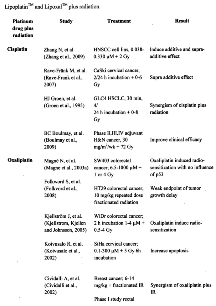
Table 2. Example of preclinical and clinical studies on oxaliplatin-based chemoradiotherapy of
colorectal cancer. (OXA = Oxaliplatin; 5FU = 5-Fluorouracil; Cape = Capecitabine; pCR = Pathological complete response; RT = Radiation)
Study Blackstock et al, 1999 Magn6 et al, 2003 Kjellstrom et al, 2005 Folkwords et al, 2008 Tippayamontri et al, 2 0 1 2 Gerard et al, 2010 Aschele et al, 2009 Roh et al, 2011 Treatment
HT29 colorectal cancer; OXA 0.48 pM; 1 or 24 h incubation; 2 or 4Gy before or after drug treatment
SW403 colorectal cancer; OXA 6.5-100 pM; 1 or 4 Gy (60CO gamma-rays, lG y/ min); RT 2h before or 12 h after or 24 h after the start of OXA
WiDr colorectal cancer; OXA 1- 4 pM; 2 h incubation; 0.5 - 4Gy HT29 colorectal cancer; OXA
10 mg/kg; d l weekly; 2Gy xlO (60CO gamma-rays, 0.6Gy/ min) RT 30 min after i.v.
HCT116 colorectal cancer; OXA 2.5-10 pM: 8 h incubation; 0.025-0.5 pM: 48 h incubation; 2.3Gy after OXA (60CO gamma- rays, 1.64Gy/min)
ACCORD-0405; n = 598; Cape 1600 mg/m2/day dl-5 weekly x5; OXA 50 mg/m2 dl weekly x5; 50Gy for 5 weeks
STAR-01, n = 747; 5FU 225 mg/m2/day dl-35; OXA 60 mg/m2 d l weekly x6; 50.4Gy in 28 daily fractions NSABP R-04; n = 1608; 5FU 225 mg/m2/day or Cape 1650 mg/m2/day dl-5 weekly x5; OXA 50 mg/m2 d l weekly x5; Outcome
Oxaliplatin induced synergistic effect with independent o f the sequence of the combination
Weak endpoints o tumor growth delay after the combined treatment
Oxaliplatin induced
radiosensitization with influence o f the level of Pt-DNA adducts
Compared Cape- 45Gy versus Cape- OXA-50Gy: pCR = 14% Vs 19%; Grade 3 toxicity = 1% Vs 25%
Compared group with OXA versus without OXA: pCR = 19% Vs 20.9%; Grade 3 toxicity 6.6% Vs 15.4%
OXA induced the supra-additive effect with no influenced of p53
Oxaliplatin induced radiosensitization
Toxicity significantly increased without affecting local tumor response
Table 2.
Example of preclinical and clinical studies on oxaliplatin-based chemoradiotherapy of colorectal cancer. (OXA = Oxaliplatin; 5FU=
5-Fluorouracil; Cape = Capecitabine; pCR=
Pathological complete response; RT=
Radiation)Study
Treatment
Outcome
Blackstock et al, HT29 colorectal cancer; OXA Oxaliplatin induced synergistic effect 1999 0.48 µM; l or 24 h incubation; 2 with independent of the sequence of
or 4Gy before or after drug the combination treatment
Magné et al, 2003 SW 403 colorectal cancer; OXA OXA induced the supra-additive 6.5-100 µM; 1 or 4 Gy (6°CO effect with no influenced of p53
gamma-rays, 1 Gy/ min); RT 2h before or 12 h after or 24 h after the start ofOXA
Kjellstrom et al, WiDr colorectal cancer; OXA 1- Oxaliplatin induced 2005 4 µM; 2 h incubation; 0.5 - 4Gy radiosensitization
Folkwords et al, HT29 colorectal cancer; OXA Weak endpoints o tumor growth 2008 10 mg/kg; dl weekly; 2Gy xlO delay after the combined treatment
(60
co
gamma-rays, 0.6Gy/ min) RT 30 min after i.v.Tippayamontri et HCTl 16 colorectal cancer; OXA Oxaliplatin induced
al, 2Ql2 2.5-10 µM: 8 h incubation; radiosensitization with influence of 0.025-0.5 µM: 48 h incubation; the level of Pt-DNA adducts
2.3Gy after OXA (60
co
gamma-rays, l .64Gy/ min)Gerard et al, 2010 ACCORD-0405; n
=
598; Cape Compared 45Gy versus Cape-1600 mg/m2/day dl-5 weekly OXA-50Gy: pCR= 14% Vs 19%; x5; OXA 50 mg/m2 dl weekly Grade 3 toxicity=
1 % Vs 25% x5; 50Gy for 5 weeksAschele et al, 2009 STAR-01, n
=
747; 5FU 225 Toxicity significantly increased mg/m2/day dl-35; OXA 60 without affecting local tumor mg/m2 dl weekly x6; 50.4Gy in response28 daily fractions
Roh et al, 2011 NSABP R-04; n = 1608; 5FU Compared group with OXA versus 225 mg/m2/day or Cape 1650 without OXA: pCR
=
19% Vs mg/m2/day dl-5 weekly x5; 20.9%; Grade 3 toxicity 6.6% Vs OXA 50 mg/m2 dl weekly x5; 15.4%Rddel etal, 2012 The German CAO/ARO/AIO,- Improve pCR 04; n = 1265; 5FU 250
mg/m2/day d l-1 4 ,22-35; OXA 50 mg/m2 d l, 8 ,2 2,29; 50.4Gy
Platinum (Pt) compounds -Cisplatin and Oxaliplatin History
In 1965, Rosenberg and his group discovered the potential of platinum-based drugs to inhibit cell division (Rosenberg, 1980). They investigated the effect of electronic current on bacterial mobility and, noticed that the cells under study formed a filamentous structure, which is a characteristic of un-dividing cells. The chemical compound causing this structure was cis- diamminedichloroplatinum (II), or cisplatin, a compound known since the 1860’s as “Peryone’s Chloride” (Rosenberg et al., 1969). After this finding, several studies o f cisplatin for cancer treatment were performed, and cisplatin was approved for clinical use in 1979 (Rosenberg et al., 1969).
The treatment with cisplatin consists of a course of intravenous injections administered every 3-4 weeks at a dose of 50-120 mg/m2 (Adelstein et al., 2003). Such treatment has a reported high efficacy for testicular, ovarian and bladder cancers, and other solid tumors (Begg, 1990). Over the years, various platinum complexes have been studied in an attempt to overcome the severe side effects of cisplatin et al., 1998). Oxaliplatin or (trans-R,R)l,2- diaminocyclohexaneoxalotoplatinum (II), is a third generation platinum-based compound in the alkylating family of anticancer agents and the first clinically available diaminocyclohexane (DACH) platinum compound. Oxaliplatin is distinctive from cisplatin and is non-cross-resistant with some platinum-resistant tumors. Although oxaliplatin was discovered 34 years ago, it only Rôdel et al, 2012 The German CAO/ARO/AIO,- lmprove pCR
04; n
=
1265; 5FU 250mg/m2/day dl-14, 22-35; OXA 50 mg/m2 dl, 8, 22, 29; 50.4Gy
Platinum (Pt) compounds -Cisplatin and Oxaliplatin
History
In 1965, Rosenberg and his group discovered the potential of platinum-based drugs to
inhibit cell division (Rosenberg, 1980). They investigated the effect of electronic current on bacterial mobility and, noticed that the cells under study formed a filamentous structure, which is a characteristic of un-dividing cells. The chemical compound causing this structure was cis-diamminedichloroplatinum (II), or cisplatin, a compound known since the 1860's as "Peryone's Chloride" (Rosenberg et al., 1969). After this finding, several studies of cisplatin for cancer treatment were performed, and cisplatin was approved for clinical use in 1979 (Rosenberg et al., 1969).
The treatment with cisplatin consists of a course of intravenous injections administered every 3-4 weeks at a dose of 50-120 mg/m2 (Adelstein et al., 2003). Such treatment has a reported high efficacy for testicular, ovarian and bladder cancers, and other solid tumors (Begg, 1990). Over the years, various platinum complexes have been studied in an attempt to overcome the severe side effects of cisplatin et al., 1998). Oxaliplatin or (trans-R,R)l,2-diaminocyclohexaneoxalotoplatinum (Il), is a third generation platinum-based compound in the alkylating family of anticancer agents and the first clinically available diaminocyclohexane (DACH) platinum compound. Oxaliplatin is distinctive from cisplatin and is non-cross-resistant with some platinum-resistant tumors. Although oxaliplatin was discovered 34 years ago, it only
gained Food and Drug Administration (FDA) approval 8 years ago, specifically for the management of advanced human colorectal cancer, for use in combination with 5-fluorouracil (5FU) and folinic acid (FA) (National Cancer Institute at the national institute of health, www.nih.gov). Unlike cisplatin, oxaliplatin is non-nephrotoxic with the main toxicity being neuropathy. Neurotoxicity is dose-limiting. Oxaliplatin is commonly given at the dose of 85 mg/m2 i.v. injection every second week (Alcindor and Beauger, 2011).
Chemical structure
The chemical structure of cisplatin and oxaliplatin in 2D and 3D are shown in Figure 1. Cisplatin has a square-planar structure with the central platinum (Pt) coordinated by two chlorides (Cl) ion ligands and other two ammonia (NH3) ligands in cis to each other.
Oxaliplatin is a much bigger organic compound, with the two amine NH3 ligands in cisplatin substituted by the (trans-R,R)l ,2-diaminocyclohexan (DACH) ligand to improve antitumor activity; and the two chloride ion ligands replaced by the oxalatobidentate ligand derived from oxalic acid to improve water solubility.
Chemistry
Cisplatin of molecular formula H6Cl2N2Pt has molecular weight 301.1 g/mol. There is
evidence suggesting that due to a high concentration (about 100 mM) of chloride ion~ outside
of cell membranes, cisplatin remains in the dichloro form until it passes through the cell membrane. Inside the cell membrane, the chloride ion concentration is about 4 mM which allows the hydrolysis process to take place (Hall et al., 2008). In aqueous solution, the
cr
ion is slowly lost and replaced by a water molecule (H2O) to form the mono-aqua product,cis-[Pt(NH3)i(OH2)Clt. Further substitution of the remaining chloroligand leads to the formation
of the diaquaplatinum complex, cis-[Pt(NH3)2(OH2)2]2\Figure 2). At physiological pH
(pH=7.87), the equation shifts and the majority of molecules present are in the diaqua form, the active form of cisplatin (Dionisi et al., 2011; Esteban-Fernandez et al., 2010).
first hydrolysis second hydrolysis
H3N, _,,..Cl H20 H3N..._ _,,..Cl 7 + H20
Pt
H3N.,,,
'--et cr
H3N/ 'OH2cr
cisplatincr
Figure 2. The hydrolysis reaction scheme of cisplatin (Reprinted with permission from Lau and
The oxaliplatin molecule has the formula CgHi4N20 4Pt and a molecular weight o f 397.3 g/mol. Oxaliplatin slowly hydrolyzes in water according to following reaction: [(DACH)Pt(C20 4)] + 2H20 -> [(DACH)Pt(H20 )]2+ + C2042+(Esteban-Femandez et al., 2010). The oxalate group can be shifted by nucleophile groups such as CT or HCO3' and H20 (Figure 3).
Upon entering the cell, cisplatin and oxaliplatin are biotransformed to their reactive species. Through non-enzymatic reactions, the chloride atom of cisplatin and oxalate group of oxaliplatin are displaced by H20 forming biaquated biotransfoimation products that readily react with nucleophiles in the cells, including DNA, RNA and protein (Hall et al., 2008).
Figure 3. The hydrolysis reaction scheme of oxaliplatin. Permission license # 3034930431133 (Kweekel et al., 2005).
Mechanism of action
Interaction with the cell
o f platinum drugs. In the extracellular environment, platinum drugs can be associated with water molecules as explained previously. These may enter the cell or cross-react with extracellular proteins such as serum albumin, reducing its bioavailablility. Platinum drugs can
oxalate
Oxaliplatin Dichloro intermediate Diaquo interm ediate
Figure 4 illustrates the mechanisms affecting and controlling the cellular accumulation The oxaliplatin molecule has the formula C8H14N2O4Pt and a molecular weight of 397.3 g/mol. Oxaliplatin slowly hydrolyzes in water according to following reaction: [(DACH)Pt(C2O4)]
+
2H2O -> [(DACH)Pt(H2O)]2++
C20/\Esteban-Femandez et al., 2010). The oxalate group can be shifted by nucleophile groups such ascr
or HCO3" and H2O (Figure 3).Upon entering the cell, cisplatin and oxaliplatin are biotransformed to their reactive species. Through .non-enzymatic reactions, the chloride atom of cisplatin and oxalate group of oxaliplatin · are displaced by H2O forming biaquated biotransformation products that readily react with nucleophiles in the cells, including DNA, RNA and protein (Hall et al., 2008).
H . /0
C(
N...__,
/o-c
+2cr
Pt 1 ____. N/"-o-
c -oxalate H'o
2+Oxaliplatin Oichloro intermediate Diaquo intermediate
Figure 3. The hydrolysis reaction scheme of oxaliplatin. Permission license # 3034930431133
(Kweekel et al., 2005).
Mechanism of action Interaction with the cell
Figure 4 illustr~tes the mechanisms affecting and controlling the cellular accumulation of platinum drugs.
In
the extracellular environment, platinum drugs can be associated with water molecules as explained previously. These may enter the cell or cross-react with extracellular proteins such as serum albumin, reducing its bioavailablility. Platinum drugs can enter the cell by passive diffusion across the lipid bilayer as well as by fluid phase endocytosis.The main organic cation transporters such as OCTl-3, CTRl, and sodium-dependent process have been identified.
Inside the cell, platinum drugs can be deactivated by binding to the thiol-rich metallothionein (MT) or chelated by glutathione (GSH) and eftluxed from the cell via the GS-X pumps. Platinum drugs can also be entrapped in subcellular organelles such as vesicles or melanosomes in melanoma cells and then subsequently exported out from the cells (Wang and Lippard, 2005). The accumulation of platinum is a steady-state phenomenon which depends on both the rates of influx and efflux of platinum drugs. When the shift of the balance between influx and efflux occurs, an increase of the extrusion process leads to a reduction in cellular accumulation of platinum. Drugs that evade the detoxification and efflux processes can enter the nucleus and bind to DNA, eliciting apoptosis if the DNA lesion is not repaired.
Endocytosis Organic cation
Passive diffusion Pt Pt fct}--JOOmM Pt
l
r
K• {CO/} - - :U mMl
{P0/}= 9mM Pt1\
{Cf}- 4-50 mM [CO/} - - 10 mM Pt f PO/]- - 80 mM Pt Pt Pt Pt detoxification / ' Pt accumulation " Nucleus P--->
(Pt-DNAGSH, MT, Vesicles, Melanosome t adducts)
1
efflux PtInteraction with the DNA
The clinical efficacy o f platinum drugs against tumors is thought to be primarily due to the formation of platinum-DNA adducts which interfere with cellular repair and DNA replication and which therefore trigger a chain of cell regulatory events, ultimately leading to cell death (Saris et al., 1996).
On DNA, cisplatin and oxaliplatin preferentially react with the highly nucleophilic N7 position on guanine or adenine, and form coordinated covalent bonds (Figure 5). Platinum binds to DNA and causes a critical structure change in the DNA such as a bend of 45 degree, unwinding of DNA and causing destacking of the purine bases (Sharma et al., 2007).
Cisplatin and oxaliplatin are bifiinctional agents. They can bind to two sites in DNA with the following order of sequence preference: -GG- > -A G -» -GA. The resulting biadducts composed o f approximately 60-65% 1,2-intrastrand GG, 25-30% 1,2-intrastrand AG, 5-10% 1,3-intrastrand GXG and 1-3% interstrand GG (Figure 6) (Eastman, 1987). Although cisplatin and oxaliplatin form the same types o f adduct at the same sites on DNA, their structures are distinct (Sharma et al., 2007). The bulky DACH carrier ligand of oxaliplatin is thought to contribute to a much bulkier adduct than cisplatin.
Interaction with the DNA
The clinical efficacy of platinum drugs against tumors is thought to be primarily due to the formation of platinum-DNA adducts which interfere with cellular repair and DNA replication and which therefore trigger a chain of cell regulatory events, ultimately leading to cell death (Saris et al., 1996).
On DNA, cisplatin and oxaliplatin preferentially react with the highly nucleophilic N7 position on guanine or ·adenine, and form coordinated covalent bonds (Figure 5). Platinum binds to DNA and causes a critical structure c~ange in the DNA such as a bend of 45 degre~, unwinding of DNA and causing destacking of the purine bases (Sharma et al., 2007).
Cisplatin and oxaliplatin are bifunctional agents. They can bind to two sites in DNA with the following order of sequence preference: -GG-> -AG->> -GA. The resulting biadducts composed of approximately 60-65% 1,2-intrastrand GG, 25-30% 1,2-intrastrand AG, 5-10% 1,3-intrastrand GXG and 1-3% interstrand GG (Figure 6) (Eastman, 1987). Although cisplatin and oxaliplatin form the same types of adduct at the same sites on DNA, their structures are distinct (Sharma et al·., 2007). The bulky DACH carrier ligand of oxaliplatin is thought to contribute to a much bulkier adduct than cisplatin.
0
{'0:NH
~~~;lNH,
Guanine (G) Adenine (A)
)
cis-diammine-Pt adducts
(Trans-RR) 1,2-diaminocyclohexane-Pt adducts
Figure S. Formation of cisplatin- and oxaliplatin-DNA adducts. Permission license #