Journal of Fundamental and Applied Sciences is licensed under aCreative Commons Attribution-NonCommercial 4.0 International License.Libraries Resource Directory. We are listed underResearch Associationscategory.
CONTRIBUTION TO STUDY THE GLYCOPROTEIN LIGANDS OF THE CEREBELLAR SOLUBLE LECTIN IN HUMAN K562 TUMOUR CELLS
H. Rechreche*and J.P. Zanetta
Molecular and Cellular Biology Laboratory (MCBL), Life Sciences Faculty, Mohamed Essedik Benyahia University, Jijel, Algeria.
Received: 10 May 2016 / Accepted: 29 August 2016 / Published online: 01 September 2016
ABSTRACT
Many cancer cells over-express significantly the glycoproteins specific to the endogenous cerebellar soluble lectin or CSL. These ligands may present the same electrophoretic profiles regardless of the specie or tissue. We purified a large amount of the active CSL using an immuno-affinity chromatography, which was used to isolate the CSL ligands from human tumour K562 cell lines. After protease digestion of these ligands, we analyzed the obtained peptides using reverse phase chromatography and isolated an overrepresented group that carried N-glycans and was relatively hydrophobic. Thus, we suggested that the CSL ligands have a common pepetide sequence specifically recognized by the CSL, who could direct the production of these CSL-recognized N-glycans. Moreover, we speculated that the expression deregulation of a specific exon encoding this peptide sequence alters the glycosylation in K562 tumour cells.
Keywords: CSL; glycoprotein ligands; tumour K562 cells; N-glycosylation; peptide signal.
Author Correspondence, e-mail:horechre@yahoo.fr
doi:http://dx.doi.org/10.4314/jfas.v8i3.11
1. INTRODUCTION
The cell surface glycoconjugates, including glycoproteins, glycolipids and proteoglycans are modified in the cancerous cells [1-6]. Nevertheless, the fonction of the glycosylation
ISSN 1112-9867
alterations in cancerogenesis is still incoherent, both inactivation and activation of glycosyltransferases participate in cancer development process [7,8]. The abnormal expression of oncogenes may control the biosynthesis of directly intervening glycosylation enzymes, however, the exact mechanism of these enzymatic disturbances is still unknown [7,9]. The promising assumptions suggesting the implication of the endogenous lectins in the malignant process are suffering from the missing of obvious relation [10,11]. The biological significance of these glycosylation aberrations in the cancerous cells is not clear; It can be either a key event at the cancer origin or one of its multiple consequences. The glycoconjugates influence the metastatic capacities of the cancerous cells and change depending to the cell differentiation and the development stage [2,9,12,13]. The disturbance of the glycobiologic interactions between the lectins and their ligands, including, the membrane glycoproteins participates in the carcinogenesis process [10,14-17].
The endogenous cerebellar soluble lectin (CSL) was initially isolated from the rat cerebellum [18,19]. It recognizes the glycoproteic glycans containing the oncofoetal epitopes [20-22]. The myelin-associated glycoprotein (MAG) and PO glycoprotein are CSL ligands [23-27]. The CSL ensures the molecules bridging between cellular surface glycoproteins during various biological processes, such as the myelin compaction and the junctions construction between axons and myelin producing cells [28-31]. The transformed cells possess a definitely higher number of CSL ligands, compared to the normal cells [31,32]. The CSL ligands overexpression generates a too great number of adhesion signals and contact inhibition loss, which prevents the cellular answer (nonsense signal) [32]. However, the increase in the CSL ligands levels does not stimulate the major glycoproteins expression; there is no modification on the total glycans biosynthesis. Furthermore, there is a practically perfect similarity between the CSL ligands profiles in various cell lines or malignant tumours, whatever is the tissue origin or the specie, suggesting that the CSL ligands had a specific profile in cancer [32]. The principal objective of this work was to seek if the CSL ligands specifically overexpressed in the tumour cells presente a consensus peptide, a sequence signal for the CSL-recognized glycans biosynthesis. We first prepared the CSL ligands-enriched material from the human K562 tumour cells. Then, we isolated the CSL ligands by affinity chromatography on
CSL-immobilized column and tried to establish their peptide maps using protease digestion and reverse phase chromatography.
2. EXPERIMENTAL
2.1. Chemicals, tissues and cell lines
Avidin-labeled alkaline phosphatase (avidin-AKP), bovine serum albumin (BSA), trypsin, protease V8, nitrotetrazolium blue, 5-bromo-4-chloro-indolyl phosphate, p-tosylarginine methylester (TAME), phenylmethylsufondyle fluoride (PMSF) and the culture medium RPMI61640 were purshed from Sigma (St Louis, MO, the USA). The N-hydroxy-succinimide ester biotin (NHS-Biotin) was from Bio-Rad (Ivry sur Seine, France). The sodium dodecyl sulfate (SDS) and the acrylamide were from Appligen (Strasbourg, France). The nitrocellulose filters (0.22 µm) was from Schleicher and Schuell (Dassel, Germany). The cyanogen bromide (CNBr)-activated Sepharose 4B was from Pharmacia (Upsala, Sweden). The Vytrac C18 HPLC column was from Interchim (Montlucon, France). The acetonitrile and trifluoracteic acid were from Merck (Darmstadt, Germany). Wistar albinos little rats (18-days old) were used in our study. The females in gestation were isolated just before the low setting and regularly supervised. The K562 tumour cells derived from human erythrocyte leukemia (K562 cells) were from the Center of Cancerlogy (Fondation Bergonié, Bordeaux, France). 2.2. SDS-page and western blot analysis
Prior to electrophoresis, the proteins were pulled down with 10% trichloroacetic acid (TCA) or methanol at pH 5. After centrifugation (3.400 g for 30 min), the pellet was rinsed with methanol and solubilized in 1% SDS and the proteins concentrations were determined [33]. Then, the pellets were solubilized in the dissociation solution (62 mM Tris-HCl pH 6.8, 2% SDS, 10% glycerol, 5% lhjkj, 1% bromophenol blue) as recommended by Laemmli and heated at 90°C for 5 min [34].The total proteins (20 µg/well) were separated by SDS-PAGE (13%) and colored by immersion in 1% Coomassie Blue Brilliant R (CBB) [35].
After electrophoresis, the proteins were bloted on nitrocellulose filters in buffer (12.5 mM Tris-HCl, 96 mM glycine containing 20% methanol and 0.04% SDS) under 36 V and 400 mA [36]. To check the quality of the electrophoretic migration and the effectiveness of the transfer,
a short coloring of the blots with the red culvert (0.2% red culvert in 3% TCA) was done. The filters were cleaned three times with water and then incubated with 3% BSA in PBS (25 mM sodium phosphate pH 7.2 containing 150 mM NaCl) during 1 h at room temperature. The biotinylated CSL (Bio-CSL) was added at a final concentration of approximately 0.16 µg/ml and let to incubate during 4 h at room temperature. Then, the blots were washed with PBS during 1 h for ten times. After the second incubation with 3% BSA for 30 min, a new washing step with PBS was done for 1 h. Then, the blots were incubated with the BSA for 30 min, supplemented with the Avidin-AKP and let at 4°C (2 h). Finally, the blots were rinsed with PBS (1 h), with PBS containing 0.1% Triton X-100 for two times and with water for three times. The fixed avidin-AKP was revealed by the nitrotetrazolum blue and the 5-bromo-chloro-3-indolylphosphate [37].
2.3. Proteins extraction and immunoaffinity chromatography
Rat cerebella (3 g) were traited with a series of sequential extractions by homogenisation followed by successive centrifugations in different buffers containing 0.1 mM PMSF and 0.26 mM TAME [19]. The extractions were executed at 4°C as below: 300 ml buffer T (10 mM Tris-HCl pH 7.2: Fraction T); 150 ml buffer TN (buffer T containing 0.4 M NaCl: Fraction TN1); 100 ml buffer TN (Fraction TN2); 100 ml buffer TNM (buffer TN containing 0.5 M Mannose: Fraction TNM). The pellet coming from each extract was initially homogenized in water, to which the buffer T was added (90%). After centrifugation, the supernatant (20 ml) was pulled down with 10% TCA and the precipitated proteins were quantified [33] and analyzed by SDS-PAGE.
The extract TNM, a CSL enriched fraction was supplemented with 0.1 mM PMSF and 0.26 mM TAME, diluted four times with the buffer T and passed through an anti-CSL Sepharose 4B column, previously equilibrated with the buffer TNM. Then, the column was subjected to a series of successive washings in the following buffers: 15 ml buffer TN1/4 (10 mM Tris-HCl pH 7.2, 0.1 M NaCl); 15 ml buffer TNM1/4 (buffer TN1/4 containing 0.5 M Mannose); 15 ml buffer TN1/4; 15 ml buffer PBS1/2 (2.5 mM sodium phosphate pH 7.2, 75 mM NaCl). The lectin elution was initially carried out by increasing the ionic force with buffer PBSX3 (75 mM sodium phosphate pH 7.2 and 450 mM NaCl), and then by pH
reduction with 0.2 M glycine-HCl pH 2. Aliquots corresponding to the various peaks were taken in order to quantify the total proteins, check the purity and the integrity of the lectin by SDS-PAGE and test their activity [33, 38].
2.4. CSL activity measurement
We evaluated the CSL activity by testing its capability to fix cell surface glycans of rat erythrocytes (agglutination test). The animal blood was recuperated in the 50 mM citrate buffer pH 7.2 and centrifuged (1800 g for 10 min). The pellets were traited with 1% glutaraldehyde in PBS and washed ten times with PBS. A series of dilutions of the purified lectin were been done in concave-bottomed wells. After addition of 100 µl of blood red cells, the plates were left at room temperature for 2 h.
2.5. Proteins biotinylation
After an exhaustive dialysis against PBS, the CSL was keept at pH 9 until use. Six hundreds µg of CSL were added to 10 µl of NHS-Biotin and let to react at 4°C for 1 h, under intermittent agitation. After blocking of the reagent excess by 100 µl of 0.2 M glycine in 0.2 M dipotassic phosphate, the Triton X-100 was added at a final concentration of 0.1%. The biotinylated CSL (Bio-CSL) was purified by exclusion chromatography (Ultrogel AcA 202 column) using PBS containing 0.1% Triton X-100. To check the Bio-CSL quality, three types of tests were corried out. Firstly, ELISA micro-titration plates were impregnated with Bio-CSL, dried at 37°C, saturated with BSA, washed several times and supplemented with Avidin-APK. The fixed Bio-CSL was detected by addition of p-nitophenylphosphate and absorption measurement at 405 nm. Secondly, the lectin integrity was estimated by SDS-PAGE and revelation with CBB or silver nitrate. Finally, the Bio-CSL activity was evaluated using the agglutination test.
2.6. Cells culture and proteins extraction
The K562 cells were cultivated in RPMI-1640 medium containing 10% SVF, to which we added penicillin (100 U/ml) and streptomycin (1mg/ml). The cells were kept at 37°C in an air atmosphere containing 5% CO2 and the culture medium was changed twice per week. The confluent K562 cells (4.68 g) were washed three times and subjected to a series of sequential extractions [19]. The following buffers were successively used: 300 ml buffer T (10 mM
Tris-HCl, pH 7.2); 150 ml buffer TN (buffer T containing 0.4 M NaCl) (Fraction TN1); 100 ml buffer TN (Fraction TN2); 100 ml buffer TNM (buffer TN containing 0.5 M Mannose); 100 ml buffer TNTM (buffer TNM containing 0.5% Triton X-100); 50 ml water. All steps were performed under conditions identical to those described for the extraction of cerebellum proteins. The proteins were quantified [33] and analyzed by SDS-PAGE.
2.7. Affinity chromatography
The purified CSL (4 mg) was immobilized on CNBr-activated Sepahrose 4B (1.25 g). The gel was extensively rinsed with 500 ml 1 mM HCl on sintered glass and then supplemented with the CSL preparation. The mixture thus obtained was kept at 4°C under slow agitation for one night and the remainder of the gel active sites was blocked by adding 1 ml 1 M ethanolamine. The crude proteins extracts (TN, TNM) from the K562 cells were diluted four times in buffer T, complemented with 0.26 mM TAME and 0.1 mM PMSF, and passed through the CSL-immobilized column previously equilibrated in buffer TN1/4 (20 ml/h). After washing with the buffer TN1/4, the retained material was eluted with 40 ml buffer TN, 30 ml buffer TN and 15 ml water. An aliquot of each peak (1/10) was pulled down with 10% TCA and used for the proteins quantification and SDS-PAGE analysis.
2.8. Proteolytic hydrolysis and reverse phase chromatography
The CSL ligands preparation was pulled down with 10% TCA and suspended in 0.01% SDS. The obtained proteins (50 µg ) were treated separately either by trypsin (1µg in 25 mM Tris-HCl pH 7.7 at 37°C) or by proteinase V8 (1µg in 50 mM ammonium carbonate pH 4 at room temperature). The reactions were blocked by proteins denaturation in acidic medium. The obtained peptides were separated using the reverse phase chromatography in which the stationary phase consisted of silica particles coated with a covalently bounded 18-atoms hydrocarbon chains (diameter of 5 µm), while a water/acetonitrile mixture represented the mobile phase. After the column equilibration (250×4.6 mm, 300 A°) with 0.1% trifluoro-acetic acid (TFA) in water, the sample was injected using a micro-syringe. The elution was done by acetonitrile increasing gradient (1 ml/min) and detected by absorption at 205 or 226 nm.
The peptides fractions separated by reverse phase chromatography were lyophilized in a vacuum centrifuge, the pellets thus obtained were included in 20 µl H2O and aliquots (1 µl) were deposited onto a nitrocellulose filters as a dot. The glycosylated peptides were revealed by addition of Bio-CSL and Avidin-AKP.
3. RESULTS AND DISCUSSION 3.1. Active csl purification
The purification of the active CSL represents an important step of the CSL ligands isolation. The obtaining of relatively large amount of CSL is necessary for the preparation of the CSL-based affinity column, allowing isolating the CSL ligands. In fact, we isolated three isoforms of CSL from the cerebella of young rats; their respective molecular weights were 31.5 kDa, 33 kDa and 45 kDa [19]. These soluble and polyvalent lectins possess a very high specificity for hybrid glycans, whose precise structure is not yet completely known. The dissociation constant of these endogenous glycans has been estimated at least at 10-8M [38]. We purified the CSL on two essential steps: a specific solubilization by mannose and an immunoaffinity chromatography [19]. Given that the rat cerebella are rich in membranous CSL ligands between the 14th and the 18th days of the postnatal life, we used the18-days old rats in our study. During the homogenization of the tissues in non-denaturing conditions, the main part of the CSL was bound to its insoluble glycoprotein ligands, while most of the soluble proteins were excluded [19]. Therefore, we supplemented the extraction buffers with mannose in order to specifically solubilize the CSL by detaching it from its endogenous ligands.
Our results indicated that the characteristic doublet of CSL (MW: 31.5 kDa and 33 kDa) was detected only during the proteins extraction by mannose solubilization (Fig. 1, wells f and g). At this purification step, the 45 kDa subunit was still undetectable; it was present in very small quantities and strongly contaminated with proteins of similar molecular weight. The lectins solubilization by mannose resulted in a great CSL enrichment. However, It also allowed the solubilizing of a large number of proteases, which can limit the lifetime of the immuno-affinity columns.
We chose the immunoaffinity chromatography to purify the CSL, it is the best-adapted technique for the proteins purification [39,40]. Indeed, we have isolated two CSL subunits (31.5 kDa and 33 kDa) in a pure state and under relatively mild elution conditions (Fig. 2, well e). The 45 kDa subunit appears to be more strongly retained, because it was only eliminated from the column by acidic elution (200 mM glycine-HCl pH 2.5) flowed by washing with PBSX3 (Fig. 2, well g).
Fig.1.SDS-PAGE profiles of total proteins from rat cerebellum extracts. 15 μg of total proteins are used in each well, electophoretically analyzed and CBB stained. a) Molecular
weight marker (kDa); b) fraction T; c) fraction TN1; d) fractions TN2; f) and g) fractions TNM.The arrows indicate the CSL subunits (33 and 31.5 kDa)
Therefore, we suggested that the 45 kDa CSL subunit was precipitated at this acidic pH and re-solubilized when the pH gone up [41].The differential elution of the various CSL subunits means that each subunit may constitute a specific homo-polymer. The CSL, a polyvalent molecule, is an agglutinin, capable to bind rat erythrocytes. Our CSL preparations have
a b c d e f
g
97
67
45
showed a more or less levels of activities, but there was a constant relationship between the proteins quantity and their agglutinant capacity (Fig. 3). The agglutination activity was detectable at a starting concentration of 0.5 µg/ml of purified lectin.
Fig. 2.SDS-PAGEcontrol of the CSL preparation obtained by immunoaffinity chromatography. a) Molecular weights marker (kDa); b) material deposited on the column (fraction TNM); c) flow through; d) washing with TN; e) and f) successive fractions eluted with PBSX3; g) elution with glycine-HCl.The arrows indicate the CSL subunits (33 and 31.5
kDa)
Fig. 3. Measurement of the CSL activity by the agglutination tests. Successive dilutions (1-10) of the purified CSL from different preparations (A-D) were prepared and added to ELISA
wells containing erythrocytes. The well E1corresponds to PBS instead of the lectin. The arrows indicate the maximum of the CSL dilution, which cause the agglutination
1 2 3 4 5 6 7 8 9 10
A
B
C
D
E
E
3.2. Isolation of the csl ligands
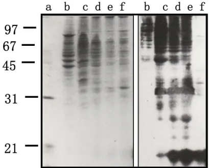
We used the CSL-immobilized Sepharose 4B gel to purify the CSL ligands from the Fraction TN1, which is detergent free and relatively rich on the CSL ligands. The absence of the Triton X-100, a non-ionic detergent helped us in fallowing the proteins sorting from the column. After a slow adsorption of the material on the affinity column, we extensively washed the column with buffer TN and buffer TN/4. The elution of the CSL ligands was done using four successive solutions: TN, TNM, water and ammonium formiate. The buffers sequence (TN/4-TN-TNM) was useful to take down specifically the CSL ligands. The water rinsing leads us to decrease the effect of the buffer TNM. In order to elute the very strongly fixed CSL ligands, we used the ammonium formiate at low pH, which was eliminated by prolonged lyophilization.As many cancerous cells, the K562 cells are very rich in CSL ligands[32].We observed varying profiles of CSL ligands form one fraction to another when the CSL ligands expression in the different fractions from the sequential proteins extraction of K562 cells was analyzed by Western blotting using the Bio-CSL and Avidin-AKP and (Fig. 4). While a very small part of these ligands was solubilized in the buffer T under a weak ionic force (Fig. 4: b), a larger one was present in the fractions with a strong ionic force and without detergents (Fig. 4: c, d and e). By contrast, a considerable quantity of CSL ligands was recovered by strong ionic force and detergent (Fig. 4: f).
Fig. 4. Electrophoretic control of the proteins contained in the K562 cells extracts. A:
SDS-PAGE (CBB Staining); B: Western blot (CSL ligands revealed with Bio-CSL and avidin-AKP). a) Molecular weights marker (kDa); b) fraction T; c) fraction TN1; d) fraction
TN2; e) fraction TNM; f) H2O
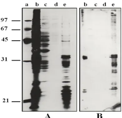
These membranous CSL ligands were completely different from the soluble ones; they showed low molecular weights and specific electrophoretic profiles. These results agree with the data observed in CHO cells [30], suggesting that the soluble CSL ligands did not derive from the membranous ligands by a proteolysis, since they had a higher Molecular weights. Moreover, we suggested that the soluble ligands participate in the extracellular matrix formation and stamps where the CSL could be a bridging molecule as it was shown in the CHO cells [30]. The comparison between the SDS-PAGE (silver nitrate colouring) of total proteins and the western blotting (Bio-CSL and Avidin-AKP revelation) of CSL ligands showed an extremely similar profiles (Fig. 5 A and B). This observation indicated that the CSL ligands were predominant in the initial material. Thus, the CSL affinity chromatography was very efficient in CSL ligands specific isolation. However, compared to the total proteins present in Fraction TN1, the ligands amounts were minor components. Previous works have showed a strong over-expression of the CSL ligands in the tumour cells compared to the
A B
a
b c d e f b c d e f
97
67
45
31
21
normal cells [31,32].
Fig. 5. Electrophoretic control of the CSL ligands isolated by CSL-affinity chromatography. A:
SDS-PAGE (silver staining); B: Western blot (CSL ligands revealed with Bio-CSL and avidin-AKP). a) molecular weights marker (kDa); b) non-retained fraction; c) elution with
TN/4; d) elution with TN; e)elution with TNM
3.3.Seeking for a sequence common to the csl ligands
The CSL ligands may possess a consensus sequence which acts as a peptide signal for their own glycans biosynthesis. Therefore, we tried to identify a group of over-represented peptides by CSL ligands digestion using suitably chosen proteases. It is difficult to know if the CSL ligands are present or not in the normal cells, however, only a few number of them were implicated in the adhesion process. This may implies the expression regulation of exons encoding these sequences, which was altered in the tumour cells and could be responsible of over-producing the CSL glycoprotein ligands [42,43]. The chromatograms of the CSL ligands digestion with protease V8 or trypsin showed two major peaks, beside a various minor peaks (data not shown). This result was expected regardless the initial assumption. Using the Bio-CSL test, We clearly demonstrated that those major peaks were CSL-recognized glycopeptides (Fig. 6). These results agree with a previous study who reports that the two
isoforms of the myelin-associated glycoprotein (MAG) are CSL ligands [44].
Fig. 6. Revelation of the CSL-recognized peptides by dot blot. The spots A (1-10) are the
peptide fractions 1-10; the spots B (1-10) are the peptide fractions 11-20; the spots C5and C6 correspond to 1 and 0.5μg of MAG not recognized by the CSL; the spots C7 and C8 correspond to 1 and 0.5μg of MAG recognized by CSL; the spots C9 and C10 correspond to 1
and 0.5 ug of bovine serum albumin. The spots B2 and B3 (strongly revealed) correspond to the major peaks observed inreverse phase chromatography
Moreover, the hydrophobic character of these glycopeptides indicates that the polypeptide chains were mainly responsible of the separation of the protease digestion products. Therefore, we expected that this common peptide sequence if exist should be hydrophobic and closed to the N-glycosylation sites of the CSL-recognized ligands. The isolation of a large quantity of these glyco-peptides should allow us to study their sequences and check our initial assumption about the existence of a peptide signal necessary to the CSL-recognized glycans biosynthesis. The N-glycans biosynthesis is directed by the presence of the sequence: Asn-X-(Ser/thr), however, the precise regulation process implicated in the N-glycosylation is still unknown [45,46]. It has been suggested that the N-glycosylation concerns only the accessible asparagine. The alternative splicing is implicated in the expression of a well-studied proteins family, the Cell Adhesion Molecules [47]. The diversity of the polypeptide chains could be a key to explain the differences of the glycosylation profiles of the CSL ligands in human K562 tumour cells [42].
1 2 3 4 5 6 7 8 9 10
A
B
C
B2 B3 C7 C84. CONCLUSION
We identified a glycoproteins family that specifically recognize the CSL in human tumour cells. Indeed, we showed an over-represented peptides group, which probably come from all CSL-recognized glycoproteins. Our results suggest that the glycoprotein ligands of the CSL share a common sequence near the N-glycosylation site. This sequence may be a peptide signal directing the biosynthesis of the CSL-recognized glycans in tumour cells and could originate from a translation deregulation of a particular exon encoding this sequence. However, given that the glycans part is not negligible, we cannot definitely confirm the existence of this peptide sequence common to these glycoproteins. The knowledge of this signal sequence may allow us to understand the malignant process of carcinogenesis. Indeed, obtaining antibody against this sequence would lead to identify and insolate glycoproteins containing CSL-recognized glycans. Moreover, we can prepare specific oligonucleotides to screen and analyse expression of these sequences.
5. ACKNOWLEDGEMENTS
List here those individuals who provided help during the research (e.g., providing language help, writing assistance or proof reading the article, etc.).
6. REFERENCES
[1] Hakomori S.I. Aberrant glycosylation in cancer cell membranes as focused on glycolipids: overview and perspectives. Cancer Res., 1985, 45(6), 2405-14.
[2] Andrews P.W. Human teratocarcinoma stem cells: Glycolipid antigen expression and modulation during differentiation. J Cell Biochem. l987, 35(5), 321-32.
[3] Krogerus L and Andersson L.C. Différent lectin-binding patterns in primary breast cancers and their metastases. Cancer, 1990, 66(8), 1802-09.
[4] Wang Y, Ao X, Vuong H, Konanur M, Miller F.R, Goodison S, Lubman D.M. Membrane glycoproteins associated with breast tumor cell progression identified by a lectin affinity approach. J Proteome Res., 2008, 7(10): 4313-25, doi: 10.1021/pr8002547.
[5] Liang Y.J, Ding Y, Levery S.B, Lobaton M, Handa K, Hakomori S.I. Differential expression profiles of glycosphingolipids in human breast cancer stem cells vs. cancer non-stem cells. Proc Natl Acad Sci USA, 2013, 110(13): 4968-73, doi: 10.1073/pnas.1302825110.
[6] Shah P, Wang X, Yang W, Toghi Eshghi S, Sun S, Hoti N, Chen L, Yang S, Pasay J, Rubin A, Zhang H. Integrated Proteomic and Glycoproteomic Analyses of Prostate Cancer Cells Reveal Glycoprotein Alteration in Protein Abundance and Glycosylation. Mol Cell Proteomics, 2015, 14 (10): 2753-63, doi: 10.1074/mcp.M115.047928.
[7] Foster C.S. Functional aspects of glycoprotein N-linked oligosaccharide processing by human tumours. Br J Cancer, 1990, 62, 57-63.
[8] Bassagañas S, Allende H, Cobler L, Ortiz MR, Llop E, de Bolós C, Peracaula R. Inflammatory cytokines regulate the expression of glycosyltransferases involved in the biosynthesis of tumor-associated sialylated glycans in pancreatic cancer cell lines. Cytokine, 2015, 75(1): 197-206, doi: 10.1016/j.cyto.2015.04.006.
[9] Hakomori S.I. Aberrant glycosylation in tumors and tumor-associated carbohydrate antigens. Adv. Cancer Res., 1989, 52, 257-331.
[10] Lotan R and Raz A. Endogenous lectins as mediators of tumor cell adhesion. J Cell Biochem., 1988, 37(1), 107-17.
[11] Rechreche H, Mallo G.V, Montalto G, Dagorn J.C, Iovanna J.L. Cloning and expression of the mRNA of human galectin-4, an S-type lectin down-regulated in colorectal cancer. Eur J Biochem., 1997, 248(1): 225-30.
[12] Feizi T. Carbohydrate differentiation antigens. Probable ligands for cell adhesion molecules. Trends Biochem. Sci., 1991, 16(3), 84-6.
[13] Kayser K, Hoeft D, Hufnagi P, Caselitz J, Zick Y, André S, Kaltner H, Gabius H.J. Combined analysis of tumor growth pattern and expression of endogenous lectins as a prognostic tool in primary testicular cancer and its lung metastases. Histol Histopathol., 2003, 8(3): 771-9.
[14] Gabius H.J and Kayser K. Elucidation of similarities of sugar receptor (lectin) expression of human lung metastases from histogenetically different types of primary tumors. Anticancer Res., 1989, 9(6), 1599-604.
[15] Glaves D, Gabius H.J and Weiss L. Site-associated expression of endogenous tumor lectins. Int. J. Cancer, 1989, 44(3), 506-11.
[16] Vidal-Vanaclocha F, Barbera-Guillem E, Weiss L, Glaves D, Gabius H.J. Quantitation of endogenous lectin expression in 3LL tumors, growing subcutaneously and in the kidneys of mice. Int J Cancer, 1990, 46(5): 908-12.
[17] Sakuma K, Aoki M, Kannagi R. Transcription factors c-Myc and CDX2 mediate E-selectin ligand expression in colon cancer cells undergoing EGF/bFGF-induced epithelial-mesenchymal transition, Proc Natl Acad Sci USA., 2012, 109(20): 7776-81, doi: 10.1073/pnas.1111135109.
[18] Zanetta J.P, Dontenwill M, Meyer A, Roussel G. Isolation and immunohistochemical localization of a lectin-like molecule from the rat cerebellum. Brain Res., 1985, 349(1-2): 233-43.
[19] Zanetta J.P, Meyer A, Kuchler S, Vincendon G. Isolation and immunochemical study of a soluble cerebellar lectin delineating its structure and function. J Neurochem., 1987, 49(4): 1250-7.
[20] Abo T and Balch C.M. A differentiation antigen of human NK and K cells identified by a monoclonal antibody (HNK-1). J. Immunol., 1981, 127 (3), 1024-9.
[21] Kruse J, Mailhammer R, Wernecke H, Faissner A, Sommer I, Goridis C, Schachner M. Neural cell adhesion molecules and myelin-associated glycoprotein share a common carbohydrate moiety recognized by monoclonal antibodies L2 and HNK-1. Nature, 1984, 311(5982): 153-5.
[22] Bollensen E, Schachner M. The peripheral myelin glycoprotein P0 expresses the L2/HNK-1 and L3 carbohydrate structures shared by neuraladhesion molecules. Neurosci Lett., 1987, 82(1): 77-82.
[23] Kuchler S, Fressinaud C, Sarlieve L.L, Vincendon G, Zanetta J.P. Cerebellar soluble lectin is responsible for cell adhesion and participates in myelin compaction in cultured rat oligodendrocytes. Dev Neurosci., 1988, 10(3): 199-212.
[24] Kuchler S, Herbein G, Sarlieve L.L, Vincendon G, Zanetta J.P. An endogenous lectin "CSL" interacts with glycoprotein components in peripheral nervous system myelin. Cell Mol
Biol.,1989, 35(5): 581-96.
[25] Zanetta J.P, Panetta A, Kuchler S, Marschal P, Zaepfel M, Meyer A, Badache A, Reeber A, Lehmann S and Vincendon G. Role of an endogenous mannosyl-lectin in myelination and stabilization of myelin structure. NATO ASI Series, 1990, H 43, 433-50.
[26] Calderon R.O, Maggio B, Neuberger T.J, DeVries G.H. Modulation of Schwann cell Po glycoprotein and galactocerebroside by the surface organization of axolemma. J Neurosci Res., 1995, 40(3):349-58.
[27] Lopez P.H. Role of myelin-associated glycoprotein (siglec-4a) in the nervous system. Adv Neurobiol., 2014, 9:245-62, doi: 10.1007/978-1-4939-1154-7_11.
[28] Perraud F, Kuchler S, Gobaille S, Labourdette G, Vincendon G, Zanetta J.P. Endogenous lectin CSL is present on the membrane of cilia of rat brain ependymal cells. J Neurocytol., 1988, 17(6):745-51.
[29] Lehmann S, Kuchler S, Theveniau M, Vincendon G, Zanetta J.P. An endogenous lectin and one of its neuronal glycoprotein ligands are involved in contact guidance of neuron migration. Proc Natl Acad Sci USA., 1990, 87(16): 6455-9.
[30] Lehmann S, Kuchler S, Badache A, Zaepfel M, Meyer A, Zanetta JP. Involvement of the endogenous lectin CSL in adhesion of Chinese hamster ovary cells. Eur J Cell Biol., 1991, 56(2): 433-42.
[31] Zanetta J.P, Staedel C, Kuchler S, Zaepfel M, Meyer A, Vincendon G. Malignant transformation in hepatocytes is associated with the general increase of glycoprotein ligands specifically binding to the endogenous lectin CSL. Carbohydr Res., 1991, 213:117-26.
[32] Maschke S, Robert J, Coindre J.M, Kuchler S, Vincendon G, Zanetta J.P. Malignant cells have increased levels of common glycoprotein ligands of the endogenous cerebellar soluble lectin CSL. Eur J Cell Biol., 1993, 62(1): 163-72.
[33] Lowry O.H, Rosebrough N.J, Farr A.L, Randall R.J. Protein measurement with the Folin phenol reagent. J Biol Chem., 1951, 193(1): 265-75.
[34] Laemmli U.K. Cleavage of structural proteins during the assembly of the head of bacteriophage T4. Nature, 1970, 227(5259): 680-5.
fractionation by affinity chromatography on concanavalin A. Brain Res., 1975, 83(2):337-48. [36] Towbin H, Staehelin T, Gordon J. Electrophoretic transfer of proteins from polyacrylamide gels to nitrocellulose sheets: procedure and some applications. Proc Natl Acad Sci U S A., 1979, 76(9): 4350-4.
[37] Leary J.J, Brigati D.J, Ward D.C.Rapid and sensitive colorimetric method for visualizing biotin-labeled DNAprobes hybridized to DNA or RNAimmobilized on nitrocellulose: Bio-blots. Proc Natl Acad Sci USA., 1983, 80(13): 4045-9.
[38] Marschal P, Reeber A, Neeser J.R, Vincendon G, Zanetta J.P. Carbohydrate and glycoprotein specificity of two endogenous cerebellar lectins. Biochimie, 1989, 71(5): 645-53.
[39] Moser A.C, Hage D.S. Immunoaffinity chromatography: an introduction to applications and recent developments. Bioanalysis, 2010, 2(4): 769-90, doi: 10.4155/bio.10.31.
[40] Qu H, Qu B, Wang X, Zhang Y, Cheng J, Zeng W, Liu S, Wang Q, Zhao Y. Rapid, sensitive separation of the three main isoflavones in soybean using immunoaffinity chromatography. J Sep Sci., 2016, 39(6):1195-201, doi: 10.1002/jssc.201501052.
[41] Henning A.K, Albrecht D, Riedel K, Mettenleiter T.C, Karger A. An alternative method for serum protein depletion/enrichment by precipitation at mildly acidic pH values and low ionic strength. Proteomics, 2015, 15(11): 1935-40, doi: 10.1002/pmic.201400257.
[42] Ruhaak L.R, Taylor S.L, Stroble C, Nguyen U.T, Parker E.A, Song T, Lebrilla C.B, Rom W.N, Pass H, Kim K, Kelly K, Miyamoto S. Differential N-Glycosylation Patterns in Lung Adenocarcinoma Tissue. J Proteome Res., 2015, 14(11): 4538-49, doi: 10.1021/acs.jproteome.5b00255.
[43] Jiang K, Li W, Zhang Q, Yan G, Guo K, Zhang S, Liu Y. GP73 N-glycosylation at Asn144 reduces hepatocellular carcinoma cell motility and invasiveness. Oncotarget. 2016 Mar 16. doi: 10.18632/oncotarget.8120.
[44] Badache A, Burger D, Villarroya H, Robert Y, Kuchler S, Steck AJ, Zanetta JP. Carbohydrate moieties of myelin-associated glycoprotein, major glycoprotein of the peripheral nervous system myelin and other myelin glycoproteins potentially involved in cell adhesion. Dev Neurosci., 1992, 14(5-6): 14:342-50.
[45] Pinho S.S, Oliveira P, Cabral J, Carvalho S, Huntsman D, Gärtner F, Seruca R, Reis C.A, Oliveira C. Loss and recovery of Mgat3 and GnT-III Mediated E-cadherin N-glycosylation is a mechanism involved in epithelial-mesenchymal-epithelial transitions. PLoS One, 2012 7: e33191.
[46] Levy-Ontman O, Fisher M, Shotland Y, Weinstein Y, Tekoah Y, Arad S.M. Genes involved in the endoplasmic reticulum N-glycosylation pathway of the red microalga Porphyridium sp: a bioinformatic study. Int J Mol Sci., 2014, 15:2305-26.
[47] Weledji E.P, Assob J.C. The ubiquitous neural cell adhesion molecule (N-CAM). Ann Med Surg (Lond), 2014, 3: 77-81.
How to cite this article:
Rechreche H. and Zanetta J.P. Contribution to study the glycoprotein ligands of cerebellar soluble lectin in human K652 tumor cell lines. J. Fundam. Appl. Sci., 2016, 8(3), 856-874.