HAL Id: hal-03123656
https://hal.archives-ouvertes.fr/hal-03123656
Submitted on 25 Mar 2021HAL is a multi-disciplinary open access archive for the deposit and dissemination of sci-entific research documents, whether they are pub-lished or not. The documents may come from teaching and research institutions in France or abroad, or from public or private research centers.
L’archive ouverte pluridisciplinaire HAL, est destinée au dépôt et à la diffusion de documents scientifiques de niveau recherche, publiés ou non, émanant des établissements d’enseignement et de recherche français ou étrangers, des laboratoires publics ou privés.
OSIP1 is a self-assembling DUF3129 protein required to
protect fungal cells from toxins and stressors
Nicolas Valette, Julien Renou, Alexis Boutilliat, Antonio José
Fernández-gonzález, Valérie Gautier, Philippe Silar, Christophe Guyeux,
Jean-claude Charr, Stéphane Cuenot, Christophe Rose, et al.
To cite this version:
Nicolas Valette, Julien Renou, Alexis Boutilliat, Antonio José Fernández-gonzález, Valérie Gautier, et al.. OSIP1 is a self-assembling DUF3129 protein required to protect fungal cells from toxins and stressors. Environmental Microbiology, Society for Applied Microbiology and Wiley-Blackwell, 2021, �10.1111/1462-2920.15381�. �hal-03123656�
1
OSIP1 is a self-assembling DUF3129 protein required to protect fungal cells from toxins
1
and stressors
2 3
Running title: OSIP1 prevent cell wall stress
4 5
Nicolas Valettea, Julien Renoua, Alexis Boutilliata, Antonio José Fernández-Gonzáleza,
6
Valérie Gautierb,Philippe Silarb, Christophe Guyeuxc, Jean-Claude Charrc, Stéphane Cuenotd,
7
Christophe Rose, Eric Gelhayea, Mélanie Morel-Rouhiera#.
8 9
a
Université de Lorraine, INRAe, Interactions Arbres/Micro-organismes (IAM), UMR 1136,
10
F-54000 Nancy, France
11 b
Université Paris Diderot, Sorbonne Paris Cité, Laboratoire Interdisciplinaire des Energies de
12
Demain (LIED), 75205 Paris, France
13 c - 14 - on, France 15 d
Institut des Matériaux Jean Rouxel, Université de Nantes, 2 rue de la Houssinière, 44322
16
Nantes Cedex 3, France
17 e
Université de Lorraine, AgroParisTech, INRAE, UMR Silva, Nancy, 54000, France.
18 19
#
Corresponding author: Mélanie Morel-Rouhier, Université de Lorraine, UMR1136
INRA-20
Université de Lorraine "Interactions Arbres/Micro-organismes", Faculté des Sciences et
21
Technologies BP 70239, F-54506 Vandoeuvre-lès-Nancy Cedex, France.
22
Melanie.Morel@univ-lorraine.fr, phone: +33 3 72 74 51 62.
23 24
Keywords: OSIP1, fungi, cell wall, stress, oak extractives, secretome
2
Summary (200 mots)
26
Secreted proteins are key players in fungal physiology and cell protection against external
27
stressing agents and antifungals. OSIP1 is a fungal-specific protein with unknown function.
28
By using Podospora anserina and Phanerochaete chrysosporium as models, we combined
29
both in vivo functional approaches and biophysical characterization of OSIP1 recombinant
30
protein. Our data showed an increased sensitivity of the P. anserina OSIP1 mutant to both
31
caspofungin and oak-extractives. This correlated with the weakened extracellular matrix
32
produced by the mutant compared to the wild type, as highlighted by SEM imaging. This
33
alteration quantitatively modified the global secretome of P. anserina grown in presence of
34
wood, such as proteins associated to the cell-wall integrity signaling pathway. Since the
35
recombinant OSIP1 form P. chrysosporium self-assembled as fibers and was capable of
36
gelation, these results argue for a structural role of OSIP1 proteins in fungi at the cell wall or
37
within the matrix confering cell protection against external toxic compounds. These data
38
could be of great interest for increasing protein secretion in a context of lignocellulosic
39
biomass degradation, such as improving the efficiency of antifungals that could be trapped
40
within the extracellular matrix.
41 42 43 44
3 Introduction
45
During evolution, fungi had to adapt to environmental constraints. The secretome, i.e. the
46
proteins secreted in the extracellular medium, is a good marker of fungal physiology and
47
trophic modes. Indeed, the secretome is involved in the first steps of the symbiosis or
48
infection establishment and plays essential roles in plant biomass degradation (Kämper et al.,
49
2006; Bouws et al., 2008; Vincent et al., 2012). Fungal secretomes are composed of
50
degradative enzymes such as proteases, lipases, Carbohydrate-Active enZymes (CAZymes),
51
and ligninolytic enzymes for some wood-decaying species (Zhu et al., 2016; Pellegrin et al.,
52
2015). This degradative system has been intensively studied due to its important application
53
in lignocellulose biomass valorization. In plant-associated fungi, other proteins can be
54
secreted to modulate plant immunity and establish symbiosis or allow pathogenic infection
55
(Plett et al., 2011; Pazzagli et al., 1999; Frias et al., 2011; Baccelli et al., 2014). Some of
56
them, the hydrophobins, are involved in the attachment of fungal structures to different kinds
57
of surfaces and the development of hyphae at the water/air interface (Wessels et al., 1991;
58
Wessels, 1996). In fungal pathogens, hydrophobins might act as virulence factors to enhance
59
fungal infection (Ruocco et al., 2015; Kubicek et al., 2008), while in symbiotic associations,
60
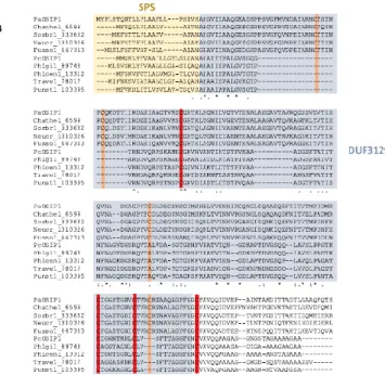
these proteins could be involved in mycorrhizae formation (Plett et al., 2012). Most of the
61
studied hydrophobins are directed to the extracellular medium through the secretory pathway.
62
However, they often remain associated with the fungal cell wall and can be found inside
63
fruiting bodies and on the surfaces of hyphae, spores and conidia (Dynesen et al., 2003;
64
Linder, 2009). Many other secreted proteins have been identified but remain of unknown
65
function. This is the case for SSP (for Small Secreted Proteins with sequence less than 300
66
amino acids) (Alfaro et al., 2014). The percentage of SSP-coding genes in the genomes of
67
saprophytic fungi such as Phanerochaete chrysoporium, Trametes versicolor or Aspergillus
68
fumigatus is similar to the one of the ectomycorrhizal fungus Laccaria bicolor (between 2 and
4
3 % of the predicted gene models) (Pellegrin et al., 2015; Valette et al., 2016). At the protein
70
level, SSPs represent between 4 and 12% of the proteins identified in the secretomes of
71
various Aspergillus species grown on sugar beet pulp or wheat bran (Valette et al., 2016).
72
However, only few have been functionally characterized. One SSP of the lignolytic fungus
73
Pleurotus ostreatus is involved in the regulation of the lignolytic system by modulating
74
expression and activity of aryl-alcohol oxidases, aryl-alcohol dehydrogenases and versatile
75
peroxidases (Feldman et al., 2017) and in the transition from primary to secondary
76
metabolism, development, aging, and fruiting body initiation (Feldman et al., 2019).
77
In a previous analysis, we have highlighted the up-regulation of various SSP-coding genes of
78
the lignolytic fungus Phanerochaete chrysosporium in presence of oak extractives (Thuillier
79
et al., 2014, Fernández-González et al., 2018). Oak extractives are mainly composed of
80
phenolic compounds and flavonoids (Zhang et al., 2015; Fernández-González et al., 2018).
81
These molecules are released from wood during the degradative process and can be toxic for
82
cells by various mechanisms such as metal and free radical scavenging activity, direct
83
interaction with enzymes, perturbation of ionic homeostasis and disruption of membrane and
84
cell wall integrity (Valette et al., 2017). One of these up-regulated genes retained our attention
85
because the corresponding protein was also detected at high amount in the secretome of
86
another white rot fungus Trametes versicolor grown on oak wood chips (Deroy et al.,
87
unpublished). This protein was thus named OSIP1 for Oak Stress Induced Protein. It shows
88
no sequence homology with characterized proteins in the databases. In this study we used two
89
fungal models to decipher the role of OSIP1: Podospora anserina for functional analysis
90
because genetic engineering is easy, contrary to P. chrysosporium or T. versicolor; and P.
91
chrysoporium for the biochemical analysis because only PcOSIP1 was successfully produced
92
as a recombinant protein.
93 94
5 Results
95
Comparative genomic analysis reveals that OSIP1 is widespread in fungi 96
OSIP1 sequences from the ascomycete P. anserina (PaOSIP1: ProtID JGI 208230, a.k.a.
97
Pa_5_3780 according to the P. anserina genome project) and the basidiomycete P.
98
chrysosporium (PcOSIP1: ProtID JGI 2981896) were used as templates to search for fungal
99
sequences using the BlastP search tool onto the whole fungal JGI database (Mycocosm from
100
Joint Genome Institute). The sequence of P. anserina P209725 (ProtID JGI 209725) was also
101
used as template since it displays 38% similarity with PaOSIP1). A total of 1057 protein
102
sequences were retrieved and analyzed by clustering. These sequences grouped into nine
103
clusters (Fig. 1A). PaOSIP1 and PcOSIP1 belonging to cluster 1, this latter was thus named
104
OSIP1 cluster (species and accession numbers are available in Table S1). It gathers sequences
105
from both ascomycetes (Pezizomycotina) and basidiomycetes (Agaricomycotina and
106
Pucciniomycotina). Moreover, this analysis revealed that OSIP1 is present in genomes of
107
wood decay fungi, and also in mycorrhizal, pathogenic fungi and other saprotrophs (Fig. 1A).
108
Although OSIP1 was found induced by oak extractives in lignolytic fungi (Thuillier et al.,
109
2014; Deroy, unpublished), this genomic analysis showing that OSIP1 is present in fungi with
110
various trophic modes, argues against its direct involvement in the lignolytic process and
111
suggests a more general role in fungal physiology.
112 113
OSIP1 sequences contain a DUF3129 domain, like some appressoria-specific proteins
114
OSIP1 sequences exhibit a signal peptide of secretion, a conserved DUF3129 domain and a
115
variable C-terminal tail (Fig. 1B). In ascomycetes, 8 cysteinyl residues are conserved, while
116
only four have been detected in the analyzed basidiomycete sequences (Fig. 1B and Fig S1).
117
The DUF3129 domain is not restricted to OSIP1 sequences. It can be identified in both
118
ascomycetes and basidiomycetes and was retrieved in 442 sequences from the Pfam 31.0
6
database. Although most of the proteins having the DUF3129 are annotated as
120
uncharacterized proteins, some of them are described as being related to CAS1
121
(Colletotrichum gloeosporioides appressoria-specific protein), MAS (Magnaporthe
122
appressoria-specific protein), gEgh16 from Blumeria graminis proteins, all being putatively
123
involved in fungal cell wall remodeling. A phylogenetic analysis was performed with the
124
sequences from the OSIP1 cluster and the 442 sequences containing the DUF3129 domain
125
(Fig. 1C). PaOSIP1 clusters with ascomycete sequences but independently of CAS1 from C.
126
gloeosporioides, MAS3 from M. grisea and gEgh16 from B. graminis. Among
127
basidiomycetes, sequences from Agaricomycotina with lignolytic, saprotrophic or
128
mycorrhizal lifestyles (Group A) cluster independently from sequences of pathogenic
129
basidiomycetes from Pucciniomycotina and Agaricomycotina (Group B) (details are given in
130
Table S1).
131 132
PaOSIP1 maintains cell wall integrity under caspofungin stress 133
To functionally characterize OSIP1, the OSIP1 knock-out mutant was generated in P.
134
anserina (PaOSIP1Δ) (Fig S2). PaOSIP1Δ was tested for its ability to grow on various carbon
135
sources (cellobiose, fructose, cellulose, pectin, glucose) and various biomasses (whatman
136
paper, hay, miscanthus, wood chips). No differences in growth, sporulation, nor
appressoria-137
like structure formation (as described in Brun et al., 2009) was highlighted between the wild
138
type and the PaOSIP1Δ mutant strains in the tested conditions (data not shown). Because the
139
DUF3129 domain is present in cell wall remodeling proteins, cell wall destabilizing agents
140
were thus tested. No phenotype was observed for Congo red that prevents glucan microfibril
141
bl l b b β-1,3 glucans (Nodet et al., 1990) nor Calcofluor white, which
142
binds chitin. By contrast, a significant deleterious growth phenotype was observed for the
143
mutant compared to the wild type in presence of caspofungin (Fig. 2). Caspofungin is a cell
7
wall-targeting antifungal compound extensively used in clinical settings for the treatment of
145
infections caused by diverse fungi. Caspofungin inhibits the synthesis of β-1,3-glucan, a
146
crucial cell wall component for many fungi, by targeting the β-1,3-glucan synthase (encoded
147
by fks1) in a non-competitive way (Van Den Bossche, 2002; Aguilar-Zapata et al., 2015).
148
Functional complementation of PaOSIP1Δ by PaOSIP1 restored the growth defect confirming
149
the role of PaOSIP1 in protecting cell wall under caspofungin treatment.
150 151
PaOSIP1Δ mutant is affected in extracellular matrix production
152
To check whether caspofungin sensitivity was due to a thinner cell wall in the mutant
153
compared to the wild type, SEM imaging was performed after cryosection of the hyphae (Fig.
154
3). The whole reconstituted images are shown as supplementary data. The thickness of the
155
cell wall was measured (n>50) based on the microscopic images. No difference was detected
156
between the WT and the mutant, nor between the caspofungin and the control condition. The
157
measured cell wall thickness of hyphae cross-sections was around 150 nm for all conditions
158
analysed. Interestingly, the main difference highlighted by comparing SEM images of both
159
mutant and WT strains was the extracellular matrix (ECM) density. Fungal extracellular
160
matrix is mainly composed of carbohydrates and proteins in complex interactions. This matrix
161
is strongly reduced in PaOSIP1Δ even in the absence of caspofungin (Fig. 3). Since this
162
matrix was shown to prevent drugs from reaching their cellular targets (Mitchell et al., 2016),
163
this could explain why PaOSIP1Δ showed an increased sensitivity to the antifungal
164
caspofungin.
165 166
Recombinant OSIP1 self-assembles as fibers and forms a gel
167
The heterologous Escherichia coli system was tested for recombinant PaOSIP1 and PcOSIP1
168
productions. Only PcOSIP1 expressed in E. coli was successfully produced. After
8
purification, 3 mg of pure protein per liter of bacterial culture were obtained. To check
170
whether the protein was correctly folded, far-UV circular dichroism analysis was performed.
171
Spectrum of PcOSIP1 revealed secondary structures, mainly alpha helices as highlighted by a
172
positive band at 190 nm and two negative bands at 208 and 222 nm (Fig. 4A).
173
After purification, PcOSIP1 rapidly self-assembled as big oligomers in Tris-NaCl buffer (30
174
mM Tris-HCl pH 8.0, 200 mM NaCl), as shown by dynamic light scattering (DLS) analysis
175
(Fig. 4B). Huge hydrodynamic radii (Rh) of 789.3 ± 388 nm and 2 281 ± 880 nm were
176
respectively measured for 56.7% and 28.7% of the PcOSIP1. This aggregation was not
177
observed when the protein was dialyzed in 50 mM phosphate buffer pH 8.0 directly after
178
purification, suggesting that this process was driven by the physicochemical properties of the
179
buffer. To check whether this aggregation was due to protein instability and thus precipitation
180
or rather a specific organization, the macromolecular structures of PcOSIP1 was analyzed by
181
Atomic Force Microscopy (AFM). In Tris-NaCl buffer, PcOSIP1 was able to self-assemble
182
into fibers with a mean diameter of 1.5 ± 0.2 nm (Fig. 4C). In phosphate buffer, a crown
183
structure was evidenced. This crown organization could be the transient states of PcOSIP1
184
fibril formation, since such structure was already described in transient states of the human
α-185
synuclein fibril formation that contributes to Parkinson's disease (Lashuel et al., 2002 ; Apetri
186
et al., 2006).
187
All the experiments described above were performed directly or few days after protein
188
purification. Interestingly, storing PcOSIP1 in Tris-NaCl buffer in the freezer led to the
189
formation of a gel, that was quite compact and elastic (Fig. 4D). This specific feature was
190
already described for other proteins as α-synuclein that can form gels in buffer at pH 7.4 in
191
the presence of NaCl (Semerdzhiev et al., 2018). These atypical properties of self-assembly
192
and jelly structure formation support a putative structural role of OSIP1 protein within the
193
fungal cell wall or the extracellular matrix.
9 195
Oak-extractives affect PaOSIP1Δ growth
196
During wood degradation, extractives act as important stressors for fungal cells (Thuillier et
197
al., 2014 ; Valette et al., 2017 ; Fernández-González et al., 2018). Some of them can act
198
directly on cell wall integrity. For example, similarly to caspofungin, cinnamaldehyde and
199
b β-1,3-glucan synthesis within the fungal cell wall (Bang et al., 2000;
200
Piotrowski et al., 2015). Because OSIP1 gene expression was induced by oak
extractive-201
induced stress both in P. chrysosporium (Thuillier et al., 2014) and P. anserina (data not
202
shown), this condition was used to test PaOSIP1Δ growth phenotype. The results showed a
203
growth delay of PaOSIP1Δ compared to the wild type in presence of wood extractives, which
204
was observable from 5 days (Fig. 5). This phenotype was partially restored by functional
205
complementation with PaOSIP1. These results support the hypothesis that PaOSIP1 could
206
participate in cell wall protection against extractives toxicity.
207 208
The deletion of OSIP1 strongly modifies the composition of the secretome and the
cell-209
wall related proteins of P. anserina in presence of oak sawdust
210
To analyze how the deletion of PaOSIP1 affects the fungal physiology in a context of biomass
211
degradation, a proteomic analysis was performed for the PaOSIP1Δ mutant grown in presence
212
of oak sawdust, in comparison with the wild type strain. The comparative analysis of the
213
secreted proteins reveals strong modification of the global secretome of PaOSIP1Δ mutant
214
compared to the wild type strain (Fig. 6A). Indeed, over the 250 proteins detected, 150 were
215
more abundant (Protein abundance index (PAI) fold>2) or specifically identified in the mutant
216
compared to the wild type, while this number was only 41 in the case of the wild type.
217
By looking at the various classes of enzymes that were more abundant in the mutant
218
secretome compared to the WT, no clear specificity was observed except for proteases that
10
were less represented (Fig. 6B). Globally, more than half (around 60%) of the detected
220
proteins of a specific class were more abundant in the secretome of the mutant compared to
221
the wild type. It is likely that the deletion of OSIP1 globally affected the secretion process of
222
the enzymes.
223
Glycoside hydrolases (GH) were the most abundant proteins with a total of 66 GH detected in
224
both secretomes. By looking at the individual GH families, we showed again that the increase
225
in protein abundance is not restricted to specific families, but was observed for many of them
226
(Fig. 6C). However, it is interesting to note that 16 GH families (over the total of 29) were
227
specifically detected in the mutant strain, especially GH5, GH11 and GH43 with at least 3
228
isoforms.
229
A high number of cell wall-related proteins, in particular glucan-acting enzymes have also
230
been found more abundant in the secretome of the mutant strain (Fig. 6D). Moreover, many
231
Wall Stress responsive Component (WSC) proteins have been detected. These proteins serve
232
as sensors of external stress cues upstream of cell wall integrity (CWI) pathway in
233
Saccharomyces cerevisiae (Verna et al., 1997) and Aspergilli (Futagami et al., 2011; Dichtl et
234
al., 2012). In line with this observation, respectively 3 and 6 DUF1996-containing proteins
235
were found more abundant and specifically detected in the mutant among the 10 detected in
236
total (Supplementary data). These proteins are of unknown function, however the DUF1996
237
domain has been associated to fungal stress sensing and response (Tong et al., 2016a and
238 2019). 239 240 Discussion 241
In this study, we characterized a new fungal protein, which participates in cell wall fitness
242
under stress. The comparative genomic analysis revealed that such proteins are widely present
243
in fungi, suggesting their involvement in a general process of stress rescue. All analyzed
11
OSIP1 possess a domain of unknown function DUF3129 that was previously identified by
245
few studies in proteins of both plant and insect pathogens (Shang et al., 2016). DUF3129 is an
246
expanded gene family highly expressed during infection in nematode-trapping fungi that form
247
adhesive branches and adhesive knobs (Andersson et al., 2014). The role of this domain was
248
attributed to the cell wall remodeling for fungal penetration to host cuticules with an unclear
249
mechanism (Justesen et al., 1996 ; Xue et al., 2002 ; Grell et al., 2003 ; Cao et al., 2012).
250
More recently, seven DUF3129 proteins of the insect pathogenic fungus Metarhizium
251
robertsii were found localized to cellular lipid droplets mediating their degradation and
252
subsequently controlling appressorial turgor required for infection (Huang et al., 2019).
253
However, nothing has been described concerning the role of these DUF3129-containing
254
proteins in saprophytic fungi. The jelly structure of the recombinant PcOSIP1 and the
255
reduction of the extracellular polysaccharide network observed for the PaOSIP1 mutant
256
strongly suggest the involvement of OSIP1 in the formation of such adhesive structures in
257
fungi. The weakened extracellular matrix of the mutant could thus be responsible for the
258
higher susceptibility of the fungus to both caspofungin and oak extractives.
259
This phenomenom has been already described in A. fumigatus, where the downregulation of a
260
hydrophobin gene by a polyphenolic compound resulted in a weakened extracellular matrix
261
and therefore increased the susceptibility of the fungi to antifungal drugs (Luo et al., 2018).
262
Hydrophobins are small (100–120 aa) secreted proteins characterized by the presence of eight
263
highly conserved cysteine residues and the ability to self-assemble as amyloid-like structure
264
and forms rodlets (Ball et al., 2020). Amyloids serve diverse purposes for structure, adhesion
265
and defence in microorganisms (Shanmugam et al., 2019) and can be evidenced in vitro using
266
fluorescent tool as thioflavin T, which binds to the beta sheet-rich structure characteristic of
267
amyloid-like structure (Groenning, 2010). PcOSIP1 does not assemble under amyloid
268
structure since no thioflavin T fluorescence signal was detected in any of the conditions of
12
temperatures and buffers tested (data not shown). We have shown that PcOSIP1 was rich
α-270
helices, while amyloid ll b β-sheets. Moreover, AFM revealed that it
271
did not form rodlets. These experimental data, coupled to sequence analysis, allowed us to
272
confirm that OSIP1 is a new self-assembling protein, that does not belong to the well-known
273
class of hydrophobins.
274
In the context of lignocellulosic biomass degradation, the structural property of OSIP1 may be
275
the key point explaining the way by which it participates in fungal stress resistance by
276
protecting the cell wall. In accordance, the comparative secretome analysis of P. anserina
277
grown on oak sawdust revealed that the PaOSIP1 mutant highly expresses WSC proteins
278
compared to the wild type in this condition. WSC proteins are localized to the cell wall and
279
the plasma membrane and act as sensors upstream of the cell-wall integrity pathway. In
280
particular, WSC-1 may function in regulating cell wall biogenesis through the MAK-1
281
pathway in Neurospora crassa (Maddi et al., 2012). Single deletions of the five wsc genes of
282
Beauveria bassiana resulted in significant, but differential, increases in cellular sensitivity to
283
cell wall perturbation, oxidation, high osmolarity, and metal ions (Tong et al., 2016b). In
284
Aspergillus fumigatus, deletions of wsc1 caused an increased in sensitivity to caspofungin but
285
no change in cellular sensitivity to other cell wall perturbation, alkaline pH and high
286
temperature (Dichtl et al., 2012). In the nematode-trapping fungus Monacrosporium
287
haptotylum, a gene cluster of 5 secreted proteins that are adjacent in the M. haptotylum
288
genome (cluster 74) is highly (>10-fold) upregulated during infection (Andersson et al.,
289
2013). This cluster gathers two genes coding for WSC proteins, one gene containing the
290
DUF3129 domain and two SSP-coding genes (Meerupati et al., 2013). This suggests a
291
functional link between these proteins. Additionally to WSC proteins, the PaOSIP1mutant
292
highly expresses DUF1996-containing proteins. In B. bassiana, DUF1996-containing proteins
293
localize in vacuoles and play significant roles in the response to cell-wall perturbation, high
13
osmolarity, oxidation, fungicidal and multiple metal stress (Tong et al., 2016a). The absence
295
of OSIP1 in P. anserina grown in presence of oak sawdust strongly affected the whole
296
secretome of the fungus, likely because of the cell wall weakness. Indeed, the functionality of
297
the cell wall integrity (CWI) and secretory systems are connected and coordinately respond to
298
exogenous stresses through the modulation of the cell periphery and secretion (Malavazi et
299
al., 2014).
300
Taking together, these data strongly suggest that OSIP1 proteins prevent cell wall stress.
301
Consequently, its absence affects the cell wall associated signaling pathway, leading to a
302
deregulation of the secretion process. These data could be of great interest for both the
303
improvement of protein secretion particularly in a context of lignocellulosic biomass
304
degradation, and the limitation of fungal pathogenicity, for which the fungal cell wall has a
305
crucial role (Gow et al., 2017).
306 307 Experimental procedures 308 Growth conditions 309
The P. anserina strain used in this study was derived from the S strain (Rizet, 1952; Boucher
310
et al., 2017). Standard culture conditions, media compositions and genetic methods for this
311
fungus have already been described (Rizet, 1941; Silar, 2013) and are available at
312
https://podospora.i2bc.paris-saclay.fr. Growth kinetics of the wild type and PaOSIP1∆ strains
313
were done in M2-Agar medium and M2-Agar medium supplemented with caspofungin (500
314
ng/ml) and oak extractives (2 mg/ml) for 10 days at 27 °C. Oak (Quercus petraea) acetonic
315
extract preparation has been performed as described previously (Fernández-González et al.,
316
2018).
317 318
Analysis of OSIP1 sequences
14
OSIP1 sequences were searched within all fungal genomes available in the Joint Genome
320
Institute database (Mycocosm https://genome.jgi.doe.gov/programs/fungi/index.jsf) using
321
BlastP with a cut off of Evalue=10-5. Sequences of OSIP1 of Podospora anserina ((PaOSIP1
322
(208230 JGI) and Phanerochaete chrysosporium (ProtID 2981896 JGI) have been used as
323
templates. Another sequence close to PaOSIP1 was added as a template (P209725 (ProtID
324
209725 JGI)). Evolutionary analyses were conducted in MEGA7 using the Neighbor-Joining
325
method (Kumar et al., 2016). The tree is drawn to scale, with branch lengths in the same units
326
as those of the evolutionary distances used to infer the phylogenetic tree. The evolutionary
327
distances were computed using the Poisson correction method and are in the units of the
328
number of amino acid substitutions per site. The analysis involved 628 amino acid sequences.
329
All ambiguous positions were removed for each sequence pair. There were a total of 467
330
positions in the final dataset. To carry out the clustering, the Laplacian eigenmap technique
331
(Belkin and Niyogi, 2003) was applied with Gaussian mixture model (Reynolds, 2015) as
332
follows. After sorting the sequences in alphabetical order, the similarity of each sequence pair
333
were obtained from the score provided during a pairwise sequence alignment using the
334
Needleman-Wunsch (Needleman and Wunsch, 1970) dynamic programming algorithm from
335
Biopython (Cock et al., 2009) module (pairwise2 function). Default values for gap open and
336
extend penalties were chosen with blosum62 matrix for amino acid substitution, leading to a
337
matrix M of integers. A similarity matrix S has been deduced by dividing each row by its
338
maximum, and by computing the identity matrix minus this one. The normalized Laplacian
339
associated with the similarity matrix has been computed as follows: L = D-1(D-S), where D is
340
the diagonal matrix whose element in position (i,i) is the sum of the i-th row in S. Eigenvalues
341
of L have then been computed and sorted in ascending order thanks to the numpy library
342
(Oliphant, 2006), and the N-th first eigenvalues have only be considered, where N is such that
343
the increase between the N-th and N+1-th eigenvalue is lower than 1%. Associated
15
eigenvectors have then been clusterized according to a Gaussian mixture model (Reynolds,
345
2015), and the model selection (number of Gaussians) has been performed according to the
346
Bayesian Information Criterion (BIC, (Schwarz, 1978)). To sum up, Laplacian eigenmap
347
allowed us to map the similarity matrix in a low dimensional space of points, each point being
348
associated to one amino acid sequence. This cloud points has been considered as the
349
superposition of a given number of gaussian trends (the clusters), this number being
350
determined thanks to the BIC criterion of parcimony. For further information about this
351
sequence clustering technique, see, e.g. Bruneau et al. (2018).
352 353
Deletion of PaOSIP1 in Podospora anserina
354
l P P (P _ _ 8 ) “ l k ” w ( l ).
355
protocol is based on the generation of two DNA fragments carrying a resistance marker
356
fl k w ’ ’ -coding sequence of the genes by two successive PCR
357
reactions. In the first step, a 832 pb-l ’-non-coding region of PaOSIP1 and a 962 pb-long
358
’ w P -amplified from the S strain DNA with the PaOSIP1-A/ PaOSIP1-B, and
359
PaOSIP1-C/ PaOSIP1-D primer pairs respectively. At the same time, the hygromycin
360
resistance marker was amplified with PaOSIP1-E and PaOSIP1-F from the pBC-hygro vector
361
(Silar, 1995). Primers sequences are given in Fig. S2. In a second step, the second round of
362
PCR using primers PaOSIP1-A and PaOSIP1-F, and PaOSIP1-D and PaOSIP1-E enabled to
363
k w ’ ’ . w P w
364
used to transform a mus51::phleoRstrain, in which the mus51gene encoding one of the
365
subunit of the non-homologous end joining dimer is replaced with a phleomycin resistance
366
gene. Three crossing-over events between the two cassettes and the P. anserina genome
367
enabled the deletion of PaOSIP1. Three hygromycin resistant transformants were selected.
368
They were crossed with the wild-type S strain, and one homokaryotic hygromycin resistant
16
and phleomycin-sensitive descendant was selected as the PaOSIP1::hygroRstrain or
370
PaOSIP1∆. Its genotype was confirmed by Southern blot analyses using digoxigenin labeled
371
probes (Fig. S2). For functional complementation tests, PaOSIP1 coding sequence was cloned
372
into pAKS-GenetR vector and expressed in PaOSIP1∆. The presence of the gene was checked
373
by PCR and three transformants were selected for functional complementation tests. They all
374
restored the mutant phenotype, thus, only the results for one of them are presented.
375 376
SEM imaging of PaOSIP1 hyphal network
377
Cloning of PcOSIP1
378
Phanerochaete chrysosporium mycelium was harvested from liquid cultures in TK medium
379
supplemented with oak extractives as previously described (Thuillier et al., 2014). Total RNA
380
was extracted and purified using the RNeasy plant minikit (Qiagen) according to the
381
f ’ uctions. RNA was treated with DNase I during purification as
382
f ’ l. A l f w f
383
by precipitating RNA with 2 M LiCl. RNAs were reverse transcribed using the masterscript
384
kit (5 prime) following the manufacturer's protocol. The PCR reactions to amplify PcOSIP1
385
(Prot ID 2981896 in the Joint Genome Institute database v2.2 (previously identified as Prot ID
386
4474 in v2.0 of P. chrysosporium genome annotation)), have been performed with Herculase
387
Taq (Agilent technologies) for cloning into the pEt26b (Novagen) vector for His-tagged
388
protein production in Escherichia coli. The sequence was amplified without the predicted
389
signal peptide of secretion using the following primers (for:
390
CCCCCATATGGCTATTATCACGCCCGCG and rev:
391
CCCCGCGGCCGCTGCTTGGAGCTCCTCATC).
392 393
17
Heterologous expression of PcOSIP1 in Escherichia coli and purification of the
394
recombinant protein
395
Expression of recombinant PcOSIP1 was performed in E. coli Rosetta2 (DE3) strain
396
containing pLysS plasmid (F– ompT hsdSB(rB– mB–) gal dcm (DE3) pRARE2 (CamR)). The 397
bacteria were cultivated in LB medium supplemented with 50µg/ml kanamycin and 50µg/ml
398
chloramphenicol at 37°C. At OD600 of 0.6, the expression of the recombinant proteins was 399
induced by adding 0.1 mM isopropyl ß-D-1 thiogalactopyranoside (IPTG) during 4h. Cells
400
were harvested by centrifugation and resuspended in 30 mM Tris-HCl pH 8.0, 500 mM NaCl
401
buffer and stored at -20°C. The purification of His-tagged PcOSIP1 was performed by affinity
402
chromatography on IMAC columns (Sigma Aldrich) from the soluble fraction obtained after a
403
30 min centrifugation (27,000 x g) of cells lysed by sonication. The washing buffer was 30
404
mM Tris–HCl pH 8.0, 2 M NaCl in a first step and 30 mM Tris–HCl pH 8.0, 500 mM NaCl
405
and 10 mM imidazole in a second step. The elution buffer was 30 mM Tris–HCl pH 8.0, 500
406
mM NaCl, 250 mM imidazole. Both proteins were dialyzed against a 30 mM Tris-HCl pH
407
8.0, 500 mM NaCl buffer by ultrafiltration on YM10 membranes, concentrated and loaded on
408
Sephadex 75 16/600 column (AKTA purifier) equilibrated with 30 mM Tris-HCl, 200 mM
409
NaCl. The purified protein was finally concentrated and analyzed on 15% SDS-PAGE gel to
410
check the purity. The concentration of the protein was determined by BC assay (interchim).
411 412
Circular dichroism (CD)
413
Due to the incompatibility of Tris buffer, which absorbs between 180 and 260 nm, the
414
PcOSIP1 spectrum was recorded exclusively in phosphate buffer. Directly after purification,
415
PcOSIP1 was dialyzed in 50 mM phosphate buffer pH 8.0 using dialysis membrane
416
(Spectra/Por, MWCO 6-8 000). Circular Dichroism spectra of PcOSIP1 was obtained in 50
417
mM phosphate buffer pH 8.0 at 25 °C in a quartz cuvette (1-mm path length) from 180 to 260
18
nm with a bandwidth of 1 nm using a Chirascan Plus spectropolarimeter (Applied
419
Photophysics, Ltd, UK). The mean residue ellipticity [θ]MR was calculated using Pro-Data
420
Viewer (Applied Photophysics, Ltd, UK) software and expressed in deg. cm2.dmol-1 per
421
residue.
422 423
Dynamic Light Scattering (DLS)
424
The homogeneity of solutions, the aggregation state and particle sizes were analyzed by
425
granulometry on a Zetasizer Nano-S model (Malvern Instruments, Malvern, UK). The protein
426
solution was analyzed by DLS at a final concentration of 4mg/ml either in 50 mM phosphate
427
buffer pH8.0 or 30 mM Tris-HCl, 200 mM NaCl buffer. The supernatant of each sample was
428
gently transferred into a quartz cuvette of 12 µl and the particle size measurements were
429
performed in triplicate at 37°C, with alight diffusion at 173°. The data were collected in
430
automatic mode and analyzed using the associated software DTS version 4.2 (Malvern
431
Instruments).
432 433
Atomic Force Microscopy (AFM)
434
PcOSIP1, either in 50 mM phosphate buffer (pH 8.0) or 30 mM Tris-HCl and NaCl 200 mM
435
buffer (pH 8.0), was analyzed at a starting protein concentration of 6 mg/ml. The protein
436
solutions were carefully dialyzed to remove NaCl and diluted 10 times just prior to AFM
437
observations. A glass coverslip was cleaned with a piranha treatment and washed in ultrapure
438
water, before being dried in a stream of nitrogen gas. A tiny droplet of each diluted protein
439
solution was deposited onto the glass coverslip heated at 20°C to promote a rapid drying
440
(within 2 minutes) while avoiding the formation of concentration gradients on the substrate
441
(Zykwinska et al., 2014). The sample was then immediately imaged by AFM. A
442
NanoWizard® Atomic Force Microscope (JPK, Germany) operating in intermittent contact
19
mode under ambient conditions was used to image the protein solutions deposited onto the
444
glass coverslip. A standard rectangular cantilever (Nanosensors NCL-W) was employed for
445
imaging (scan rate of 0.5 Hz), with a free resonance frequency of 174 kHz and a curvature
446
radius of the tip of 10 nm. In order to check the reproducibility of the observed morphology,
447
all samples were scanned at least on three different zones. Each sample was investigated using
448
fresh tips previously cleaned by UV-ozone treatment. The height measurements were done
449
using JPK Data Processing software (JPK, Germany).
450 451
LC–MS/MS protein identification
452
P. anserina wild type and PaOSIP1∆ strains were cultivated in flasks containing 1 g of oak
453
sawdust and 10 ml of M2 medium without any carbon source for 1 month at 25°C. For each
454
strain, three independent cultures were pooled before protein extraction. Proteins from the
455
whole sample (sawdust containing mycelium and secretome) were extracted with 10 ml of 50
456
mM sodium acetate pH4.5 buffer for 1.5 h under shaking at 4°C. The sample was centrifuged,
457
concentrated with centricon filter membrane (5 kDa) until around 3 ml and precipitated with
458
cold acetone (80%). 10 µg of proteins was loaded on 12% SDS-PAGE gel. After a short
459
migration (0.5 cm) in the stacking gel, the gels were stained with Coomassie blue and each
460
electrophoresis track was cut into two 2-mm-wide strips. Proteomic identification was
461
performed at the Plate-f ’A l P q P -Ouest (PAPPSO, INRA,
462
Jouy-en-Josas, France; http://pappso.inra.fr/), according to a protocol described in Navarro et
463
al. (2010). Briefly, the digestion of the proteins contained in the gel strips was carried out
464
according to a standard trypsinolysis process, using modified trypsin (Promega,
465
Charbonnières-les-Bains, France). Peptide analysis was performed by Ultimate 3000
466
RSLCnano liquid chromatography (Thermo Fisher Scientific, Waltham, Massachusetts, USA)
467
coupled to a Q-exactive mass spectrometer (Thermo Fisher Scientific) using electrospray
20
ionization. Peptide attribution and protein annotation were performed by comparing mass
469
spectrometry data to predicted proteins in the genomes of P. anserina as well as an internal
470
contaminant database, using X!Tandem Cyclone software (X!Tandem, Jouy-en-Josas,
471
France). The protein annotation was completed manually by BlastP using both the NCBI
472
(https://blast.ncbi.nlm.nih.gov) and JGI Mycocosm
473 (https://mycocosm.jgi.doe.gov/Podan3/Podan3.home.html) databases. 474 475 Acknowledgements 476
This work was supported by a grant overseen by the French National Research Agency
477
(ANR) as part of the "Investissements d'Avenir" program (ANR-11-LABX-0002-01, Lab of
478
Excellence ARBRE) and the Region Lorraine Research Council. We thank Thomas Bacchetta
479
for experimental help, Sylvie Cangemi for expert technical assistance, Christophe Rose (UMR
480
SILVA-SILVATECH INRAE) for microscopy and Alexandre Kriznik for CD data
481
(UMR7365 UL/CNRS IMoPA, Nancy, France). Proteomics analyses were performed on the
482
PAPPSO platform (http://pappso.inra.fr) which is supported by INRA (http://www.inra.fr),
483
the Ile-de-France regional council (https://www.iledefrance.fr/education-recherche ), IBiSA (
484
https://www.ibisa.net ) and CNRS ( http://www.cnrs.fr ).
485 486
The authors declare no competing interests.
487 488
References
489
Aguilar-Zapata, D., Petraitiene, R., and Petraitis, V. (2015) Echinocandins: The Expanding
490
Antifungal Armamentarium. Clin Infect Dis 61 Suppl 6:S604-11.
491 492
21
Alfaro, M., Oguiza, J.A., Ramírez, L., and Pisabarro, A.G. (2014) Comparative analysis of
493
secretomes in basidiomycete fungi. J Proteomics 102: 28–43.
494 495
Andersson, K.M., Meerupati, T., Levander, F., Friman, E., Ahrén, D., and Tunlid, A. (2013)
496
Proteome of the nematode-trapping cells of the fungus Monacrosporium haptotylum. Appl
497
Environ Microbiol 79: 4993-5004.
498 499
Andersson, K.M., Kumar, D., Bentzer, J., Friman, E., Ahrén, D., and Tunlid, A. (2014)
500
Interspecific and host-related gene expression patterns in nematode-trapping fungi. BMC
501
Genomics 15: 968.
502 503
Apetri, M.M., Maiti, N.C., Zagorski, M.G., Carey, P.R., and Anderson, V.E. (2006)
504
f α-Synuclein Oligomers: Characterization by Raman and Atomic Force
505
Microscopy. J Mol Biol 355 : 63–71.
506 507
Baccelli, I., Luti, S., Bernardi, R., Scala, A., and Pazzagli, L. (2014) Cerato-platanin shows
508
expansin-like activity on cellulosic materials. Appl Microbiol Biotechnol 98: 175–184.
509 510
Ball, S.R., Kwan, A.H., and Sunde, M.(2020) Hydrophobin Rodlets on the Fungal Cell Wall
511
Curr Top Microbiol Immunol 425: 29-51.
512 513
Bang, K.H., Lee, D.W., Park, H.M., and Rhee, Y.H. (2000) Inhibition of fungal cell wall
514
synthesizing enzymes by trans-cinnamaldehyde. Biosci Biotechnol Biochem 64: 1061-1063.
515 516
22
Belkin, M., and Niyogi, P. (2003) Laplacian eigenmaps for dimensionality reduction and data
517
representation. Neural Comput 15: 1373-1396.
518 519
Boucher, C., Nguyen, T.S., and Silar P. (2017) Species delimitation in the Podospora
520
anserina/P. pauciseta/P. comata species complex (Sordariales). Cryptogamie, Mycologie 38:
521
485-506.
522 523
Bouws, H., Wattenberg, A., and Zorn, H. (2008) Fungal secretomes— ’ lb x f
524
white biotechnology. Appl Microbiol Biotechnol 80: 381-388.
525 526
Brun, S., Malagnac, F., Bidard, F., Lalucque, H., and Silar, P. (2009) Functions and regulation
527
of the Nox family in the filamentous fungus Podospora anserina: a new role in cellulose
528
degradation. Mol Microbiol 74: 480-96.
529 530
Bruneau, M., Mottet, T., Moulin, S., Kerbiriou, M., Chouly, F., Chrétien, S., and Guyeux, C.
531
(2018) A clustering package for nucleotide sequences using Laplacian Eigenmaps and
532
Gaussian Mixture Model. Computers in Biology and Medicine, Elsevier 93: 66-74.
533 534
Cao, Y., Zhu, X., Jiao, R., and Xia, Y. (2012) The Magas1 gene is involved in pathogenesis
535
by affecting penetration in Metarhizium acridum. J Microbiol Biotechnol 22 : 889–893.
536 537
Cock, P.A., Antao, T., Chang, J.T., Chapman, B.A., Cox, C.J., Dalke, A., et al. (2009)
538
Biopython: freely available Python tools for computational molecular biology and
539
bioinformatics. Bioinformatics 25: 1422-1423.
540 541
23
Dichtl, K., Helmschrott, C., Dirr, F., and Wagener, J. (2012) Deciphering cell wall integrity
542
signalling in Aspergillus fumigatus: identification and functional characterization of cell wall
543
stress sensors and relevant Rho GTPases. Mol Microbiol 83: 506-19.
544 545
Dynesen, J., and Nielsen, J. (2003) Surface hydrophobicity of Aspergillus nidulans
546
conidiospores and its role in pellet formation. Biotechnol Prog 19: 1049-1052.
547 548
Feldman, D., Kowbel, D.J., Glass, N.L., Yarden, O., and Hadar, Y. (2017) A role for small
549
secreted proteins (SSPs) in a saprophytic fungal lifestyle: Ligninolytic enzyme regulation in
550
Pleurotus ostreatus. Sci rep 7: 14553.
551 552
Feldman, D., Amedi, N., Carmeli, S., Yarden, O., and Hadar, Y. (2019) Manipulating the
553
expression of Small secreted protein 1 (Ssp1) alters patterns of development and metabolism
554
in the white-rot fungus Pleurotus ostreatus. Appl Environ Microbiol 85: e00761-19.
555 556
Fernández-González, A.J., Valette, N., Kohler, A., Dumarçay, S., Sormani, R., Gelhaye, E.,
557
and Morel-Rouhier, M. (2018) Oak extractive-induced stress reveals the involvement of new
558
enzymes in the early detoxification response of Phanerochaete chrysosporium. Environ
559
Microbiol 20: 3890-3901.
560 561
Frías, M., González, C., and Brito, N. (2011) BcSpl1, a cerato-platanin family protein,
562
contributes to Botrytis cinerea virulence and elicits the hypersensitive response in the host.
563
New Phytol 192: 483-495.
564 565
24
Futagami, T., Nakao, S., Kido, Y., Oka, T., Kajiwara, Y., Takashita, H., et al. (2011) Putative
566
stress sensors WscA and WscB are involved in hypo-osmotic and acidic pH stress tolerance in
567
Aspergillus nidulans. Eukaryot Cell 10: 1504-1515.
568 569
Gow, N.A.R., Latge, J.P., and Munro, C.A. (2017) The Fungal Cell Wall: Structure,
570
Biosynthesis, and Function. Microbiol Spectr 5(3).
571 572
Grell, M.N., Mouritzen, P., and Giese, H. (2003) A Blumeria graminis gene family encoding
573
proteins with a C-terminal variable region with homologues in pathogenic fungi. Gene 311:
574
181–192.
575 576
Groenning, M. (2010) Binding mode of Thioflavin T and other molecular probes in the
577
context of amyloid fibrils—current status. J Chem Biol 3: 1–18.
578 579
Huang, W., Hong, S., Tang, G., Lu, Y., and Wang, C. (2019) Unveiling the function and
580
regulation control of the DUF3129 family proteins in fungal infection of hosts. Philos Trans R
581
Soc Lond B Biol Sci 374: 20180321.
582 583
Justesen, A., Somerville, S., Christiansen, S., and Giese, H. (1996) Isolation and
584
characterization of two novel genes expressed in germinating conidia of the obligate biotroph
585
Erysiphe graminis f. sp. hordei. Gene 170: 131–135.
586 587
Kämper, J., Kahmann, R., Bölker, M., Ma, L.J., Brefort, T., Saville, B.J., et al. (2006) Insights
588
from the genome of the biotrophic fungal plant pathogen Ustilago maydis. Nature 444: 97–
589
101.
25 591
Kubicek, C.P., Baker, S.E., Gamauf, C., Kenerley, C.M., and Druzhinina, I.S. (2008)
592
Purifying selection and birth-and-death evolution in the class II hydrophobin gene families of
593
the ascomycete Trichoderma/Hypocrea. BMC Evol Biol 8: 4.
594 595
Kumar, S., Stecher, G., and Tamura, K. (2016) MEGA7: Molecular Evolutionary Genetics
596
Analysis version 7.0 for bigger datasets. Mol Biol Evol 33: 1870-1874.
597 598
Lashuel, H.A., Petre, B.M., Wall, J., Simon, M., Nowak, R.J., Walz, T., and Lansbury, P.T.
599
Jr. (2002) Alpha-synuclein, especially the Parkinson's disease-associated mutants, forms
pore-600
like annular and tubular protofibrils. J Mol Biol 322: 1089-1102.
601 602
Linder, M.B. (2009) Hydrophobins: Proteins that self assemble at interfaces. Curr Opin
603
Colloid Interface Sci 14: 356–363.
604 605
Luo, J., Wang, K., Li, G.S., Lei, D.Q., Huang, Y.J., Li, W.D., et al. (2018)
3,5-606
Dicaffeoylquinic Acid Disperses Aspergillus Fumigatus Biofilm and Enhances Fungicidal
607
Efficacy of Voriconazole and Amphotericin B. Med Sci Monit 24: 427-437.
608 609
Maddi, A., Dettman, A., Fu, C., Seiler, S., and Free, S.J. (2012) WSC-1 and HAM-7 are
610
MAPK-1 MAP kinase pathway sensors required for cell wall integrity and hyphal fusion in
611
Neurospora crassa. PLoS ONE 7: e42374.
612 613
26
Malavazi, I., Goldman, G.H., and Brown N.A. (2014) The importance of connections between
614
the cell wall integrity pathway and the unfolded protein response in filamentous fungi. Brief
615
Funct Genomics 13: 456-470.
616 617
Meerupati, T., Andersson, K.M., Friman, E., Kumar, D., Tunlid, A., and Ahrén, D. (2013)
618
Genomic mechanisms accounting for the adaptation to parasitism in nematode-trapping fungi.
619
PLoS Genet. 9: e1003909.
620 621
Mitchell, K.M., Zarnowski, R., and Andes, D.R. (2016) Fungal Super Glue: The Biofilm
622
Matrix and Its Composition, Assembly, and Functions PLoS Pathog 12: e1005828.
623 624
Navarro, D., Couturier, M., Damasceno da Silva, G.G., Berrin, J-G., Rouau, X., Asther, M.,
625
and Bignon, C. (2010) Automated assay for screening the enzymatic release of reducing
626
sugars from micronized biomass. Microbial Cell Fact 9: 58.
627 628
Needleman, S., and Wunsch, C. (1970) A general method applicable to the search for
629
similarities in the amino acid sequence of two proteins. J Mol Biol 48 : 443-53.
630 631
Nodet, P., Capellano, A., and Fèvre, M. (1990) Morphogenetic effects of Congo red on
632
hyphal growth and cell wall development of the fungus Saprolegnia monoka. J Gen Microbiol
633
136: 303-310.
634 635
Oliphant, T.E. (2006) A guide to NumPy, USA: Trelgol Publishing.
636 637
27
Pazzagli, L., Cappugi, G., Manao, G., Camici, G., Santini, A., and Scala, A. (1999)
638
Purification, characterization, and amino acid sequence of cerato-platanin, a new phytotoxic
639
protein from Ceratocystis fimbriata f sp platani. J Biol Chem 274: 24959–24964.
640 641
Pellegrin, C., Morin, E., Martin, F.M., and Veneault-Fourrey, C. (2015) Comparative analysis
642
of secretomes from ectomycorrhizal fungi with an emphasis on Small-Secreted Proteins.
643
Front Microbiol 6: 1278.
644 645
Piotrowski, J.S., Okada, H., Lu, F., Li, S.C., Hinchman, L., Ranjan, A., et al. (2015)
Plant-646
derived antifungal agent poacic acid targets b-1,3-glucan. Proc Natl Acad Sci 112: E1490-7.
647 648
Plett, J.M., Kemppainen, M., Kale, S.D., Kohler, A., Legué, V., Brun, A., et al. (2011) A
649
secreted effector protein of Laccaria bicolor is required for symbiosis development. Curr Biol
650
21: 1197–1203.
651 652
Plett, J.M., Gibon, J., Kohler, A., Duffy, K., Hoegger, P.J., Velagapudi, R., et al. (2012)
653
Phylogenetic, genomic organization and expression analysis of hydrophobin genes in the
654
ectomycorrhizal basidiomycete Laccaria bicolor. Fungal Genet Biol 49 : 199–209.
655 656
Ragni, E., Fontaine, T., Gissi, C., Latgè, J.P., and Popolo, L. (2007) The Gas family of
657
proteins of Saccharomyces cerevisiae: characterization and evolutionary analysis. Yeast 24:
658
297-308.
659 660
Reynolds, D. (2015) Gaussian mixture models. Encyclopedia of biometrics 827-832.
28
Rizet, G. (1941) Sur l'analyse génétique des asques du Podospora anserina. C. R. Acad. Sci.
662
Paris 212: 59-61.
663 664
Rizet, G. (1952) Les phénomènes de barrage chez Podospora anserina. I. Analyse génétique
665
des barrages entre souches S and s. Rev Cytol Biol Veg 13: 51-92.
666 667
Ruocco, M., Lanzuise, S., Lombardi, N., Woo, S.L., Vinale, F., Marra, R., et al. (2015)
668
Multiple roles and effects of a novel Trichoderma Hydrophobin. Mol Plant Microbe Interact
669
28: 167–179.
670 671
Schwarz, G.E. (1978) Estimating the dimension of a model. Annals of Statistics 6: 461–464.
672 673
Semerdzhiev, S.A., Lindhoud, S., Stefanovic, A., Subramaniam, V., van der Schoot, P., and
674
Claessens, M.M.A.E. (2018) Hydrophobic-Interaction-Induced Stiffening of Alpha-Synuclein
675
Fibril Networks. Phys Rev Lett 120: 208102.
676 677
Shang, Y., Xiao, G., Zheng, P., Cen, K., Zhan, S., and Wang, C. (2016) Divergent and
678
convergent evolution of fungal pathogenicity. Genome Biol Evol 8: 1374–1387.
679 680
Shanmugam, N., Baker, M.O.D.G., Ball, S.R., Steain, M., Pham, C.L.L., and Sunde, M.
681
(2019) Microbial functional amyloids serve diverse purposes for structure, adhesion and
682
defence. Biophys Rev 11: 287-302.
683 684
Silar, P. (1995) Two new easy-to-use vectors for transformations. Fungal Genet News l 42:
685
73.
29 687
Silar, P. (2013) Podospora anserina: from laboratory to biotechnology. In: Genomics of
Soil-688
and Plant-Associated Fungi. P. K. M. Benjamin A. Horwitz, Mala Mukherjee, Christian P.
689
Kubicek (ed). Heidelberg New York Dordrecht London: Springer, pp. 283-309.
690 691
Thuillier, A., Chibani, K., Belli, G., Herrero, E., Dumarçay, S., Gérardin, P., et al. (2014)
692
Transcriptomic responses of Phanerochaete chrysosporium to oak acetonic extracts: Focus on
693
a new glutathione transferase. Appl Environ Microbiol 80: 6316–6327.
694 695
Tong, S.M., Chen, Y., Zhu, J., Ying, S.H., and Feng, M.G. (2016b) Subcellular localization of
696
five singular WSC domain-containing proteins and their roles in Beauveria bassiana
697
responses to stress cues and metal ions. Environ Microbiol Rep 8: 295-304.
698 699
Tong, S.M., Chen, Y., Ying, S.H., and Feng, M.G. (2016a) Three DUF1996 proteins localize
700
in vacuoles and function in fungal responses to multiple stresses and metal ions. Sci Rep 6:
701
20566.
702 703
Tong, S.M., Wang, D.Y., Gao, B.J., Ying, S.H., Feng, M.G. (2019) The DUF1996 and WSC
704
domain-containing protein Wsc1I acts as a novel sensor of multiple stress cues in Beauveria
705
bassiana. Cell Microbiol 21: e13100.
706 707
Valette, N., Benoit-Gelber, I., Falco, M.D., Wiebenga, A., de Vries, R.P., Gelhaye, E., and
708
Morel-Rouhier, M. (2017) Secretion of small proteins is species-specific within Aspergillus
709
sp, Microb Biotechnol 10: 323–329.
710 711
30
Valette, N., Perrot, T., Sormani, R., Gelhaye, E., and Morel-Rouhier, M. (2017) Antifungal
712
activities of wood extractives. Fungal Biol Rev 31: 113–123.
713 714
Van Den Bossche, H. (2002) Echinocandins - an update. Expert Opinion Ther Pat 12:
151-715
167.
716 717
Verna, J., Lodder, A., Lee, K., Vagts, A., and Ballester, R. (1997) A family of genes required
718
for maintenance of cell wall integrity and for the stress response in Saccharomyces cerevisiae.
719
Proc Natl Acad Sci USA 94: 13804-13809.
720 721
Vincent, D., Kohler, A., Claverol, S., Solier, E., Joets, J., Gibon, J., et al. (2012) Secretome of
722
the free-living mycelium from the ectomycorrhizal basidiomycete Laccaria bicolor. J
723
Proteome Res 11: 157-171.
724 725
Wessels, J.G., De Vries, O.M., Asgeirsdottir, S.A., and Schuren, F.H. (1991) Hydrophobin
726
genes involved in formation of aerial hyphae and fruit bodies in Schizophyllum. Plant Cell 3:
727
793-799.
728 729
Wessels, J.G.H. (1996) Fungal hydrophobins: proteins that function at an interface. Trends
730
Plant Sci 1: 9-15.
731 732
Xue, C., Park, G., Choi, W., Zheng, L., Dean, R.A., and Xu, J.R. (2002) Two novel fungal
733
virulence genes specifically expressed in appressoria of the rice blast fungus. Plant Cell 14:
734
2107-2119.
735 736
31
Zhang, B., Cai, J., Duan, C.Q., Reeves, M.J., and Fei He, F. (2015) A review of polyphenolics
737
in oak woods. Int J Mol Sci 16: 6978-7014.
738 739
Zhu, N., Liu, J., Yang, J., Lin, Y., Yang, Y., Ji, L., et al. (2016) Comparative analysis of the
740
secretomes of Schizophyllum commune and other wood-decay basidiomycetes during
solid-741
state fermentation reveals its unique lignocellulose-degrading enzyme system. Biotechnol
742
Biofuels 9: 42.
743 744
Zykwinska, A., Guillemette, T., Bouchara, J-P., and Cuenot, S. (2014) Spontaneous
self-745
assembly of SC3 hydrophobins into nanorods in aqueous solution. Biochemica et Biophysica
746 Acta 1844: 1231-1237. 747 748 Figure legends 749
Fig. 1: Comparative genomics of fungal OSIP1
750
Sequences were retrieved from the whole fungal JGI database (Mycocosm from Joint
751
Genome Institute) using BlastP search tool with PcOSIP1, PaOSIP1 and P209725 as
752
templates. (A) A total of 1057 sequences have been retrieved (cut off of Evalue=10-5) and
753
clustered as described in Experimental procedures part. The number of sequences found per
754
trophic mode is indicated by various colors for each cluster. (B) Sequence alignment of
755
ascomycete OSIP1 sequences from P. anserina, Chaetomium thermophilum, Sordaria
756
brevicollis and Fusarium solani. The signal peptide of secretion is colored in yellow, the
757
DUF3129 domain in blue gray. The eight conserved cysteinyl residues are highlighted (in red
758
those that are both conserved in basidiomycete and ascomycete sequences and in orange those
759
that are specifically conserved in ascomycetes sequences) (C) Evolutionary relationship of
760
OSIP1 and DUF3129 containing proteins. The evolutionary history of OSIP1 cluster
32
sequences and DUF3129 containing sequences retrieved from the Pfam database, was inferred
762
using the Neighbor-Joining method. Basidiomycete sequences are highlighted in gray. The
763
functionally characterized proteins are reported. Sc: Saccharomyces cerevisiae, Mg:
764
Magnaporthe grisea, Pc: Phanerochate chrysosporium, Pa: Podospora anserina, Bg:
765
Blumeria graminis, Cg: Colletotrichum gloeosporioides. Because some of
DUF3129-766
containing proteins are annotated as GAS-like proteins in the pfam database, GAS1
767
(Glycolipid Anchored Surface) from Saccharomyces cerevisiae that has been well studied
768
(Ragni et al., 2007) and the GAS1 sequence of P. chrysosporium, were added to the analysis.
769 770
Fig. 2: Growth phenotype of PaOSIP1 in presence of caspofungin.
771
Wild type, PaOSIP1 mutant and a complemented strain (PaOSIP1_PaOSIP1) were grown
772
in M2 medium as control (A) and M2 supplemented with caspofungin (500 ng/ml) (B) for 10
773
days at 27°C (n=3). The pictures show fungal growth after 10 days.
774 775
Fig. 3: SEM images of PaOSIP1 and WT hyphal network.
776
Cryosections of mycelium were obtained as described in material and methods and visualized
777
by scanning electron microscopy. Images shown in the figure correspond to the top left
778
quarter of the whole images shown in supplemental data. The bar scale corresponds to 5 µm.
779
The extracellular matrix has been manually colored in yellow, the merged images are shown
780
in the middle panels. The single colored ECM is presented on the right panels.
781 782
Fig. 4: Self-assembly of PcOSIP1.
783
(A) Circular dichroïsm analysis of PcOSIP1 secondary structure. The spectrum was recorded
784
in 50 mM phosphate buffer pH 8.0 with 66 µM of protein. PcOSIP1 spectrum shows signals
785
at 190, 208 and 222 nm (B) Dynamic light scattering analysis of the oligomerization state of