Kent Academic Repository
Full text document (pdf)
Copyright & reuse
Content in the Kent Academic Repository is made available for research purposes. Unless otherwise stated all content is protected by copyright and in the absence of an open licence (eg Creative Commons), permissions for further reuse of content should be sought from the publisher, author or other copyright holder.
Versions of research
The version in the Kent Academic Repository may differ from the final published version.
Users are advised to check http://kar.kent.ac.uk for the status of the paper. Users should always cite the published version of record.
Enquiries
For any further enquiries regarding the licence status of this document, please contact: researchsupport@kent.ac.uk
If you believe this document infringes copyright then please contact the KAR admin team with the take-down information provided at http://kar.kent.ac.uk/contact.html
Citation for published version
Patlatzoglou, Konstantinos and Chennu, Srivas and Boly, Melanie and Noirhomme, Quentin and
Bonhomme, Vincent and Brichant, Jean-Francois and Gosseries, Olivia and Laureys, Steven
(2018) Deep Neural Networks for Automatic Classification of Anesthetic-Induced Unconsciousness.
In: Lecture Notes in Computer Science. Brain Informatics: International Conference, BI 2018,
DOI
https://doi.org/10.1007/978-3-030-05587-5_21
Link to record in KAR
https://kar.kent.ac.uk/69530/
Document Version
Deep Neural Networks for Automatic Classification of
Anesthetic-Induced Unconsciousness
Konstantinos Patlatzoglou1()[0000-0002-5888-8490], Srivas Chennu1,2[0000-0002-6840-2941],
Mélanie Boly3, Quentin Noirhomme4, Vincent Bonhomme5,6,
Jean-Francois Brichant7, Olivia Gosseries8 and Steven Laureys8 1 University of Kent, Chatham Maritime, Kent, United Kingdom
{kp356,sc785}@kent.ac.uk
2 University of Cambridge, Cambridge, United Kingdom
3 Department of Neurology and Department of Psychiatry, University of Wisconsin, Madison,
Wisconsin, USA
4 Faculty of Psychology and Neuroscience, Maastricht University, Maastricht, Netherlands. 5 GIGA - Consciousness, Anesthesia and Intensive Care Medicine Laboratory, University and
CHU University Hospital of Liege, Liege, Belgium.
6 Department of Anesthesia and Intensive Care Medicine, CHU University Hospital of Liege,
Liege, Belgium.
7 Department of Anesthesia, University of Liege, Liege, Belgium. 8 Coma Science Group, GIGA Consciousness
University and University Hospital of Liège, Liège, Belgium
Abstract. Despite the common use of anesthetics to modulate consciousness in the clinic, brain-based monitoring of consciousness is uncommon. We com-bined electroencephalographic measurement of brain activity with deep neural networks to automatically discriminate anesthetic states induced by propofol. Our results with leave-one-participant-out-cross-validation show that convolu-tional neural networks significantly outperform multilayer perceptrons in dis-crimination accuracy when working with raw time series. Perceptrons achieved comparable accuracy when provided with power spectral densities. These find-ings highlight the potential of deep convolutional networks for completely au-tomatic extraction of useful spatio-temporo-spectral features from human EEG. Keywords: Consciousness, Anesthesia, EEG, Deep learning
1
Introduction
In the United States alone, 60,000 people receive general anesthesia (GA) every day for surgery [1]. Despite the obvious fact that GA fundamentally modulates brain ac-tivity, brain monitoring is not routine practice in the operating room, and is limited to proprietary systems which have produced mixed results, in part due to considerable inter-individual variability [2]. Recent research into electroencephalographic (EEG) signatures of propofol-induced unconsciousness have highlighted the potential for improved brain monitoring [1, 3].
One of the challenges encountered in deploying novel EEG metrics of conscious-ness at the bedside is automation, in that they require expert analysis or interpretation of the data. To work towards addressing this challenge, we apply recent developments in artificial intelligence research, deep neural networks in particular, to the challenge of fully automated feature learning from EEG to detect states of unconsciousness due to propofol anesthesia. As there is no state-of-the-art deep learning model or reference dataset for EEG classification, we compare the performance of two widely used mod-els, multilayer perceptrons (MLP) and convolutional neural networks (cNN), in their ability to discriminate states of unconsciousness from only 1 second of raw EEG data. With leave-one-participant-out-cross-validation, we show that cNNs achieve nearly 90% accuracy and significantly outperform MLPs, and generalize to data from partic-ipants unseen during network training.
2
Methods
2.1 Dataset Collection
The data used in this work were acquired from a propofol anesthesia study [4], in which the experimental design is described in detail. Briefly, the study was approved by the Ethics Committee of the Faculty of Medicine of the University of Liege, with participants giving written informed consent. Moreover, physical examination and medical history were obtained, in order to assure of any potential issues during anes-thesia (e.g. pregnancy, trauma, surgery, mental illness, drug addiction, asthma, motion sickness).
Fifteen-minute spontaneous high-density electroencephalography (hd-EEG, 256 channel Hydrocel GSN) was recorded from 9 participants (mean age 22± 2 y, 4 males) during propofol anesthesia, at three different levels of consciousness, from fully awake, to mild sedation (slow response to command) and clinical unconscious-ness (no response), as depicted in Fig. 1. Sedation procedure was monitored, while computer-controlled intravenous infusion was used to estimate effect-site concentra-tions of propofol. The level of behavioral consciousness was confirmed with the Ramsay scale, see [4] for details.
Fig. 1. Experimental design of the propofol anesthesia study. Participants underwent anesthetic induction into progressively deeper states of unconsciousness measured by behavior.
3
2.2 EEG Pre-processing
Minimal pre-processing steps were applied to the original data, in order to simulate a real-world scenario where deep learning could be applied to EEG data in real-time. Although raw EEG recordings tend to be noisy, the selection of the workflow was based on the notion of an automated feature extraction done by deep learning, along with a potential practical value of such implementation within a clinical context, where manual intervention and a priori knowledge of the signal would be infeasible.
Two different representations were extracted from the datasets, to compare the ef-fects of using the raw time series versus a spectral representation. The latter has often been used in similar studies as a useful feature in EEG classification [5–8].
Raw Data Representation. For reducing the computational complexity of the deep learning pipeline, 20 electrodes of EEG data were examined, located as per the 10-20 system, namely: Fp1, Fp2, F7, F3, Fz, F4, F8, T3, C3, Cz, C4, T4, T5, P3, Pz, P4, T6, O1, Oz, and O2. Data were segmented into 1 second non-overlapping epochs and band-pass filtered between 0.5-40 Hz using a window FIR design (firwin, scipy). The vertex (Cz) electrode was the online reference, which was replaced by the average activity of all the 19 channels. Finally, the time series were down-sampled to 100 Hz, resulting in 100 samples per epoch. No manual artefact or bad channel rejection was performed other than the removal of the first 10 seconds of recording, which con-tained large unstable drifts. All pre-processing steps were implemented using the MNE-python library with default settings, unless specified otherwise.
Power Spectral Density Representation. To generate spectral representation of the EEG, raw data processed as above were submitted to the periodogram function (scipy) to obtain the power spectral density (PSD) of each channel and epoch. 201 points were used to compute the PSD, which resulted in 100 frequency bins (one-sided spectrum, dc coefficient removed). Importantly, this ensured that the dimen-sionality of the data was identical with both raw and PSD representations. The result-ing dimension of each instance (epoch) was a 20 x 100 (channels x time sam-ples/frequency bins) 2D-array for both representations.
Finally, the data were normalized by epoch using the scikit-learn library, before feed-ing them into the deep learnfeed-ing networks. This can be thought as normalizfeed-ing the whole scalp activity for each epoch and participant independently. Although there are many ways to normalize the data (e.g. by time sample or by channel), this way was considered more appropriate in terms of its physical interpretation and practical appli-cation, as only data from the current epoch is required for applying the normalization.
2.3 Deep Learning Architectures
Two deep learning architectures were compared, as a way to investigate the suitability of such algorithms in classifying states of consciousness and extracting relevant
fea-tures from the EEG. Convolutional neural networks (cNN) are a class of feed-forward networks that have become very interesting for end-to-end EEG research (both for analysis and interpretation of data) during the recent years. This architecture has shown to be very efficient in analyzing raw data (mostly from images), as it reveals spatial features across different levels of abstraction, using the convolution operation over local segments of the data [9]. In contrast, the Multilayer perceptron (MLP) net-work is a naïve implementation of a deep learning model, which can be used as a baseline for comparison (cNN can be thought as an MLP with a specialized structure). Our aim here was not to optimize each network for the given task, but rather to compare them fairly, to reveal the computational advantages of each design. Hence the two models were compared with respect to their architectural sizes, which can be thought as the number of neurons/trainable parameters within each functional layer.
Convolutional Neural Network. The architecture of the cNN is a sequential model based on a simple design used in computer vision for hand-written digit classification (mnist example, Keras). The first functional layer (feature extraction) is a sequence of two convolutional layers, followed by a max-pooling and a dropout layer. The second functional layer (classification), consists of a fully connected layer, followed by a dropout layer and three softmax units (one for each conscious state). As a reference size, the original number of feature maps and hidden neurons were used, namely 32 for the 1st convolutional layer, 64 for the 2nd convolutional layer and 128 neurons for
the 3rd dense layer. The patch window for max pooling was 2x2. Dropout rates were
0.25 and 0.5, respectively. Convolution windows were chosen with kernels 1x5 and 5x10 (1x1 strides), with the first layer only extracting temporal information (no pad-ding used). Finally, all activation functions were relu units (except output layer). The model was trained using the categorical cross-entropy loss function and the Adadelta optimizer. Initialization of network weights was done with the Xavier uniform initial-izer. The cNN architecture is summarized in Fig. 2.
Fig. 2. Convolutional neural network architecture (reference size) for classifying the three conscious states: wakefulness, sedation, loss of consciousness. Raw EEG or PSD epochs were used as an input tensor.
5
Multilayer Perceptron. We employed a sequential MLP model designed to match the number of output neurons in each functional layer of the cNN (rather than equalis-ing network layers). This ensured that the computational cost of each design was comparable in terms of training time. Both functional layers of the MLP consist of fully connected layers, followed by a dropout layer (2nd layer includes the three
soft-max units). The number of hidden units for the 1st layer was based on the number of
neurons after the flattening in the cNN architecture (22016 for the reference size), while for the 2nd layer was kept the same. Activation functions, dropout rates and
other model parameters during training were also kept the same with respect to the cNN. The MLP architecture is summarized in Fig. 3.
Fig. 3. Multilayer perceptron network architecture (reference size) for classifying the three conscious states. Raw EEG or PSD epochs were used as input tensors, after flattening the 2D-array into a 2000-dimensional vector.
2.4 Experiments
Twelve experiments were done in total for the 2 x 2 x 3 combinations of data repre-sentations (Raw vs PSD), deep learning architectures (MLP vs cNN) and 3 different network sizes – small, reference and large, in order to compare performance of the models. The number of feature maps and neurons of the fully connected layers for each architecture and network size are listed below.
Table 1. Network Sizes
Network Size cNN MLP
Small (16, 32, 64) (11008, 64) Reference (32, 64, 128) (22016, 128) Large (64, 128, 256) (44032, 256)
To evaluate model performance, EEG data were divided into training and test sets. Previous studies have divided data from each participant proportionally into training and test sets [6, 10, 11]. However, an ideal but hard goal would be to generalize and predict states of consciousness in unseen participants. With this goal in mind, leave-one-participant-out cross validation (LOPOCV) was used for the training and testing
of the models, with each participant contributing 2700 instances on average (9 partic-ipants, 3 states, 15x60 1-sec epochs ≈ 24300 total instances). Each instance was la-beled with one-hot encoding as the target vector, indicating one of the three sedation states. Training was done with a batch size of 100 and for 10 runs (epochs). Models were evaluated by their accuracy, computed as the percentage of epochs correctly predicted in the left-out participant. All experiments were implemented in Python 3 using Keras/Tensorflow on a CUDA NVIDIA GPU (Tesla P100).
3
Results
3.1 Architecture Comparison
The results from our 2 x 2 experimental design (Raw/PSD X cNN/MLP) were similar for all three network sizes, and are summarized below. Reported figures and accura-cies are for the reference size networks depicted in Figs. 2 and 3.
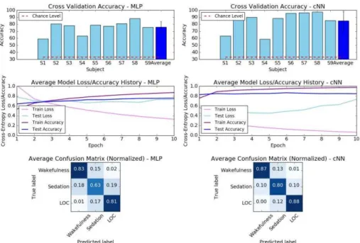
Raw Data. With raw EEG input, the MLP achieved an average accuracy of 75.45% across participants, with the cNN achieving 86.05% (Fig. 4). These accuracies are significantly higher than the chance level accuracy of 33.33%. Cross-entropy loss on the test set did not significantly decrease after the first epoch. Overall, the cNN was able to achieve better accuracies for each state of consciousness and participant.
Fig. 4. MLP vs cNN (reference size) comparison for raw EEG classification of the three con-scious states. Cross validation accuracies, average model loss and confusion matrices are shown for each architecture.
7
As seen in Fig. 4, the confusion matrices suggest that Wakefulness and LOC were not often confused. The intermediate state of Sedation was hardest to predict, due to indi-vidual variability in response to propofol. However, this would not present a problem in the clinical context, where anesthetic induction is much more rapid [10].
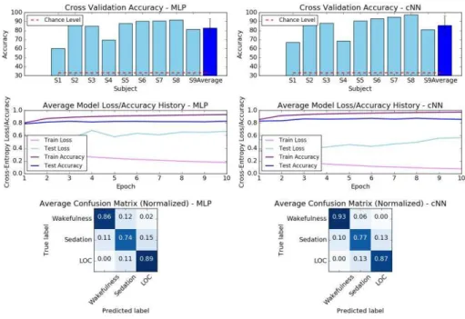
Power Spectral Density. With PSD input, the two architectures were equally capable in classifying states of consciousness (Fig. 5). In particular, the MLP performed better than when provided with raw time series as input, but the cNN did not (MLP: 83.4%, cNN: 87.35%). Importantly, cross-entropy loss revealed that the models converged faster using the PSD representation.
Fig. 5. MLP vs cNN (reference size) comparison for EEG classification of the three conscious states, using the PSD representation.
To understand the changes in the underlying EEG signal driving these accuracies, we visualized the PSDs in each state of consciousness (Fig. 6). As expected, we observed a decrease in alpha oscillations in Sedation, followed by the emergence of high-alpha oscillations during LOC.
Fig. 6. Power spectral density (uV2 /Hz, dB) of the EEG epochs, divided by the sedation phases
of the experiment. Representative frontal (Fz) and parietal (Pz) electrodes are shown.
3.2 Statistical Analysis – ANOVA Model
As a final step, a three-way ANOVA (type 2) was performed on the accuracies ob-tained in all twelve experiments across 2 architectures, 2 data representations and 3 model sizes, as summarized in Fig. 7 and detailed in Table 2.
Fig. 7. Three-way ANOVA for the comparison of the data representations, architectures and network sizes. Error bars indicate 95% confidence interval.
Table 2. ANOVA Table
Sum_sq df F Pr(>F)
Architecture 1230.94 1 10.601 0.0015
9
Model Size 14.34 2 0.061 0.9401
Architecture-Data Representation 351.13 1 3.024 0.0852
Architecture-Model Size 6.21 2 0.026 0.9735
Data Representation-Model Size 6.18 2 0.026 0.9737
Architecture-Data Representation-Model Size 0.85 2 0.003 0.9963
Residual 11146.71 96
The results of the ANOVA indicated that network architecture (cNN/MLP) was the strongest contributor to model performance (F = 10.6, p=0.0015), while data repre-sentation (Raw/PSD) also had a significant but weaker effect (F = 5.34, p=0.0229), driven by the improved accuracy of MLPs with PSD data.
In terms of resource utilization, the cNN was also better than the MLP, as the latter had a significantly larger number of parameters to learn (e.g. 46,872,579 in MLP vs 2,921,219 in cNN, for the reference network size). cNN was also faster to train by ~18%. Furthermore, a repetition of the above experiments with an alternative compar-ison using the same number of trainable parameters (rather than the same number of neurons) in each architecture, gave a much more prominent difference in accuracies, with the MLP performing much worse. Finally, we also verified that increasing epoch size from 1s through to 10s did not improve performance of either model.
4
Discussion
Our findings highlight the capability ofpotential for deep learning of human EEG to discover and utilize generalizable features for automatic identification of conscious-ness during anesthesia. Further, we have shown that modern cNNs significantly out-perform fully connected MLPs, potentially due to their ability to extract more effec-tive spatio-temporo-spectral features from the raw signal. This notion is supported by the fact that MLPs performed as well as cNNs when given PSD data as input.
Though this study aimed to conduct a comparative analysis rather than hyperpa-rameter optimization to maximize accuracy, the fact that cNNs were able to perform very well given only with 1 second of raw EEG data despite the lack of such optimi-zation suggests that they could find utility in real-world applications for assessment and monitoring of consciousness.
Acknowledgements We acknowledge funding from the UK Engineering and Physical Sciences Research Council [EP/P033199/1],
t
he Belgian National Fund for Scientific Research, theEuropean Commission, the Human Brain Project, the Luminous project, the French Speaking Community Concerted Research Action, the Belgian American Educational Foundation, the Wallonie-Bruxelles Federation, the European Space Agency, the University and University Hospital of Liège (Belgium). This research was undertaken with the support of the Alan Turing Institute (UK Engineering and Physical Sciences Research Council Grant EP/N510129/1).
References
1. Purdon, P.L., Pierce, E.T., Mukamel, E.A., Prerau, M.J., Walsh, J.L., Wong, K.F.K., Salazar-Gomez, A.F., Harrell, P.G., Sampson, A.L., Cimenser, A., Ching, S., Kopell, N.J., Tavares-Stoeckel, C., Habeeb, K., Merhar, R., Brown, E.N.: Electroencephalogram signatures of loss and recovery of consciousness from propofol. Proc. Natl. Acad. Sci. (2013). doi:10.1073/pnas.1221180110
2. Avidan, M.S., Jacobsohn, E., Glick, D., Burnside, B.A., Zhang, L., Villafranca, A.,
Karl, L., Kamal, S., Torres, B., O’Connor, M., Evers, A.S., Gradwohl, S., Lin, N.,
Palanca, B.J., Mashour, G.A.: Prevention of Intraoperative Awareness in a High-Risk Surgical Population. N. Engl. J. Med. (2011). doi:10.1056/NEJMoa1100403
3. Chennu, S., O'Connor, S., Adapa, R., Menon, D.K., Bekinschtein, T.A.: Brain Connectivity Dissociates Responsiveness from Drug Exposure during Propofol-Induced Transitions of Consciousness. PLoS Comput. Biol. 12, 1–17 (2016). doi:10.1371/journal.pcbi.1004669
4. Murphy, M., Bruno, M.-A., Riedner, B.A., Boveroux, P., Noirhomme, Q., Landsness, E.C., Brichant, J.-F., Phillips, C., Massimini, M., Laureys, S., Tononi, G., Boly, M.: Propofol Anesthesia and Sleep: A High-Density EEG Study. Sleep. 34, 283–291 (2011). doi:10.1093/sleep/34.3.283
5. Schirrmeister, R.T., Springenberg, J.T., Fiederer, L.D.J., Glasstetter, M., Eggensperger, K., Tangermann, M., Hutter, F., Burgard, W., Ball, T.: Deep learning with convolutional neural networks for EEG decoding and visualization. Hum. Brain Mapp. 38, 5391–5420 (2017). doi:10.1002/hbm.23730
6. Stober, S., Cameron, D.J., Grahn, J. a: Using Convolutional Neural Networks to Recognize Rhythm Stimuli from Electroencephalography Recordings. Neural Inf. Process. Syst. 2014. 1–9 (2014)
7. Howbert, J.J., Patterson, E.E., Stead, S.M., Brinkmann, B., Vasoli, V., Crepeau, D., Vite, C.H., Sturges, B., Ruedebusch, V., Mavoori, J., Leyde, K., Sheffield, W.D., Litt, B., Worrell, G.A.: Forecasting seizures in dogs with naturally occurring epilepsy. PLoS One. (2014). doi:10.1371/journal.pone.0081920
8. Park, Y., Luo, L., Parhi, K.K., Netoff, T.: Seizure prediction with spectral power of EEG using cost-sensitive support vector machines. Epilepsia. (2011). doi:10.1111/j.1528-1167.2011.03138.x
9. Krizhevsky, A., Sutskever, I., Hinton, G.E.: ImageNet Classification with Deep Convolutional Neural Networks. Adv. Neural Inf. Process. Syst. 1–9 (2012). doi:http://dx.doi.org/10.1016/j.protcy.2014.09.007
10. Juel, B.E., Romundstad, L., Kolstad, F., Storm, J.F., Larsson, P.G.: Distinguishing Anesthetized from Awake State in Patients: A New Approach Using One Second Segments of Raw EEG. Front. Hum. Neurosci. (2018). doi:10.3389/fnhum.2018.00040
11. Korshunova, I., Kindermans, P.-J., Degrave, J., Verhoeven, T., Brinkmann, B.H., Dambre, J.: Towards improved design and evaluation of epileptic seizure predictors. IEEE Trans. Biomed. Eng. 9294, 1–1 (2017). doi:10.1109/TBME.2017.2700086