AVIS
Ce document a été numérisé par la Division de la gestion des documents et des archives de l’Université de Montréal.
L’auteur a autorisé l’Université de Montréal à reproduire et diffuser, en totalité ou en partie, par quelque moyen que ce soit et sur quelque support que ce soit, et exclusivement à des fins non lucratives d’enseignement et de recherche, des copies de ce mémoire ou de cette thèse.
L’auteur et les coauteurs le cas échéant conservent la propriété du droit d’auteur et des droits moraux qui protègent ce document. Ni la thèse ou le mémoire, ni des extraits substantiels de ce document, ne doivent être imprimés ou autrement reproduits sans l’autorisation de l’auteur.
Afin de se conformer à la Loi canadienne sur la protection des renseignements personnels, quelques formulaires secondaires, coordonnées ou signatures intégrées au texte ont pu être enlevés de ce document. Bien que cela ait pu affecter la pagination, il n’y a aucun contenu manquant.
NOTICE
This document was digitized by the Records Management & Archives Division of Université de Montréal.
The author of this thesis or dissertation has granted a nonexclusive license allowing Université de Montréal to reproduce and publish the document, in part or in whole, and in any format, solely for noncommercial educational and research purposes.
The author and co-authors if applicable retain copyright ownership and moral rights in this document. Neither the whole thesis or dissertation, nor substantial extracts from it, may be printed or otherwise reproduced without the author’s permission.
In compliance with the Canadian Privacy Act some supporting forms, contact information or signatures may have been removed from the document. While this may affect the document page count, it does not represent any loss of content from the document.
Étude des interactions entre les boucles D et T chez
les ARNs de transfert (ARNts)
par Félix Doyon
Département de Biochimie (Bio-informatique) Faculté des arts et des sciences
Mémoire présenté à la Faculté des études supérieures en vue de l'obtention du grade de Maître ès Sciences (M.Sc.)
en Bio-informatique
Août, 2007
2008 MARS 1 3
Faculté des études supérieures
Ce mémoire intitulé:
Étude des interactions entre les boucles D et T chez les ARNs de transfert (ARNts)
présenté par: Félix Doyon
a été évalué par un jury composé des personnes suivantes:
. président-rapporteur
Serguei Chteinberg, directeur de recherche
Résumé
Afin d'élucider les contraintes générales imposées sur la structure des b,?ucles D et T (région DT) chez les ARNs de transfert (ARNts), nous avons sélectionné, à partir d'une librairie combinatoire d'ARNts où plusieurs nucléotides étaient randomisés, une série d'ARNts suppresseur in vivo. L'analyse de ces séquences démontre que, parmi les nucléotides randomisés, les plus conservés sont les nucléotides 54 et 58 de la boucle T. La plupart du temps, ils forment une combinaison U54-A58 qui permet la formation d'une paire de base reverse-Hoogsteen. De façon surprenante, les autres séquences arborent quant à eux soit G54-A58, soit G54-G58. Nous avons pu montrer par modélisation moléculaire que ces deux alternatives permettent la formation de paires de base très similaires à la paire de base reverse-Hoogsteen U-A et qui pourraient la remplacer dans le contexte d'un ARNt fonctionnel. Ceci élève la paire de base reverse-Hoogsteen 54-58 au rang des éléments structuraux importants ayant un impact sur la fonction de la molécule. Nous suggérons que son rôle principal est de préserver la conformation du dinucléotide 59-60 et par conséquent de maintenir l'architecture générale en forme de « L» des ARNts. Dans un autre ordre d'idée, on peut remarquer que toutes les séquences tombent dans deux catégories, chacune caractérisée par un type particulier de séquence. L'analyse de ces deux groupes nous a permis de proposer deux arrangements pour la structure de la région DT.
Nous avons nommé le premier « piège à purine spécifique». Il est représenté par 12 séquences et correspond à une version généralisée des interactions entre les boucles D et T, où la purine 18 de la boucle D s'intercale entre la paire de base reverse-Hoogsteen U54-A58 et la purine 57 de la boucle T. L'identité de la purine 18 est contrôlée par un appariement spécifique avec le nucléotide 55 : elle est un G ou un A selon si ce dernier est un U ou un G, respectivement. Nous avons nommé le deuxième· arrangement «piège à purine non-spécifique ». Il est représenté par 16 séquences c'est un arrangement qui n'a jamais été décrit auparavant dans la littérature. Il est caractérisé par l'intercalation de la purine 18 entre deux paires de base reverse-Hoogsteen U54-A58 et A55-C57. Par contre, il nécessite la présence d'un huitième nucléotide dans la boucle T et, à l'inverse du « piège à purine spécifique », la purine 18 ne forme pas d'interactions avec le nucléotide 55. Ainsi, nous avons décrit et explicité la logique derrière certains éléments de structure chez l'ARNt, et ce savoir, appliqué à de nouvelles molécules, nous permettra d'approfondir nos connaissances de la structure d l'ARN.
Abs"tract
To elucidate the general constraints imposed on the structure of the D- and T-Ioops in functional tRNAs, active suppressor tRNAs were selected in vivo from a combinatorial tRNA gene library in which several nucleotide positions of these loops were randomized. Analysis of the nucleotide sequences of the selected clones demonstrates that among the randomized nucleotides, the most conservative are nucleotides 54 and 58 in the T-Ioop. In most cases, they make the combination U54-A58, which allows the formation of the normal reverse Hoogsteen base pair. Surprisingly, other clones have either the combination A58 or G54-G58. However, molecular modeling shows that these purine-purine base pairs can very closely mimic the reverse-Hoogsteen base pair U-A and thus can replace it in the T -Ioop of a functional tRNA. This places the reverse Hoogsteen base pair 54-58 as one of the most important structural aspects of tRNA functionality. We suggest that the major role of this base pair is to preserve the conformation of dinucleotide 59-60 and, through this, to maintain the general architecture of the tRNA L-form. With only one exception, ail of the clones fall into two groups, each characterized by a distinct sequence pattern. Analysis of these two groups has allowed us to suggest two different types of nucleotide arrangement in the DT region. The first type, the so-called specific purine trap, is found in 12 individual tRNA clones and represents a generalized version of the standard D-T loop
interaction. It consists of purine 18 sandwiched between the reverse-Hoogsteen basepair U54-A58 and purine 57. The identity of purine 18 is restricted by the specifie base-pairing with nucleotide 55. Depending on whether nucleotide 55 is U or G, purine 18 should be, respectively, G or A. The second structural type, the so-called non-specifie purine trap, corresponds to the nucleotide sequence pattern found in 16 individual tRNA clones and is described here for the first time. It consists of purine 18 sandwiched between two reverse-Hoogsteen base-pairs U54-A58 and A55-C57 and, unlike the specifie purine trap, requires the T-Ioop to
contain an extra eighth nucleotide. Since purine 18 does not form a base-pair in the nonspecific purine trap, both purines, G18 and A 18, fit to the structure equally weil. The important role of both the specifie and non-specifie purine traps in the formation of the tRNA L-shape is discussed.
Table des matières
RÉSUMÉ ... 111
ABSTRACT ... V
TABLE DES MATIÈRES ... ,VIl
LISTE DES TABLEAUX, ...
x
LISTE DES FIGURES ... XlREMERCiEMENTS ... XIV
CHAPITRE 1 : INTRODUCTION ... 1 1.1 STRUCTURE DE L'ARN ... 4 1.1.1 Le nucléotide _________________________________________________________________________________________________________________ .7 1.1.2 La structure primaire de l'ARN ________________________________________________________________________________________ .8 1.1.3 La structure secondaire de l'ARN ____________________________________________________________________________________ .9 1.1.4 La structure tertiaire de l'ARN _______________________________________________________________________________________ ) 1
1.2 L'ARN DE TRANSFERT (ARNt) ... 12 1.2.1 La structure tertiaire en forme de «L» et les deux centres fonctionnels ____________________________ )2 1.2.2 La région charnière et les nucléotides la composant. ______________________________________________________ )4 1.2.3 Les interactions intéressantes relevées à ce jour ___________________________________________________________ )5
1.3 DIFFÉRENTES MÉTHODES D'ANALYSE DE LA STRUCTURE DE L'ARN ... .16 1.3.1 La cristallographie de rayon-X _______________________________________________________________________________________ ) 7 1.3.2 La résonnance magnétique nucléaire (RMNt ________________________________________________________________ ) 7 1.3.3 La modélisation moléculaire interactive _________________________________________________________________________ )8
1.3.4 Les librairies combinatoires D'ARNt ... )8
1.4 HYPOTHÈSE ET OB .. IECTIFS ...••.•••...•.••••...•...••... 19
CHAPITRE 2 : IMPORTANCE OF THE REVERSE HOOGSTEEN BASE PAIR 54-58 FOR TRNA FUNCllON (published
articleL ...
212.1 INTRODUCTION ...•.•.••...•...•.•...•....••...•... 22
2.2 MATERIALS & METHODS ...•...•.•...•...•.•.•.•...•...••••...••.... 26
2.2.1 Strains ... .26
2.2.2 Construction of the combinatoriallibrary and selection of suppressor tRNAs ... .26
2.2.3 Sequencing ... 27
2.2.4 Measurement of the -Galactosidase activity ... .27
2.2.5 Presence of the Suppressor tRNAs in the cytosol and the aminoacylation level ... .28
2.2.6 Computer modeling ... 29
2.3 RESLIL TS •••••••••.•...•••••••••.••.•...•.•.•••••••.•.•.••.•.•.•.•.•.••••••....•...••••.•••••••.••••...•.•.•.••.••. .29
2.3.1 The library Design ... .29
2.3.2 Cloning and selection of functional clones ... .30
2.3.3 Analysis of the nucleotide sequences ... .32
2.3.4 Modeling of the tRNA structures ... .39
2.4 DiSCUSSiON ....•.•.•...••.•••••••...••••••...•••...•..•...••••... 40
2.5 ACKNOWLEDGEMENTS ... 48
CHAPITRE 3: APPORT SCIENTIFIQUE DE L'ARTICLE ... 49
CHAPITRE 4 : SPECIFIC AND NON-SPECIFIC PURINE TRAP IN THE T-LOOP OF NORMAL AND SUPPRESSOR TRNAS (published
articleL ...
534.1 INTRODUCTION ...•...•... 54
4.2.1 The selected clones __________________________________________________________________________________________________ 61 4.2.2 Type 1: a quasi normal pattern ____________________________________________________________________________________ 63
4.2_3 Extension of the quasi normal pattern for T8-tRNAs. ____________________________________________________ 66 4.2.4 The specifie purine trap in the T-Ioop of the Type 1 tRNAs ____________________________________________ 70
4.2.5 Purine trap and reverse-Hoogsteen base-pair U54-A58 _______________________________________________ 71 4.2.6 Type Il as a non-specifie purine trap ______________________________________________ .. __________________________ .73
4.2.7 Stability of the non-specifie purine trap and its dependence on the identity of certain nucleoti des ______________________________________________________________________________________________________ 0 _______________ 000 _ 079
4.3 DISCUSSION .••••.•....••..•••••••••••••.•••••.••... _ ..• _ .•...• _ •.•.•....•.•.•.••.•.. _ ...•.•.•...•...•...••.••..•.••.•. 80
4.4 METHODS ...••.•... _ ...•.••.••..•..•.••.•• _ .• __ .. _ .. _ .•• _ ..•.••..•..•...•... _ •..••...•...•..• _ •..•.. 93
4.4.1 Cloning, selection and measuring the suppressor tRNA activity ____________________________________ 93 4.4.2 Computer modeling _____________________________________________________________ . ______ . ______ . _______________________ 93 4.5 ACKNOWLEDGEMENTS •...•...•...•...••..•...••.•...•.•..•.•.•....••.•..•...•...•...•.••.•..•.•..•• 94
CHAPITRE 5 : APPORT SCIENTIFIQUE DE L'ARTICLE ... 95
CHAPITRE 6 : CONCLUSiON ... 99
BI BLIOG RAPHIE ... 101
ANNEXE 1 : ACCORD DES COAUTEURS DE L'ARTICLE 1 ... .. ANNEXE Il : ACCORD DES COAUTEURS DE L'ARTICLE
2 ... ..11
Liste des tableaux
Table 2.1 The nucleotide sequences and the j3-galactosidase activity of the
selected tRNA clones ... 33 Table 4.1 Sequences of the D-and T-Ioops and suppressor activities of the
Liste des figures
Figure 1.1 Structure tridimensionnelle du ribosome contenant des ARNts. _______
.4
Figure 1.2 Structure polymérique des acides nucléiques .. ____________________________________ .6 Figure 1.3 Structure d'un nucléotide de l'ARN . _______________________________________________________ .7 Figure 1.4 Différents éléments de structure secondaire retrouvés dans l'ARN. 10 Figure 1.5 Structures secondaire (A) et tertiaire (8) de l'ARN de transfert .. _______ .14 Figure 2.1 The standard tRNA L-form .. _____________________________________________________________________ .24 Figure 2.2 Construction of the tRNA gene library. __________________________________________________ .25 Figure 2.3 Northern blot showing the presence in the cytosol and the level ofaminoacylation of sorne suppressor tRNAs., _________________________________________ 35 Figure 2.4 Juxtaposition of the bases in RH-GA, RH-GG and other alternative
base pair candidates for replacement of RH-UA. _________________________________ 38 Figure 2.5 The model of the structure of the DT region for clone K31 (red)
superimposed on the corresponding region in yeast tRNAPhe
(g reen). ________________________________________________________________________________________________________ 40 Figure 4.1 The structure of the DT region in the context of the tRNA L-shape. 57 Figure 4.2 The design of the tRNA gene library (K-library) [ZDS2003L _____________ 59 Figure 4.3 The nucleotide arrangements in two inter-Ioop base-pairs G18-U
(ljJ )55 and A 18-G55 . _________________________________________________________________________________ 65 Figure 4.4 The general view of the DT region in the Type 1 and Il tRNAs. _________ .69
Fjgure 4.5 The detailed structure of the non-specific purine trap. _______________________ J5 Figure 4.6 Comparison of the model of the non-specific purine trap and the
Figure 4.7 Comparison of the model of the non-spedfic purine trap and
À l'homme qui représente la Science pour moi, Serguei Chteinberg.
Remerciements
J'aimerais remercier mon directeur de recherche, Serguei Chteinberg, pour sa patience, sa compréhension et la confiance qu'il m'a accordée.
J'aimerais aussi remercier tous les membres du laboratoire, particulièrement mes coauteurs Ekaterina Zagryadskaya et Jianhong Chen.
J'aimerais aussi remercier ma copine Julie, sans l'aide et le support de laquelle je n'aurais probablement pas terminé ce mémoire.
Finalement, j'aimerais remercier le programme stratégique de bourse de formation des IRSC en bio-informatique, les bourses d'excellence bit, ansi que le programme de bourse du Fonds de la Recherche en Santé du Québec (FRSQ) pour leur support financier.
L'humanité se questionne depuis bien longtemps sur la question de l'origine de la vie. Plusieurs différentes théories ont vu le jour à travers les différentes époques, allant du Créationnisme à la théorie du « Big Bang )}. Plus près de la réalité que l'on connaît aujourd'hui, beaucoup de découvertes ont été faites dans le domaine de la biologie depuis l'hypothèse de l'évolution émise par Darwin en 1859. Le 20e siècle a depuis été particulièrement prolifique pou r l'avancement de la Science. La découverte de la structure de l'ADN par Watson et Crick a généré un engouement sans précédent, non seulement chez la communauté scienti'fique mais aussi pour la société en général [WC1953]. Ce grand pas vers la compréhension de ce que nous sornmes a ensuite ouvert la porte à de nombreuses découvertes dont certaines ont révolutionné notre vision de l'organisme vivant et son fonctionnement. Pour n'en nommer que quelques-unes, il y eut la détermination de la structure du premier ARN de transfert, il y a plus de 30 ans [QSMKSR1974] et, plus récemment, la détermination de la structure du ribosome [BNHMS2000, WBCMWCVHR2000, VYBLECN2001].
À ce jour, on sait que l'acide désoxyribonucléique (ADN) est la molécule encodant la presque totalité des processus vitaux. Il a été établi qu'il doit d'abord être transcrit en acide ribonucléique (ARN), ce qui s'effectue avec la collaboration essentielle de l'ARN polymérase. Par la suite, l'ARN est à son tour traduit en protéine par l'entremise du complexe ribosomal. Cette synthèse de peptides est un
processus élaboré qui implique une multitude de participants, allant des acides aminés jusqu'aux différentes enzymes. Bien que la science ait apporté de nombreuses réponses au cours des dernières décennies, beaucoup reste encore à éclaircir. D'ailleurs, nous découvrons et caractérisons sans cesse de nouveaux joueurs dans le processus de la synthèse protéique. Leurs fonctions recèlent de
nombreux mystères que les scientifiques tentent d'élucider encore à ce jour.
Le séquençage en entier du génome humain, en 2004, a entre autre permis d'estimer le nombre total de gènes entre 20000 et 25000 [IHGSC2004]. Suite à cette révélation, il a été admit que plusieurs des gènes séquencés correspondent fort probablement à des protéines. Néanmoins, certains de ces gènes codent pour des ARNs et nous savons déjà que ceux-ci jouent un rôle prépondérant dans plusieurs processus biologiques. Le plus évident de ces rôles est de servir de messager (ARNm) entre l'ADN et le ribosome, qui est le centre de la synthèse des protéines. D'autre part, l'ARN joue un rôle au niveau de la composition du complexe ribosomal (ARNr). Il est également impliqué dans le transport des acides aminés destinés à être incorporés lors de la synthèse des protéines (ARNt). Par conséquent, l'ARN, sous ses différentes formes, assure plusieurs fonctions vitales à la cellule et de nos jours encore on lui découvre de nouveaux rôles et applications. Un des plus récents exemples est sans doute la découverte et
l'utilisation des siRNAs, qui révolutionne présentement le monde de la biologie moléculaire [HB1999, EHLYWT2001].
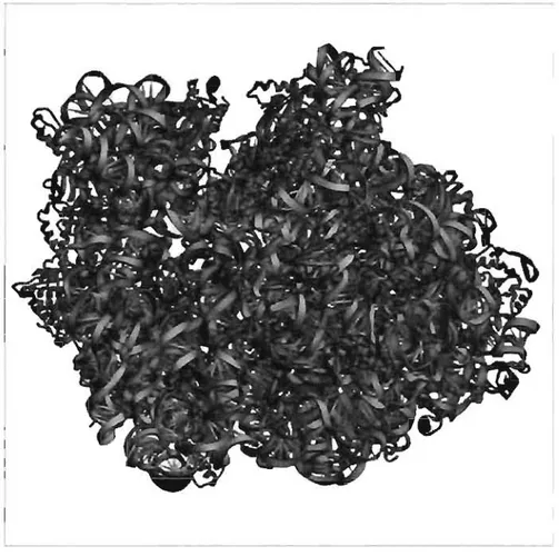
D'un point de vue structurel, les ARNs sont des molécules hautement organisées. La figure 1.1 est un exemple tridimensionnel de plusieurs molécules d'ARN et illustre bien leur complexité. Leur arrangement spatial est si crucial que leurs fonctions ne peuvent être assurées que dans des conditions bien précises, telles qu'un repliement adéquat de la molécule et ce, dans une fenêtre de temps bien précise. On comprend alors pourquoi il est primordial de bien comprendre les différents aspects qui régissent le repliement des ARNs. Ces connaissances nous permettront éventuellement d'être en mesure de mieux cerner leurs fonctions et d'en comprendre les mécanismes.
Figure 1.1 Structure tridimensionnelle du ribosome contenant des ARNts. L'ARN est
représenté sous forme de bâtonnets (représentant les bases) et de rubans (représentant le squelette sucre-phosphate). La grosse sous-unité (238) est en gris, la petite (168) en cyan, l'ARN 58 en mauve, alors que les ARNts sont en jaune, orange et rouge. Les protéines ribosomales sont aussi présentes en magenta (grosse sous-unité) et en mauve foncé (petite sous-unité) [YYBLECN2001].
1.1 STRUCTURE DE L'ARN
À la base, l'ARN est un polymère formé d'un squelette sucre-phosphate et
de quatre différentes bases azotées: l'adénine, la cytosine, la guanine et l'uracile (Figure 1.2). Il existe des molécules d'ARN de toutes les tailles: de l'ARN de
transfert (ARNt), qui est composé habituellement d'environ 76 nucléotides, jusqu'au ribosome, pour lequel la petite sous-unité à elle seule comporte plus de 1500 nucléotides chez les procaryotes et quelques centaines de plus chez les eucaryotes [BA2002].
L'ARN ressemble beaucoup à l'ADN, mis à part quelques différences déterminantes: dans un premier temps, le sucre du nucléotide est un ribose plutôt qu'un désoxyribose. Dans un deuxième temps, l'uracile remplace la thymine de l'ADN. En effet, la figure 1.2 montre que l'ADN possède un groupement hydroxyl en position 3' de son pentose, alors que l'ARN en possède aussi un en position 2'. L'ARN dispose, tout comme les protéines, d'une structure hautement hiérarchisée: la structure primaire correspond à la séquence nucléotidique composant la molécule; la structure secondaire correspond à l'appariement Watson-Crick entre les différents nucléotides; tandis que la structure tertiaire correspond à l'arrangement adopté par la molécule en trois dimensions. Dépendamment du type d'ARN et de ses fonctions, certains éléments de structure sont conservés et nécessaires au bon fonctionnement de la molécule. Nous voyons plus en détails ces différents aspects dans les sections 1.1.2, 1.1.3 et 1.1.4.
A
RK\ B \ ' H; \ ( l /1"l/~~
l '\
,1
\N-
"N~.J '\" Adernine O=P-OCfi, ' ' 1 () Gu nmW
0- (.';:
~ Cytosi\,
neo=~
-
'H,~
N
~'
()
:.
~
,:(0 \\ \
G ' () , , " t·1 \ uamne 1 \ 1 1 .... \'. p-OCI1, ~ -... "J;;;:A. .... 1W
'
0 ,,- ' Nil, "-ci \ i 0 , 01H
l~
1
NH \l
,
A
Thymine (1 - ~ - ()(~ 0 "'~ n Uraello,
~
o , H DNA '. >. \Figure 1.2 Structure polymérique des acides nucléiques. Pour les deux polymères,
l'extrémité 5' est située en haut alors que l'extrémité 3' est en bas. A) Polymère d'ARN illustrant les 4 bases azotées de l'ARN, l'adénine, la cytosine, la guanine et l'uracile, B)
polymère d'ARN illustrant les 4 bases azotées de l'ARN, l'adénine, la cytosine, la guanine et la thymine. Les différences entre l'ADN et l'ARN sont illustrées en jaune. On peut y voir que la thymine est remplacée par l'uracile dans l'ADN et que l'ARN possède un groupement hydroxyl en 2'. [KTF].
1.1.1 Le nucléotide
Le nucléotide est l'unité de base du polymère que sont les acides nucléiques. Il est formé de trois parties, soit d'une base azotée, d'un pentose, et
d'un groupement phosphate (Figure 1.3). Les bases canoniques (ou
conventionnelles) de l'ADN et de l'ARN sont des purines (adénine et guanine) ou des pyrimidines (cytosine, thymine, uracile). Le pentose est un ribose chez l'ARN tandis qu'il est un désoxyribose chez l'ADN. Le nucléotide possède trois arêtes hydrophiles via lesquelles il forme des ponts hydrogènes (pont-H) avec d'autres nucléotides et constitue ainsi des paires de bases. Ces trois arêtes sont la face Watson-Crick, la face Hoogsteen et la face Sucre [LW2001]. Dans une hélice double régulière, qui est un élément de structure fréquemment retrouvé, les bases se font face par leur côté Watson-Crick, mais une multitude de paires de bases dites non-Watson-Crick sont possibles.
o
- IlO -
P
-O
1
-
1
o
CH OH OHFigure 1.3 Structure d'un nucléotide de l'ARN. Le nucléotide est formé d'une base azotée (représentée par un rectangle bleu), d'un ribose (en noir; les carbones sont numérotés de l' à 5') et d'un groupement phosphate (en vert) [ND].
Plusieurs nucléotides peuvent être modifiés au stade post-transcriptionnel. Par exemple, dans l'ARNt, la boucle T comporte une pseudouridine (41) en position 55. Ce nucléotide s'apparente à l'uracile, mais où le lien entre le sucre et la base se fait entre le C1 du sucre et le C5 de la base plutôt qu'avec le N1 [CG2000]. Un autre nucléotide modifié communément rencontré est l'inosine. Il s'agit en fait d'une guanine qui a subi une désamination. L'inosine est impliquée dans l'hypothèse Wobble, selon laquelle le nucléotide en 5' de l'anticodon jouit d'une plus grande liberté conformationnelle, lui permettant de former des paires de bases non-conventionnelles. Cette hypothèse expliquerait l'existence de 40 AR Nt pour 60 codons [WC1966]. Cet exemple démontre bien qu'il est clair que ces nucléotides modifiés jouent un rôle important tant au niveau de la structure que de la fonction. Ainsi, dû à leur importance relative, on les retrouve souvent dans les centres fonctionnels des molécules d'ARN.
1.1.2 La structure primaire de l'ARN
La structure primaire consiste en la séquence nucléotidique de l'ARN, c'est-à-dire la suite des nucléotides la composant, de l'extrémité 5' jusqu'à l'ex~rémité 3'. L'ADN est d'abord transcrit en ARN, qui subit ensuite plusieurs étapes de maturation au cours desquelles il est épissé et où certains nucléotides peuvent être modifiés [GE2000] (tel que décrit à la section 1.1.1).
1.1.3 La structure secondaire de l'ARN
La structure secondaire consiste en l'appariement Watson-Crick des nucléotides (section 1.1.1). Ce positionnement forme différents domaines qui peuvent être composés d'hélices doubles, de tige-boucles (<< hairpin loop »), de renflements (<< bulges ») et de boucles internes (<< internai loop »). Les tige-boucles sont constituées d'une hélice double bouclée par un petit nombre de nucléotides non-appariés. Les renflements correspondent à un ou plusieurs nucléotides non appariés dans un seul des brins d'une hélice double. Les boucles internes quant à elles sont formées de nucléotides non-appariés dans les deux brins d'une hélice double. Les renflements et les boucles internes confèrent une certaine flexibilité à la région. Les nucléotides les composants sont souvent impliqués dans les interactions au niveau de la structure tertiaire. La Figure 1.4 montre quelques-uns de ces motifs structurels.
Deux forces primordiales interviennent au niveau de la structure secondaire de l'ARN et ont un impact sur sa structure et ses fonctions. Celle ayant le plus grand impact est régit par des interactions hydrophobes et favorise l'empilement des orbitales TT des cycles aromatiques des bases azotées l'un par-dessus l'autre. Ce phénomène est appelé empilement (<< stacking ») et joue un énorme rôle dans la stabilité de la molécule d'ARN [FPHT1983].. Le deuxième élément primordial à
intervenir au niveau de la structure secondaire de l'ARN est le pont-Ho Ce type d'interaction implique le partage d'un atome d'hydrogène par deux atomes électronégatifs. C'est d'ailleurs ce qui détermine la complémentarité entre l'adénine et l'uracile, et entre la guanine et la cytosine [J1997]. La présence de ces types d'interactions entre nucléotides a une grande influence au niveau de l'arrangement tridimensionnel de la molécule d'ARN et joue donc indirectement un rôle certain dans sa capacité à assurer ses fonctions de façon adéquate.
Bulgc Loop (right)
InternaI Loop
Figure 1.4 Différents éléments de structure secondaire retrouvés dans l'ARN. Plusieurs éléments de structure peuvent être retrouvés au sain de l'ARN. Notamment, les tige-boucle, les régions d'empilement, les renflements, les boucles internes et les boucles multiples [SG2005].
1.1.4 La structure tertiaire de l'ARN
La structure tertiaire de l'ARN diffère grandement de celle de l'ADN. Dans le cas de l'ADN, la structure tertiaire est majoritairement constituée de longues hélices doubles régulières. Dans le cas de l'ARN, il a plutôt tendance à replier ses éléments de structure secondaire l'un sur l'autre, de façon très compacte, un peu à la manière des protéines. Ces éléments de structure secondaire sont les hélices doubles, les tige-boucles, les régions connectrices simple brin, les paires de bases tertiaires et/ou non-Watson-Crick. Il n'est pas rare de voir des éléments distants en termes de structure secondaire se rapprocher dans la structure tertiaire et interagir ensemble.
La façon dont les ARNs se replient demeure un mystère, mais plusieurs motifs structuraux ont été déterminés. Ces différents éléments de structure pourraient former un ensemble de « blocs» structuraux bien défini à partir duquel les ARN adoptent la multitude de structures qu'on leur attribue. Les interactions de type tertiaire impliquent aussi parfois les atomes qui ne sont pas impliqués dans l'appariement entre les bases, c'est-à-dire les atomes du ribose et du groupement phosphate [HBH2005]. D'autres recherches sur la nature des nucléotides qui composent la molécule d'ARN ainsi que sur leurs interactions sont bien entendues nécessaires afin de pouvoir élucider les mécanismes de repliement de l'ARN. De
plus, l'étude de l'arrangement spatial en relation avec la composition des molécules d'ARN est essentielle afin de permettre d'identifier certaines causes du mauvais repliement des ARNs. Bien que les récents développements technologiques dans ce domaine de recherche aient permis de grandes percées, beaucoup reste encore à être élucidé quant aux ARN.
1.2 L'ARN DE TRANSFERT (ARNt)
À ce jour, les différentes études portant sur l'ARNt ont permis de déterminer qu'il est un participant important du processus de synthèse des protéines. En effet, les ARNts sont impliqués au niveau du ribosome et agissent en tant que molécules adaptatrices entre l'ARN messager et la séquence de la protéine nouvellement synthétisée. C'est donc dire qu'ils ont une fonction vitale au sein de notre organisme. Sa structure, bien qu'elle ait été séquencée, comporte de nombreux éléments déterminants dont le rôle et l'importance relative restent encore à déterminer.
1.2.1 La structure tertiaire en forme de «L» et les deux centres fonctionnels
Afin que l'ARNt puisse jouer son rôle au sein de la synthèse protéique, deux centres fonctionnels ont été identifiés: l'anticodon et l'extrémité acceptrice. Ces centres interagissent respectivement avec l'ARN messager et le ribosome, sur son
site de réaction peptidyltransférase. Pour que ces interactions soient possibles, la molécule d'ARNt doit adopter une structure particulière et précise afin que ses deux centres fonctionnels soir bien positionnés pour lui permettre d'assumer ses fonctions cellulaires vitales.- C'est donc dire que les structures secondaire et tertiaire sont extrêmement importantes pour que la fonctionnalité des ARNtssoit
conservée.
À ce sujet, la Figure 1.5 illustre une molécule d'ARNt sous sa forme secondaire et sous sa forme tertiaire. La structure secondaire est dite en « feuille de trèfle ». Cette appellation est due à la présence de trois tige-boucles (D, T et anticodon) et d'une quatrième tige, dite acceptrice. Cette dernière forme un domaine coaxial avec la tige-boucle T (domaine Il) et elle a la .capacité et le rôle de lier l'acide aminé. La tige-boucle de l'anticodon forme quant à elle un domaine coaxial avec la tige boucle 0 (domaine 1).
L'orientation mutuelle de ces deux domaines est d'une importance primordiale afin que les ARNts puissent assumer correctement leur fonction. Il a été déterminé que les deux domaines coaxiaux doivent être orientés de façon perpendiculaire, ce qui confère la fornie de « L » à la structure tertiaire de l'ARNt. Les domaines 1 et Il se retrouvent ainsi aux extrémités du « L », soit au bout de la
la figure 1.5(B). Cette structure tridimensionnelle rigide universelle est très conservée chez les ARNt cytosoliques [SHBIS1998].
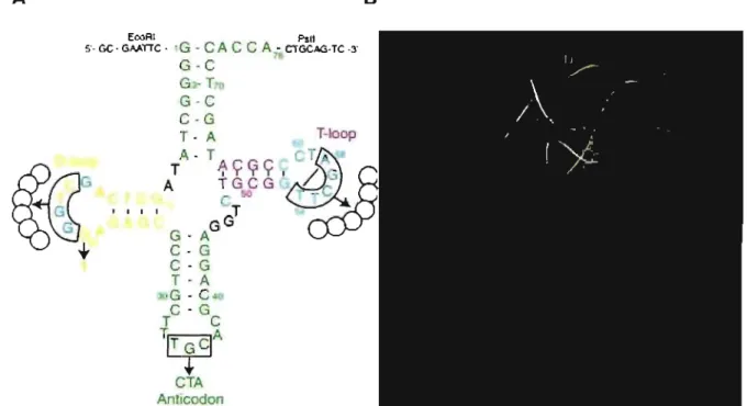
A
EcoRI PsU
5'- GC - GAATIC - 'G . CAC C A 7-CTGCAG-TC -3'
G-C G T G-C C·G T· A T-Ioop
~
G
' "...
A- T~
TA
T ~ÇCfÇ , / 3 A ~G!iCCG -_C , T GG G - A C- G C · G T - A ,G - C n C · G T C TITGCJA+
CTA Anllcodon BFigure 1.5 Structures secondaire (A) et tertiaire (8) de l'ARN de transfert. La tige acceptrice (ACC) ainsi que la tige-boucle de l'anticodon (AlC) sont en vert, la tige-boucle D en jaune et la tige-boucle T en magenta dans les deux structures. Les nucléotides en cyan sont ceux composant la région DT. Les nucléotides encadrés dans la structure secondaire sont les nucléotides qui ont été mutés dans la librairie-K [ZDS2003].
1.2.2 La région charnière et les nucléotides la composant
Les deux centres fonctionnels sont situés à une distance précise et cruciale de 70
A
l'un de l'autre [SSA 1994]. D'un point de vue tridimensionnel, seule la forme en « L» permet d'obtenir cette distance. Cette particularité suggère doncque la conformation adoptée par la région charnière au coude du «L» joue indubitablement un rôle primordial dans ce maintien et par conséquent dans la fonctionnalité de la molécule, qui constitue le sujet principal de ce travail.
La région charnière de la molécule est majoritairement composée des nucléotides des boucles D et T, et sera référée tout au long de ce mémoire en tant que région DT. Plusieurs des nucléotides de la région DT sont extrêmement conservés, ce qui suggère fortement leur implication dans la fixation des deux domaines hélicoïdaux. Par exemple, parmi les onze nucléotides de la région DT, huit sont invariables, du moins chez les ARNts cytosoliques. Les trois autres possèdent seulement un degré limité de variabilité: le nucléotide 57 doit être une purine, alors que les deux nucléotides formant l'appendice, 59 et 60, sont des pyrimidines dans la plupart des cas [SHBIS1998]. De plus, la région DT est composée d'interactions conservées entre ces nucléotides, malgré qu'il soit difficile d'évaluer l'importance relative de chacune de ces interactions ponctuelles.
1.2.3 Les interactions intéressantes relevées
à
ce jourPlusieurs interactions révélées par la détermination de la structure de l'ARNt semblent dignes d'intérêt. Tout d'abord, mentionnons la paire de base universelle dite « reverse-Hoogsteen » U54-A58. Cette paire est formée par l'interaction entre
le premier et le cinquième nucléotide de la boucle T. Puis, une autre intéressante interaction dans la région DT implique le nucléotide 18 de la boucle D qui s'empile entre les nucléotides 57 et 58 de la boucle T. Cet arrangement permet de former une paire de base non-Watson-Crick avec le nucléotide 55. On dénote aussi une paire de base tertiaire, mais de type Watson-Crick cette fois. Elle est formée par l'interaction des nucléotides 19 et 56 au-dessus du nucléotide 57.
La région DT est également composée d'autres éléments. Parmi ceux-ci, on y retrouve l'appendice, qui est formé de deux nucléotides non-appariés en 3' du nucléotide 58. Il est situé entre la paire de base « reverse-Hoogsteen» et la dernière paire de bases de la tige T. L'appendice vient s'empiler sur la dernière couche du domaine 1. Cette couche est elle-même constituée d'une paire de base tertiaire formée par les nucléotides 15 et 48. Un autre élément présent digne de mention est un motif « U-turn » qui est régi par le nucléotide 4J55.
1.3 DIFFÉRENTES MÉTHODES D'ANALYSE DE LA STRUCTURE DE L'ARN
Même si le présent ouvrage se concentre sur l'analyse de la structure de l'ARNt, il est important de mentionner les différents procédés par lesquels ces structures ont été obtenues. Plusieurs méthodes ont été développées au fil des années, chacune possédant ses avantages et ses limites. Ensemble, ces
différents outils se sont complémentés de façon à nous permettre d'obtenir une banque de structures qui correspond au point de départ de notre analyse.
1.3.1 La cristallographie de rayon-X
La cristallographie est une méthode basée sur l'analyse du patron de diffraction d'un cristal pur provenant d'une molécule. Ce patron de diffraction est obtenu en faisant passer un faisceau de rayons X à travers le cristal. La toute première macromolécule à être cristallisée à la fin des années cinquante fût la protéine myoglobine. Plus tard, cette technique s'est raffinée et a permis, entre autres, de déterminer la structure tridimensionnelle de l'ARNthe.
1.3.2 La résonnance magnétique nucléaire (RMN)
Cette autre technique permet la détermination de la structure de macromolécules biologiques. Dépendamment de leur environnement physico-chimique, différents protons résonnent à des fréquences différentes. Ce « déplacement chimique» peut être enregistré et analysé. Il permet de distinguer les atomes selon leur environnement physico-chimique à l'intérieur de la molécule, et d'ainsi obtenir différentes informations à propos de la molécule à l'étude. Cette méthode permet d'obtenir, avec une haute résolution, la structure de molécules mal structurées (pour lesquelles la structure secondaire n'est pas stable). Par
contre, en comparaison avec la méthode de cristallographie de rayon-X, la RMN ne peut être utilisée que pour des molécules relativement petites [L2001].
1.3.3 La modélisation moléculaire interactive
Cette méthode capitalise sur l'existence de structures tridimensionnelles et fait intervenir le jugement humain. L'objectif est d'analyser in silico certains éléments ponctuels de ces structures. Cette technique englobe aussi les différentes méthodes informatiques de prédiction de structures et de leurs comportements à différentes températures et dans différents environnements. La modélisation moléculaire interactive est un outil prédictif qui doit être utilisé de façon très critique. Par opposition avec les méthodes physico-chimiques de détermination de la structure, il ne constitue pas une preuve en soi mais plutôt un argument en faveur d'un modèle.
1.3.4 Les librairies combinatoires D'ARNt
Les librairies combinatoires sont aussi connues sous l'appellation « évolution dirigée ». Elles constituent une méthode puissante qui permet d'obtenir des séquences et des structures qui ne sont pas retrouvées dans' la nature. L'avantage indéniable de cette méthode est qu'elle permet l'obtention d'une plus
grande variété de séquences qui n'ont pas été favorisées a priori, comme c'est le
cas lorsque la technique de mutagénèse dirigée est utilisée.
1.4 HYPOTHÈSE ET OBJECTIFS
Malgré la connaissance de sites d'interaction sur la région DT de l'AR Nt, plusieurs questions restent à ce jour sans réponse. Par exemple, pourquoi la paire de base « reverse-Hoogsteen » est-elle absolument nécessaire afin de préserver la fonctionnalité de la molécule? En quoi est- elle si spécifique et qu'est-ce qui la rend unique? Ces questions ne peuvent êtres résolues par une approche biochimique standard. C'est pourquoi nous avons choisi de coupler une analyse théorique poussée qui impliquera de la modélisation moléculaire in silico à la collecte de données qui se fera de manière biochimique par l'entrernise de librairies combinatoires d'ARNts.
L'objectif global de mon projet consiste à déterminer les règles qui gouvernent le repliement des ARNs structurés en trois dimensions. Plus spécifiquement, mon objectif est de déterminer les conditions nécessaires et suffisantes à la formation de motifs d'ARN. L'ARNt est une molécule-modèle de premier choix pour réaliser cette étude. En effet, beaucoup de données de
séquence et de structure sont disponibles. De plus, c'est une molécule assez complexe du point de vue structural tout en ayant une taille raisonnable.
CHAPITRE 2: Importance of the reverse Hoogsteen
base pair 54-58 for tRNA function
Ekaterina 1. Zagryadskaya, Félix R. Doyon, Sergey V. Steinberg
Département de Biochimie, Université de Montréal, Mtl, Qc H3C 3J7, Canada
001: 10.1093/nar/gkg448 Nucleic Acids Research, 2003, Vol. 31, No. 143946-3953
To elucidate the general constraints imposed on the structure of the D- and T-Ioops in functional tRNAs, active suppressor tRNAs were selected in vivo fram a combinatoricil tRNA gene library in which several nucleotide positions of these loops were randomized. Analysis of the nucleotide sequences of the selected clones demonstrates that among the randomized nucleotides, the most conservative are nucleotides 54 and 58 in the T-Ioop. In most cases, they make the combination U54-A58, which allows the formation of the normal reverse Hoogsteen base pair. Surprisingly, other clones have either the combination A58 or G54-G58. However, molecular modeling shows that these purine-purine base pairs can very closely mimic the reverse Hoogsteen base pair U-A and thus can replace it in the T-Ioop of a functional tRNA. This places the reverse Hoogsteen base pair 54-58 as one of the most important structural aspects of tRNA functionality. We suggest that the major role of this base pair is to preserve the conformation of
dinucleotide 59-60 and, through this, to maintain the general architecture of the tRNA L-form.
2.1 INTRODUCTION
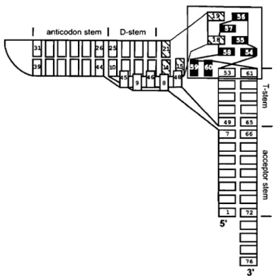
One of the most conservative elements in the tRNA tertiary structure is the region at the outer corner of the tRNA L-form, where the T-Ioop interacts with the D-Ioop. This region, which we will henceforth cali the DT region, is comprised of the whole T-Ioop, the first base pair of the T-stem 53-61 and nucleotides 18 and 19 of the D-Ioop, which interact, respectively, with nucleotides 55 and 56 of the T-Ioop (Fig. 1). Out of 11 nt of the DT region, only three, 57, 59 and 60, show a limited variability: 57 is always a purine, while 59 and 60 are pyrimidines in most cases [SHBIS1998]. The other 8 nt of this region are invariable in cytosolic tRNAs. The DT region is involved in several important tRNA functions. First, it plays a major role in maintaining the perpendicular arrangement of the two helical domains called the L-form, which provides the proper juxtaposition of the two functional centers, the acceptor terminus and the anticodon. Also, this region is important for correct and efficient maturation of the termini of the molecule [TLAD1994, AKT1995, NTK1999]. Finally, it harbors recognition elements for interaction with different tRNA-binding enzymes, including some aminoacyl-tRNA synthetases [MF1988a, MFJS1991, PU1992, BYTC1994, PWU2000, NMAVGS2001].
The tertiary structure of the DT region is of special interest and has been the subject of a number of studies [QR1976, dBK1983, RCWEEEG1987, OAK1995]. The presence of such elements as the U-turn between T54 and C55, the unusual non-Watson-Crick base pairs T54-A58 and G18-Y55, the mutual intercalation of fragments 57-58 and 18-19, the bulging of nucleotides 59-60 and the interaction of phosphate 60 with the arnino graup of C61 makes this region one of the most structurally diversified in the whole tRNA. This diversity raises questions concerning the raie played by each of these elements in the structure of the DT region and of the whole molecule and the lirnits within which these elements could be modified without destroying tRNA structure and function. These questions become even more important in view of the recent finding that rRNA also contains motifs resembling the structure of the DT region [NF2002]. Thus, elucidation of the raie and the sequence requirements for formation of the elements constituting t~lis region in such a relatively small molecule as tRNA would contribute to understanding of structure-function relationships in other RNAs and RNA-pratein complexes, including the ribosome. To address this prablem, we here undertook an analysis of the general constraints imposed on the structures of the D- and T-loops in a tRNA functioning in vivo. For this, we selected suppressor tRNAs fram a specially designed combinatorial tRNA library in which a number of positions in the D- and T-Ioops were randomized. Analysis of the nucleotide sequences of the
successful tRNA clones sheds light on the role of particular elements of the DT region in the global tRNA structure.
3'
Figure 2.1 The standard tRNA L-form. Rectangles represent individual nucleotides. The DT region at the outer corner of the molecule is boxed. Cross-hatched and filled rectangles represent nucleotides of the D- and T-Ioop, respectively. Unpaired nucleotides as weil as nucleotides at the beginning and the end of the helical regions are numbered in accordance with the standard tRNA nomenclature [SHBIS1998]. Nucleotides of the anticodon loop, non-stacked nucleotides of the D-Ioop and nucleotide 47 are not shown. There are two base pairs, G18-Y55 and G19-C56, formed between the D- and T-Ioops. The reverse Hoogsteen base pair T54-A58, whose structure is seen in Figure 2.4, is formed within the T-Ioop. Dinucleotide 59-60 bulges from the double helical stem between base pairs G53-C61 and T54-A58. Nucleotide 59 stacks on the tertiary base pair 15-48, constituting the last layer of the D/anticodon helical domain. This interaction fixes the perpendicular arrangement of the two helical domains called the L-form.
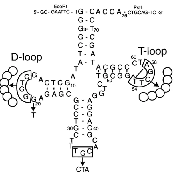
EcoRI Pstl
5'- GC - GAATTC - 1
G - CAC C A -
CTGCAG-TC -3'G - C
76G3-
T70G-C
C-G
D-Ioop
T - A
60T-Ioop
A- TACGCCCTA
8T
I I I I I / G
CG
A
TGCGG
ACTCG1Q
C
50TC~
G
I I I 1T
G
GA
GAGC
GG
G-A
C-G
C-G
T-A
30G - C40
C-G
T
C
~T
GelA
i-CTA
Figure 2.2 Construction of the tRNA gene library. In the nucleotide sequence of E. coli
tRNAAlauGc, each of the two enclosed regions, 16-19 in the D-Ioop and 54-58 in the T-Ioop, was replaced by six fully randomized positions, while nucleotide G20 and the anticodon TGC (boxed) were replaced by T20 and CTA, respectively. Nucleotides 54 and 58, which form the reverse Hoogsteen base pair in the T -Ioop, are connected by a line. The EcoRI and Pst! restriction sites that are seen flanking the 5' and 3' termini were used for cloning the library into the pGFIB-1 plasmid.
2.2 MATE RIALS & METHODS
2.2.1 Strains
The Escherichia coli strains TOP10 (Invitrogen) and XAC-1 (F' /ac/373/acZu118am
proB+/F l1(1ac-proB)x111 na/A rif argEamara) were used, respectively, for cloning and
selection of the suppressor tRNAs. The XAC-1 strain contains amber mutations in the genes /acZ and argE [NMKAM1986].
2.2.2 Construction of the combinatorial library and selection of suppressor tRNAs
The template oligonucleotide coding for the combinatorial tRNA library (Fig. 2) was synthesized at BioCorp Inc. (Montreal, Canada) ampli'Fled by PCR to produce the double-stranded DNA using primers 5'- CGMTICGGGGCTATA- 3' and 5'-GACTGCAGTGGTGGAGT-3', and cloned into plasrnid pGFIB-1 using
EcoR/ and Pst/ restriction sites, as described previously [BSFECC 1998]. This
plasmid provides a constitutive high level expression of a cloned tRNA gene [MM1986]. Ali enzymes were from New England Biolabs. Of 20 ml of the ligation mixture, 5 ml was electroporated into competent TOP10 cells, providing 4.5 X 10⁶ colonies, i.e. about a quarter of the sequence cornplexity of the library. The plasrnid
DNA was recovered using the Qiafilter Midiprep kit (Qiagen) and then transformed into competent XAC-1 cells. The positive clones were selected as blue colonies when grown on LB-agar containing ampicillin (100 mg/ml) and X-Gal (200 ml of the 20 rng/ml solution spread on top of each 150 X 15 mm plate). The plasmid ONA of these clones was extracted and retransformed into the XAC-1 cells to confirm the dependence of the phenotype on the presence of the plasmid. The ability of the selected tRNAs to suppress the nonsense mutation in gene argE was checked by plating the retransformed XAC-1 cells on minimal A medium without arginine.
2.2.3 Sequencing
Sequencing of the selected tRNA genes was performed on the LI-COR DNA sequencing system (Département de Biochimie, Université de Montréal) using primers 5'- GCTICTTTGAGCGAACGATCAAAAA T AAGT -3' and 5'-GGGTITTCCCAGTCACGACGTIGTAAAACG-3' labeled at the S'-end with IROye 800 (LI-COR Biosciences).
2.2.4 Measurement of the ~-Galactosidase activity
~-Galactosidase activity of clones with suppressor tRNA genes was determined as described by Miller [M1972] using overnight cultures grown in A
medium containing 0.4% glucose and, 1 mM MgS04 to an A600 of 0.8-0.9. The
values were obtained by averaging the measurements from three independent cultures and calculated as a percentage of the activity of the control tRNAAlasu+.
2.2.5 Presence of the Suppressor tRNAs in the cytosol and the aminoacylation level
To preserve the aminoacylated form of the tRNAs, the total cellular RNA was extracted under acidic conditions, as described previously [BSFECC1998]. To obtain the deacylated tRNA, 4 mg of the total RNA was mixed with 1.5 ml of 0.5 M Tris (pH 9.0), incubated for 30 min at 3rC and deposited on an acid polyacrylamide gel (6.5% polyacrylamide, 8 M urea, 0.1 M sodium acetate) together with the untreated fraction. The gel was run for 24 h at 300 V at 4°C in 0.1 M sodium acetate, after which the part of the gel around the xylene cyanol dye was transferred by electroblotting to a Hybond-N nylon membrane (Amersham). The membrane was hybridized with two radiolabeled ONA probes, one complementary to region 26-44 of the suppressor tRNAs, consisting of the anticodon stem and loop, and the other to region 34-53 of the E. coli 5S rRNA. The 5S rRNA probe was
used to monitor the amount of total RNA in each sample. The hybridization was performed overnight at 3rC in 7% SOS, 0.25 M Na2HP04 (pH 7.4), 1 mM EOTA
2.2.6 Computer modeling
Preliminary modeling was done interactively, using the InsightlllDiscover package (Version 2000; Accelrys Inc., San Diego, CA). The X-ray structure of the yeast tRNAPhe [SM2000] was used as a starting conformation. The presumed structures of RH-GA or RH-GG were appended to the T-stem replacing base pair U54-A58. The other randomized nucleotides were arranged in a way to resemble the structure of the DT region in the normal tRNAs and, at the same time, to pravide a reasonable system of hydragen bonds and base-base stacking interactions. Each model was submitled to unrestrained energy minimization using the AMBER force field [PCCRCFSSWK1995] until an energy minimum was reached. Visualizations were done in a Silicon Graphies 02 computer.
2.3 RESLIL TS
2.3.1 The library Design
The library was built fram E.coli tRNAAlaUGC as a scaffold (Fig. 2.2). The choice was determined by the fact that the most important tRNAAla identity element
for the cognate alanyl-tRNA synthetase, the G3-U70 base pair, was located in the acceptor stem, Le. neither in the DT region nor in the anticodon, the sites that were modified in this study [MF1988, HS 1988]. This would minimize the role of interaction with a particular aminoacyl-tRNA synthetase as a factor in the tRNA selection. To enable the tRNAs to recognize the amber stop codon UAG, the anticodon TGC in the gene was replaced by CTA. Ali five nucleotides of region 54-58 of the T-Ioop, which were known to be involved in conserved interactions either within the loop or with nucleotides of the D-Ioop, were fully randomized. Correspondingly, four nucleotides 16-19 of the D-Ioop, which could be involved in interactions with the T-Ioop, were also fully randomized. To prevent nucleotide G20 from substituting for either G18 or G19 in their interactions with the nucleotides of the T-Ioop, it was replaced by T20. To stimulate the formation of alternative structural patterns in the DT region, we added one and two nucleotides to the randomized regions of the and D-Ioops, respectively. Thus, in the design, the T-loop contained 8 nt, one more than in the standard tRNA structure, while the D-loop had 10 nt, which is not unprecedented for the cytosolic tRNAs [SHBIS1998]. Each loop had six randomized positions, providing for the total sequence complexity of a library of 1.7 X 10⁷ variants.
The tRNA gene library was synthesized chemically, amplified by PCR and cloned into the pGFIB-1 plasmid, as described previously [BSFECC1998]. The selection of active suppressor tRNA clones was done in the XAC-1 strain of E. coli, which had nonsense amber mutations in genes laeZ and argE. A successful suppression of the first mutation in the presence of 5-bromo-4-chloro-3-indolyl
13-0-galactopyranoside (X-Gal) provides blue colonies, which was used for the primary identification of functional tRNA clones. Out of 3 X 10⁴ clones screened, several dozen positive clones were selected, whose suppressor activity was confirmed by a subsequent retransformation and by suppression of the second mutation in geneargE, which converts the arginine-dependent cells into prototrophs. The
13-galactosidase activity was evaluated quantitatively for each clone and compared to that of the control tRNAAla su+. The latter tRNA was derived from the normal
tRNAAla by changing the anticodon from UGC to CUA and cloned in the same plasmid as the other suppressor tRNAs. The nucleotide sequences of the selected tRNA clones, as deduced from their genes, are presented in Table 2.1. Only the sequences of those clones whose activity was at least 1 % of the control are given. Comparison to the original design revealed six clones with a nucleotide deletion in the T-Ioop and three clones with a deletion in the O-Ioop, providing, respectively, for a seven mernber T-Ioop or a nine member O-Ioop. In two clones, K25 and K30, mutations affected the nonrandomized part of the T-Ioop, deleting, respectively, U59 and C60. No other mutation outside the randomized regions was found. For
eight clones arbitrarily chosen from Table 2.1, the in vivo level of the suppressor tRNA and of its aminoacylated form was determined by acid polyacrylamide gel electrophoresis followed by hybridization with a specifie probe complementary to the anticodon stem and loop. For ail suppressor tRNAs tested, the level in cytosol was detectable, although relatively low compared to that of tRNAAlasu+ (Fig. 3). For each clone, most of the tRNA was found in the aminoacylated form.
2.3.3 Analysis of the nucleotide sequences
ln the experiments described above, on average, only one in every 1000 clones showed a detectable level of nonsense suppression activity. This indicated that the nucleotide sequence space available for the DT region was rather small. A systematic analysis of the sequences of the selected clones could help to reveal the ru les imposed on the structure of this region in functional tRNAs.
Clone TI-tRNAs K30 K25 K3 K15 K29 K6 T8-tRNAs K26 Kl8 KI K7 K24 K5 K20 K27 K19 K2 K23 K9 KI7 K4 K28 K21 K31 KlO K14 K13 K16 K32 D-loop 16 19 1 1 AGUGAGG.n.UA AGGAACGCUA AGAACGAAUA AGGCAUAUUA AGGAAAAAUA AGAGGGAGUA 16 19 1 1 AGAACGACUA AGAACAAAUA AGGAGAACUA AGGACAAAUA AGAAAAACUA AGCGAAGAUA AGGAGAUCUA AGUG..n.AAUUA AGA-CAACUA AGAAAGACUA AGUAAGGUUA AGAGCGAAUA AGAGGCCAUA AGA-CGGGUA AGGGCA..l\AU.n. AGUGAAAGUA AG.n.GAGGGUA AGA-AGGAUA AGGAGGGAUA AGAGGAAAUA AGGGGGAUUA AGUCGGUAUA T-loop 54 58 1 1 1!CCJ®l>.U 1!~C 1!~UC 1!G.~UC 1!GG~UC ~CAC~UC 54 58 1 1 UAAACAUC - -UAAACAUC UAACCAUC UAACCAUC UAGCCAUC - -1!AGCC~UC 1!AGCC~UC 1!AGCC~UC 1!AUAC~UC 1!GACG~UC 1!GCC~UC ~ACGC:a.UC GAGCCAUC - -GCACAAUC - -GCAGCAUC - -GCCACAUC - -GCCCAAUC - -~UACC~UC ~GACC~UC ~UACC~UC ~UCAA~UC ~UCGA~UC p-Galactosidase activity (%) 10.8 :t 1.1 17.7 :t 6.2 4.1 :t 0.3 11.0 :t 1.4 5.1 :t 1.0 25.0:t 5.0 3.9 :t 0.8 2.5 :t 0.6 1.3 :t 0.1 1.3 :t 0.2 6.0:t 0.8 1.7 :t 0.3 3.2 :t 0.2 9.9 :t 2.0 2.0:t 0.4 7.9 :t 1.7 5.9 :t 1.1 1.3 :t 0.3 6.5 :t 1.8 1.1 :t 0.1 2.9 :t 0.6 2.8 :t 0.5 6.3 :t 0.8 5.9 :t 2.2 4.1 :t 0.5 1.3 :t 0.2 4.8 ± 1.2 38.5 :t 1.8
Table 2.1 The nucleotide sequences and the p-galactosidase activity of the selected tRNA clones. The sequences are deduced from the genes. Only the 0- and T-Ioops, where the sequences differ one from the other, are given. Positions of nucleotides 16-19 in the D-Ioop and 54-58 in the T-Ioop mark the beginning and the end of the each randomized region. Nucleotides forming the RH base pair in the T-Ioop are underlined. The activity oftRNAAlasu+ is taken as 100%.
We started the analysis with the 'quasi-normal' clones, those that had the normal 7 nt in the T-Ioop. Henceforth, we will cali such molecules T7-tRNAs, in contrast to T8-tRNAs which have 8 nt in this loop. Analysis showed that in T7-tRNAs, the fifth position of the T-Ioop was always occupied by A and was the only invariable position in both randomized regions (Table 2.1). The second most conservative nucleotide was the first one of the T-Ioop, which in ail sequences except K6 was U. The presence of U and A, respectively, in the first and the fifth position of the T-Ioop would allow the formation of the reverse Hoogsteen base pair U54-A58 (RH-UA), as in the normal tRNAs. Although in the normal tRNAs U54 is always modified to T, it is not yet known whether it is the case in the selected tRNAs. On the other hand, because this modification does not interfere with the ability of the base to form hydrogen bonds, its absence would not affect the formation of base pair U54-A58. Another conservative feature consistent with wild-type tRNA is the presence of a purine in position 57 of ail but one T7 -tRNA. Other randomized nucleotides, including ail 6 nt in the D-Ioop, were essentially more diversified and did not seem to provide for a common structural pattern.
Among T8-tRNAs, ha If of the sequences (11 out of 22) also contained U in the first position of the T -Ioop (Table 2.1). If this U plays the sa me role as it does in T7 -tRNAs, there should be an A a few nucleotides later that is able to form RH-UA .with this U. Generally, this A could occupy either the fifth or the sixth position of the
T-Ioop, depending on the position of the additional eighth nucleotide. Analysis showed that in those T8-tRNAs whose T-Ioop started with U the only other conservative nucleotide was the A occupying the sixth position of the same loop. Thus, the formation of RH-UA by these two nucleotides would place the additional nucleotide in the region between them.
Clona Tris .68 rRNA . . -tRNA tRNA A.la K1 la K3 K4 K5 Ka lU lUI su+ - + - + - + - + - + - + - + - + - + -"" - - - -pi -- -~ - - ~~
Figure 2.3 Northern blot showing the presence in the cytosol and the level of arninoacylation of sorne suppressor tRNAs. For each clone the - and + lanes correspond to the samples not treated and treated with Tris. In the - lanes the aminoacylated and deacylated forms of the suppressor tRI\lA move as individual bands, while in the + lanes the total tRNA is deacylated and the suppressor tRNA moves as one band. In ail - lanes the bands corresponding to the aminoacylated form of the tRNA are much larger than those corresponding to the deacylated form and are comparable to the bands in the + lanes, representing the total amount of the suppressor tRNA. This indicates that in ail clones most of the tRNA is present in the aminoacylated form. A smaller size of the bands of the suppressor tRNAs compared to tRNAAlasu+ indicates a notably lower presence of the selected tRNAs in the cytosol. 53 rRNA was visualized to monitor the amount of total RNA in each sample. Because the signal from 53 rRNA was much stronger than that from suppressor tRNAs, the upper and lower parts of the same membrane have been exposed, respectively, for 4 h and overnight. The nucleotide sequence of clone K8, due to its low l3-galactosidase activity, is not included in Table 2.1, but is available upon request.
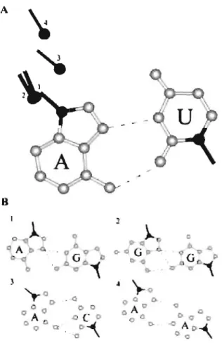
ln ail other T8-tRNAs, the first position of the T-Ioop was occupied by G (Table 2.1). If this G plays a structural role analogous to that played by U, its possible partner would occupy either the fifth or the sixth position of the T-Ioop. Neither of the two positions was conserved in these sequences: the fifth nucleotide was allowed to be either C or A, while the sixth was either A or G. To explore the abilities of both the fifth and the sixth nucleotides to pair with the first G, we looked for possible arrangements of three different dinucleotide combinations, GC, GA and GG, that would be close to the arrangement of U and A in RH-UA. For Ge, we did not find any satisfactory arrangement. However, for both cornbinations GA and GG we found arrangements that are presented in Figure 2.4. In these arrangements, the G that is equivalent to U in RH-UA donates two hydrogen atoms for formation of hydrogen bonds with atom N7 of the other purine. This purine can be either A or G. In the latter case, an additional hydrogen bond can be formed between N2-H and
06
of the first and second G residues, respectively. The two arrangements GA and GG are superimposable in the sensethat if one overlaps the positions of the glycosidic bonds of the first nucleotides, the glycosidic bonds of the second nucleotides in both arrangements would occupy about the same position. ln the same sense, these two arrangements are fairly close to RH-UA. Accommodation of any of these arrangements based on the standard RH-UA would require a shift and rotation of one of the bases by only 1.5A
and 20°,respectively. The refore , a replacement of RH-UA in the T-Ioop by either GA or GG would require only relatively minor changes in the positions of the neighboring nucleotides. To reflect the closeness of these GA and GG arrangements to RH-UA, we will cali them RH-GA and RH-GG, respectively.
Further analysis revealed a few additional nucleotide combinations like CA and AA seen in Figure 2.4 that cou Id also be arranged relatively closely to RH-UA while having a reasonable system of hydrogen bonds. Still, ail these additional combinations were more distant from RH-UA than RH-GA or RH-GG and, therefore, their incorporation into the T-Ioop instead of RH-UA wou Id cause greater changes in the conformation of the whole DT region. This latter aspect was expected to render these combinations less preferable in this place than RH-GA or RH-GG.
A B ~ p
.l
o
\)}.
0 (l A q G O CI 0- ,,4-0- 0 oi
i
0 o cr :\ .s).
",-00- --0 p 0 0 0 0 o AO a c Q A II C- o a ° O D Q . 00- \ 0- AD"",°0
Figure 2.4 Juxtaposition of the bases in RH-GA, RH-GG and other alternative base pair candidates for replacement of RH-UA. (A) Positions of the glycosidic bonds in the alternative base pairs compared to that in RH-UA. In each base pair the position of the glycosidic bond corresponding to the base on the right is superimposed on that of U in RH-UA. The glycosidic bond of the other nucleotide will thus occupy a particular place depending on the structure of the base pair. The numbers indicating particular positions of the glycosidic bonds correspond to the base pairs in (B).
The fact that GG and GA can be accommodated close to RH-UA, while Ge
cannot, makes the sixth rather than the tifth nucleotide of the T-Ioop in T8-tRNAs the most probable partner to form a base pair with the G occupying the tirst position of this loop. As to the other randomized nucleotides in both loops of